Leucopenia and treatment efficacy in advanced nasopharyngeal carcinoma
Bạn đang xem bản rút gọn của tài liệu. Xem và tải ngay bản đầy đủ của tài liệu tại đây (613.52 KB, 8 trang )
Su et al. BMC Cancer (2015) 15:429
DOI 10.1186/s12885-015-1442-3
RESEARCH ARTICLE
Open Access
Leucopenia and treatment efficacy in advanced
nasopharyngeal carcinoma
Zhen Su1†, Yan-Ping Mao1†, Pu-Yun OuYang1, Jie Tang1, Xiao-Wen Lan1 and Fang-Yun Xie1,2*
Abstract
Background: Leucopenia or neutropenia during chemotherapy predicts better survival in several cancers. We
aimed to assess whether leucopenia could be a biological measure of treatment and a marker of efficacy in
advanced nasopharyngeal carcinoma (ANPC).
Methods: We retrospectively analyzed 3826 patients with ANPC who received chemoradiotherapy. Leucopenia
was categorised on the basis of worst grade during treatment according to the National Cancer Institute Common
Toxicity Criteria version 4.0: no leucopenia (grade 0), mild leucopenia (grade 1–2), and severe leucopenia (grade
3–4). Associations between leucopenia and survival were estimated by Cox proportional hazards model.
Results: Of the 3826 patients, 2511 (65.6 %) developed mild leucopenia (grade 1–2) and 807 (21.1 %) developed
severe leucopenia (grade 3–4) during treatment; 508 (13.3 %) did not. A multivariate Cox model that included
leucopenia determined that the hazard ratios (HR) of death for patients with mild and severe leucopenia were 0.69
[95 % confidence interval (95 %CI) 0.56-0.85, p < 0.001] and 0.75 (95 %CI 0.59-0.95, p = 0.019), respectively; the HR
of distant metastasis for patients with mild and severe leucopenia were 0.77 (95 %CI 0.61-0.96, p = 0.023) and 0.99
(95 %CI 0.77-1.29, p = 0.995), respectively. Leucopenia had no effect on locoregional relapse.
Conclusions: Our results indicate that mild leucopenia during chemoradiotherapy is associated with improved
overall survival and distant metastasis–free survival in ANPC. Mild leucopenia may indicate appropriate dosage of
chemotherapy. We can identify the patients who may benefit from chemotherapy if they experienced leucopenia
during the treatment. Prospective trials are required to assess whether dosing adjustments based on leucopenia
may improve chemotherapy efficacy.
Keywords: Leucopenia, Advanced nasopharyngeal carcinoma, Chemoradiotherapy, Survival, Treatment efficacy
Background
Nasopharyngeal carcinoma (NPC) is a distinct type of
head and neck cancer. The incidence rate is as high as
20–30 per 100,000 populations in endemic areas of
southern China and Southeast Asia [1–3]. Radiotherapy
(RT) is the primary treatment, plus chemotherapy when
needed according to clinical stage. With the development of diagnostic imaging, chemotherapy regimens,
targeted drugs, and radiotherapeutic techniques, especially the application of IMRT (Intensity Modulated
* Correspondence:
†
Equal contributors
1
Sun Yat-sen University Cancer Center, State Key Laboratory of Oncology in
South China, Collaborative Innovation Center for Cancer Medicine,
Guangzhou 510060, China
2
Department of Radiation Oncology, Sun Yat-sen University Cancer Center;
State Key Laboratory of Oncology in South China; Collaborative Innovation
Center for Cancer Medicine, Guangzhou 510060, China
Radiation Therapy), survival of NPC has improved significantly [4–6]. However, 10–20 % of patients with advanced NPC (ANPC) develop distant metastasis after
radical chemoradiotherapy, rendering distant metastases
the main reason for treatment failure. To reduce the occurrence of distant metastasis, different timings of
chemotherapy is recommended for ANPC according to
NCCN (National Comprehensive Cancer Network)
guidelines [7]. In 2014 version of NCCN guidelines , the
categories of evidence for induction or adjuvant chemotherapy of NPC has changed [7]. Category of induction
chemotherapy of NPC changed from category 2A to category 3. Category of adjuvant chemotherapy “cisplatin +
RT followed by cisplatin/5-FU changed from category 1
to category 2A and “cisplatin + RT followed by carboplatin/5-FU changed from category 2A to category 2B.
© 2015 Su et al.; licensee BioMed Central. This is an Open Access article distributed under the terms of the Creative Commons
Attribution License ( which permits unrestricted use, distribution, and
reproduction in any medium, provided the original work is properly credited. The Creative Commons Public Domain
Dedication waiver ( applies to the data made available in this article,
unless otherwise stated.
Su et al. BMC Cancer (2015) 15:429
Bone marrow suppression is a common adverse reaction of cytotoxic drugs and could be a biological measure of drug activity and might predict treatment efficacy
[8, 9]. Leucopenia or neutropenia during treatment is a
common phenomenon of bone marrow suppression.
Some studies reported that leucopenia or neutropenia is
a prognostic factor predicting better clinical outcome in
several solid tumors, e.g., breast cancer [10–12], colorectal cancer [13, 14], advanced gastric cancer [15–17], lung
cancer [18–20], and Hodgkin’s lymphoma [21]. Others
have reported different results [22, 23]. However, the predictive (ie, estimation of the chance of benefit from
chemotherapy) or prognostic (ie, estimation of the chance
of survival) role of leucopenia in advanced nasopharyngeal
carcinoma have not been established.
We aimed to investigate the association between
leucopenia during treatment and survival of ANPC and
to provide evidences, through rigorous statistical analysis
of a large series of subjects with ANPC, of the utility of
leukocyte count as a surrogate marker of drug efficacy.
Methods
Patients and methods
We retrospectively collected 3939 newly diagnosed ANPC
patients from January 2005 to December 2010 treated
in the Nasopharyngeal Carcinoma Department of Sun
Yat-Sen University Cancer Center. 113 paitents were excluded owing to different reasons, abnormal liver function,
abnormal kidney function, unsatisfactory blood sugar control and so on. 3826 patients were involved in the study.
The Sun Yat-Sen University Cancer Center Institutional
Review Board (IRB) and ethics committee reviewed and
approved the study. The study was retrospective. Patient
records were anonymized and de-identified prior to
analysis.
Pretreatment evaluation included complete patient history, physical examination, hematology and biochemistry
profiles, nasopharynx and neck magnetic resonance imaging (MRI), chest radiography, abdominal ultrasound,
bone emission computed tomography (ECT), and chest or
abdomen computed tomography (CT) when necessary.
Page 2 of 8
leucopenia was based on the lowest recorded leukocyte
count for a given patient between the first day of treatment administration and 1 week after the end of treatment, and was graded according to the National Cancer
Institute Common Toxicity Criteria version 4.0. Patients
were classified as having no leucopenia (grade 0), mild
leucopenia (grade 1–2), and severe leucopenia (grade 3–4).
Indications for using granulocyte colony–stimulating
factor (G-CSF) were not specified; it was generally used
in grade 3–4 or febrile leucopenia, and was not used
for prophylaxis.
Follow-up
Patients were regularly followed after RT until death or
their last follow-up appointment. Clinic visits were
scheduled every three months in the first three years,
every six months during the fourth to fifth years, and
once a year after the fifth year. Patients underwent physical examination and nasopharyngoscopy on each visit.
Nasopharynx and neck MRI, chest radiography, abdominal ultrasound, and ECT were performed after RT or
according to clinical indications. The follow-up duration
was calculated from the first day of therapy to the day of
death or the day of last examination.
Statistical analysis
We estimated the following endpoints (interval to the
first defining event): overall survival (OS), locoregional
relapse–free survival (LRFS), and distant metastasis–free
survival (DMFS). Survival curves were estimated using
the Kaplan-Meier method and compared using the logrank test. Multivariate analyses were performed using the
Cox proportional hazards model. We used chi-square
tests and Kruskal–Wallis H tests to assess the statistical
significance of associations between categorical variables
and the three groups. All statistical tests were 2-tailed;
p < 0.05 was considered statistically significant. All tests
were conducted using IBM SPSS version 20.0.0 (IBM
Corporation, Armonk, NY, USA).
Results
Patient characteristics
Treatment
The treatment strategy for all patients was based on National Comprehensive Cancer Network Guidelines [24,
25]. All patients were treated with intensity-modulated
RT (IMRT) or conventional RT (CRT) with chemotherapy; the radiation techniques and chemotherapy regimens have been described previously [26, 27].
Laboratory measurements
We performed leukocyte and neutrophil counts for all
patients within two weeks before therapy and at least
once weekly during treatment. The most severe grade of
Table 1 lists the patient characteristics. We studied 3826
patients (2873 male; 953 female). The median age at diagnosis for male patients was 46 years (range 20–84 years);
that for female patients was 44 years (range 20–76 years).
CRT and IMRT were administered to 2583 and 1243 patients, respectively. Induction chemotherapy (IC) was administered to 1073 patients, concurrent chemotherapy
(CC) to 1291 patients, IC plus CC (IC + CC) to 1255 patients, and CC plus adjuvant chemotherapy (CC + AC)
to 207 patients. We administered <4 and ≥4 chemotherapy cycles to 2364 (61.8 %) and 1462 (38.2 %)
patients, respectively. No significant differences were
Su et al. BMC Cancer (2015) 15:429
Page 3 of 8
Table 1 Patient characteristics according to grade of leucopenia
Variable
All
Absent leucopenia
Mild leucopenia
Severe leucopenia
Total
3826
508(13.3)
2511(65.6)
807(21.1)
male
2873(75.1)
421(82.9)
1936(77.1)
516(63.9)
female
953(24.9)
87(17.1)
575(22.9)
291(36.1)
Gender
P value
<0.001
Age(years)
0.105
<45
1982(51.8)
242(47.6)
1325(52.8)
415(51.4)
> = 45
1844(48.2)
266(52.4)
1186(47.2)
392(48.6)
= < 10 × 10^9/L
3448(90.1)
424(83.5)
2278(90.7)
746(92.4)
>10 × 10^9/L
378(9.9)
84(16.5)
233(9.3)
61(7.6)
Leukocyte count
<0.001
Pathological type(WHO)
0.692
I
83(2.2)
11(2.2)
55(2.2)
17(2.1)
II
203(5.3)
23(4.5)
143(5.7)
37(4.6)
III
3540(92.5)
474(93.3)
2313(92.1)
753(93.3)
T-classification
0.720
T1
172(4.5)
22(4.3)
109(4.3)
41(5.1)
T2
272(7.1)
29(5.7)
185(7.4)
58(7.2)
T3
1871(48.9)
271(53.3)
1221(48.6)
379(47.0)
T4
1511(39.5)
186(36.6)
996(39.7)
329(40.8)
N0
517(13.5)
86(16.9)
335(13.3)
96(11.9)
N1
1978(51.7)
252(49.6)
1313(52.3)
413(51.2)
N2
1043(27.3)
133(26.2)
680(27.1)
230(28.5)
N3
288(7.5)
37(7.3)
183(7.3)
68(8.4)
N-classification
0.09
Clinical stage
0.222
III
2094(54.7)
295(58.1)
1369(54.5)
430(53.3)
IV
1732(45.3)
213(41.9)
1142(45.5)
377(46.7)
CRT
2583(67.5)
329(64.8)
1741(69.3)
513(63.6)
IMRT
1243(32.5)
179(35.2)
770(30.7)
294(36.4)
Radiotherapy
0.004
Chemotherapy
<0.001
IC
1073(28.0)
198(39.0)
697(27.8)
178(22.1)
CC
1291(33.7)
202(39.8)
878(35.0)
211(26.1)
IC + CC
1255(32.8)
98(19.3)
804(32.0)
353(43.7)
CC + AC
207(5.4)
10(2.0)
132(5.3)
65(8.1)
NO
3029(79.2)
403(79.3)
2069(82.4)
557(69.0)
YES
797(20.8)
105(20.7)
442(17.6)
250(31.0)
Paclitaxel
<0.001
Chemotherapy cycles
<0.001
<4
2364(61.8)
400(78.7)
1575(62.7)
389(48.2)
>=4
1462(38.2)
108(21.3)
936(37.3)
418(51.8)
Abbreviations: CRT: conventional radiotherapy; IMRT: intensity modulated radiation therapy; IC: Induction chemotherapy; CC: concurrent chemotherapy;
AC: adjuvant chemotherapy; WHO: world health organization
Su et al. BMC Cancer (2015) 15:429
Page 4 of 8
observed for age, T classification, N classification, and
clinical stage. There were significant differences in pretreatment leukocyte count, type of chemotherapy,
chemotherapy cycles, type of RT, sex, and paclitaxel use
(yes or no) in the compared groups (all p < 0.05). Patients who developed leucopenia during treatment had
lower pretreatment leukocyte counts (p < 0.001). More
female patients developed leucopenia (female vs. male,
90.1 % vs. 85.3 %, p < 0.001); patients using paclitaxel
were likely to develop severe leucopenia (31.4 % vs.
18.4 %, p < 0.001).
The median OS was 52.6 months (range 3.07–113.0
months); 10.9 % of patients (417/3826) developed locoregional relapse, 16.5 % (633/3826) developed distant metastases, and 19.0 % (727/3826) died. The 5-year OS,
LRFS, and DMFS rates for the entire population were
80.70 %, 87.9 %, and 82.1 %, respectively.
During treatment, 2511 patients (65.6 %) developed
mild leucopenia (grade 1–2) and 807 patients (21.1 %)
developed severe leucopenia (grade 3–4); the remaining
508 (13.3 %) did not develop leucopenia.
Survival analyses including leucopenia
Table 2 shows the univariate analysis of the baseline and
clinical characteristics as prognostic factors, including
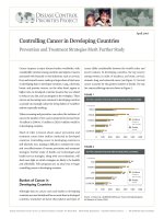
leucopenia. Kaplan–Meier curves according to severity of
leucopenia showed that better OS and DMFS were predicted for patients with leucopenia and that leucopenia had
no significant effect on LRFS (Fig. 1). The 5-year OS rate in
patients with no leucopenia, mild leucopenia, and severe
leucopenia was 75.5 %, 81.9 %, and 80.5 %, respectively
(mild vs no leucopenia, p = 0.001; severe vs no leucopenia,
p = 0.03; mild vs severe, p = 0.314). The 5-year DMFS rate
in patients with no leucopenia, mild leucopenia, and severe
leucopenia was 79.7 %, 83.7 %, and 78.9 %, respectively
(mild vs. no leucopenia, p = 0.038; severe vs no leucopenia,
p = 0.927; mild vs severe, p = 0.007). The 5-year LRFS rate
in patients with no leucopenia, mild leucopenia, and severe
Table 2 Univariate analysis of survival for patients with ANPC
All population
Variable
Cycles <4 population
Cycles > =4 population
OS
DMFS
OS
DMFS
OS
DMFS
HR(95 %CI)
0.70(0.57-0.86)
0.79(0.63-0.98)
0.73(0.57-0.92)
0.87(0.66-1.14)
0.56(0.38-0.86)
0.56(0.37-0.86)
p
0.001
0.038
0.009
0.309
0.007
0.008
Leucopenia
Mild VS Absent
Severe VS Absent
HR(95 %CI)
0.77(0.60-0.97)
1.01(0.78-1.31)
0.86(0.63-1.16)
1.10(0.79-1.54)
0.59(0.38-0.90)
0.73(0.46-1.14)
P
0.030
0.927
0.320
0.554
0.016
0.166
HR(95%CI)
0.91(0.76-1.09)
0.77(0.64-0.93)
0.85(0.66-1.08)
0.78(0.61-1.02)
0.98(0.75-1.28)
0.77(0.58-1.09)
p
0.314
0.007
0.191
0.069
0.887
0.058
Mild VS Severe
Gender
HR(95 %CI)
0.62(0.51-0.75)
0.69(0.57-0.84)
0.63(0.50-0.79)
0.71(0.55-0.91)
0.61(0.45-0.84)
0.67(0.49-0.2)
P
<0.001
<0.001
<0.001
0.007
0.002
0.015
HR(95 %CI)
1.84(1.59-2.14)
1.09(0.93-1.27)
1.93(1.59-2.34)
1.14(0.94-1.39)
1.73(1.36-2.19)
1.01(0.77-1.30)
P
<0.001
0.304
<0.001
0.191
<0.001
0.941
Age
T-classification
HR(95 %CI)
1.27(1.14-1.40)
1.09(0.99-1.22)
1.27(1.11-1.45)
1.11(0.96-1.27)
1.26(1.07-1.49)
1.08(0.91-1.27)
P
<0.001
0.092
0.001
0.157
0.007
0.375
HR(95CI)
1.56(1.43-1.70)
1.65(1.50-1.81)
1.70(1.51-1.90)
1.75(1.54-1.97)
1.39(1.21-1.60)
1.52(1.32-1.76)
P
<0.001
<0.001
<0.001
<0.001
<0.001
<0.001
N-classification
Radiotherapy
HR(95 %CI)
0.80(0.68-0.94)
0.91(0.76-1.08)
0.88(0.70-1.10)
1.03(0.82-1.30)
0.69(0.54-0.89)
0.75(0.58-0.97)
P
0.008
0.273
0.264
0.764
0.005
0.030
Abbreviations: OS: overall survival; DMFS: distant metastasis-free survival; HR: hazard ratio; CI: confidence interval; ANPC: advanced nasopharyngeal carcinoma
Su et al. BMC Cancer (2015) 15:429
Page 5 of 8
leucopenia was 88.9 %, 87.4 %, and 88.6 %, respectively (all
p > 0.05 for any two compared groups).
We performed multivariate analysis to investigate
whether leucopenia could be a marker of improved OS
and DMFS (Table 3). Leucopenia and other prognostic
factors, i.e., age, sex, T classification, N classification, pathological type, type of chemotherapy, paclitaxel use, and type
of RT were included in the multivariate analysis, which determined that leucopenia, sex, T classification, and N classification were independent prognostic factors for OS and
DMFS. Compared to patients without leucopenia, the hazard ratios (HRs) of death for patients with mild and severe
leucopenia were 0.69 [95 % confidence interval (95 %CI)
0.56-0.85, p < 0.001] and 0.75 (95 %CI 0.59-0.95, p = 0.019),
respectively. The HR of distant metastasis for patients with
mild and severe leucopenia were 0.77 (95 %CI 0.61-0.96,
p = 0.023) and 0.99 (95 %CI 0.77-1.29, p = 0.995), respectively. When we compared patients with mild leucopenia to
patients with severe leucopenia, the HRs of death and distant metastasis were 0.93 (95 %CI 0.77-1.11, p = 0.416) and
0.77 (95%CI 0.64-0.93, p = 0.006), respectively.
When pretreatment leukocyte count (≤10 × 109/L
vs. >10 × 109/L) was included in the Cox model, leucopenia remained significant for OS (mild leucopenia:
HR = 0.70, 95 %CI 0.57-0.86, p = 0.001; severe leucopenia: HR = 0.76, 95 %CI 0.59-0.97, p = 0.026) and DMFS
(mild leucopenia: HR = 0.77, 95 %CI 0.61-0.96, p = 0.023;
severe leucopenia: HR = 0.99, 95 %CI 0.77-1.30, p = 0.995).
Tables 2 and 3 depict the subgroup analysis results for
patients who received <4 and ≥4 chemotherapy cycles.
Mild and severe leucopenia tended to be associated with
improved survival in patients who received <4 or ≥4
chemotherapy cycles.
Fig. 1 Kaplan–Meier survival curves of (a) Overall Survival, (b)
Locoregional Relapse-free Survival, and (c) Distant Metastasis-free
Survival according to severity of leucopenia
Discussion
In this study, we found that survival was improved in patients who developed leucopenia during chemoradiotherapy for ANPC. Patients with mild leucopenia had better
OS and DMFS than those with severe leucopenia. Leucopenia was an independent prognostic factor for OS and
DMFS in patients who received <4 and ≥4 chemotherapy
cycles. This is the first instance that has been reported in
pretreated ANPC.
As far as we know, leucopenia or neutropenia indicates
that the chemotherapeutic agent dose is sufficient to cause
bone marrow suppression and an anti-tumor effect [8, 9].
The absence of leucopenia or neutropenia indicates an absent or weak biological effect of chemotherapy, likely indicating that the dose is too low. On the other hand, severe
leucopenia may indicate overdosage. High-dose chemotherapy does not improve survival, and impairs patient
quality of life [28]. We speculate that moderate-dose
chemotherapy, as evidenced by moderate toxicity, is the
optimal treatment, correlating with better survival than
Su et al. BMC Cancer (2015) 15:429
Page 6 of 8
Table 3 Multivariate analysis of survival for patients with ANPC
All population
Variable
Cycles <4 population
Cycles > =4 population
OS
DMFS
OS
DMFS
OS
DMFS
HR(95 %CI)a
0.69(0.56-0.85)
0.77(0.61-0.96)
0.70(0.55-0.89)
0.88(0.63-1.23)
0.73(0.46-1.15)
0.61(0.40-0.94)
P
<0.001
0.023
0.003
0.452
0.174
0.025
HR(95 %CI)a
0.75(0.59-0.95)
0.99(0.77-1.29)
0.82(0.60-1.11)
0.79(0.61-1.03)
0.97(0.74-1.27)
0.84(0.53-1.32)
P
0.019
0.995
0.204
0.083
0.828
0.446
HR(95 %CI)a
0.93(0.77-1.11)
0.77(0.64-0.93)
0.85(0.66-1.09)
0.90(0.68-1.19
0.71(0.46-1.07
0.73(0.56-0.96)
P
0.416
0.006
0.204
0.469
0.108
0.026
Leucopenia
Mild VS Absent
Severe VS Absent
Mild VS Severe
Gender
HR(95 %CI)
0.67(0.55-0.81)
0.70(0.58-0.86)
0.66(0.52-0.84)
0.73(0.57-0.93)
0.66(0.48-0.91)
0.68(0.49-0.94)
P
<0.001
<0.001
0.001
0.013
0.010
0.019
HR(95 %CI)
1.82(1.57-2.12)
1.05(0.89-1.23)
1.88(1.55-2.28)
1.09(0.89-1.34
1.71(1.34-2.17)
1.03(0.81-1.33)
P
<0.001
0.532
<0.001
0.367
<0.001
0.783
Age
T-classification
HR(95 %CI)
1.49(1.35-1.66)
1.33(1.19-1.47)
1.51(1.33-1.72)
1.36(1.19-1.56)
1.49(1.26-1.76)
1.27(1.08-1.51)
P
<0.001
<0.001
<0.001
<0.001
<0.001
0.005
HR(95CI)
1.77(1.62-1.93)
1.78(1.62-1.97)
1.92(1.71-2.16)
1.92(1.68-2.18)
1.56(1.35-1.80)
1.63(1.40-1.90)
P
<0.001
<0.001
<0.001
<0.001
<0.001
<0.001
N-classification
Abbreviations: OS: overall survival; DMFS: distant metastasis-free survival; HR: hazard ratio; CI: confidence interval; ANPC: advanced nasopharyngeal carcinoma
a
Adjusted for age (<45 and ≥45 years old), sex, T classification (T1/T2/T3/T4), N classification (N0/N1/N2/N3), pathological type, type of radiotherapy, type of
chemotherapy, and paclitaxel use
under- or overdosage. Colleoni et al. [29] found that patients who received level II doses (65-84 % of the prescribed dose) had longer disease-free survival (DFS) and
OS than patients who received higher (level I: >85 % of
the prescribed dose) or lower (level III: <65 % of the prescribed dose) doses (p = 0.07, p = 0.03, respectively). Additionally, Brunetto et al. reported that there was no
difference in OS for patients whose dose had been reduced compared to patients whose dose had been maintained [30]. Nakatat al. [17] and Shitara et al. [15] both
found that patients with mild neutropenia had better outcomes than those with severe neutropenia; others have reported that patients who developed grade 2–3 leucopenia
or neutropenia had significantly better prognosis than
those with grade 4 leucopenia or neutropenia [16, 17, 31].
Our results agree with these results. In other words, mild
leucopenia or neutropenia might be a barometer of the
appropriate chemotherapeutic dosage to obtain sufficient
anti-tumor effect in a patient, leading to improved clinical
outcome; however, severe leucopenia or neutropenia
might be a marker of overdosage and suboptimal survival.
However, there are differing findings: Kim et al. [22]
reported that neutropenia was not a significant prognostic indicator of improved progression-free survival and
OS (p = 0.180, p = 0.698, respectively) in stage I-IIIB
breast cancer. Kumpulainen et al. [23] drew a wholly different conclusion, where the 10-year DFS in FIGO
(International Federation of Obstetrics and Gynecology)
stage IC-IV disease was 45 % in patients with lower
leukocyte counts (<2.5 × 10⁹/L) and 66 % in patients
with higher leukocyte counts (≥2.5 × 10⁹/L) (p < 0.05).
The probable reason is that the different disease stages
might obscure the impact of leucopenia. Most studies
and ours studied patients with advanced-stage disease.
Several reports have stated that pretreatment high
leukocyte or neutrophil count might be a poor prognostic indicator and that leucopenia or neutropenia are less
likely to occur during treatment [32, 33]. However, in
our multivariate analysis, which included this factor,
leucopenia remained significant for OS and DMFS.
Due to the retrospective nature of our study, there are
some limitations. First, the chemotherapy regimens and
Su et al. BMC Cancer (2015) 15:429
dose varied. Second, patients were identified from 2005 to
2010, and the normal range of hematological profiles may
have varied. Third, although G-CSF was not used for
prophylaxis, it would nevertheless affect the severity of
leucopenia. Fourth, we only analyzed leucopenia, a sign of
myelosuppression. Taking hemoglobin and platelet inhibition into account might reflect the relationship between
myelosuppression and prognosis more accurately.
Page 7 of 8
3.
4.
5.
6.
Conclusions
Leucopenia during chemoradiotherapy of ANPC is
strongly associated with better OS and DMFS; mild
leucopenia indicates better survival than severe leucopenia. This may indicate that mild leucopenia is a surrogate marker for adequate chemotherapeutic dose. We
can identify the patients who may benefit from chemotherapy if they experienced leucopenia during the treatment. The chemotherapy dose should not only depend
on the body surface area, but also be based on its toxic
effects. Prospective trials are required to assess whether
dosing adjustments based on leucopenia may improve
chemotherapy efficacy.
Abbreviations
ANPC: Advanced nasopharyngeal carcinoma; CRT: Conventional radiotherapy;
IMRT: Intensity modulated radiation therapy; NCCN: National comprehensive
cancer network; G-CSF: Granulocyte colony-stimulating factor; MRI: Magnetic
resonance imaging; ECT: Emission computed tomography; CT: Computed
tomography; IC: Induction chemotherapy; CC: Concurrent chemotherapy;
AC: Adjuvant chemotherapy; HR: Hazard ratio; CI: Confidence interval;
OS: Overall survival; DMFS: Distant metastasis-free survival; LRFS: Locoregional
relapse–free survival; DFS: Disease-free survival; FIGO: International federation
of obstetrics and gynecology; WHO: World health organization.
Competing interests
The authors declare that they have no competing interests.
Authors’ contributions
SZ checked data, drafted the manuscript and performed the statistical
analysis. OYPU, TJ and LXW collected the data. XFY participated in the
design of the study. MYP conceived of the study, and participated in its
design and coordination and helped to draft the manuscript. All authors
read and approved the final manuscript. Both Zhen Su and Yan-Ping Mao
contributed equally to this manuscript.
Acknowledgements
This work was supported by grants from the National Natural Science
Foundation of China (No. 81201746), Planned Science and Technology
Project of Guangdong Province (2012B031800092), Medical Science
Foundation of Guangdong Province (No. B2012135), Cultivating Foundation
of Education-bureau of Guangdong Province (No. LYM11001).
7.
8.
9.
10.
11.
12.
13.
14.
15.
16.
17.
18.
19.
20.
Received: 16 October 2014 Accepted: 15 May 2015
21.
References
1. Chen L, Mao Y, Xie F, Liu L, Sun Y, Tian L, et al. The seventh edition of the
UICC/AJCC staging system for nasopharyngeal carcinoma is prognostically
useful for patients treated with intensity-modulated radiotherapy from an
endemic area in China. Radiother Oncol. 2012;104(3):331–7.
2. Perri F. Locally advanced nasopharyngeal carcinoma: current and emerging
treatment strategies. World Journal of Clinical Oncology. 2011;2(12):377.
22.
23.
Adham M, Kurniawan AN, Muhtadi AI, Roezin A, Hermani B, Gondhowiardjo
S, et al. Nasopharyngeal carcinoma in Indonesia: epidemiology, incidence,
signs, and symptoms at presentation. Chin J Cancer. 2012;31(4):185–96.
Peng G, Wang T, Yang KY, Zhang S, Zhang T, Li Q, et al. A prospective,
randomized study comparing outcomes and toxicities of intensity-modulated
radiotherapy vs. conventional two-dimensional radiotherapy for the treatment
of nasopharyngeal carcinoma. Radiother Oncol. 2012;104(3):286–93.
Lai SZ, Li WF, Chen L, Luo W, Chen YY, Liu LZ, et al. How does intensitymodulated radiotherapy versus conventional two-dimensional radiotherapy
influence the treatment results in nasopharyngeal carcinoma patients? Int J
Radiat Oncol Biol Phys. 2011;80(3):661–8.
Baujat B, Audry H, Bourhis J, Chan AT, Onat H, Chua DT, et al.
Chemotherapy in locally advanced nasopharyngeal carcinoma: an individual
patient data meta-analysis of eight randomized trials and 1753 patients. Int
J Radiat Oncol Biol Phys. 2006;64(1):47–56.
Pfister DG, Spencer S, Brizel DM, Burtness B, Busse PM, Caudell JJ, et al.
Head and neck cancers, Version 2.2014. Clinical practice guidelines in
oncology. J Natl Compr Canc Netw. 2014;12(10):1454–87.
Kvinnsland S. The leucocyte nadir, a predictor of chemotherapy efficacy?
Br J Cancer. 1999;80(11):1681.
Gurney H. How to calculate the dose of chemotherapy. Br J Cancer.
2002;86(8):1297–302.
Mayers C, Panzarella T, Tannock IF. Analysis of the prognostic effects of
inclusion in a clinical trial and of myelosuppression on survival after
adjuvant chemotherapy for breast carcinoma. Cancer. 2001;91(12):2246–57.
Poikonen P, Saarto T, Lundin J, Joensuu H, Blomqvist C. Leucocyte nadir as
a marker for chemotherapy efficacy in node-positive breast cancer treated
with adjuvant CMF. Br J Cancer. 1999;80(11):1763–6.
Saarto T, Blomqvist C, Rissanen P, Auvinen A, Elomaa I. Haematological
toxicity: a marker of adjuvant chemotherapy efficacy in stage II and III breast
cancer. Br J Cancer. 1997;75(2):301–5.
Sunaga T, Suzuki S, Kogo M, Kurihara T, Kaji S, Koike N, et al. The association
between neutropenia and prognosis in stage III colorectal cancer patients
receiving adjuvant chemotherapy. Eur J Cancer Care. 2013:n/a-n/a.
Shitara K, Matsuo K, Takahari D, Yokota T, Inaba Y, Yamaura H, et al.
Neutropaenia as a prognostic factor in metastatic colorectal cancer patients
undergoing chemotherapy with first-line FOLFOX. Eur J Cancer.
2009;45(10):1757–63.
Shitara K, Matsuo K, Takahari D, Yokota T, Shibata T, Ura T, et al. Neutropenia
as a prognostic factor in advanced gastric cancer patients undergoing
second-line chemotherapy with weekly paclitaxel. Ann Oncol.
2010;21(12):2403–9.
Yamanaka T, Matsumoto S, Teramukai S, Ishiwata R, Nagai Y, Fukushima M.
Predictive value of chemotherapy-induced neutropenia for the efficacy of
oral fluoropyrimidine S-1 in advanced gastric carcinoma. Br J Cancer.
2007;97(1):37–42.
Nakata B, Tsuji A, Mitachi Y, Yamamitsu S, Hirata K, Takeuchi T, et al. Moderate
neutropenia with S-1 plus low-dose cisplatin may predict a more favourable
prognosis in advanced gastric cancer. Clin Oncol-Uk. 2006;18(9):678–83.
Jang SH, Kim SY, Kim JH, Park S, Hwang YI, Kim DG, et al. Timing of
chemotherapy-induced neutropenia is a prognostic factor in patients with
metastatic non-small-cell lung cancer: a retrospective analysis in
gemcitabine-plus-platinum-treated patients. J Cancer Res Clin Oncol.
2013;139(3):409–17.
Pallis AG, Agelaki S, Kakolyris S, Kotsakis A, Kalykaki A, Vardakis N, et al.
Chemotherapy-induced neutropenia as a prognostic factor in patients with
advanced non-small cell lung cancer treated with front-line docetaxel–gemcitabine chemotherapy. Lung Cancer-J Iaslc. 2008;62(3):356–63.
Di Maio M, Gridelli C, Gallo C, Shepherd F, Piantedosi FV, Cigolari S, et al.
Chemotherapy-induced neutropenia and treatment efficacy in advanced
non-small-cell lung cancer: a pooled analysis of three randomised trials.
Lancet Oncol. 2005;6(9):669–77.
Klimm B, Reineke T, Haverkamp H, Behringer K, Eich HT, Josting A, et al. Role
of hematotoxicity and sex in patients with Hodgkin’s lymphoma: an analysis
from the German Hodgkin Study Group. J Clin Oncol. 2005;23(31):8003–11.
Kim J, Park J, Kim D, Kim J, Kim Y, Nam J, et al. Is chemotherapy-induced
neutropenia a prognostic factor in patients with ovarian cancer? Acta
Obstet Gyn Scan. 2010;89(5):623–8.
Kumpulainen EJ, Hirvikoski PP, Johansson RT. Neutropenia during adjuvant
chemotherapy of breast cancer is not a predictor of outcome. Acta Oncol.
2009;48(8):1204–6.
Su et al. BMC Cancer (2015) 15:429
Page 8 of 8
24. Forastiere AA, Ang KK, Brizel D, Brockstein BE, Burtness BA, Cmelak AJ, et al.
Head and neck cancers. J Natl Compr Canc Netw. 2008;6(7):646–95.
25. Forastiere AA, Ang K, Brizel D, Brockstein BE, Dunphy F, Eisele DW, et al.
Head and neck cancers. J Natl Compr Canc Netw. 2005;3(3):316–91.
26. Liang SB, Sun Y, Liu LZ, Chen Y, Chen L, Mao YP, et al. Extension of local
disease in nasopharyngeal carcinoma detected by magnetic resonance
imaging: improvement of clinical target volume delineation. Int J Radiat
Oncol Biol Phys. 2009;75(3):742–50.
27. Ma J, Liu L, Tang L, Zong J, Lin A, Lu T, et al. Retropharyngeal lymph node
metastasis in nasopharyngeal carcinoma: prognostic value and staging
categories. Clin Cancer Res. 2007;13(5):1445–52.
28. Brandberg Y. Quality of life in women with breast cancer during the first
year after random assignment to adjuvant treatment with marrowsupported high-dose chemotherapy with cyclophosphamide, thiotepa,
and carboplatin or tailored therapy with fluorouracil, epirubicin, and
cyclophosphamide: scandinavian breast group study 9401. J Clin Oncol.
2003;21(19):3659–64.
29. Colleoni M, Price K, Castiglione-Gertsch M, Goldhirsch A, Coates A, Lindtner
J, et al. Dose–response effect of adjuvant cyclophosphamide, methotrexate,
5-fluorouracil (CMF) in node-positive breast cancer. International Breast
Cancer Study Group. Eur J Cancer. 1998;34(11):1693–700.
30. Brunetto AT, Carden CP, Myerson J, Faria AL, Ashley S, Popat S, et al. Modest
reductions in dose intensity and drug-induced neutropenia have no major
impact on survival of patients with non-small cell lung cancer treated with
platinum-doublet chemotherapy. J Thorac Oncol. 2010;5(9):1397–403.
31. Cameron DA, Massie C, Kerr G, Leonard RC. Moderate neutropenia with
adjuvant CMF confers improved survival in early breast cancer. Br J Cancer.
2003;89(10):1837–42.
32. Chang H, Gao J, Xu BQ, Guo SP, Lu RB, Li G, et al. Haemoglobin, neutrophil
to lymphocyte ratio and platelet count improve prognosis prediction of the
TNM staging system in nasopharyngeal carcinoma: development and
validation in 3237 patients from a single institution. Clin Oncol-Uk.
2013;25(11):639–46.
33. He JR, Shen GP, Ren ZF, Qin H, Cui C, Zhang Y, et al. Pretreatment levels of
peripheral neutrophils and lymphocytes as independent prognostic factors in
patients with nasopharyngeal carcinoma. Head Neck. 2012;34(12):1769–76.
Submit your next manuscript to BioMed Central
and take full advantage of:
• Convenient online submission
• Thorough peer review
• No space constraints or color figure charges
• Immediate publication on acceptance
• Inclusion in PubMed, CAS, Scopus and Google Scholar
• Research which is freely available for redistribution
Submit your manuscript at
www.biomedcentral.com/submit