A small molecular agent YL529 inhibits VEGF-D-induced lymphangiogenesis and metastasis in preclinical tumor models in addition to its known antitumor activities
Bạn đang xem bản rút gọn của tài liệu. Xem và tải ngay bản đầy đủ của tài liệu tại đây (2.25 MB, 12 trang )
Xu et al. BMC Cancer (2015) 15:525
DOI 10.1186/s12885-015-1451-2
RESEARCH ARTICLE
Open Access
A small molecular agent YL529 inhibits
VEGF-D-induced lymphangiogenesis and
metastasis in preclinical tumor models in
addition to its known antitumor activities
Youzhi Xu1,2†, Wenjie Lu1,2†, Peng Yang3†, Wen Peng2,4†, Chunting Wang2, Manli Li2, Yan Li2, Guobo Li2,
Nana Meng2, Hongjun Lin2, Lixin Kan2, Siying Wang2, Shengyong Yang2, Luoting Yu2* and YingLan Zhao2*
Abstract
Background: The lymph node metastasis is a key early step of the tumor metastatic process. VEGFD-mediated tumor
lymphangiogenesis plays a key role, since down-regulation of p-VEGFR-3 could block the lymph node metastasis.
YL529 has been reported to possess potent anti-angiogenesis and antitumor activities; however, its roles in
tumor-associated lymphangiogenesis and lymphatic metastasis remain unclear.
Method: We investigated the effect of YL529 on tumor-associated lymphangiogenesis and lymph node metastasis
using in vitro lymph node metastasis models and in vivo subcutaneous tumor models in C57 BL/6 mice.
Result: We found that YL529 inhibited VEGF-D-induced survival, proliferation and tube-formation of Human Lymphatic
Endothelial Cells. Furthermore, in established in vitro and in vivo lymph node metastasis models using VEGF-D-LL/2 cells,
YL529 significantly inhibited the tumor-associated lymphangiogenesis and metastasis. At molecular level, YL529
down-regulated p-VEGFR-3, p-JNK and Bax while up-regulated Bcl-2.
Conclusion: YL529 provided the therapeutic benefits by both direct effects on tumor cells and inhibiting
lymphangiogenesis and metastasis via the VEGFR-3 signaling pathway, which may have significant direct clinical
implications.
Keywords: YL529, Lymphangiogenesis, Metastasis, VEGF-D, VEGFR-3
Background
Tumor metastasis is the key and final cause of cancer
mortality [1–3]. It usually occurs via the hematogenic and
the lymphogenic metastasis routes [4, 5], and the latter is
considered to be a very important route contributing to
metastasis of solid tumors [6].
Tumor lymphangiogenesis is regulated by many important factors, such as vascular endothelial growth factor
(VEGF) and its receptor subtype members (VEGFRs).
VEGFR-1 ~ 3 are almost exclusively located on the surface
* Correspondence: ;
†
Equal contributors
2
State Key Laboratory of Biotherapy and Cancer Center, West China Hospital,
West China Medical School, and Collaborative Innovation Center for
Biotherapy, Sichuan University, 17#, 3rd Section, Ren min South Road,
Chengdu 610041, China
Full list of author information is available at the end of the article
of vascular endothelial cells and lymphatic vessels in normal tissues and are up-regulated only during embryonic
and tumor. Among all of VEGFs and VEGFRs, the Vascular endothelial growth factor D (VEGF-D) is indispensable
for development of the lymphatic system and VEGFR-3
has also been implicated as the major effectors of lymphangiogenesis and regulates lymphatic vessel growth
[1, 7]; VEGF-C and VEGF-D are the ligands for the
tyrosine kinases VEGFR-3 and VEGFR 2, which mainly
regulates the growth of lymphatic vessels via their receptor VEGFR-3, and partly regulates the growth of blood
vessels via VEGFR-2 [8, 9]. Since literatures have showed that
tumor-induced lymphangiogenesis driven by VEGF-C and
VEGF-D-induced activation of VEGFR-3 could promote regional lymph node metastasis in multiple solid tumors
© 2015 Xu et al. This is an Open Access article distributed under the terms of the Creative Commons Attribution License
( which permits unrestricted use, distribution, and reproduction in any medium,
provided the original work is properly credited. The Creative Commons Public Domain Dedication waiver (http://
creativecommons.org/publicdomain/zero/1.0/) applies to the data made available in this article, unless otherwise stated.
Xu et al. BMC Cancer (2015) 15:525
[10–13]. Consistently, tumor cells with up-regulated
VEGF-C and VEGF-D could increase the intratumoral
and peritumoral lymphangiogenesis and exacerbate metastasis to local lymph nodes and distant organs [14].
And some researchers have reported that VEGF-C
plays an important role in the process of lung cancer
metastasis, but there are just a few reports about
VEGF-D as the ligand for VEGFR-3 in the tumor metastasis process in vivo [1, 2]. Therefore, targeting of
VEGF-C or VEGF-D/VEGFR-3 could potentially block
the lymphatic metastasis.
Current studies suggest that therapeutic strategies targeting tumor lymphangiogenesis via the VEGF/VEGFR
kinase axis are promising approaches for the treatment
of cancer lymphogenic metastasis. However, quite a few
target therapies show toxicity and have only moderate
response rates for tumor treatment.
A number of small molecules, which could inhibit the
intrinsic tyrosine kinase activity of VEGFR, have been reported previously with a range of nanomolar potencies,
specificities, and pharmacokinetic properties [15–17]. Our
group also focused on developing a small molecular compound that potently and selectively blocks the VEGF-D/
VEGFR3 receptor system after oral administration,
suitable for the chronic therapy of VEGF-D/VEGFR3dependent pathological lymphangiogenesis. We previously
described YL529, a small molecular anti-cancer agent
synthesized by our laboratory, inhibits tumor neovascularization and cell proliferation in a panel of cell
lines and in tumor-bearing mouse models. YL529 has
demonstrable potent antitumor and anti-angiogenic
properties against human umbilical vein endothelial
cells (HUVECs) by blocking VEGF165-induced VEGFR-2
Page 2 of 12
autophosphorylation [18]. YL529 also inhibited the phosphorylation of VEGFR-3. However, it is still unclear
whether YL529 could effectively inhibit lymphangiogenesis and the associated lung and lymphatic metastasis,
or whether YL529 could potentially improve the survival
of the affected individuals.
In present study,we choose to detect the expression of
VEGF-D in VEGF-D-LL/2 cell after YL529 treatment because other researchers in our group have been successfully established the over-expression VEGF-D in Lewis
lung cancer cells VEGF-D-LL/2 cell in vitro. And it is
true that tumor metastatic models have been successfully established via injecting over-expression VEGF-D
Lewis lung cancer cells (VEGF-D-LL/2) in C57 BL/6
mice [19]. Our current study aimed to further address
these issues, and we found that YL529 blocked VEGFD-induced lymphangiogenesis and inhibited tumor
growth efficiently at the dosage of 150 mg/kg/day or
even less, as a result, affected mice have significantly
better survival rate. This novel finding may have significant direct clinical implications.
Methods
Preparation of YL529
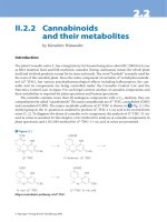
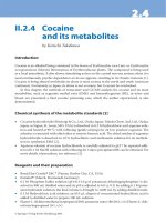
The route of synthesis of YL529 (N-methyl-4-(4-(3(trifluoromethyl)benzamido) phenoxy)picolinamide4methylbenzenesulfonate) was provided by State Key
Laboratory of Biotherapy, Sichuan University (Sichuan,
China), its structural formula is shown in Fig. 1a, YL529
were analyzed and identified by high performance liquid
chromatography (HPLC, Waters, MA, USA) and nuclear
magnetic resonance (NMR). For all in vitro assays, YL529
was prepared as stock solution in dimethyl-sulphoxide
Fig. 1 The chemical structure of YL529 and its interaction with VEGFR3. a The chemical structure of YL529; b and c The interactions between
YL529 and VEGFR3 were shown in 3-D structure and 2-D map
Xu et al. BMC Cancer (2015) 15:525
(DMSO) at final DMSO concentration 0.05 % (V/V) and diluted in the cell culture medium. For in vivo studies, YL529
was dissolved in ultrapure water with 0.5 % sodium carboxymethylcellulose (CMC-Na) combined with 2 % Tween-20,
then administered by oral gavages at 10 ml/kg/day.
Homology modeling and molecular docking methods
The sequence of human VEGFR3 (Genbank accession
number: AAO89505.1) was extracted from the NCBI protein sequence database. The crystal structure of VEGFR2
(PDB code: 4ASD) was used as a template for human
VEGFR3 homology modeling; the sequence identity between VEGFR2 and VEGFR3 is larger than 70 %. The
homology model of VEGFR3 was generated using the
MODELER program implemented in Discovery Studio
(DS) 3.1 software package.
The established VEGFR3 homology model was then used
for the following docking study. A sphere containing the
key residues in VEGFR3 (including CYS930, ALA877,
VAL859, PHE929, LEU1044, LYS879, ASP1055, CYS1054,
VAL910, GLU896, ILE899, and LEU900) was defined as
the binding site. GOLD 5.0 was used for molecular docking since it was an excellent docking program. Gold Score
was selected as the score function; number of dockings
was set as 30; and the other parameters were set as default. The docking results were shown in Fig. 1b and c.
Antibody and reagents
The primary antibodies VEGF-D, LYVE-1, β-actin were
purchased from Santa Cruz Biotechnology (Santa Cruz,
CA); VEGFR-3/phospho-VEGFR-3, JNK phospho-JNK,
Bcl-2 and Bax antibodies were purchased from Cell
Signaling Laboratories (Beverly, MA). The terminal
deoxynucleotidyl transferase mediated nick-end labeling (TUNEL) assay kit was purchased from Promaga
Company (Madison, WI). DMSO, Tween-20, Gelatin and
CMC-Na were purchased from Sigma Chemical Co, (St.
Louis, MO); Matrigel was purchased from BD Pharmingen
(La Jolla, CA).
Cell culture
Human lymphatic endothelial cells (HLECs) were purchased from the ScienCell™ Research Laboratories and
cultured in endothelial cell medium (ECM) containing
5 % fetal bovine serum (FBS), 1 % endothelial cell growth
supplement (ECGS), 100 IU/mL penicillin and 100 μg/mL
streptomycin. Lewis lung carcinoma LL/2 cells were obtained from American Type Culture Collection (ATCC)
and cultured in DMEM containing 10 % FBS, 100 IU/mL
penicillin and 100 μg/mL streptomycin. Subcultures were
performed with trypsin-EDTA. All cells were incubated in
an atmosphere of 5 % CO2 at 37 °C.
Page 3 of 12
The establishment of VEGF-D high-expressing Lewis lung
cell lines
The transfected murine LL/2 Lewis lung carcinoma cells
with the pcDNA3.1 (+) expression vector containing mouse
VEGF-D (VEGF-D-LL/2 cells) or pcDNA3.1 (vector alone)
were transfected as reported before obtained in our laboratory [20–22]. The cell lines were maintained in an
atmosphere of 5 % CO2 at 37 °C in cell culture medium
DMEM supplemented with 10 % fetal bovine serum.
Western blot analysis was performed to analyze the expression of recombinant VEGF-D of these cells.
Cell viability assay
Cell viability assays were performed using CCK-8 kit [23].
Briefly, VEGF-D-LL/2 cells, parental LL/2 cells and HLECs
were treated with various concentrations of YL529 in
96-well culture plates for 48 h. CCK-8 was added to the
cells and the plate was incubated for an additional 2–4 h.
The optical density (OD) was then measured at 450 nm
using a Spectra MAX M5 microplate spectrophotometer
(Molecular Devices, Sunnyvale, CA).
Scratch-induced migration assay
The anti-metastasis effect of YL529 was determined using
a scratch-induced cell migration assay in vitro [24, 25].
Briefly, 1 × 10⁵ HLECs were plated in 6 well plates and
synchronized by serum-free ECM medium for 8 h, then
used a micropipette tip to create a 2 mm-wide linear gap.
Cells were washed to remove non-adherent cells and
further incubated with fresh ECM medium containing
or lacking YL529. An inverted microscope (Carl Zeiss,
Germany) was used to photograph after YL529 treatment
for another 48 h.
Transwell migration assay
Transwell migration assay was adopted to indicate the
anti-metastasis effect of YL529 [26]. Briefly, the transwell
were pre-coated with 25 % Matrigel Matrix containing
growth factors (BD Biosciences, Bedford, MA) for 30 min
at 37 °C. And then the bottom chambers were filled with
600 μl ECM medium and the top chambers were seeded
with 4 × 10⁴ cells HLECs (100 μl/well). The top and bottom chambers were incubated for another 24 h. Then cells
on the top surface of the membrane were scraped with a
cotton swab. Cells on the bottom side of the membrane
were fixed with 4 % paraformaldehyde and stained with
0.1 % crystal violet (Sigma-Aldrich, USA). An inverted
microscope (Zeiss, Axiovert 200, Germany) was used to
obtain the images and the invading cells were quantified.
Tube formation assay
The antilymphogenic effects of YL529 were analyzed
in vitro using a tube-formation assay in HLECs. Briefly,
96-well plate was pre-incubated with 100 μl per well of
Xu et al. BMC Cancer (2015) 15:525
Matrigel Matrix at 37 °C for 30 min, HLECs were seeded
at a density of 3 × 10⁴ cells per well in ECM medium.
And after cultured in the presence or absence of designed
concentrations of YL529 on Matrigel Matrix at 37 °C another 6 h, tube formation by endothelial cells was evaluated and photographed under an inverse microscope
(Zeiss, Axiovert 200, Germany).
Western blot analysis
Standard western blot analysis was performed [23]. Briefly,
Cell lysates were washed with phosphate buffered saline
(PBS) and lysed in RIPA (radioimmunoprecipitation assay)
buffer. Then lysates were centrifuged at 12000 g for
30 min at 4 °C. The Bio-Rad Protein Assay kit (Bio-Rad
Laboratories) was used to determine the samples protein concentration according to the manufacturers’ recommendations. The lysates were dissolved in 5 × SDS
sample buffer and denatured, then subjected to 6 % to
12 % SDS-PAGE (sodium dodecyl sulfate polyacrylamide
gel electrophoresis) according to molecular weight and
transferred onto PVDF (polyvinylidene fluoride) (Bio-Rad,
Hercules, CA) membranes. Membranes were blocked for
1 h in 5 % dried milk in TBS/T at room temperature and
incubated overnight at 4 °C with the primary antibodies
and horseradish peroxidase-conjugated secondary antibodies. Protein bands were visualized with enhanced
chemiluminescent substrate (Amersham Biosciences Corp.,
Piscataway, NJ).
Pharmacokinetic characteristics analyses of YL529 in mice
C57 BL/6 mice (n = 3 per time point) were administered
150 mg/kg YL529 orally. Blood samples of the mice were
collected at appropriate intervals and the plasma concentration of YL529 was analyzed by HPLC (Waters,
MA, USA). The pharmacokinetic characteristics and parameters were analyzed using Pharmacokinetic Software
of Drug and Statistics (DAS, edited and published by the
Mathematical Pharmacology Professional Committee of
China, Shanghai, China).
Effect of YL529 on lymph metastasis in mice syngeneic
models
Seven-week-old female C57 BL/6 mice (Beijing animal
center, Beijing, China) were used. VEGE-D-LL/2 cells
(1 × 10⁶) were injected intramuscularly and subcutaneously in hind limb of mice. The mice were randomized
into 5 groups (n = 10 per groups) and orally administered
37.5, 75 and 150 mg/kg/day YL529, vehicle and saline
alone (N.S.), respectively for 14 days when tumors became
visible the 10th day after cells were implanted. Tumor
growth and mice weight was monitored every 3 days.
When animals were sacrificed with CO2 gas at the end
of drug administration, tumor tissues, lung organs and
local lymph nodes were removed, weighted and calculated
Page 4 of 12
by manual. Tumor size was determined by measuring
the largest and perpendicular diameters every 3 days,
tumor volume was calculated using the formula: volume
(mm3) = 0.5 × length × width2. In addition, the same subcutaneously tumor models were used to monitor the survival time of experimental mice after drug administration.
All animals experiments performed were in accordance
with the guidelines of the institute’s Animal Care and Use
Committee of Sichuan University (Chengdu, China).
Histological analysis for tumor tissue
Tumor tissues from vehicle and 150 mg/kg YL529 groups
were fixed in 4 % paraformaldehyde, dehydrated and
embedded in paraffin [27]. Sections of these tissues were
subsequently incubated with LYVE-1 and corresponding
second antibodies and visualized using peroxidase-DAB.
TUNEL staining was also performed for fixed tissues
(Promega, USA). Quantification was done as described
[28]. Briefly, the microvessel counting was done in representative 200× fields or three high-power (400×) fields
of the highest vascular density.
Safety profile of YL529 in vivo
To evaluate the safety profile of YL529 (150 mg/kg) in vivo,
we have observed the gross measures such as weight loss,
life span, behavior and feedings. Moreover, we have also
investigated the mortality and clinical signs of C57BL/6
mice throughout oral administration period. In addition,
blood samples and heart, liver, spleen, lung, and kidney
tissues of mice were collected; the histopathological,
serum biochemistry and hematological analysis were done
to observe the possible pathological changes. In addition,
we also evaluated the safety profiles of YL529 after oral
administration with high dose (6000 mg/kg) for 14 days.
Statistical analysis
Data was expressed as mean ± SD/SEM. SPSS (SPSS, IL,
USA) is used for statistical analysis. Statistical differences were
considered significant when p < 0.05. Survival curves were
constructed according to the Kaplan-Meier method, and
the survivals were compared by means of the log-rank test.
Results
Molecular modeling and kinase inhibition profile of YL529
The novel multi-kinase small-molecule inhibitor YL529
have been identified by the computer-aided drug design
(CADD), chemical synthesis and high-throughput screening (HTS) methods in our lab, and in vitro kinase binding
assay showed that YL529 inhibited VEGFR2 activity at
10 μM,and YL529 significantly inhibited VEGFR-3 activity
by 97 % at the same concentration [18]. In this study, we
used computer simulation and computer-based molecular
docking methods to further explore the interaction modes
of YL529 with the kinase domain of VEGFR-3, and Fig. 1
Xu et al. BMC Cancer (2015) 15:525
depicts a possible binding configuration of YL529 with
VEGFR-3. Based on these studies, we can see that the
N-methylpicolinamide of YL529 forms strong hydrogenbond interactions with the CYS930 residue in the hinge
region of VEGFR-3. The N atom in amide moiety of
YL529 forms another important hydrogen-bond interaction with the GLU896 in the DFG region of VEGFR-3.
Additionally, YL529 also forms pi-related interactions
with ASP1055, LYS879 and VAL859, consistent with
what we have previously observed in YL529-VEGFR-2
interaction [18].
Page 5 of 12
YL529 inhibits proliferation, migration, invasion, and tube
formation of HLECs in vitro
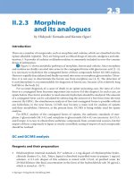
CCK-8 assay was used to evaluate the potential proliferation inhibition effect of HLECs after treated with YL529
or vehicle for 48 h. Our data showed that YL529 inhibited
the proliferation of HLECs, with IC50 value of 5.5 μM.
We next examined the effects of YL529 on HLECs migration using a wound healing migration assay [19]. The
results showed that YL529 markedly decreased the number of migrating HLECs, as shown in Fig. 2a. At 2.5 μM,
YL529 could inhibit the migration of cells by 44.3 %, and
Fig. 2 The effects of YL529 on HLECs. a Wound assay. Cells were wounded and migrated cells were quantified by manual counting; b Transwells
assay. Invaded cells were stained and quantified; c Tube formation assay. 100×, Mean ± SEM, n = 3, *p < 0.05, **p < 0.01, ***p < 0.001
Xu et al. BMC Cancer (2015) 15:525
Page 6 of 12
when the concentrations increased to 5 μM or 10 μM,
the inhibition of cells migration was increased to 66.8 %
or 79.8 %, respectively.
To further measure the effect of YL529 on HLECs
invasion, we used a transwell assay and measured the
number of HLECs that passed through a membrane barrier following treatment with various concentrations of
YL529 [29]. As shown in Fig. 2b, compared with vehicle,
2.5 μM YL529 could inhibit the invasion of cells by
28.0 %, and when concentrations of YL529 increased to
5 μM or 10 μM, the inhibition of cell invasion was increased to 65.9 % or 90.2 %, respectively.
To further understand the mechanism of the antilymphogenic effect of YL529, the antilymphogenic tube
formation effect of YL529 was analyzed with cultured
HLECs in vitro [30]. As shown in Fig. 2c, YL529 treatment at 10 μM for 6 h strongly inhibited the formation
of tube-like structures. Quantification showed that YL529
treatment inhibited tube formation by 85.7 %. In great
contrast, HLECs without YL529 treatment spread and
aligned with each other and formed a rich meshwork of
branching capillary-like tubules within 6 h (Fig. 2c).
YL529 inhibited the proliferation of VEGF-D-LL/2 cells
VEGF-D-LL/2 cells were previously established by transfection with the pcDNA3.1 (+) expression vector containing mouse VEGF-D [31]. Similar to the previous data
from our group, this study uses a VEGF-D antibody that
detects the mature form of VEGF-D, because the fully
mature cleaved form of 21 kD has the greatest affinity
for the receptors, and can bind and activate not only
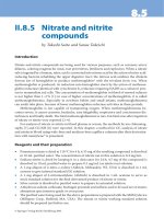
VEGFR-2 but also VEGFR-3. As shown in Fig. 3a, western
blot analysis showed that the expression level of VEGF-D
(21 kD) in VEGF-D-LL/2 cells was higher than the cells
that were transfected with null-vectors (pcDNA-LL/2).
This result confirmed that VEGF-D protein was constitutively up-regulated in VEGF-D-LL/2 cells. When VEGFD-LL/2 cells and parental LL/2 cells were treated with
YL529 or vehicle for 48 h, the calculated corresponding
IC50 value was 7.2 μM and 9.6 μM, respectively. And we
found that YL529 potently inhibited the proliferation of
VEGF-D-LL/2 cells,
Potential molecular mechanism of the effects of YL529 on
HLECs and VEGF-D-LL/2 cells
To gain in-depth insight into the molecular mechanism
of anti-lymphangiogenesis effects of YL529, the expression levels of VEGFR-3/p-VEGFR-3 in HLECs were analyzed by western blotting in vitro. As shown in Fig. 3b,
in HLECs, YL529 potently inhibited the phosphorylation
of VEGFR-3 after 2 h of treatment, even though no change
of the total expression of VEGFR-3 was observed. This
data may provide a molecular mechanism of the previously
observed specific effects of YL529 on HLECs.
Fig. 3 Western blotting to probe the molecular mechanisms of
effects of YL529 in vitro. a p-VEGFR3 in HLECs was detected after
YL529 treatment by western blotting analysis. b The overexpression
of VEGF-D in VEGF-D-LL/2 cells was confirmed by western blotting
analysis. The molecular weight of full-length VEGF-D was 21 KD.
c VEGF-D-LL/2 cell were treated with YL529, p-JNK/JNK, Bax, Bcl-2
were analyzed by western blotting analysis
Moreover, as shown in Fig. 3c, YL529 decreased the expression level of p-JNK and Bax while increased the expression level of Bcl-2 in VEGF-D-LL/2 cells, which may explain
the observed effects of YL529 on VEGF-D-LL/2 cells.
Pharmacokinetics of YL529 in vivo
To determine the pharmacokinetic characteristics of YL529
in mice, C57 BL/6 mice were treated with YL529 orally and
the key pharmacokinetic parameters of YL529 were summarized in Table 1, i.e., after oral administration of YL529
at 150 mg/kg, the peak plasma concentration (Cmax)
was 18.03 μg/ml, the time-to-peak concentration (Tmax)
was 2 h, the half-life (t1/2z) was 4.11 h and the AUC0→∞
Xu et al. BMC Cancer (2015) 15:525
Page 7 of 12
Table 1 Pharmacokinetic parameters of YL529 after oral
administration with single dose of 150 mg/kg in mice
Pharmacokinetic parameter
Value
AUC(0-∞)(mg/L*h)
170.95
AUC(0-t)(mg/L*h)
167.32
t1/2β(h)
6.03
T1/2α(h)
1.39
t1/2z(h)
4.11
Cmax(mg/L)
Tmax(h)
18.03
2.00
Data were expressed as the mean ± SD compared with vehicle. SPSS (SPSS, IL)
software was used for statistical analysis (n = 3; *p < 0.05)
was 170.95 mg/L · h, which suggested that the oral absorption and bioavailability and the pharmacokinetics
of YL529 in mice are highly desirable.
YL529 inhibited tumor growth in VEGF-D-LL/2 tumors
syngeneic models and extend the life span of affected
mice
Based on the safety profile and the in vivo pharmacokinetics of YL529, chronic oral administration of YL529 at
daily dosages of 37.5 ~ 150 mg/kg/day was chosen to treat
the mice with VEGF-D-LL/2 tumors (syngeneic s.c. and
muscle models). We found that YL529 inhibited tumor
growth in dose-dependent manner in both models.
Specifically, in s.c. model, as shown in Fig. 4a, there
was a remarkable tumor volume reduction (66.22 %),
Fig. 4 Effect of YL529 on tumor growth and survival in s. c. model. The mice bearing tumors subcutaneously were treated with YL529 or controls
orally. a YL529 resulting in significant tumor growth inhibition; b Recovered tumor weights were significantly smaller in YL529 treated animals;
c There was no significant difference of body weight after YL529 treated; d A significant increase in survival in YL529 treated mice was observed
(Log-rank). Mean ± SEM, n = 10, *p < 0.05, **p < 0.01
Xu et al. BMC Cancer (2015) 15:525
compared with the vehicle-treated group, and the final
tumor weights showed similar pattern (Fig. 4b), while
there was no loss of body weight at this dosage (Fig. 4c).
Page 8 of 12
Furthermore, survival experiment in vivo showed that
six out of ten animals in N.S. and vehicle groups died at
29 days (Mice were regarded as sacrificed when the
Fig. 5 Effects of YL529 on tumor growth, lung and lymph node metastasis in muscle model. The muscle metastasis models were treated with
YL529 or controls orally. a YL529 resulting in significant tumor growth inhibition; b There was no significant difference of body weight after
YL529 treated; c and d Percentage of mice with lung and lymph node metastasis; e Mean volume of auxiliary lymph nodes were harvested from
mice. Mean ± SEM, n = 10, *p < 0.05, **p < 0.01
Xu et al. BMC Cancer (2015) 15:525
Page 9 of 12
Table 2 The organs parameters of YL529 after oral administration
6000 mg/kg YL529 with a single dose in SD rats
Index
Vehicle
YL529
Vehicle
YL529
Female(♀)
Female (♀)
Male (♂)
Male (♂)
Heart (%)
0.41 ± 0.08
0.42 ± 0.09
0.43 ± 0.05
0.42 ± 0.06
Liver (%)
3.11 ± 0.10
3.13 ± 0.09
3.09 ± 0.10
3.09 ± 0.13
Spleen (%)
0.23 ± 0.03
0.25 ± 0.03
0.23 ± 0.09
0.24 ± 0.04
Lung (%)
0.43 ± 0.05
0.43 ± 0.06
0.44 ± 0.11
0.42 ± 0.09
Kidney (%)
0.37 ± 0.06
0.38 ± 0.09
0.36 ± 0.08
0.38 ± 0.09
Data were expressed as the mean ± SD compared with vehicle. SPSS (SPSS, IL)
software was used for statistical analysis (n = 10; *p < 0.05)
diameter of tumors reached about 20 mm), while in
YL529 treated groups, the life span of the mice have
been significantly extended (Fig. 4d, *p < 0.05, by logrank test), i.e., 90 % of mice survived to 40 days and
60 % of mice in 150 mg/kg group survived to 54 days
when we finished the survival observation.
Similar results were observed in the muscle model
(Fig. 5a) (*p < 0.05). Moreover, it is worthy to stress that
YL529 also significantly reduced the number of mice
with lung metastasis and inguinal lymph node metastasis
in the tumor muscle model, while there was no loss of
body weight at this dosage at the end of drug administration (Fig. 5b). similarly, as shown in Fig. 5c, d and e
(*p < 0.05, **p < 0.01), the percent of mice with local
lymph node metastasis in vehicle group could reach
80 % ~ 90 %, while reaching only 50 % in 37.5 mg/kg
group and 20 % in 150 mg/kg group, respectively.
Safety profile of YL529 in a preclinical study
Currently, side effects are the prevalent shortcomings of
anticancer drugs. In our current study, mice treated with
YL529 at the daily dosage of 150 mg/kg for 18 days
showed no obvious body weight loss or tissue damage,
which is consistent with our previous reported [18]. Furthermore, we further evaluated the toxicity in SD rats
treated by YL529 at the daily dosage of 6000 mg/kg for
14 days, and we found that there were no obvious weight
changes of major organs (heart, liver, spleen, lung, kidney
and brain) (Table 2), or obvious pathological damage, or
other adverse effects (Data not shown).
YL529 inhibited lymphangiogenesis in tumor tissues in
additional to induce tumor cell apoptosis in vivo
The immunohistochemical analysis and TUNEL apoptosis assays and LYVE-1 staining were performed to directly evaluate whether YL529 could inhibit the tumor
lymphangiogenesis and induce tumor cell apoptosis in vivo.
As shown in Fig. 6a, YL529 remarkably decreased the
amount of lymphangiogenesis in tumor tissues at 150 mg/
kg for 14 days in VEGF-D-LL/2 tumor syngeneic model, as
indicated by the LYVE-1 staining (34.71 % higher than the
vehicle group in VEGF-D-LL/2 muscle model). Moreover, TUNEL assay found that there were a dramatic higher
Fig. 6 Immunohistochemical analysis of LYVE-1 and TUNEL assay in vivo. Sections from vehicle group and 150 mg/kg YL529-treated tumor were
collected. (a) LYVE-1 and (b) TUNEL were detected in the VEGF-D-LL/2 tumor model. Quantitative of the mean LYVE-1 and TUNEL-positive area
counted at × 200. Mean ± SEM, 200×, n = 6, *p < 0.05, **p < 0.01
Xu et al. BMC Cancer (2015) 15:525
number of apoptotic tumor cells in the treated group
(20-fold increase vs. vehicle), in s.c. model (Fig. 6b).
Discussion
Metastasis, particularly via the lymphatic system, is a
common prognostic factor and critical process in the
spread of solid cancer [2]. The VEGF/VEGFR pathway has
been widely studied because VEGFR expression is strongly
correlated with tumor metastasis progression and poor
prognosis. Therefore, this pathway has been pursued as a
therapeutic strategy for inhibition of lymphangiogenesis
and metastatic in tumors,and some animal studies have
shown that the expression of VEGF-D promotes lymphangiogenesis and metastatic spread of tumor cells via
lymphatic in papillary thyroid carcinoma, lung cancer
and gastric cancer [32–34]. However, most studies are
based on cultured tumor cells. Little in vivo research
has been done to correlate the high VEGF-D levels and
lymph node metastasis in lung carcinoma, thus, it remains
unclear whether expression of VEGF-D in culture accurately represents the in vivo situation.
Furthermore, since VEGF-D plays a remarkable role in
up-regulating lymphangiogenesis and regional lymph node
metastasis, other researchers in our group has established
the mouse lymph node metastasis model by transfecting
high expression VEGF-D into LL/2 Lewis lung carcinoma
cells, and found that VEGF-D mainly bind to VEGFR-3 in
lymphatic endothelial cells in vivo [31], and further study
demonstrated that VEGFR-3 contributes to autocrine effects through VEGF-D-LL/2 cells [20].
YL529 was developed as a potential multikinase anticancer agent in our laboratory using CADD, HTS, de
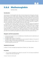
Fig. 7 The proposed underlying mechanisms of YL529
Page 10 of 12
novo synthesis and has been reported to have a variety of
pharmacological effects such as inhibiting tumor growth
via antiangiogenesis and anti-proliferation in our previously study, especially it could block the activities of human umbilical vein endothelial cells (HUVEC) stimulated
with VEGF165 previously [18]. And some researchers have
reported that VEGFR-2 can also be expressed on lymphatic endothelial cells, while its expression levels were less
than VEGFR-3 in lymphatic ECs, because VEGFR2 is
the major effector of angiogenesis and regulates blood
vessel growth of vascular ECs, not lymphatic ECs [35–37].
However, it is still possible that some of its inhibitory activities can be assigned to VEGFR-2, because YL529 is a
multikinases inhibitor.
To further address some caveats, we determined that
YL529 inhibits the proliferation and migration of highexpressing VEGF-D Lewis lung carcinoma LL/2 cells. Similar effects of YL529 were also observed in HLECs, which
endogenously express high level of VEGFR-3. Consistent
with this idea, we found that the anti-proliferation effect of
YL529 was dependent on high VEGF-D expression, since
YL529 has no as obvious effect on the parental LL/2
cells, which has low level of endogenous VEGF-D expression (Data not shown). Interestingly, high expression of
VEGF-D has been found to correlate with the up-regulation
of Fra-1, a member of the AP-1 family of transcription factors [38]. And AP-1 interacts with Fos and Jun protein family members, and the latter is regulated by ERK1/2 and
JNKs, respectively.
Mechanistically, our result showed that YL529 downregulation of p-VEGFR-3 in HLECs cell in a dose-dependent
manner, which is consistent with the computer-aided drug
Xu et al. BMC Cancer (2015) 15:525
design (CADD) and molecular reverse docking results.
Furthermore, we found that YL529 also inhibited the
phosphorylation of JNK1/2, while the total protein expression of JNK1/2 was not changed. Along this line,
YL529 can also decrease the expression level of Bax and
upregulate Bcl-2, which supported the idea of a potential
pro-apoptotic effect of YL529. Taken together, it is reasonable to propose that YL529 could not only downregulated the levels of p-VEGFR-3, but also blocked the
VEGFR-3 signaling pathway by interfering with the expression levels of p-JNK and induced apoptosis via
down-regulating Bax and up-regulating Bcl-2.
These results are further supported by the fact that
YL529 inhibited the tumor-associated lymphangiogenesis
and metastasis in mice harboring VEGF-D-LL/2 Lewis
carcinoma in vivo, and by immunohistochemical staining
showed that YL529-treated tumors displayed less LYVE-1
positive vessels compared with vehicle.
Our data has confirmed that YL529 exhibits the characteristics of blocking tumor metastasis at several different
time points in the process of tumor lymphangiogenesis for
the first time: (I) YL529 inhibits proliferation and induces
apoptosis of in high expression VEGF-D tumor cells;
(II) YL529 downregulates the expression of VEGFR-3 on
HLECs and blockades the VEGFR-3 signaling pathways by
interfering with the activation of JNK1/2; (III) YL529 interrupts and inhibits HLECs proliferation, invasion and
tube formation; (IV) YL529 inhibits the tumor-associated
lymphangiogenesis; (V) YL529 suppresses tumor cell migration and induces apoptosis. Moreover, there is a closely
associated between lymph node metastasis and VEGFR-3
expression in several lewis lung carcinoma models. The
binding of VEGF-D and VEGFR-3 could promote migration and proliferation of HLECs through JNKs signaling
pathways. So blockade of the VEGFR-3 pathways could efficiently inhibit tumor lymphangiogenesis and metastasis.
These results support the hypothesis that the therapeutic
effect of YL529 on tumor lymphangiogenesis and metastasis could be attributed to direct inhibition of lymphangiogenesis in lung tumor models induced by VEGF-D and
through down-regulation of VEGFR-3.
Conclusions
In conclusion, our results indicated that YL529 significantly inhibited the tumor-associated lymphangiogenesis
and metastasis induced by VEGF-D in the established
VEGF-D over-expressing Lewis lung carcinoma model
(Fig. 7). Tumor growth was significantly retarded and
prolonged life span was observed after YL529 treatment
in tumor-bearing mice. We have demonstrated for the
first time that YL529 suppresses tumor lymphangiogenesis
and lymphatic metastasis by down-regulation of VEGF-D
in VEGF-D-LL/2 cancer cells and inhibiting neogenesis
and tube formation of lymphatic endothelial cells directly
Page 11 of 12
through the VEGFR-3 pathway. Our findings suggest that
YL529 may be an attractive agent against lymphangiogenesis and metastasis. Moreover, YL529 has a novel chemical
structure that is different from VEGFR-3 inhibitors in
clinical use. In addition, YL529 was well tolerated by the
host animal in therapeutically beneficial doses, making it a
promised candidate for phase I clinical trials as an antilymphatic metastasis drug.
Abbreviations
HLECs: Human lymphatic endothelial cells; VEGF: Vascular endothelial growth
factor; VEGF-C/D: Vascular endothelial growth factor-C/D; HUVECs: Human
umbilical vein endothelial cells; HPLC: High performance liquid chromatography;
NMR: Nuclear magnetic resonance; DMSO: Dimethyl-sulphoxide;
CMC-Na: Carboxymethylcellulose; ECM: Endothelial cell medium; FBS: Fetal
bovine serum; ATCC: American type culture collection; OD: Optical density;
PBS: Phosphate buffered saline; RIPA: Radioimmunoprecipitation assay;
PVDF: Polyvinylidene fluoride; SDS-PAGE: Sodium dodecyl sulfate
polyacrylamide gel electrophoresis; Cmax: The peak plasma concentration;
Tmax: The time-to-peak concentration; t1/2z: The half-life; TUNEL: Terminal
deoxynucleotidyl transferase mediated nick-end labeling.
Competing interests
The authors declare that they have no competing interests.
Authors’ contributions
YZ X, WJ L, P Y, W P, CT W, YL Z, LT Y and SY Y were responsible for
experimental design, interpretation of the results and writing the manuscript.
WJ L, YZ X, ML L, Y L and GB L performed the experimental procedures. YZ
X, WJ L, P Y, NN M, SY W and HJ L were responsible for the analysis of the
data. LX K provided purely writing assistance. All authors read and approved
the manuscript.
Acknowledgements
This work was supported by National Science & Technology Major Project
(2011ZX09102-001-013 and 2012ZX09501), National Natural Sciences
Foundation of China (81272459 and 81402947), Natural Sciences Foundation
of Anhui Province (1508085QH162), and the Grants for Scientific Research of
BSKY from Anhui Medical University (XJ201315).
Author details
1
Department of Pathophysiology, School of Basic Medicine, Anhui Medical
University, 81#, Mei Shan Road, Hefei 230032, China. 2State Key Laboratory of
Biotherapy and Cancer Center, West China Hospital, West China Medical
School, and Collaborative Innovation Center for Biotherapy, Sichuan
University, 17#, 3rd Section, Ren min South Road, Chengdu 610041, China.
3
Department of Transfusion, the First Affiliated Hospital of Anhui Medical
University, 81#, Mei Shan Road, Hefei 230032, China. 4Department of
Oncology, The People’s Hospital of Guizhou Province, 83#, Zhong Shan East
Road, Guiyang 550004, China.
Received: 13 October 2014 Accepted: 19 May 2015
References
1. Hanahan D, Weinberg RA. Hallmarks of cancer: the next generation. Cell.
2011;144:646–74.
2. Stacker SA, Williams SP, Karnezis T, Shayan R, Fox SB, Achen MG.
Lymphangiogenesis and lymphatic vessel remodelling in cancer. Nat Rev
Cancer. 2014;14:159–72.
3. Stacker SA, Caesar C, Baldwin ME, Thornton GE, Williams RA, Prevo R, et al.
VEGF-D promotes the metastatic spread of tumor cells via the lymphatics.
Nat Med. 2001;7:186–91.
4. Karaman S, Detmar M. Mechanisms of lymphatic metastasis. J Clin Invest.
2014;124:922–8.
5. Cao Y. Emerging mechanisms of tumour lymphangiogenesis and lymphatic
metastasis. Nat Rev Cancer. 2014;5:735–43.
6. Alitalo A, Detmar M. Interaction of tumor cells and lymphatic vessels in
cancer progression. Oncogene. 2014;31:4499–508.
Xu et al. BMC Cancer (2015) 15:525
7.
8.
9.
10.
11.
12.
13.
14.
15.
16.
17.
18.
19.
20.
21.
22.
23.
24.
25.
26.
Baldwin ME, Halford MM, Roufail S, Williams RA, Hibbs ML, Grail D, et al.
Vascular endothelial growth factor D is dispensable for development of the
lymphatic system. Mol Cell Biol. 2005;25:2441–9.
Smith NR, Baker D, James NH, Ratcliffe K, Jenkins M, Ashton SE, et al. Vascular
endothelial growth factor receptors VEGFR-2 and VEGFR-3 are localized
primarily to the vasculature in human primary solid cancers. Clin Cancer
Res. 2010;16:3548–61.
Achen MG, Jeltsch M, Kukk E, Mäkinen T, Vitali A, Wilks AF, et al. Vascular
endothelial growth factor D (VEGF-D) is a ligand for the tyrosine kinases
VEGF receptor 2 (Flk1) and VEGF receptor 3 (Flt4). Proc Natl Acad Sci U S A.
1998;95:548–53.
Karnezis T, Shayan R, Caesar C, Roufail S, Harris NC, Ardipradja K, et al. VEGF-D
promotes tumor metastasis by regulating prostaglandins produced by the
collecting lymphatic endothelium. Cancer Cell. 2012;14:181–95.
Zhang L, Zhou F, Han W, Shen B, Luo J, Shibuya M, et al. VEGFR-3 ligandbinding and kinase activity are required for lymphangiogenesis but not for
angiogenesis. Cell Res. 2010;20:1319–31.
Skobe M, Hawighorst T, Jackson DG, Prevo R, Janes L, Velasco P, et al.
Induction of tumor lymphangiogenesis by VEGF-C promotes breast cancer
metastasis. Nat Med. 2007;7:192–8.
Han KY, Chang JH, Dugas-Ford J, Alexander JS, Azar DT. Involvement of
lysosomal degradation in VEGF-C-induced down-regulation of VEGFR-3.
FEBS Lett. 2014;588:4357–63.
Nwogu CE, Yendamuri S, Tan W, Kannisto E, Bogner P, Morrison C, et al. Lung
cancer lymph node micrometastasis detection using real-time polymerase
chain reaction. correlation with vascular endothelial growth factor expression.
J Thorac Cardiov Sur. 2013;145:702–7.
Hecht JR, Trarbach T, Hainsworth JD, Major P, Jäger E, Wolff RA, et al.
Randomized, placebo-controlled, phase III study of first-line oxaliplatin-based
chemotherapy plus PTK787/ZK 222584, an oral vascular endothelial growth
factor receptor inhibitor, in patients with metastatic colorectal adenocarcinoma.
J Clin Oncol. 2011;29:1997–2003.
Podar K, Anderson KC. Emerging therapies targeting tumor vasculature in
multiple myeloma and other hematologic and solid malignancies. Curr
Cancer Drug Targets. 2011;11:1005–24.
Wilhelm SM, Dumas J, Adnane L, Lynch M, Carter CA, Schütz G, et al.
Regorafenib (BAY 73–4506): a new oral multikinase inhibitor of angiogenic,
stromal and oncogenic receptor tyrosine kinases with potent preclinical
antitumor activity. Int J Cancer. 2011;129:245–55.
Xu Y, Lin H, Meng N, Lu W, Li G, Han Y, et al. YL529, a novel, orally available
multikinase inhibitor, potently inhibits angiogenesis and tumor growth in
preclinical models. Brit J Pharmacol. 2013;169:1766–80.
Shibuya M. Vascular Endothelial Growth Factor (VEGF) and Its Receptor (VEGFR)
Signaling in Angiogenesis: A Crucial Target for Anti- and Pro-Angiogenic
Therapies. Genes Cancer. 2011;2:1097–105.
Jiang QQ, Fan LY, Yang GL, Guo WH, Hou WL, Chen LJ, et al. Improved
therapeutic effectiveness by combining liposomal honokiol with cisplatin in
lung cancer model. BMC cancer. 2008;8:242.
Hu J, Ye H, Fu A, Chen X, Wang Y, Chen X, et al. Deguelin–an inhibitor to
tumor lymphangiogenesis and lymphatic metastasis by downregulation of
vascular endothelial cell growth factor-D in lung tumor model. Int J Cancer.
2010;127:2455–66.
Hirakawa S, Brown LF, Kodama S, Paavonen K, Alitalo K, Detmar M. VEGF-Cinduced lymphangiogenesis in sentinel lymph nodes promotes tumor
metastasis to distant sites. Blood. 2007;109:1010–7.
Xu YZ, Zheng RL, Zhou Y, Peng F, Lin HJ, Bu Q, et al. Small molecular
anticancer agent SKLB703 induces apoptosis in human hepatocellular
carcinoma cells via the mitochondrial apoptotic pathway in vitro and
inhibits tumor growth in vivo. Cancer Lett. 2011;313:44–53.
Liang S, Fu A, Zhang Q, Tang M, Zhou J, Wei Y, et al. Honokiol inhibits
HepG2 migration via down-regulation of IQGAP1 expression discovered by
a quantitative pharmaceutical proteomic analysis. Proteomics. 2011;10:1474–83.
Goldberg L, Kloog Y. A Ras Inhibitor Tilts the Balance between Rac and Rho
and Blocks Phosphatidylinositol 3-KinaseaDependent Glioblastoma Cell
Migration. Cancer Res. 2006;66:11709–17.
Agarwal A, Covic L, Sevigny LM, Kaneider NC, Lazarides K, Azabdaftari G, et al.
Targeting a metalloprotease-PAR1 signaling system with cell-penetrating
pepducins inhibits angiogenesis, ascites, and progression of ovarian cancer.
Mol Cancer Ther. 2008;7:2746–57.
Page 12 of 12
27. Liu JY, Wei YQ, Yang L, Zhao X, Tian L, Hou JM, et al. Immunotherapy of
tumors with vaccine based on quail homologous vascular endothelial
growth factor receptor-2. Blood. 2003;102:1815–23.
28. Mikalsen LT, Dhakal HP, Bruland OS, Nesland JM, Olsen DR. Quantification of
angiogenesis in breast cancer by automated vessel identification in CD34
immunohistochemical sections. Anticancer Res. 2011;31:4053–60.
29. Petrovic N, Schacke W, Gahagan JR, O’Conor CA, Winnicka B, Conway RE,
et al. CD13/APN regulates endothelial invasion and filopodia fonnation.
Blood. 2007;110:142–50.
30. Hertel J, Hirche C, Wissmann C, Ebert MP, Höcker M. Transcription of the
vascular endothelial growth factor receptor-3 (VEGFR3) gene is regulated
by the zinc finger proteins Sp1 and Sp3 and is under epigenetic control:
transcription of vascular endothelial growth factor receptor 3. Cell Oncol
(Dordr). 2014;37:131–45.
31. Wen J, Fu AF, Chen LJ, Xie XJ, Yang GL, Chen XC, et al. Liposomal honokiol
inhibits VEGF-D-induced lymphangiogenesis and metastasis in xenograft
tumor model. Int J Cancer. 2009;124:2709–18.
32. Yasuoka H, Nakamura Y, Zuo H, Tang WH, Takamura Y, Miyauchi A, et al.
VEGF-D expression and lymph vessels play an important role for lymph node
metastasis in papillary thyroid carcinoma. Modern pathol. 2005;18:1127–33.
33. Matsumoto M, Roufail S, Inder R, Caesar C, Karnezis T, Shayan R, et al. Signaling
for lymphangiogenesis via VEGFR-3 is required for the early events of
metastasis. Clin Exp Metastasis. 2013;30:819–32.
34. Zhao YC, Ni XJ, Wang MH, Zha XM, Zhao Y, Wang S. Tumor-derived VEGF-C,
but not VEGF-D, promotes sentinel lymph node lymphangiogenesis prior to
metastasis in breast cancer patients. Med Oncol. 2012;29:2594–600.
35. Cleaver O, Melton DA. Endothelial signaling during development. Nat Med.
2003;9:661–8.
36. McCarty M, Wey J, Stoeltzing O, Liu W, Fan F, Bucana C, et al. ZD6474, a
vascular endothelial growth factor receptor tyrosine kinase inhibitor with
additional activity against epidermal growth factor receptor tyrosine kinase,
inhibits orthotopic growth and angiogenesis of gastric cancer. Mol Cancer
Ther. 2004;3:1041–8.
37. Gaengel K, Genove G, Armulik A, Betsholtz C. Endothelial-mural cell signaling
in vascular development and angiogenesis. Arterioscl Throm Vas Biol.
2009;29:630–8.
38. Debinski W, Slagle-Webb B, Achen MG, Stacker SA, Tulchinsky E, Gillespie GY,
et al. VEGF-D is an X-linked/AP-1 regulated putative onco-angiogen in human
glioblastoma multiforme. Mol Med. 2001;7:598–608.
Submit your next manuscript to BioMed Central
and take full advantage of:
• Convenient online submission
• Thorough peer review
• No space constraints or color figure charges
• Immediate publication on acceptance
• Inclusion in PubMed, CAS, Scopus and Google Scholar
• Research which is freely available for redistribution
Submit your manuscript at
www.biomedcentral.com/submit