Nghiên cứu bệnh nhiễm độc tố botulin của vi khuẩn clostridium botulinum trên vịt tại một số tỉnh đồng bằng sông cửu long tt tiếng anh
Bạn đang xem bản rút gọn của tài liệu. Xem và tải ngay bản đầy đủ của tài liệu tại đây (636.1 KB, 27 trang )
MINISTRY OF EDUCATION AND TRAINING
CAN THO UNIVERSITY
DOCTOR THESIS SUMMARY
Major: PATHOLOGY AND TREATMENT OF ANIMALS
Code: 62 64 01 02
NGUYEN THU TAM
STUDY ON BOTULIN POISONING DISEASE OF
Clostridium botulinum ON DUCKS IN THE
MEKONG DELTA, VIETNAM
Can Tho- 2020
THIS STUDY WAS ACCOMPLISHED
AT CANTHO UNIVERSITY
SCIENCE INSTRUCTOR: ASSOC. PROF. NGUYEN ĐUC HIEN
The thesis was defended with the doctoral examination
committee at the school level
Place:…………………………………., Can Tho University
At…. …. day …… month ….. year ……….
Opponent 1:
Opponent 2:
Opponent 3:
The thesis could be found at the library:
1. Learning Resource Center, Can Tho University.
2. National Library of Vietnam.
LIST OF PUBLISHED SCIENTIFIC ARTICLES
1. Nguyen Thu Tam, Dang Ngoc Le. 2014. Survey on the leisons in
mice after injected the intestinal suspension originated from the
ducks infected botulin toxin. Can Tho University – Journal of
Science, Agriculture Edition 2014: 107-110.
2. Nguyen Thu Tam, Ly Thi Lien Khai, Nguyen Duc Hien. 2016.
The isolation and identification of Clostridium spp. in the field soil in
Phu Tan and Chau Phu district, An Giang province. Science and
Technology Journal of Agriculture and Rural Development, 11:7377.
3. Nguyen Thu Tam, Tran Thi Phan, Nguyen Duc Hien. 2016. The
isolation and identification of Clostridium botulinum from
limberneck ducks in Tan Phu and Tri Ton district, An Giang
province. Science and Technology Journal of Agriculture and Rural
Development, 11:147-150.
4. Nguyen Thu Tam, Nguyen Duc Hien, Ho Thi Viet Thu. 2016.
Diagnosis of “Cum can” disease on ducks via the experiment in
mice. Can Tho University – Journal of Science, Agriculture Edition
2016: 125-130.
5. Nguyen Thu Tam, Nguyen Duc Hien, Ho Thi Viet Thu. The
isolation and identification of Clostridium botulinum on snails (Pila
conica) and crabs (Somannia theplusa) in Can Tho city, An Giang
and Kien Giang province. Can Tho University – Journal of Science,
Agriculture Edition 2016: 131-134.
Chapter 1 INTRODUCTION
1.1 The imperative of this study
The Mekong Delta area has interlacing river systems,
tropical climate, a wide area of rice cultivation, aquatic animals, and
so on. Therefore, it is convenient for raising free-grazing ducks. The
number of free-grazing ducks is about 31.5 million, got 70% of a
total of ducks in this region as well as 40% of a total of ducks in
Vietnam. The free-grazing method has some benefits such as taking
natural feed or spilled rice after harvesting to significantly reduce
costs in the livestock. However, it has potential risks due to
uncontrol the grazing environment, and diseases can outbreak
severely. Recently, “Limberneck” disease or “Cum Can” (in
Vietnamese) has been one of the common diseases that occurred in
the free-grazing ducks in the Mekong Delta. This disease is in the
waterfowl caused by botulin toxin of Clostridium botulinum; thus, it
is also called the botulism disease.
Clostridium botulinum is completely anaerobic and
producing spores with the oval shapes. This pathogen usually exists
in the soil, especially in the sedimentary mud areas, mollusks
corpses, and in the anaimal intestine. It can produce severe
botulinum neurotoxin to destroy all the central nervous system
(Todar, 2009). Ducks infected with this toxin show some symptoms
such as paralysis of the neck, eyelids, wings, legs, and a high rate of
mortality; it causes a significant loss for the farmers (Rocke and
Friend, 1998).
In human and veterinary medicine, the botulism disease has
been studied in humans, poultry, waterfowl. However, the research
of the botulinum disease as well as risk factors, bio-characteristics of
C. botulinum are limited in the Mekong Delta and also in Vietnam.
Research is necessary to do for supplying information about C.
botulinum and the botulism disease in the free-grazing ducks in the
Mekong Delta. Therefore, the study “Study on botulin poisoning
disease of Clostridium botulinum on ducks in the Mekong Delta”
was carried out.
1.2 The aim of this study
- Determination of the frequency of the botulism disease on
the free-grazing ducks in the Mekong Delta.
1
- Determination of the prevalence of Clostridium botulinum
and the type of botulin toxin on the free-grazing ducks.
- Determination of the prevalence of C. botulinum in the
grazing environment.
- Determination of the pathogenicity of isolated C. botulinum
strains originated in the Mekong Delta.
1.3 The new scientific distributions
- Scientific pieces of evidence firstly about the prevalence of
the botulism disease on the free-grazing ducks in the Mekong Delta.
- Determination of botulin type in the infected ducks in the
Mekong Delta.
- Determination of botulin types of C. botulinum in the
infected ducks in the Mekong Delta
- Application of the mouse bioassay to indicate the botulin
infection due to Clostridium botulinum in ducks in Vietnam.
1.4 The scientific meaning of this study
It is the first study that systematic research about the
botulism disease on the free-grazing ducks in the Mekong Delta,
Vietnam. From those results, a scientific process can be formed for
diagnosing this disease as well as preventing and treatments of the
botulism disease on the free-grazing ducks in the Mekong Delta and
Vietnam.
2
Chapter 3 RESEARCH CONTENTS AND METHODS
3.1 The research materials
3.1.1 The research period and places: This study was carried out
from October 2013 to October 2018.
3.1.2. Research places
3.1.2.1. Sample collected places
Samples were collected in An Giang, Can Tho, Hau Giang, and
Kien Giang province of the Mekong Delta, Vietnam.
3.1.2.2. Samples analysis and the experiment on mice
The isolation, identification and toxin examination of C. botulinum
were done at the Specialized Veterinary laboratory 3, Department of
Veterinary Medicine, College of Agriculture, Can Tho University.
3.1.2.3. The field-experiment
The toxin test of C. botulinum on laying ducks were done at
Vemedim Corporation Company, Thoi Thanh ward, Thoi Lai District,
Can Tho city.
3.2 The research equipments
Equipments and requirements
- The questionnaire (Appendix 1).
- The information was collected from the Statistical Yearbook
and Sub-Department of Animal Health about the total of
poultry/waterfowl population, climate, duck breedings, and diseases
in the sample collection places.
- Microbiology stuffs
Chemicals and media
- Alcohol 96o, alcohol 70o, distiled water, cedar oil, Crystal
violet, Lugol, Safranine, Bromocresole purple, Gelatin Phosphat
Buffer… (Merck, Germany); sheep blood (Nam Khoa, Vietnam).
- Examined antibiotics: amikacin, ampicillin, amoxicillin,
ceftiofur, cephalexin, doxycycline, florfenicol, fosfomycin,
marbofloxacin, norfloxacin (Oxoid, Bristish)
- Mac Farland 0, 5 (Biorad)
- Media: NB (Nutrient broth, Merck, Germany), TSA (Tryptis
Soy Agar, Merck, Germany), CMM (Cooked Meat Media, Oxoid,
Bristish), EYA (Egg Yorlk Agar, Merck, Gemany), Thioglycollate
(Merck, Germany), SFP Agar Base (Difco, USA), MHA (Mueller
Hinton Agar, Merck, Germany).
3
- Carbohydrate tests: Lactose, glucose, maltose, saccarose
(Merck, Germany).
- Biochemical test kit: API 20A (Biorad, USA).
- TPGY broth: 5% Trypticase, 0.5% Pepton, 0.4% Glucose, 2%
Yeast extract, 0.1% Sodium thioglycolate; CMM (Cooked Meat
Medium, Oxoid, Bristish).
- Antitoxins: type C, D, E (10UI/ml) (Statens Serum Institute,
Denmark).
3.3 Research contents
3.3.1. Content 1: The botulism disease on the free-grazing ducks in the
Mekong Delta
3.3.1.1 The research aim: Evaluation of the frequency of the botulism
disease on the free-grazing ducks in the Mekong Delta.
3.3.1.2 The research objects: The free-grazing ducks infected
botulism disease in the Mekong Delta. Those ducks included meat
ducks and laying ducks. The meat duck were raised around 4-12
weeks old while laying ducks were chosen after 12 weeks old.
3.3.1.3 The research method
a. The livestock situation of ducks in the Mekong Delta
The data were collected via the retrospective investigation
from the Statistic Department of Sub-Department of Animal Health
in those provinces including the total number of free-grazing ducks,
advantages or disadvantages of natural condition for raising those
ducks from 2012 to 2014.
b. The prevalence of the botulism disease on the freegrazing ducks in the Mekong Delta
- The cross-sectional investigation was done to clarify the
prevalence of the botulism disease on ducks in 4 provinces.
- The number of samples were showed in Table 3.1.
Table 3.1 The distribution of collected samples in 4 provinces
Places
An Giang
Can Tho
Hau Giang
Kien Giang
Total
No. of meat ducks
20,000
19,000
18,000
22,000
79,000
4
No. of laying ducks
25,800
28,700
25,350
31,200
108,505
Step 1. Investigation
Coporating with local veterinarians and owners to collect
information about the number of duck flocks, health condition,
nervous performances, activities. If ducks showed symptoms of the
botulism disease such as ruffled feathers, less eating, weak legs,
paralysis of neck/eyelids/wings/legs, researcher used the
questionnaires to collect more data and bought the duck samples.
Step 2: Collecting samples
Speciments were collected on alive or just died ducks. (1) To
alive ducks: blood was withdrawed 5-10ml from the neck veins;
ducks were examined the gross lesions and also collected the
intestinal content and liver. (2) To just died ducks: ducks were
dissected to check the gross lesions and collected samples as in alive
ducks. In each duck flocks, it collected 1-5 ducks.
3.3.1.4 Observed factors
+ The ratio of the botulism disease on the free-grazing ducks
in the Mekong Delta.
+ The frequency of clinical symptoms of the botulism disease
on ducks.
+ The frequency of lesions of the botulism disease on ducks.
3.3.2. Content 2: The isolation of C. botulinum and determination of
botulin toxin on the free-grazing ducks infected botulism disease.
3.3.2.1 The research aim: Determination of the prevalence of C.
botulinum and botulin toxin types on the free-grazing ducks.
3.3.2.2. The research objects: The sera and speciments of infected
ducks in Content 1.
3.3.2.3 The research method
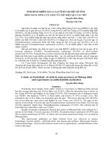
a. The isolation of C. botulinum from speciments of
infected ducks
The isolation of C. botulinum from speciments of infected
ducks was carried out following Lindstrom and Korkeala (2006) and
having modifications. This method was showed in Fig 3.1.
5
Contents
in
intestine
and
liver
Culture
on the
blood
agar
and SFP
Soil,
water,
crabs,
snails
CMM
media
Typical
colonies
Gram
stain
(bacilli,
Gr+, spores)
Biochemical
characteristics
by
usingAPI 20A
Clostridium
botulinum
and
keeping
strains
in CMM/
thioglycolate
Subculture
(identical
colonies)
(All steps were done and incubated in the anaerobic condition at 37oC/24-48h)
Fig 3.1. The method of isolation and identification of Clostridium botulinum (Lindstrom and
Korkeala, 2006) (having modification)
Fig 3.2 C. botulinum colonies on blood agar
Fig 3.3 C. botulinum colonies on SFP
6
Fig 3.4 The spores of C. botulinum under the microscope (X 100)
b. The biochemical characteristics of C. botulinum: those
characteristics were examined by using API 20A kit (following
the manufacture)
Table 3.2 The biochemical characteristics of C. botulinum using API20A kit
c. Determination of botulin toxin of C. botulinum
- Sera: sera were melt and filtrated via 0.45µm filter to collect
the clear supernatant.
- Experimental animals: SPF mice that bought in Pasteur
Institute, Ho Chi Minh were raised in 3 days to adopt the
environment before using in the experiment.
- Experimental design
Table 3.3 The experimental design
Group
Group I
Group II
Control
No. of examined
mice
400
400
20
Dose (ml/mouse)
0,5
0,5
0,5
- The filtrated supernatants were check the aseptic by culturing
on blood agar and SFP medium. Those samples were incubated at
37oC, 24h.
- The serum used in this study must be aseptic.
Examined results
Mice in Group I showed symptoms such as paralysis of
legs/eyelids, ruffled feathers, abdominal breathing or died while mice
7
in Group II and Control were healthy without abnormal symptoms; it
indicated that botulin toxin was in that serum sample.
d. Determination of botulin toxin type
The sera having botulin toxin were chosen to determine the toxin
types. The sera were filtrated and divided into 3 equal parts to test
with 3 kinds of antitoxins including C, D and E type with
concentration of 1:1. Each suspension of serum and antitoxin was
injected to 2 mice. The standard antitoxin was diluted with the
biophysical saline at the concentration of 1/100 before testing. The
experimental distribution was described in Table 3.4. Determination
of botulin toxin types was summarized in Fig 3.6.
Table 3.4 The experimental design to determine the botulin toxin types
No. of
Type C
Type D
Type E
Control
samples
(mice)
(mice)
(mice)
(mice)
1
2
2
2
2
Dose/mouse
(ml)
1
Type C: serum + antitoxin type C; Type D: serum + antitoxin type D; Type E: serum
+ antitoxin type E; Putting those tests at the room temperature in 30-60min (CDC,
1998); Control (without antitoxin): injected the biophysical saline 0,9%.
8
Duck serum (botulism infected)
Filtration (0.45µm filter), divided into 2 parts
Specimens
suspension
9(qua lưới lọc
Without heat treatment
Step
1
With heat treatment at 100oC/10min
Group II: IP injected:
0.5 ml/mouse
(2 mice/sample)
Group I: IP injected:
0.5 ml/mouse
(2 mice/sample)
Died or abnormal mice
Healthy mice
Autopsy, recording abnormal symptoms,
and checking the aseptic of bacteia
suspension
Botulin toxin was destroyed by the heat
Positive (having toxin in the serum)
Positive sera in Step 1 was filtrated via 0.45µm filter and divided into 3 parts to
examine the antitoxins
Serum + Antitoxin type C
Step
2
IP injected: 2 mice
(1ml/mouse)
Serum + Antitoxin type D
IP injected: 2 mice
(1ml/mouse)
Serum + Antitoxin type E
IP injected: 2 mice
(1ml/mouse)
(1ml/con)
Healthy mice
Healthy mice
Healthy mice
thường
Specimens has the toxin of
C. botulinum type C
Specimens has the toxin of
C. botulinum type D
Specimens has the toxin of
C. botulinum type E
Fig 3.5: Determination of botulin type in the duck serum (CDC, 1998)
9
Examined results
After 7 days observed, mice were healthy. It indicated that sera
had the corresponding of toxin types; toxin in serum was neutralized
by the added antitoxin.
Mice in the control group: healthy without abnormal symptoms.
3.3.2.4. Observed factors
- The prevalence of C. botulinum in specimens.
- The rate of sera having toxin.
- The prevalence of toxin types.
- The frequency of clinical symptoms on mice infected with
botulin toxin.
- The frequency of gross lesions on mice infected with botulin
toxin.
3.3.3. Content 3: Determination of risk factors causing the botulism
disease on the free-grazing ducks in the Mekong Delta
3.3.3.1 The research aim: Evaluation of the prevalence of C.
botulinum and the pathogenicity of isolated strains from the grazing
environment.
3.3.3.2 The research objects: the environment samples (soil, water,
crabs, snails)
3.3.3.4 The research method
a. Sample collection
At the same time of collecting botulism infected ducks, the
environment samples were collected in the same fields. The number
of samples were showed in Table 3.5.
Table 3.5 The environment samples collected in this study
Place
An Giang
Can Thơ
Hau Giang
Kien Giang
Total
No. of soil
samples
159
141
144
156
600
No. of water
samples
159
141
144
156
600
No. of crab
samples
63
42
50
61
216
No. of snail
samples
106
94
96
104
400
Total
645
563
582
646
2,436
- Soil and water were collected in one field. Wet fields: taking
soil at the depth of 5-10 cm. Wet fields with mud: taking the mud on
the surface of the field. Each sample was 25-30g. Water samples:
taking 50-100ml and keeping in the sterile tubes.
- Crabs, snails were collected in one field. Those crabs or
snails were small or just died, and kept in the sterile tubes.
All samples were kept in the cool conditions (Franciosa et al., 1996).
10
b. The isolation of C. botulinum from the environment
samples
The method was same as the decription in 3.3.2.3.
3.3.3.5. Observed factors
- The prevalence of C. botulinum in the soil and water.
- The prevalence of C. botulinum in crabs and snails.
3.3.4. Content 4: The pathogenicity of C. botulinum isolated from
infected ducks
3.3.4.1 The research aim: Determination of pathogenicity of
isolated C. botulinum strains
3.3.4.2 The research objects: isolated C. botulinum strains in
Content 1 and Content 3.
3.3.4.3. The research method
a. Antimicrobial susceptibility of C. botulinum
The disk-diffusion method of Kirby-Bauer was used in this
study (Bauer et al., 1966). The sensibility of C. botulinum to
antibiotics were determined following the standards of Clinical and
Laboratory Standards Institute (CLSI, 2019).
- Method: the suspension of C. botulinum was adjusted with
MacFarland 0.5 to equal 108CFU/ml and spread on MHA medium.
After that, antibotics dics were put on the media how to be 2.5-3.5cm
each other and 2cm to the edge of the petri disk. Those samples were
incubated at 37oC, 24h with CO2. After 24h, the zone diameter of
antibiotics were measured to determine the susceptibility.
Table 3.6: The standard of zone diameter of antibiotics (CLSI, 2019)
Zone diameter (mm)
Content
Antibiotics
(μg)
Sensitive
Intermediate
Amikacin
30
≥ 17
15-16
Ampicillin
10
≥ 17
14-16
Amoxicillin
25
≥ 18
14-17
ceftiofur
30
≥21
18-20
Cephalexin
30
≥ 18
15-17
Doxycycline
30
≥13
10-12
Florfenicol
30
≥19
15-18
Fosformycin
200
≥16
13-15
Marbofloxacin
5
≥18
16-17
Norfloxacin
10
≥17
13-16
11
Resistant
≤ 14
≤ 13
≤ 13
≤17
≤ 14
≤9
≤14
≤12
≤15
≤12
b. The experiment of botulin toxin on ducks
- Suspension of botulin toxin
The process followed the method of Cook et al. (1998) and
was showed in Fig 3.6
Selected C. botulinum colonies on TSA
(from strains caused mice dead)
Subculture
TPGY broth
Anaerobic incubation at 37oC/24h
CMM (Cooked Meat Medium)
Abaerobic incubation at 35oC/5days
Get 10 ml from CMM
Centrifuge 12.000 rpm, 15min/4oC
Collecting and filtrating supernatant
Examined samples with botulin
Fig 3.6The preparation method of botulin toxin
Determination of LD50 on ducks
The toxin suspension was prepared following the above
method.
A total of 90 ducks were used to determine the LD50 via
intravenous and oral injection.
Group I: C. botulinum was oral injection at the dose of 3ml,
5ml, 7ml, 10ml. Each dose was done with 3 ducks and one toxin
suspension.
Group II: C. botulinum was intravenous injection at the dose
of 1 ml, 2ml, 3 ml, 4ml, 5ml, 6ml to the wing veins. được tiêm trực
12
tiếp vào tĩnh mạch cánh vịt. Each dose was done with 3 ducks and
one toxin suspension.
The experimental design was showed in Table 3.7.
Table 3.7 The experimental design to determine LD50 of botulin toxin
Experiment
Injection route
Intravenous
Dose (ml)
1
2
3
4
5
6
3
No. of examined samples
3
3
3
3
3
3
3
No. of examined ducks
9
9
9
9
9
9
9
Oral
5
3
9
7
3
9
10
3
9
LD50 was determined for each examined sample and got the
average value. The formula of LD50 calculation was follwed Reed
and Muench (1938).
LD50 = -
x (b-a) + a
A: Death ratio (%) next over 50%.
B: Death ratio (%) next under 50%.
a: Diluted concentration of A
b: Diluted concentration of B
Experimental ducks: 140 days old, small size, bright
feathers, clear eyes, agility. The cages and raising environment were
prepared well for living and testing. Before testing, ducks were kept
in experimental cages 2 days.
The injection of botulin toxin to ducks
The toxin suspension was prepared following Fig 3.5., and
injected following Table 3.6. After IV or PO with the toxin
suspension, ducks were observed the clinical symptoms. Those ducks
was observed about diet, laying rate, symptoms and been necrospy at
7 post-injection days to examine lesions.
Observed symptoms
After injection, ducks were observed the clinical symptoms.
The information was resorded 3 times/day (6h, 12h, 18h after
infection, and untill ducks were death)
The toxin suspension was prepared following Fig 3.5., and
injected following Table 3.6. After IV or PO with the toxin
suspension, ducks were observed the clinical symptoms. Those ducks
was observed about diet, laying rate, symptoms and been necrospy at
7 post-injection days to examine lesions.
The clinical symptoms were observed including movement,
activities, feathers, squealing, chased reaction, breathing, feces,
swimming.
13
Observed lesions
Ducks that were nearly death or just died, were dissected to
determined the lesions in the organ systems such as the nervous
system(brain), respiratory system (trachea, lung), circulation system
(heart), digestive system (esophagus, proventriculus, gizzard, small
intestine, large intestine, anal), skin, musscle, connective tissues,
lympho and the liver, spleen, kidneys. Those picture of lesions were
captured to make the data.
3.3.4.3. Observed factors
- The mortality rate of ducks after 7 days of injection with the
typical symptoms of botulin poisoning.
- The frequency of typical symptoms of the botulism disease
caused by C. botulinum.
- The frequency of lesions in the internal organs.
3.6 Data analysis
Data were collected and basically analyzed by using Microsoft
Excel. Data were also statistically analyzed by using Chi-quare
Yates and Fixer’s test, Minitab 16.0 software with the confidence
level 95%.
14
Chapter 4 RESULTS AND DISCUSSION
4.1. The prevalence of the botulism disease on the free-grazing
ducks in the Mekong Delta
4.1.1. The botulism disease on the free-grazing ducks in the
Mekong Delta
The prevalence of the botulism disease was showed in Table 4.1.
Table 4.1. The prevalence of the botulism disease on the free-grazing ducks
Botulism infected ducks
No. of
Province
No. of examined ducks
flocks
No. of ducks
Ratio (%)
An Giang
53
45,800
558
1.22
Can Tho
47
44,700
497
1.11
Hau Giang
48
43,350
547
1.26
Kien Giang
52
53,200
633
1.19
Total
200
187,050
2,235
1.19
The results of Table 4.1 showed that ducks were infected
with botulism disease at a little higher rate in Hau Giang province
(1.26%). However, the infected rate was not significantly different
among those provinces (P>0.05). It indicated that the free-grazing
ducks in the Mekong Delta could be infected botulin toxin of C.
botulinum at the same risk. This disease causes the paralysis of neck,
eyelids, wings, and legs; it called “Cum can” disease in those
provinces.
4.1.2. The prevalence of the botulism disease on the free-grazing
ducks in the Mekong Delta by breedings
Table 4.2 The rate of the botulism disease on the free-grazing ducks by breedings
Breeds
No. of examined ducks No. of infected ducks
Meat duck
99,676
905
Laying duck
87,374
1,330
Total
187,050
2,235
Ratio (%)
0.91a
1.52b
1.19
Ducks were raised on the field from 4 to 12 weeks old, after
that, those ducks were selected for raising to collect eggs in many
years. Therefore, laying ducks started at 12 weeks old. In Table 4.2,
laying ducks were infected with botulism disease (1.52%) higher
than meat ducks were (0.91%) (P<0.05). Laying ducks had been
raising for a long time; they could contact with the pathogens
frequently to be infected or consume the feed contaminated the toxin.
Those results were similar to the reports from Boroff and Reilly
(1959), Gross and Smith (1971). Haagsma (1987) also indicated that
the waterfowl infected the botulin toxin was recovered after
treatment without remaining immune response.
15
4.1.3 The clinical symptoms of ducks infected with botulism
disease
Table 4.3. The frequency of clinical symptoms on botulism infected ducks (n=2,235)
Symptom
No. of ducks
Ratio (%)
Lost appettie, hairiness, less activities
1,532
68.55
Neck paralysis
1,965
87.92
Eyelids paralysis, mydriasis
2,013
90.07
Leg paralysis
1,783
79.78
Greenish feces – Diarrhea
1,586
70.96
Blood diarrhea
768
34.36
The results of Table 4.3 showed that symptoms of neck
paralysis, eyelids paralysis, leg paralysis were at a high rate of
87.92%, 90.07%, 79.78% respectively. The nervous toxin (botulin
toxin) destroys the central nervous system and depresses the
neuromuscular, motor neurons including oral and ocular muscles.
The infected ducks were weak legs to be fewer activities, and
paralysis of neck, eyelids. Shin (2010) reported an outbreak of
botulism poisoning on ducks and wild birds; the most popular
symptoms were observed in 2,000 infected cases such as the
paralysis of necks, eyelids. Besides, blood diarrhea also occurred on
a few infected ducks (34.36%).
4.1.4. The gross lesions on ducks infected botulism disease
Table 4.4 The frequency of gross lesions on ducks infected botulism disease (n= 420)
Lesions
No. of ducks
Ratio (%)
Pulmonary edema – hemorrhage
362
86.19
Hemorrhage liver
401
95.48
Hemorrhage kidney
57
13.57
Swolen and hemorrhage spleen
40
9.52
Gas - empty of food in the intestine
387
92.14
Hemorrhage was the most frequent lesions occurred on the
infected ducks. Table 4.4 showed the results that lesions on the lung
and liver were the highest rate 95.48%, 86.19% respectively. The
intestines that had gas and was no food inside, was also at the high
rate (92.14%). This results were similar to the research of Jensen and
Duncan (1980). In that study, the botulin toxin from waterfowl
caused the experimental ducks symptoms such as pulmonary edema,
respiratory paralysis, and hemorrhage in lungs and livers.
16
4.2 The isolation of C. botulinum and determination of botulin
toxin on botulism infected ducks
4.2.1. The isolation of C. botulinum from the specimens of
botulism infected ducks
Table 4.5 The prevalence of C. botulinum in the specimens of botulism infected ducks
Specimens
Feces
Liver
Province
No. of
No. of
No. of
No. of
Ratio
examined
positive
examined
positive
(%)
samples
samples
samples
samples
An Giang
52
28
53.05
53
26
Can Tho
49
21
42.86
49
21
Hau Giang
50
27
54.00
50
19
Kien Giang
58
30
51.72
59
25
Total
209
106
50.72
116
91
Ratio
(%)
49.06
42.86
38.00
42.37
43.13
Although the prevalence of C. botulinum in feces (50.72%)
seemed to be higher than that in livers (43,13%), there was no
significant difference between those specimens (P>0.05). Moreover,
the prevalence of C. botulinum in those specimens was not
remarkably different among those provinces (P>0.05) (Table 4.5).
4.1.2 Determination of toxin in the sera of botulism infected
ducks via the experiment on mice
4.1.2.1 The results of toxic test on mice
Table 4.6 The percentage of mice infected botulin toxin from the sera of botulism infected
ducks
Group I (n = 200)
Group II (n = 200)
Control group (n = 20)
Conditions
No. of
Ratio
No. of
Ratio
No. of
Ratio
mice
(%)
mice
(%)
mice
(%)
Death
126
63.00
0
0
0
0
Abnormal (*)
74
37.00
0
0
0
0
Normal
0
0
200
100
20
100
Infected disease
200
100
0
0
0
0
(*): mice with symptoms such as moody, skip eating, diarrhea, abdominal breath
The results exhibited that mice in Group I (without heat
treatment) were died (63%) and abnormal (37%); mice in Group II
(with heat treatment) were healthy. It indicated that there was botulin
toxin in sera of botulism infected ducks as well as the decription of
CDC (1998). Therefore, the free-grazing ducks that were neck
paralysis was dueto botulin toxin of C. botulinum.
17
4.1.2.2 The determination of botulin toxin type via the neutral
reaction on mice
Table 4.7 The results of determination of botulin toxin type (n=126)
Type C
Type D
Type E
Type C+D
Type C+E
Control
No.
No.
No.
No.
No.
No.
Results
Ratio
Ratio
Ratio
Ratio
Ratio
Ratio
of
of
of
of
of
of
(%)
(%)
(%)
(%)
(%)
(%)
samples
samples
samples
samples
samples
samples
(1)
(2)
(3)
49
26
51
38.89
20.64
40.48a
72
22
32
57.14
17.46
25.40b
65
25
36
51.59
19.84
28.57b
5
3.97
(1) No neutralization (died mice), (2) Partial neutralization (mice with
death), (3) healthy mice
0
0
0
0
12
100
2
1.59
clinical symptoms, no
The results of Table 4.7 showed that the botulin toxin type C
in ducks’ sera was present at the highest rate (40.48%) followed by
type E (28.57%), type D (25,40%) (P<0.05). It indicated that the
botulism disease on the free-grazing ducks in the Mekong Delta was
mainly caused by the botulin toxin type C of C. botulinum.
Moreover, there was a combination of botulin toxin type C + type D
(3.97%), and type C+ type E (1.59%).
4.3 Determination of risk factors causing the botulism disease on
the free-grazing ducks in the Mekong Delta
4.3.1 The prevalence of C. botulinum in soil and water at the
grazing field with botulism infected ducks
Table 4.8 The prevalence of C. botulinum in the field soil and water
No. of
Soil
Water
Province
examined
No. of positive
Ratio
No. of positive
samples
samples
(%)
samples
An Giang
159
20
12.58a
25
Can Tho
141
22
15.60ab
25
Hau Giang
144
27
18.75ab
31
Kien Giang
156
36
23.08b
37
Total
600
105
17.50
118
Different letters in one column are statistical differences (P<0.05)
Ratio
(%)
15.72
17.17
21.53
23.72
19.67
The prevalence of C. botulinum was 17.5% insoild, and
19.67% in water. In Kieng Giang province, C. botulinum was present
at a high rate in both soil and water (23.08% and 23.72%) (Table
4.8). Those samples were collected in three districts (Giong Rieng,
Tan Hiep and An Biên) with 116,086 ha of rice fields in total.
Therefore, it was estimated that 27,721 ha of rice fields could be
contamined with C. botulinum. According to husbandry practices,
farmers usually raise the free-grazing ducks in the area having many
rice fields. Ducks consume spilled rice and aquatic feed on those
18
fields. Therefore, those practices have potential risks for disease to
outbreak on the free-grazing ducks in the Mekong Delta.
4.3.2 The prevalence of C. botulinum on crabs and snails
Table 4.9 The prevalence of C. botulinum on crabs and snails at the grazing field
Crabs
Snails
No. of
No. of
No. of
No. of
Province
Ratio
examined
positive
examined
positive
(%)
samples
samples
samples
samples
An Giang
63
5
7.94
106
4
Can Tho
42
3
7.14
94
3
Hau Giang
50
4
8.00
96
2
Kien Giang
61
6
9.84
104
3
Total
216
18
8.33a
400
12
Different letters in one row are statistical differences (P<0.05)
Ratio
(%)
3,77
3,19
2,08
2,88
3,00b
The results of Table 4.9 showed that the prevalence of C.
botulinum was 8.33% in crabs higher than that in snails (3,00%)
(P<0.05). Crabs and snails are crustaceans living in water of rice
fields and could have C. botulinum in their intestine (Dohms, 2008).
Moreover, Clostridium spp. usually present in sedimentary lands,
swamps, flooded areas. It is a reason to impact the prevalence of this
pathogen in crabs and snails (Wosbeser et al.,1987).
4.4 The pathogenicity of isolated C. botulinum originated from
infected ducks and the environment
4.4.1 The antimicrobial susceptibility testing of C. botulinum
Table 4.10 The susceptibility of isolated C. botulinum strains to antibiotics
Clostridium botulinum (n=241)
Sensitive
Intermediate
Resistant
Antibiotics
Abr
No. of
Ratio
No. of
Ratio
No. of
Ratio
samples
(%)
samples
(%)
samples
(%)
Amikacin
Ak
139
57.68
81
33.61
21
8.71
Ampicillin
Am
0
0
212
87.97
29
12.03
Amoxicillin
Ax
31
12.86
189
78.42
21
8.71
Ceftiofur
Cf
235
97.51
6
2.49
0
0
Cephalexin
Cp
58
24,07
160
66.39
23
9.54
Doxycycline
Dx
237
98.34
4
1.66
0
0
Florfenicol
FFc
241
100
0
0
0
0
Fosformycin
Fos
241
100
0
0
0
0
Marbofloxacin
Ma
241
100
0
0
0
0
Norfloxacin
Nr
232
96.27
9
3.73
0
0
The results of Table 4.10 exhibited that of 10 antibiotics used
in this study, there were six antibotics showing a high sensibility (95100%) (Table 4.10). Among them, C. botulinum was sensitive 100%
to florfenicol, fosformycin, and marbofloxacin. This result was
similar to the report of Nguyen Đuc Hien (2012) that C. botulinum
19
was also sensitive to norfloxacin, fosfomycin, and ceftiofur.
Although the treatment using botulin antitoxin of C. botulinum gets
high efficiency, it loses the economic values in the poultry husbandry
(Dohms, 2008). Farmers could choose the cheap and effective
antibiotics to treat or control the outbreak of this disease.
4.4.2 The multiple resistance of isolated C. botulinum strains
against antibiotics
Of 241 isolated C. botulinum strains, there was 42/241 strains
showed resistance against 2-4 antibotics with 11 combined
phenotypes. C. botulinum was resistant at the highest rate against 2
antibiotics (9.54%). The common phenotypes was Am-Ax (10/241
strains). Moreover, there were six strains that were resistant against 4
antibiotics in this study.
Table 4.11 The multiple resistance of isolated C. botulinum against antibiotics (n=241)
No. of resistant
No. of
No. of
Phenotypes
Total
Ratio (%)
antibiotics
phenotype
strains
2
6
3
4
4
Total
1
11
Ak-Am
Ak-Ax
Ak-Cp
Am-Ax
Am-Cp
Ax-Cp
4
3
2
10
3
1
Ak-Am-Ax
Ak-Am-Cp
Ak-Ax-Cp
Am-Ax-Cp
Ak-Am-Ax-Cp
1
2
2
8
6
23
9.54
13
5.39
6
42
2.49
4.4.3 The experiment of botulin toxin testing on ducks
Table 4.12 The results of botulin toxin testing on ducks after 7 days injection
No. of
No. of
No. of
samples
samples
No. of
Ratio
Injection
examined
causing
causing
ducks
(%)
samples
mice
mice
death
abnormal
Exp. group
IV/PO
90
32
23
71.88
9
Control group No injection
8
0
0
0
Ratio
(%)
28.13
0
The results of Table 4.12 showed that 100% samples caused
abnormal effects on experimental ducks; among of them, 23/32
samples caused ducks death (71.88%). It indicated that the toxic
ability of botulin toxin from isolated C. botulinum was high. It
was similar to the research from Nguyen Duc Hien and Nguyen
20
Manh Hung (2012) as well as from Notermans et al. (1980),
Jensen and Ducan (1980). Ducks were IV injected died much
more than than via PO.
Table 4.13 The clinical symptoms of botulin toxin test on ducks (n=90)
No. of ducks having
Symptoms
Control
anormal symptoms
Moody, less activities
0/8
90
Ruffled feather, loos feather
0/8
90
Eyelid paralysis, mydriasis
0/8
60
Neck paralysis
0/8
83
Leg paralysis
0/8
55
Less eating
0/8
90
Less laying
0/8
90
Greenish feces – diarrhea
0/8
32
Ratio
(%)
100
100
67.19
92.19
60.94
100
100
35.94
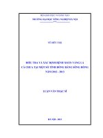
The results of Table 4.13 revealed that ducks had the typical
symptoms of botulin poisoning such as less eating, noody, ruffled
hair, less laying (100%); neck paralysis was 92.19%; eyelid
paralysis, mydriasis was 76.19%; leg paralysis was 60.94%; greenish
feces – diarrhea was 35.94% in comparison with the control group
having healthy ducks without abnormal symptoms. Those abnormal
activities on ducks were due to the effect of butulin toxin of C.
botulinum. This result got on with the report from Dohms (1987) that
the clinical symptoms of the botulism disease on ducks were mainly
the paralysis of neck, eyelid, legs. Therefore, this disease was named
“soft neck symptom” (Limberneck).
21
Fig 4.1 Ruffled feathers
Fig 4.2 Eyelid paralysis
Fig 4.4 “Limberneck”
Fig 4.3 Greenish diarrhea
Bảng 4.14 The gross lesions of the experimental ducks infected botulin toxin (n=90)
Lesions
Control
Hemorrhage lungs
Swollen and heamorrhage spleen
Heamorrhage liver
Heamorrhage kidney
Gas and empty in the intestine
0/8
0/8
0/8
0/8
0/8
No. of ducks
having lesions
73
27
83
30
80
Ratio
(%)
81.25a
29.69b
92.19a
32.81b
89.06a
Different letters in one column are statistical differences (P<0.05)
Table 4.14 showed that ducks infected botulin toxin had the
chracteristic symptoms such as hemorrhage lungs, hemorrhage in
liver, spleen at a high rate; another symptoms got a low rate. Ducks
in the control group were healthy without lesions. The results also
indicated that heamorrhage liver got the highest rate (92.19%)
follwed by gas and empty in the intestine (89.06%), heamorrhage
lung (81.25%). Johnson et al. (2010) also reported that botulin toxin
caused shortness of breath, especially type C; it explained for lesions
in lungs.
22