Mammographic microcalcifications and breast cancer tumorigenesis: A radiologic-pathologic analysis
Bạn đang xem bản rút gọn của tài liệu. Xem và tải ngay bản đầy đủ của tài liệu tại đây (1.85 MB, 9 trang )
Naseem et al. BMC Cancer (2015) 15:307
DOI 10.1186/s12885-015-1312-z
RESEARCH ARTICLE
Open Access
Mammographic microcalcifications and breast
cancer tumorigenesis: a radiologic-pathologic
analysis
Madiha Naseem1,2*, Joshua Murray3†, John F Hilton4†, Jason Karamchandani2,5†, Derek Muradali2,6†,
Hala Faragalla2,5, Chanele Polenz1†, Dolly Han1†, David C Bell5† and Christine Brezden-Masley1,2†
Abstract
Background: Microcalcifications (MCs) are tiny deposits of calcium in breast soft tissue. Approximately 30% of early
invasive breast cancers have fine, granular MCs detectable on mammography; however, their significance in breast
tumorigenesis is controversial. This study had two objectives: (1) to find associations between mammographic MCs
and tumor pathology, and (2) to compare the diagnostic value of mammograms and breast biopsies in identifying
malignant MCs.
Methods: A retrospective chart review was performed for 937 women treated for breast cancer during 2000–2012
at St. Michael’s Hospital. Demographic information (age and menopausal status), tumor pathology (size, histology,
grade, nodal status and lymphovascular invasion), hormonal status (ER and PR), HER-2 over-expression and presence
of MCs were collected. Chi-square tests were performed for categorical variables and t-tests were performed for
continuous variables. All p-values less than 0.05 were considered statistically significant.
Results: A total of 937 patient charts were included. About 38.3% of the patients presented with mammographic
MCs on routine mammographic screening. Patients were more likely to have MCs if they were HER-2 positive
(52.9%; p < 0.001). There was a significant association between MCs and peri-menopausal status with a mean age of
50 (64%; p = 0.012). Patients with invasive ductal carcinomas (40.9%; p = 0.001) were more likely to present with
MCs than were patients with other tumor histologies. Patients with a heterogeneous breast density (p = 0.031) and
multifocal breast disease (p = 0.044) were more likely to have MCs on mammograms. There was a positive correlation
between MCs and tumor grade (p = 0.057), with grade III tumors presenting with the most MCs (41.3%). A total of
52.2% of MCs were missed on mammograms which were visible on pathology (p < 0.001).
Conclusion: This is the largest study suggesting the appearance of MCs on mammograms is strongly associated
with HER-2 over-expression, invasive ductal carcinomas, peri-menopausal status, heterogeneous breast density and
multifocal disease.
Keywords: Microcalcifications, Breast imaging, Mammography, Tumorigenesis, Breast pathology, HER-2
* Correspondence:
†
Equal contributors
1
Department of Hematology/Oncology, St. Michael’s Hospital, 30 Bond
Street, Toronto, Ontario M5B 1W8, Canada
2
Faculty of Medicine, University of Toronto, 1 Kings College Circle, Toronto,
ON M5S 1A8, Canada
Full list of author information is available at the end of the article
© 2015 Naseem et al.; licensee BioMed Central. This is an Open Access article distributed under the terms of the Creative
Commons Attribution License ( which permits unrestricted use, distribution, and
reproduction in any medium, provided the original work is properly credited. The Creative Commons Public Domain
Dedication waiver ( applies to the data made available in this article,
unless otherwise stated.
Naseem et al. BMC Cancer (2015) 15:307
Background
Breast cancer is the most common cancer in females over
the age of 20. Breast cancer represents 26% of all newly diagnosed cancer cases in women and 14% of women are is
expected to die from it [1]. The incidence rates of breast
cancer have risen from 1982 through the early 1990s, in
part due to increased mammography screening.
The advent of mammographic screening has not only
provided us with the ability to detect potentially fatal tumors at a non-palpable stage, but it has also created the
platform to study the natural history of breast cancer in
its early stages of development [2]. One of the easily detectable mammographic anomalies, and often the earliest signs of a malignant breast disease, are tiny deposits
of calcium in the breast soft tissue, called microcalcifications (MCs) [3]. The presence of MCs was first reported
in 1913 by a German surgeon, Solomon, who conducted
a radiographic examination of a mastectomy specimen.
In 1951, a radiologist named Leborgne proposed that
MCs could be the only mammographic manifestation of
breast carcinoma [4]. Since then, active efforts have been
made by radiologists to identify MCs in mammograms
(Figure 1b), making them one of the most important
diagnostic markers of breast lesions [5].
Although MCs are also associated with benign conditions such as secretory diseases and fat necrosis, around
40% of breast cancers present with MCs and frequently,
serve as the only mammographic features indicating the
presence of a tumor [6]. X-ray diffraction and electron
microscopic analysis have revealed two distinct forms of
MCs based on their appearance and chemical composition [7]. Type I MCs are calcium oxalate crystals, while
Type II MCs are composed of another bone specific
mineral called hydroxyapatite [3]. Among the two, Type
II MCs are exclusively found in malignant breast disease,
and these crystals are known to accelerate the pathological process involved in breast cancer.
Malignant MCs have one of three appearances: crushed
stone (pleomorphic), powdery, or casting-type [8]. Previous studies have shown that patients presenting with
casting-type MCs have aggressive tumor pathology, with a
death rate five times that of patients who do not present
with such MCs [9].
Considerable progress has been made in understanding
the molecular foundations of breast carcinogenesis. However, the biogenesis of MCs and their role in breast cancer
is still understudied. MCs are shown to be associated with
the overexpression of Human Epidermal Growth Factor
Receptor Type 2, HER-2, a transmembrane protein receptor which serves as an independent poor prognostic factor
in premalignant breast lesions. Previous studies have also
examined the associations between MCs and other prognostic factors of breast cancer, such as Estrogen (ER) and
Progesterone receptor (PR) positivity. However, there is
Page 2 of 9
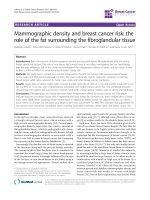
Figure 1 Mammogram and pathology report for HER-2 positive patient.
a: Digital Mammogram (Mammomat Novation, Siemens Healthcare,
Erlangen, Germany) of the left breast from a 40 year old woman with a
HER-2 positive invasive ductal carcinoma, shows a malignant appearing
mass (arrows) with numerous pleomorphic calcifications confined to
the mass, BI-RADS 5. b: Hematoxyln and eosin stained section (400×) of
poorly differentiated invasive ductal carcinoma with microcalcification
(arrowhead).
currently no consensus on the prognostic significance of
MCs in early breast cancer.
This paper presents associations between benign and
malignant mammographic MCs, and breast biomarkers,
patient demographics, and breast radiological features. It
also evaluates the utility of mammograms in identifying
MCs by comparing breast biopsy and mammogram
reports.
Methods
Ethics
Institutional research ethics board approval from St.
Michael’s Hospital was obtained for this research study.
Naseem et al. BMC Cancer (2015) 15:307
Page 3 of 9
Inclusion/exclusion criteria
Microcalcifications on pathology
All patients seen by medical oncologists at St. Michael’s
Hospital Medical Day Care Unit, diagnosed only with invasive breast cancer were included in the study. Benign
and malignant microcalcifications on mammography
and pathology for patients with invasive breast cancer
were included. Patients with non-invasive diseases were
not included, as this study’s focus is on investigating the
role of MCs in invasive disease. Patients were selected
based on availability of electronic health records, dating
back to 2000.
At St. Michael’s Hospital, Stereotactic core biopsy for
microcalcifications was obtained, and initially cut into
10 levels from each core and every other level was
stained. If MCs were found on the initial levels, nothing
more was done. If MCs were not found, the slides were
polarized to find polarizable calcium crystals. If MCs
were not found, the blocks were further x-rayed and
then cut on deeper levels with MCs until they were discovered. The radiology report was also checked as x-ray
specimen indicate if there are MCs within the cores submitted. If MCs were found in the specimen radiograph
and not in the blocks, examination of deeper levels was
conducted to assess as much tissue as possible.
For this study, pathological reports were prepared by
staff pathologists, and included if available on patient’s
electronic health record.
Data acquisition
A retrospective chart review was performed for 937
women treated for breast cancer during 2000–2012 at St.
Michael’s Hospital, Toronto, Canada. Demographic information (age and menopausal status), tumor pathology
(size, histology, grade, nodal status and lymphovascular invasion), hormonal status (ER and PR), HER-2 overexpression and presence of both benign and malignant MCs on
mammograms and pathology reports were collected for
breast cancer patients. Mammograms were obtained from
the Department of Medical Imaging at St. Michael’s Hospital,
using the Digital Mammogram using technology from
Siemens Mammomat Novation DR (2004).
Immunohistochemistry
Hormone receptor status that was collected from the
pathology reports, was determined using immunohistochemistry (IHC). ER and PR were detected with the
Ventana 6 F11 and Ventana 16 clones, respectively, with
heat retrieval pretreatment and no dilution. HMK was detected by using the Dako 34BetaE12 (reacts with cytokeratins 1,5,10,14) with heat retrieval pretreatment and a 1:0
dilution. As per the 2010 College of American Pathologists
guidelines, ≥ 1% of tumor cell nuclei must be immunoreactive to be considered ER/PR positive. The same criteria has been used in previous studies. HER-2 was
detected using the Novocastra CB11 with a 1:40 dilution. For each antibody used, appropriate second antibodies were complexed to streptavidin and chromagen.
IHC is used first for overexpression of HER-2 in genecopy ratio. As per College of American Pathologists
2013 guidelines, any case with a 2+ score on IHC is sent
for in situ hybridization, whether fluorescent in situ
hybridization (FISH) or bright field dual in situ
hybridization (DISH). IHC scores (0) and (1+) are considered negative and nothing else needs to be done. IHC
score (2+) is equivocal and needs in situ hybridization.
IHC score (3+) is considered positive and nothing else
needs to be done. These guidelines were applied to obtain study samples.
Statistical analysis
Descriptive statistics were calculated for each variable of
interest. Proportions and frequencies were calculated for
categorical variables while means and standard deviations were calculated for continuous variables.
The distribution of the presence of MCs on mammography was examined. Chi square tests were performed to
test for associations between the presence of MCs on
mammography and categorical variables, while t-tests
were performed to test for associations for continuous
variables.
The distribution of the presence of both benign and malignant MCs on pathology was examined. The presence of
MCs on mammograms was tested for association with the
over-expression of HER-2, and hormonal status of ER and
PR. All tests were two-sided and p-values less than 0.05
were considered statistically significant. No corrections for
multiple testing were done for this exploratory analysis.
Results
A total of 937 charts were reviewed for patients with stages
I-III breast cancer during 2000–2012 at St. Michael’s
Hospital, Toronto. Table 1 presents patient characteristics
for the twelve variables of interest. About 38.3% of the patients had MCs present of any type, either benign or malignant. The mean age was 58.1 years (age range 25–98
years) with most patients having either ductal (81.3%) or
lobular (9.5%) lesions; Of these, only 21.4% of patients had
evidence of lymphovascular invasion. In total, 78.2% were
ER positive while 64.9% were PR positive. Only 16.3% of
the patients were HER-2 positive.
Table 2 presents the results of the tests of association
between the presence of MCs and the other variables of
interest. Variable names appearing in bold had a significant association with the presence of MCs.
Naseem et al. BMC Cancer (2015) 15:307
Page 4 of 9
Table 1 List of patient characteristics
Patient characteristics
n (%)
Table 1 List of patient characteristics (Continued)
Mean (±SD)
Yes
51 (8.5)
Age
58.1 (13.3)
No
548 (91.5)
Tumor Size
2.5 (1.9)
Family History of Breast Cancer
Mammography Calcifications
Yes
158 (35.1)
Yes
287 (38.3)
No
282 (64.1)
No
462 (61.7)
Children
Recurrence
Yes
40 (7.9)
No
466 (92.1)
Histology
Yes
277 (59.0)
Yes-1st pregnancy ≥ 30 years
53 (11.7)
Nulliparous
124 (27.3)
HER-2
Ductal
738 (81.3)
Positive
139 (16.3)
Lobular
86 (9.5)
Negative
713 (83.4)
Other
84 (9.3)
Positive
712 (78.2)
Negative
199 (21.8)
Lymphovascular Invasion
Yes
156 (21.4)
No
574 (78.6)
Node
Positive
274 (34.3)
Negative
526 (65.8)
Tumor Grade
1
221 (25.3)
2
375 (43.0)
3
277 (31.7)
Density
Almost entirely fatty
252 (56.0)
Scattered
72 (16.0)
Very dense
17 (3.8)
Extremely dense
24 (5.3)
Heterogeneously dense
57 (12.3)
Other
28 (6.2)
Bilaterality
Yes
18 (2.9)
No
599 (97.1)
Architectural Distortion
Yes
90 (17.0)
No
438 (83.0)
Focality
Unifocal
464 (81.7)
Multifocal/Multicentric
104 (18.3)
Menopausal Status
Pre
229 (26.1)
Peri
29 (3.3)
Post
620 (70.6)
Diabetes
ER
PR
Positive
591 (64.9)
Negative
319 (35.1)
This table outlines the proportion (n) of study patients with certain demographic,
tumor pathologic, and mammographic characteristics. N = Number of patients in
the sample, SD = Standard Deviation.
Tumor pathology
The relationship between the presence of MCs and histology was significant, (p =0.001). Patients with ductal carcinoma were more likely to have MCs than were patients
with other tumor classifications (mammary, lobular,
mixed). There was no significant relationship between
MCs and lymphovascular invasion or nodal status. Among
patients with a grade III tumor, 41.3% had MCs, as opposed to 39.8% with a grade II tumor and 30.7% with a
grade I tumor. There was a positive correlation between
the presence of MCs and an increase in tumor grade,
however, this relationship was not statistically significant
(p = 0.057). There was no significant association between
the presence of MCs and mean tumor size or the rate of
tumor recurrence. Recurrence was measured using a 5 year
recurrence end-point, and there was no statistical association between having MCs on mammography and recurrence pattern (p = 0.258).
Breast biomarkers
Patients were more likely to have MCs if they had an overexpression of HER-2 (52.9%; p = 0.001). Images from a patient with overexpression of HER-2 showed the presence
of MCs in both mammographic images (Figure 1a) and
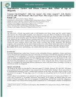
the corresponding pathology sample (Figure 1b). Conversely, neither the mammogram (Figure 2a) nor the
pathology sample (Figure 2b) displayed evidence of
MCs for patient with HER-2 negative disease. There was
Naseem et al. BMC Cancer (2015) 15:307
Page 5 of 9
Table 2 Statistical associations for the presence of MCs
on mammography
(a) Categorical variables Microcalcifications
No
n
Test statistic
Yes
(%)
n
(%)
Recurrence
Yes
21
(52.5) 19
(47.5)
No
287
(61.6) 179
(38.4)
Histology
357
(59.1) 247
(40.9)
Lobular
58
(82.9) 12
(17.1)
Other
44
(63.8) 25
(36.2)
Lymphovascular Invasion
p-value
1.28
0.258
72
(57.6) 53
(42.4)
No
334
(64.9) 181
(35.1)
Node Status
134
(62.3) 81
(37.7)
Negative
287
(63.8) 163
(36.2)
Tumor Grade
133
(69.3) 59
(30.7)
2
192
(60.2) 127
(39.8)
3
122
(58.7) 86
(41.3)
0.159
0.782
Almost entirely fatty
164
(65.1) 88
(34.9)
Scattered
44
(61.1) 28
(38.9)
Very dense
10
(58.8) 7
(41.2)
Extremely dense
16
(66.7) 8
(33.3)
Heterogeneously dense 23
(40.4) 34
(59.7)
17
(60.7) 11
(39.3)
Bilaterality
Yes
6
(50.0) 6
(50.0)
No
320
(60.4) 210
(39.6)
Architectural Distortion
Yes
47
(56.0) 37
(48.1)
No
268
(61.9) 165
(38.1)
Multifocal/Multicentric
49
(52.1) 45
(47.9)
Unifocal
271
(63.9) 153
(36.1)
Focality
Menopausal Status
Pre
111
(59.0) 77
(41.0)
Peri
9
(36.0) 16
(64.0)
Post
331
(64.1) 185
(35.9)
Diabetes
Yes
26
(60.5) 17
(39.5)
No
294
(60.1) 195
(39.9)
(43.5)
No
144
(63.6) 111
(36.4)
Children
Yes
156
(62.7) 93
(37.3)
Yes-after age 30
25
(50.0) 25
(50.0)
No
59
(53.6) 51
(46.4)
HER-2
Positive
48
(47.1) 54
(52.9)
Negative
399
(66.2) 204
(33.8)
Positive
89
(59.7) 60
(40.3)
Negative
375
(62.8) 222
(37.2)
Positive
150
(59.1) 104
(40.9)
Negative
314
(63.8) 178
(34.9)
0.057
Microcalcification
No
4.33
0.115
12.9
<0.001
0.36
0.549
1.42
0.233
Test Statistic
Yes
Mean SD
12.32 0.031
Density
(56.5) 52
(b) Continuous Variables
5.74
1
91
PR
0.08
Positive
0.001*
Yes
ER
1.98
Yes
Family History
χ2
15.1
Ductal
Other
Table 2 Statistical associations for the presence of MCs
on mammography (Continued)
Mean SD
t
Age
59.0
(13.2) 57.4
(12.3) 1.67
Tumor Size
2.2
(1.8)
(2.1)
2.5
p-value
0.100
−1.58 0.115
This table outlines statistical associations between the presence of MCs and
other variables of interest. Chi square values and p-values are outlines. P-values
< 0.05 is considered statistically significant and highlighted in bold.
no significant association between the presence of MCs
with ER (p = 0.549) and/or PR status (p = 0.233).
0.18
0.669
0.81
0.369
4.04
0.044
8.86
0.012
0.00
0.999
1.66
0.198
Patient demographics
The mean age of pre-menopausal patients was 43.0
while the mean age of peri-menopausal and postmenopausal patients was 50.0 and 64.2 respectively.
Menopausal status was recorded on the patient charts,
and the age range of all patients was 25–98 years. The
relationship between menopause and the presence of
MCs was statistically significant (p = 0.012). Among the
peri-menopausal patients, 64% had MCs present as opposed to the 41% of pre-menopausal and 35.9% of postmenopausal patients who had MCs. There was a higher
likelihood of women to have MCs if they either had no
children (43.5%) or had children after age 30 (50%).
However, this relationship was not statistically significant
(p = 0.115). There was no significant association between
MCs and mean age, family history of breast cancer, or
diabetes.
Mammographic characteristics
Patients with heterogeneously dense breasts, as reported
on mammograms were more likely to have MCs than all
Naseem et al. BMC Cancer (2015) 15:307
Page 6 of 9
architectural distortion within the breast tissue (p = 0.369),
or having bilateral breast cancer (p = 0.669).
MCs on pathology
From the total cohort of 937 patients, there were only 472
patients with MCs noted on pathology. Of these, 52.2% of
patients who did not have MCs appearing on mammography had detectable MCs on pathology samples, which
were statistically significant (Table 3) (p < 0.001).
Figure 2 Mammogram and pathology report for HER-2 negative patient.
a: Digital Mammogram (Mammomat Novation, Siemens Healthcare,
Erlangen, Germany) of the right breast from a 53 year old woman
with HER-2 negative invasive ductal carcinoma, shows a lobulated
mass (arrows) in the upper right breast, with no evidence of calcifications,
BI-RADS 4. b: 400× H&E stained section of invasive ductal carcinoma of
no special type composed of pleomorphic, mitotically active ductal
epithelial cells with sheet-like growth.
other breast densities (almost entirely fatty, scattered,
very dense, extremely dense, other) (p =0.031). Patients
with multifocal or multicentric breast cancers were more
likely to present with MCs (p = 0.044). There was no significant association between the presence of MCs with
Discussion
Mammographic MCs serve as important diagnostic
markers of benign and malignant breast lesions. However, there is still a lack of consensus regarding the genesis of MCs and their relationship with breast cancer
pathology. Our study indicates that presence of both
malignant and benign mammographic MCs is associated
with poor prognostic factors of breast cancer, and could
serve as indicators of aggressive tumor growth.
This study found a positive relationship between the
presence of MCs and over-expression of HER-2. HER-2
is a valuable therapeutic and prognostic marker in primary breast carcinomas [10]. It plays a significant role in
the HER family of receptors, normally involved in regulating breast growth and development. Over-expression
of HER-2 proto-oncogene, called c-erbB-2, is associated
with breast cancer [11]. This gene is amplified in approximately 20 to 30% of breast cancers and is associated with aggressive tumor behaviour [12]. Wang et al.
[10] conducted a retrospective study of 152 patients, and
found MCs were more common in carcinomas with
HER-2 over-expression at a prevalence of 61.6% compared to those without HER-2 at 35.4%. Similarly, Seo
et al. [13] also found an association between mammographic MCs and HER-2 over-expression in 498 patients.
Our study further confirms a greater prevalence of MCs
(52.9%) among tumors over-expressing HER-2, compared to tumors without HER-2 amplification (33.8%).
This is further reinforced by the figures, showing presence of MCs for a HER-2 positive patient (Figure 1) and
a complete absence of MCs for a HER-2 negative patient
(Figure 2). However, unlike Seo et al. [13] who found
more MCs in patients with HER-2 over-expression
under the age of 50, our results showed that the presence of MCs was independent of patient age (p = 0.100).
Our study cohort was also twice (n = 937) as large as the
population assessed by Seo et al. (n = 498). Given the
strong association, MCs could serve as early indicators
of HER-2 over-expression, warranting further molecular
investigation into their relationship.
Hormone receptor status is useful for its prognostic
significance and treatment planning in patients with advanced breast cancer. Previous studies have investigated
the association between MCs and hormone receptor
Naseem et al. BMC Cancer (2015) 15:307
Page 7 of 9
Table 3 Sensitivity of mammograms in detecting MCs in comparison to MCs identified in biopsy specimens
Categorical variables
Microcalcifications (Pathology)
No
Test statistic
Yes
n
(%)
n
(%)
No
134
(47.8)
147
(52.2)
Yes
41
(21.4)
150
(78.5)
Microcalcifications (Mammography)
χ2
p-value
32.4
<0.001
Bolded numbers indicate statistical significance. Of the 472 patients who had pathology samples available, 147 (31%) had a false negative result, where MCs were
detected in pathology samples but not in mammography.
status with variable results. Griniatsos et al. [14] found
an increased number of patients with both estrogen and
progesterone receptor positive tumors presented with
mammographic MCs. Similarly, Karamouzis et al. [15]
also found MCs in over 65% of ER positive, and over
46% of PR positive tumors. On the contrary, Ferranti
et al. [16] found an inverse relationship between mammographic MCs and hormone receptor positive lesions.
The prognostic significance of ER and PR expression has
been a matter of debate for many years. However,
current available evidence suggests that ER/PR negative
tumors have a worse prognosis altogether [17]. Similar
to Gajdos et al. [18], our study showed that the presentation of MCs is independent of hormone receptor status.
Furthermore, strong correlations between MCs and
tumor grade, tumor histology, and breast density highlight
the prognostic significance of these calcium deposits. Our
results showed that a higher prevalence of MCs was found
among patients with high grade tumors than those with
low grade tumors. This result reinforces previous studies, such as that by Palka et al. [9], which showed a
strong relationship between MCs and high grade lesions.
Conversely, Dinkel et al. [19] found this correlation to
be poor and inconclusive.
To better understand these associations, the relationship between mammographic breast densities was compared to the presence of MCs. The physical composition
of the breast varies, with different proportions of fat,
connective tissue, ductal and lobular elements contributing to differences in mammographic breast density. The
greater the number of fibroglandular tissue, the higher
the category of breast density. Our results confirmed
those of Skandalis et al. [20], who found elevated levels
of MCs in patients with heterogeneously dense breasts
and high tumor grades. We found MCs to be significantly associated with heterogeneous breast densities,
with a high prevalence of fibroglandular tissue.
The link between tumor grade and breast density
highlights some molecular factors giving rise to MCs
and contributing to tumorigenesis. Tabar and Dean [21]
propose that high-grade ductal carcinomas undergo a
process called neoductogenesis, promoting vascular
invasion, with excessive lymphatic and hematogenous
spread, leading to a worse tumor prognosis. A high
prevalence of fibroglandular breast tissue can lead to increased accumulation of versican, a proteoglycan associated with high tumor grade and invasive disease in
patients with high breast densities and mammographic
MCs [20].
Approximately 90% of ductal carcinoma in situ (DCIS)
appear as MCs, 40% of which progress to an invasive
breast cancer. Among invasive carcinomas, our study
further discovered a greater prevalence of MCs in multifocal invasive disease (47.9%) than unifocal invasive disease (36.1%). Tot et al. [22] studied the influence of
tumor focality on breast cancer survival, and found the
highest ten year survival rate to be amongst patients
with unifocal tumors. Multifocal tumors serve as poor
prognostic parameters in breast cancer, and their strong
association with MCs further reinforces the role of MCs
as poor prognostic indicators of breast cancer.
There was no significant outcome difference between
patients with MCs and those without MCs. Recurrence
was measured using a 5 year recurrence end-point, and
there was no statistical association between having MCs
on mammography and recurrence pattern (p = 0.258).
These results could also be affected by our study limitations. Our study population was limited to St. Michael’s
hospital, whereas a multi-site analysis would have allowed
for a more rigorous analysis. We also conducted a retrospective chart review, where missing data on electronic
charts could not be included in the study. Hence, as
seen in Table 1, numbers for all variables vary due to incomplete information recorded on patient charts. Inclusion of additional biomarkers, such as p53 and further
genetic analysis would have further improved our understanding of the relationship between MCs and breast
cancer.
Furthermore, MCs smaller than 130 μm were not visible
on our digital mammogram [23]. In these circumstances,
histological examination of breast tissue can reveal smaller
MCs that were missed in mammography. To test for MCs
that were missed in mammograms, we compared the presence of MCs among pathology and mammography reports
Naseem et al. BMC Cancer (2015) 15:307
for each patient. Table 3 further depicts these discrepancies.
Our study showed that 52.2% of MCs that were absent on
mammograms were visible under a histological examination. Hence, our results were influenced by the size limitations of mammograms. Also, we did not examine the
chemical composition of MCs, which would have been useful for an accurate understanding of their role in breast
tumorigenesis.
Conclusions
In summary, this study is the largest correlation analysis
performed to date, investigating the association of any
mammographic MCs in breast cancer patients with variables of breast cancer. Based on the strong associations
between MCs and poor prognostic indicators of breast
cancer, such as HER-2 over-expression, high tumor
grade, prevalence of fibroglandular tissue, and multifocal
disease, it can be suggested that MCs are strongly associated with breast cancer variables that lead to a poor
prognosis. Based on these results, MCs warrant closer
attention and follow-up. There is also a need for developing improved screening methods to detect smaller
MCs that might otherwise be missed on screening mammograms. Since biological distinctions between subtypes
of breast cancers likely reflect differences in the pathways of tumor development and disease prognosis, future studies should investigate the molecular pathways
interconnecting MC genesis with breast tumorigenesis.
Abbreviations
MC: Microcalcifications; ER: Estrogen receptor; PR: Progesterone receptor;
HER-2: Human epidermal growth factors receptor 2; SMH: St. Michael’s
Hospital; IHC: Immunohistochemistry; FISH: Fluorescent in sity hybridization;
DISH: Dual in situ hybridization.
Competing interests
The authors declare that they have no competing interests.
Authors’ contributions
CBM conceived of the study, and participated in its design and coordination
and helped to draft the manuscript. MN coordinated the study and wrote
the manuscript. JM carried out the statistical analysis and participated in
writing the manuscript. JFH, CP, and DH participated in acquiring data and
drafted the manuscript. JK provided with laboratory medicine and pathology
analysis, and drafted the manuscript. HF provided valuable information on
pathology analysis, and helped with manuscript editing. DM provided
mammography data and analysis. DCB carried out immunoassays and
participated in study analysis. All authors read and approved the final
manuscript.
Acknowledgements
The authors would like to thank the following people from St. Michael’s
Hospital for their assistance with data collection: Adiba Khan, Rebecca
Heersink, Sophie Hogeveen, Ammar Bookwala and Amanda Manoharan from
the Department of Medical Oncology. We would also like to thank the
administrative staff of Medical Day Care Unit at St. Michael’s Hospital for
allowing the study to be conducted in a timely fashion. No funding was
received for this study.
Author details
1
Department of Hematology/Oncology, St. Michael’s Hospital, 30 Bond
Street, Toronto, Ontario M5B 1W8, Canada. 2Faculty of Medicine, University of
Page 8 of 9
Toronto, 1 Kings College Circle, Toronto, ON M5S 1A8, Canada. 3Horizon
Health Network, The Moncton Hospital, 135 MacBeath Avenue, Moncton,
New Brunswick E1C 6Z8, Canada. 4Dana-Farber Cancer Institute, Brigham and
Women’s Hospital, and Harvard Medical School, 450 Brookline Avenue,
Boston, MA 02215, USA. 5Department of Laboratory Medicine and Pathology,
St. Michael’s Hospital, 30 Bond Street, Toronto, ON M5B 1W8, Canada.
6
Department of Medical Imaging, St. Michael’s Hospital, 30 Bond Street,
Toronto, ON M5B 1W8, Canada.
Received: 6 August 2014 Accepted: 20 February 2015
References
1. Breast cancer statistics. [].
2. Tabar L, Duffy SW, Vitak B, Chen HH, Prevost TC. The natural history of
breast carcinoma: what have we learned from screening? Cancer.
1999;86(3):449–62.
3. Bellahcene A, Castronovo V. Increased expression of osteonectin and
osteopontin, two bone matrix proteins, in human breast cancer. Am J
Pathol. 1995;146(1):95–100.
4. Nalawade YV. Evaluation of breast calcifications. Indian J Radiol Imaging.
2009;19(4):282–6.
5. Bansal GJ, Thomas KG. Screen-detected breast cancer: does presence of
minimal signs on prior mammograms predict staging or grading of cancer?
Clin Radiol. 2011;66(7):605–8.
6. Castronovo V, Bellahcene A. Evidence that breast cancer associated
microcalcifications are mineralized malignant cells. Int J Oncol.
1998;12(2):305–8.
7. Morgan MP, Cooke MM, McCarthy GM. Microcalcifications associated with
breast cancer: an epiphenomenon or biologically significant feature of
selected tumors? J Mammary Gland Biol Neoplasia. 2005;10(2):181–7.
8. Zunzunegui RG, Chung MA, Oruwari J, Golding D, Marchant DJ, Cady B.
Casting-type calcifications with invasion and high-grade ductal carcinoma
in situ: a more aggressive disease? Arch Surg. 2003;138(5):537–40.
9. Palka I, Ormandi K, Gaal S, Boda K, Kahan Z. Casting-type calcifications on
the mammogram suggest a higher probability of early relapse and death
among high-risk breast cancer patients. Acta Oncol. 2007;46(8):1178–83.
10. Wang Y, Ikeda DM, Narasimhan B, Longacre TA, Bleicher RJ, Pal S, et al.
Estrogen receptor-negative invasive breast cancer: imaging features of tumors
with and without human epidermal growth factor receptor type 2 overexpression.
Radiology. 2008;246(2):367–75.
11. Yarden Y. Biology of HER2 and its importance in breast cancer. Oncology.
2001;61 Suppl 2:1–13.
12. Hynes NE, Stern DF. The biology of erbB-2/neu/HER-2 and its role in cancer.
Biochim Biophys Acta. 1994;1198(2–3):165–84.
13. Seo BK, Pisano ED, Kuzimak CM, Koomen M, Pavic D, Lee Y, et al. Correlation
of HER-2/neu overexpression with mammography and age distribution in
primary breast carcinomas. Acad Radiol. 2006;13(10):1211–8.
14. Griniatsos J, Vassilopoulos PP, Kelessis N, Agelatou R, Apostolikas N. The
prognostic significance of breast tumour microcalcifications. Eur J Surg
Oncol. 1995;21(6):601–3.
15. Karamouzis MV, Likaki-Karatza E, Ravazoula P, Badra FA, Koukouras D,
Tzorakoleftherakis E, et al. Non-palpable breast carcinomas: correlation of
mammographically detected malignant-appearing microcalcifications and
molecular prognostic factors. Int J Cancer. 2002;102(1):86–90.
16. Ferranti C, Coopmans DYG, Biganzoli E, Bergonzi S, Mariani L, Scaperrotta G,
et al. Relationships between age, mammographic features and pathological
tumour characteristics in non-palpable breast cancer. Br J Radiol.
2000;73(871):698–705.
17. Hayes DF. Disease related indicators for a proper choice of adjuvant
treatments. Breast. 2011;20 Suppl 3:162–4.
18. Gajdos C, Tartter PI, Bleiweiss IJ, Hermann G, Csepel J, Estabrook A, et al.
Mammographic appearance of nonpalpable breast cancer reflects pathologic
characteristics. Ann Surg. 2002;235(2):246–51.
19. Dinkel HP, Gassel AM, Tschammler A. Is the appearance of microcalcifications
on mammography useful in predicting histological grade of malignancy in
ductal cancer in situ? Br J Radiol. 2000;73(873):938–44.
20. Skandalis SS, Labropoulou VT, Ravazoula P, Likaki-Karatza E, Dobra K, Kalofonos
HP, et al. Versican but not decorin accumulation is related to malignancy in
mammographically detected high density and malignant-appearing
Naseem et al. BMC Cancer (2015) 15:307
Page 9 of 9
microcalcifications in non-palpable breast carcinomas. BMC Cancer.
2011;11:314.
21. Tabar L, Dean PB. Thirty years of experience with mammography screening:
a new approach to the diagnosis and treatment of breast cancer. Breast
Cancer Res. 2008;10 Suppl 4:3.
22. Tot T, Gere M, Pekar G, Tarian M, Hofmever S, Hellberg D, et al. Breast
cancer multifocality, disease extent, and survival. Hum Pathol.
2011;42(11):1761–9.
23. Bick U, Diekmann F. Digital mammography: what do we and what don’t we
know? Eur Radiol. 2007;17(8):1931–42.
Submit your next manuscript to BioMed Central
and take full advantage of:
• Convenient online submission
• Thorough peer review
• No space constraints or color figure charges
• Immediate publication on acceptance
• Inclusion in PubMed, CAS, Scopus and Google Scholar
• Research which is freely available for redistribution
Submit your manuscript at
www.biomedcentral.com/submit