A novel engineered VEGF blocker with an excellent pharmacokinetic profile and robust anti-tumor activity
Bạn đang xem bản rút gọn của tài liệu. Xem và tải ngay bản đầy đủ của tài liệu tại đây (2.48 MB, 14 trang )
Liu et al. BMC Cancer (2015) 15:170
DOI 10.1186/s12885-015-1140-1
RESEARCH ARTICLE
Open Access
A novel engineered VEGF blocker with an
excellent pharmacokinetic profile and robust
anti-tumor activity
Lily Liu1, Haijia Yu1, Xin Huang2, Hongzhi Tan3, Song Li2, Yan Luo1, Li Zhang3, Sumei Jiang3, Huifeng Jia3,
Yao Xiong3, Ruliang Zhang4, Yi Huang3, Charles C Chu5,6,7 and Wenzhi Tian1*
Abstract
Background: Relatively poor penetration and retention in tumor tissue has been documented for large molecule
drugs including therapeutic antibodies and recombinant immunoglobulin constant region (Fc)-fusion proteins due
to their large size, positive charge, and strong target binding affinity. Therefore, when designing a large molecular
drug candidate, smaller size, neutral charge, and optimal affinity should be considered.
Methods: We engineered a recombinant protein by molecular engineering the second domain of VEGFR1 and a
few flanking residues fused with the Fc fragment of human IgG1, which we named HB-002.1. This recombinant
protein was extensively characterized both in vitro and in vivo for its target-binding and target-blocking activities,
pharmacokinetic profile, angiogenesis inhibition activity, and anti-tumor therapeutic efficacy.
Results: HB-002.1 has a molecular weight of ~80 kDa, isoelectric point of ~6.7, and an optimal target binding
affinity of <1 nM. The pharmacokinetic profile was excellent with a half-life of 5 days, maximal concentration of
20.27 μg/ml, and area under the curve of 81.46 μg · days/ml. When tested in a transgenic zebrafish embryonic
angiogenesis model, dramatic inhibition in angiogenesis was exhibited by a markedly reduced number of subintestinal
vessels. When tested for anti-tumor efficacy, HB-002.1 was confirmed in two xenograft tumor models (A549 and
Colo-205) to have a robust tumor killing activity, showing a percentage of inhibition over 90% at the dose of
20 mg/kg. Most promisingly, HB-002.1 showed a superior therapeutic efficacy compared to bevacizumab in the
A549 xenograft model (tumor inhibition: 84.7% for HB-002.1 versus 67.6% for bevacizumab, P < 0.0001).
Conclusions: HB-002.1 is a strong angiogenesis inhibitor that has the potential to be a novel promising drug for
angiogenesis-related diseases such as tumor neoplasms and age-related macular degeneration.
Keywords: VEGF inhibitor, VEGFR1, Recombinant Fc-fusion protein, Anti-tumor therapy, Angiogenesis
Background
Targeted tumor therapy is the focus of recent intense
drug development by the pharmaceutical industry with
the primary interests centered on antibody drugs [1].
However antibody and/or recombinant protein drugs
with molecular weights (MWs) of over 100 kDa usually
have relatively poor tumor penetration and retention
capacity for which the molecular size, charge, as well as
target binding affinity play important roles [2]. There are
* Correspondence:
1
Department of Cell Biology, Huabo Biopharm Co Ltd., Shanghai 201203,
China
Full list of author information is available at the end of the article
several barriers to large molecule transport in solid tumors due to disordered vasculature, tissue structure, as
well as extracellular matrix (ECM). These factors, which
impact penetration and retention of large molecule
drugs, have to be considered when designing new molecular constructs.
Angiogenesis, the process by which the existing vascular
network expands to form new blood vessels, is mainly mediated by vascular endothelial growth factor (VEGF),
which upon binding with VEGF receptor (VEGFR), can induce phosphorylation of the receptors expressed in the
blood vessel endothelial cells [1], thus leading to proliferation of the endothelial cells and the development of the
© 2015 Liu et al.; licensee BioMed Central. This is an Open Access article distributed under the terms of the Creative Commons
Attribution License ( which permits unrestricted use, distribution, and
reproduction in any medium, provided the original work is properly credited. The Creative Commons Public Domain
Dedication waiver ( applies to the data made available in this article,
unless otherwise stated.
Liu et al. BMC Cancer (2015) 15:170
vascular system. Under pathological conditions, VEGF-A
and other members of the VEGF family including placental growth factor (PlGF) are upregulated [3-6]. Among the
factors contributing to angiogenesis, VEGF-A is the main
ligand driving angiogenesis, making it an important target
for drug development.
Several drugs targeting VEGF have been approved for
use in the treatment of cancer [7] as well as for wet agerelated macular degeneration (AMD) [8]. Bevacizumab is
a humanized antibody targeting VEGF-A and was approved under the trade name of Avastin in 2004 for the
treatment of metastatic colon cancer [9-11] as well as several other solid tumors including lung cancers [12,13],
glioblastoma [14,15], renal cancers [16], and ovarian cancers [17-19]. The main mechanism by which bevacizumab
exerts anti-tumor activity is by preventing VEGF-A from
binding with its receptors, thus resulting in inhibition of
new blood vessel growth in tumor tissues. Bevacizumab is
a humanized IgG1 with over 90% of human and less than
10% of murine components [20]. The recommended dose
for bevacizumab is 5 mg/kg every 2 weeks, even though it
could be detected in serum for 12 weeks [21]. Bevacizumab is the first VEGF blocker proven to improve survival
by 30% in patients with metastatic colorectal cancer
[22]. However due to target limitation (only targeting
VEGF-A) as well as relatively poor tissue penetration
because of its large size, the overall impact of bevacizumab in prolonging survival was very limited [22,23],
with 5-year survival generally between 5% and 8% [23],
suggesting that VEGF-A blockade alone may not be
good enough to completely prevent tumor angiogenesis
and corresponding tumor growth.
Aflibercept (originally called VEGF-Trap) was approved
in August of 2012 under the trade name of Zaltrap for the
treatment of metastatic colon cancer, and the same molecule was approved in November of 2011 under the trade
name of Eylea for the treatment of AMD. Aflibercept is a
recombinant fusion protein consisting of the second immunoglobulin (Ig) domain of VEGFR1 and the third Ig
domain of VEGFR2, fused to the immunoglobulin constant region (Fc) portion of human IgG1 [24]. Unlike bevacizumab, aflibercept exhibits affinity for all isoforms of
VEGF and PlGF [25] and exerts robust antivascular effects by rapid regression of existing tumor vessels [26],
normalization of surviving mature vessels [27], and inhibition of new tumor vessel growth [28]. The anti-tumor
efficacy of aflibercept has been confirmed in several solid
tumor models, all demonstrating effective tumor inhibition [29]. Aflibercept has a MW of 110 kDa and has a
half-life in plasma of 4-5 days [24]. The clinical benefits
for aflibercept treatment of metastatic colon cancer patients are similar to bevacizumab [30].
It has been documented that the VEGF-binding affinity of VEGFR1 is 10 fold higher than that of VEGFR2
Page 2 of 14
[31] and the second Ig domain of VEGFR1 is critical for
VEGF binding [32]. We reasoned that a recombinant
protein composed of only the second domain (D2) of
VEGFR1 might retain sufficient VEGF binding, but also
have better bioavailability and penetration properties
due to its smaller size as compared to the previously described current generation of drugs that block VEGF.
We therefore designed an expression vector that
expressed a recombinant protein consisting of the D2
portion of VEGFR1 fused with the Fc portion of human
IgG1. This protein was extensively characterized for its
target-binding affinity, angiogenesis inhibition, and pharmacokinetic (PK) profile, as well as for its anti-tumor efficacy in several xenograft tumor models.
Methods
Engineering of recombinant proteins
HB-002.1 is a recombinant protein consisting of two components: one is the D2 domain of human VEGFR1 (Flt1)
(P134-T226) plus 5 (S129-R133) and 2 (N227, T228)
amino acids of upstream and downstream flanking sequence respectively, and the second is the Fc fragment of
human IgG1. To construct the HB-002.1 expression vector, 57 nucleotides encoding the signal peptide of mouse
IgG1 heavy chain were added to the 5' end of VEGFR1D2, a Kozak sequence was added to the 5' end of the
signal peptide sequence, and cloning sites, HindIII and
EcoRI, were added to the 5' and 3' ends of the resulting
sequence, respectively. This designed D2 expression cassette
sequence was synthesized (GenScript) and subcloned
into the HindIII and EcoRI sites of the pHB-Fc vector
(Generay, ID: X9913T).
The recombinant Flt1[2]-Fc protein contains the
VEGFR1-D2 domain (P134-T226) without the addition
of flanking region amino acids, plus the Fc fragment of
human IgG1.
All recombinant proteins were expressed and purified
from Chinese hamster ovary (CHO) cells (Cat# CCL-61,
ATCC). 5 μg of each protein were loaded on 10% SDSPAGE gels under reducing as well as non-reducing conditions. Gels were stained with 0.3% Coomassie Brilliant
Blue R-250 and destained with 20% methanol.
Western blotting and digestion of proteins with
N-glycosidase F
To validate the identity of the purified protein, Western
blotting analysis was performed [33]. Briefly, different
amounts of the purified protein (1, 0.5, 0.25 μg) were separated by electrophoresis in 4-12% Bis-Tris protein gels, and
then transferred to a polyvinylidene difluoride membrane.
The membrane was probed using antibodies specific either
for Fc fragment (horseradish peroxidase (HRP)-conjugated
rabbit F(ab’)2 anti-human IgG, Fc-fragment specific (ImmunoResearch Lab) or HRP*Polyclonal Rabbit Anti-Human
Liu et al. BMC Cancer (2015) 15:170
IgG (Fc) (Cat#C030222, Cellway-Lab, Luoyang, China)), or
for human VEGFR1 (Cat# 10136-RP02, Sino Biological Inc)
followed by incubation with secondary antibody (HRP-conjugated Affinipure F(ab')2 Fragment Goat Anti Rabbit
IgG1, F(ab')2 Fragment Specific (ImmunoResearch Lab)).
Specific bands were visualized via the ECL kit according to
the manufacturer’s instructions (Amersham).
To analyze the impact of glycosylation on protein activity, HB-002.1 protein (Lot#20130521, 3.62 mg/ml) diluted
to 0.5 mg/ml in 100 mM of ammonium bicarbonate was
incubated with N-glycosidase F (Cat#11365193001, Sigma)
(5 Unit/10 μg protein) at 37°C for 18 hours. Digested and
non-digested proteins were analyzed in 12% SDS-PAGE
under reducing and non-reducing conditions. In parallel,
the digested protein was also assayed for target binding activity, which was compared to that of the parental protein.
Target-binding assay
Target binding affinity of HB-002.1 was measured by
ELISA in Falcon 96-Well ELISA Micro Plates coated
overnight at room temperature with VEGF ligands or
PIGF (R&D Systems) in PBS (100 ng per well). Coated
plates were blocked with 3% dry fat milk in PBS-T buffer
(PBS containing 0.05% Tween-20) and then 100 μl of
serially diluted HB-002.1 or bevacizumab (Lot#:N3526,
Roche) or hIgG-Fc (Cat#:10702-HNAH, Sino Biological
Inc) (from 5 nM to 0.0024 nM) were transferred into the
plates. After incubation at room temperature for 1 hour,
plates were washed 5 times with PBS-T solution, and
then incubated with HRP-conjugated Fc-specific antibody (Cat#C030222, Cellway-Lab, Luoyang, China) at
room temperature for 1 hour. Plates were washed 5 times
with PBS-T buffer and then developed with 100 μl of
HRP-substrate solution for up to 5 minutes. The reaction
was stopped with 1 N H2SO4, and the absorbance at 450
nM was determined in a standard plate reader.
To determine the kinetic target binding affinity of HB002.1, varying amounts of VEGF-A were mixed with 0.5
nM of HB-002.1, Flt1[2]-Fc, hIgG-Fc or bevacizumab and
then incubated for 2 hours at room temperature. The mixtures were transferred to VEGF-A coated plates and incubated for 1 hour at room temperature, the non-bound
proteins in solution were washed away, and the amounts
of HB-002.1, Flt1[2]-Fc, hIgG-Fc or bevacizumab bound
to the plates were measured by HRP-conjugated rabbit
anti-human IgG-Fc antibody. The kinetic binding affinities
were analyzed according to the amounts of free VEGF
blocker in the mixtures.
VEGFR2 phosphorylation assay
4 ml of human umbilical vein endothelial cells (HUVECs)
(Cat#HUVEC-004, ALLCELLS) in complete HUVECadapted medium (Cat#H-004, ALLCELLS) were incubated
in 6 cm dishes at 37°C, 5% CO2 for 24 hours, cells were
Page 3 of 14
starved for 2 hours and then challenged for 15 minutes
with either medium alone, or VEGF-A (20 ng/ml) only, or
VEGF-A pre-incubated with varying amounts of HB002.1. Cells were washed twice with cold PBS and then
dissolved in 200 μl of lysis buffer (50 mM Tris, pH 7.4, 1%
sodium deoxycholate, 1% Triton X-100, 0.1% SDS, 1 mM
EDTA, pH 8.0, 150 mM NaCl). After centrifugation and
quantitation, equal amounts of supernatant from each
sample were subjected to Western blotting analysis using
antibodies specific either for total VEGFR2 (Cat# 2479,
Cell Signaling Technology) or for VEGFR2 phosphotyrosine (Cat# 3770S, Cell Signaling Technology).
VEGF-induced HUVEC proliferation and tube formation
assay
HUVEC proliferation in response to VEGF-A and the
impact of HB-002.1 on cell proliferation was measured
using CCK-8 kits (Cat# CK04-11, DOJINDO Laboratories) following the manufacturer's instructions. Briefly,
2000 HUVECs per well were plated in a 96-well plate,
which was incubated at 37°C for 2 hours. 100 μl of reagent solution containing 20 ng/ml of VEGF-A and
varying amounts of HB-002.1, bevacizumab or hIgG-Fc
were transferred to the plate. Cells were cultured for
72 hours at 37°C, and then CCK-8 was added to these
cultures, which were incubated for 4 additional hours
followed by spectrophotometric analysis at 450 nm.
The VEGF-induced tube formation assay was conducted
as previously described [34]. Briefly, 50 μl of HUVECs at 3
× 105/ml in culture medium were mixed with 50 μl of culture medium containing 20 ng/ml of VEGF-A plus 1000
nM HB-002.1 protein, bevacizumab or control human
IgG. The mixtures were added to 96-well plates containing 50 μl of solidified Matrigel. Plates were incubated in a
cell culture incubator at 37°C for 24 hours. Tube formation was observed using an inverted phase contrast microscope (Eclipse TS100, Nikon). Images were captured with
a CCD color camera (KP-D20AU, Hitachi) attached to the
microscope using 40x magnification plus 1.5x amplification by the CCD camera. The tube length in three different fields was measured using Image-Pro Plus software
(Version 6.0, Media Cybernetics).
Angiogenesis analysis
The impact of HB-002.1 on angiogenesis was investigated
using a transgenic zebrafish embryonic angiogenesis model
[35]. Briefly, the tested protein or control drugs were
microinjected into the common cardinal vein of zebrafish
at 48 hours post-fertilization (hpf). The subintestinal vessels
(SIVs) were visualized under a Multi-Purpose Zoom Microscope (Nikon AZ100), and the area of the SIVs at 72 hpf
was measured as mean fluoresence intensity (MFI) using
NIS-Elements D imaging software. The percentage of
angiogenesis inhibition was calculated as (MFI of vehicle
Liu et al. BMC Cancer (2015) 15:170
treated SIVs - MFI of drug treated SIVs)/MFI of vehicle
treated SIVs x 100.
Pharmacokinetic analysis
16 BALB/c mice (female, age of 4-5 weeks, body weight
of 18-20 g) received a subcutaneous (s.c.) injection of
50 μg HB-002.1 protein (~2.5 mg/kg mouse) and bled at
1, 2, 4, 6, 24, 48, 72, and 144 hours after injection. Levels
of HB-002.1 in the plasma were measured by ELISA
assay using human VEGF165 (R&D Systems) as capture
protein and HRP-anti-human Fc (Jackson ImmunoResearch Lab) as the detection antibody.
Page 4 of 14
specific for CD31 (Cat#: ab9498, Abcam) followed by goat
anti-mouse secondary antibody (Cat#: KIT5002, Fuzhou
Maixim) and goat anti-rabbit secondary antibody (Cat#:
KIT5005, Fuzhou Maixim), respectively. The microvessel
density was quantified by the visual approximation technique, which involved manual counting vessels in three
different microscope fields at 10x magnification. The histology results were analyzed by a pathologist on a singleblind basis. For tumor necrosis evaluation on H&E stained
slides, homogenous staining in pink or pale color without
cellular profiles/outline were considered necrotic cells,
while cellular profiles/outlines with dark blue nuclei were
considered healthy cells.
In vivo efficacy study
Mouse xenograft tumor models using human Colo-205
and A549 cancer cells were applied to the investigation
of the in vivo efficacy of HB-002.1. Cells purchased from
ATCC were resuspended in serum-free medium. BALB/c
nude mice were ordered from Shanghai SLAC Laboratory
Animal Co. Ltd. The animals were specific pathogen free
and approximately 4 - 5 weeks old upon arrival at PharmaLegacy Laboratories. The procedures that were applied
to animals in this protocol had been approved by PharmaLegacy Laboratories IACUC before the execution of the
study. Approximately 5 × 10⁶ cells in 200 μl of serum-free
medium/matrigel (50:50 v/v) were injected s.c. in the right
flank of each of the 70 mice for each model under
anesthesia by 3 - 4% isoflurane. When the average tumor
volume reached 100 - 200 mm3, 50 mice bearing tumors
of suitable size were randomized into 5 groups (10 mice
per group) according to tumor volume and body weight.
Mice were treated with two different doses (5 mg/kg,
20 mg/kg) of HB-002.1 or control drugs by intraperitoneal
(i.p.) injections twice weekly for four weeks except for
doxorubicin which was given only in one injection. Tumor
volume and body weight were measured twice a week
until the termination of the study. Tumor growth inhibition (TGI%) = (1-(change in mean treated tumor volume/
change in mean control untreated tumor volume)) × 100.
Tumor weight measured at time of mice sacrifice.
Histology analysis
Tumors were harvested and sectioned at the end of the experiments. Tumor sections were subsequently dewaxed
and rehydrated. After quenching endogenous peroxidase
activity, sections were immunohistochemically stained with
respective antibody. Stained sections were dehydrated in
alcohol and xylene, and then mounted. The procedure for
hematoxylin and eosin (H&E) staining of tumor sections
was as follows: dewaxing in xylene, gradient ethanol dehydration, hematoxylin staining, rinsing with tap water, counterstaining with eosin, rinsing with ethanol, gradient
ethanol dehydration, and vitrification with xylene. Immunohistochemical staining was performed using antibodies
Statistics
Statistical software used for data analysis and presentation was SAS 9.3 (SAS Institute), Prism 5 (GraphPad
Software), and Excel 11 (Microsoft). Binding curves were
calculated and presented using Prism 5 nonlinear regression least squares fit sigmoidal dose-response variable
slope (also known as four-parameter dose-response)
curves. Comparisons between different treatment groups
in HUVEC proliferation was performed using a two-way
analysis of variance (ANOVA), which included the main
effects of treatment group and log10 concentration, as
well as the treatment group x log10 concentration interaction. Upon finding a significant interaction effect, separate one-way ANOVA comparisons were carried out at
each concentration. If a significant difference was found,
then Tukey’s multiple comparisons were used. Comparisons between different treatment groups in tube formation by one-way ANOVA provided a F-test with a small
P value (P = 0.0015) supporting subsequent Tukey’s multiple comparison test. Comparisons between control (vehicle-treated) and different treatment groups for inhibition
of zebrafish angiogenesis were made by Dunnett’s multiple
comparison test. In vivo tumor volumes and weights were
expressed as mean ± standard error of the mean or geometric mean with 95% confidence interval. Comparisons
between different in vivo treatments and control PBS
treated mice for changes in tumor weights were made by
Mann-Whitney two-tailed test. For tumor volume, repeated measures (RM) ANOVA with a mixed models approach was used to determine if the treatment groups
behaved differently across time (i.e. the “group x time”
interaction). A log10 transformation of tumor volume was
used to satisfy the required underlying assumptions of this
statistical model. Since graphical analysis and theoretical
considerations suggest that tumor volume grows logarithmically, such that its rate of growth decreases over time, a
log10transformation was applied to day (specifically, log10of
Day +1), and included as a linear main effect, as well as in
the interaction term with group. The model contained one
repeated “within subjects” factor of time, a “between
Liu et al. BMC Cancer (2015) 15:170
Page 5 of 14
animals” factor of treatment group, and the group x time
interaction. Both group and time were considered fixed effects in each of the RM ANOVA models, as necessarily
was, the group x time interaction. Upon finding a significant difference, interest only focused on the comparison of
the treatment groups to control (PBS), but not amongst
each other. To calculate the statistical significance of treatments on TGI%, we calculated the ratio of tumor volume
at Day 35 relative to Day 0 for each mouse, followed by a
log transformation of this ratio to achieve normality (log
Day35/Day0), which is analytically equivalent to looking at
percent change in tumor volume, but is more suited to
conventional analysis. ANOVA was then used to compare
the mean log ratios with the Student-Newman-Keuls test
to make multiple comparisons. P < 0.05 was considered
significant. For CD31 staining of tumor sections, only
group descriptive statistics were calculated. No inferential
statistical comparisons were performed since the sample
size was so small (n = 3).
did not bind to VEGF at all, and neither did truncated
protein containing the first 2 domains (Flt1[1,2]) or that
containing domains 2 and 3 (Flt1[2,3]) [32]. Only protein
containing domains 1-3 had full VEGF binding activity
comparable to that of the whole extracellular portion of
wild type VEGFR1. This phenomenon was confirmed as
well by Barleon et al [36], revealing the requirement of
VEGF binding for the first three Ig-like domains. Based on
these studies, we designed the HB-002.1 protein in which 5
flanking amino acids (S129-R133) at the N-terminal and 2
amino acids (N227, T228) at the C-terminal of the D2 domain were included with D2 (Figure 1A). The D2 domainonly (Flt1[2]-Fc) was also expressed as a control for VEGF
binding assay.
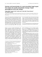
The HB-002.1 and Flt1[2]-Fc proteins were produced
in CHO cells upon transfection with the corresponding
construct. The secreted proteins were purified and
resolved in 10% SDS-PAGE gels showing MWs of HB002.1 and Flt1[2]-Fc at ~110 kDa in non-reducing
conditions, and ~45 kDa in reducing conditions (Figure 1B),
both relatively larger than the calculated MW, which
is most likely due to glycosylation since there are
two N-linked glycosylation sites in the D2 domain.
Bevacizumab resolved in the correct MW positions
(Figure 1B).
Results
Engineering and production of HB-002.1
It has been documented that the second Ig-like domain
(D2) of human VEGFR1 (Flt1[2]) is critical to VEGF
binding [32], however the purified Flt1[2] fused with Fc
A.
LS
VEGFR1-D2
hIgG1-Fc
Flt1(2)-Fc
N-Flank (5aa)
HB-002.1
B.
C-Flank (2aa)
D.
C.
Blotting with Fc-specific Ab
R
NR
hIgG-Fc
2
3
kDa
4
5
170
130
95
70
55
6
Load (µg):
1
0.5 0.25
kDa
170
130
95
70
55
1
0.5 0.25
hIgG-Fc
hIgG-Fc
hIgG-Fc
NR
R
1
Blotting with VEGFR1-specific Ab
R
Load (µg): 1
0.5 0.25
NR
kDa
1
0.5 0.25
170
130
95
70
55
43
43
43
34
34
34
25
25
25
1 4 HB-002.1;
2 5 Bevacizumab
3 6 Flt1(2)-Fc
Figure 1 Engineering and production of HB-002.1. (A) Diagram of HB-002.1 engineered structure. Illustration on top represents Flt1[2]-Fc
consisting of the D2 domain only fused with the Fc portion of human IgG1. HB-002.1 contains the D2 domain plus 5 and 2 amino acids at the
5' and 3' flanking region respectively. Signal peptide derived from the heavy chain of mouse IgG1 (LS) was included for both constructs. (B)
SDS-PAGE gel analysis. Three proteins were included in the analysis: HB-002.1 (Lane 1, 4); Bevacizumab (Lane 2, 5); Flt1[2]-Fc (Lane 3, 6). 5 μg of
each protein were loaded under reducing and non-reducing conditions. (C-D), Western blot analysis. 0.25, 0.5, and 1 μg of HB-002.1 protein and
1 μg of hIgG-Fc were resolved on 10% SDS-PAGE gels under reducing (R) or non-reducing (NR) conditions, transferred to a polyvinylidene difluoride
membrane, and probed with Fc-specific (C) or VEGFR1-specific (D) antibody (Ab). For comparison, protein MW size markers are shown in kDa.
Liu et al. BMC Cancer (2015) 15:170
Page 6 of 14
To confirm the identity of the proteins, Western blotting
was performed using antibodies specific for Fc (Figure 1C)
or VEGFR1 (Figure 1D), showing specific bands for each
specified portion of the protein at different protein loading
amounts.
HB-002.1 has strong binding affinity to VEGF-A
HB-002.1 was first analyzed for its binding affinity to
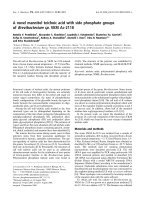
VEGF-A and compared with that of Flt1[2]-Fc and bevacizumab. The data showed that HB-002.1 had a high affinity with a half maximal effective concentration (EC50)
A.
D.
VEGF-A
4
3
2
1
0
0.001
0.01
HB-002.1
Bevacizumab
0.6
Flt1(2)-Fc
HB-002.1
Bevacizumab
hIgG-Fc
VEGF blocker (nM)
5
OD450
of 24 pM, which was 3-fold higher than that of bevacizumab (EC50 = 72 pM) (Figure 2A). As expected, Flt1[2]-Fc
only had a minimal binding activity to VEGF-A, confirming a binding requirement for the flanking sequence.
Binding activity of HB-002.1 to VEGF-B and PIGF was
also investigated by ELISA, showing a modest binding to
VEGF-B (Figure 2B) but low binding to PIGF (Figure 2C).
To determine the target-binding kinetics of HB-002.1,
equilibrium binding assays were performed in which
varying amounts of VEGF-A were mixed with 0.5 nM of
HB-002.1 or bevacizumab, and the unbound HB-002.1
0.1
1
0.4
0.2
0.0
0.01
10
0.1
conc. nM
VEGF-B
B.
E.
0.5
rhVEGFR1-Fc
HB-002.1
hIgG-Fc
0.4
OD450
1
10
100
VEGF-165 (nM)
Non-reducing
D
ND
Reducing
D
ND
0.3
0.2
0.1
0.0
0.1
1
10
100
1000
Protein conc. (nM)
PIGF
C.
F.
0.5
OD450
0.4
0.3
rhVEGFR1-Fc
HB-002.1
hIgG-Fc
0.2
0.1
0.0
0.1
1
10
100
1000
Protein conc. (nM)
Figure 2 Target binding activity of HB-002.1. Target binding activity of intact as well as deglycosylated HB-002.1 was analyzed by ELISA. (A) Binding
to VEGF-A was compared to bevacizumab and Flt1(2). hIgG-Fc was used as negative control. (B-C) Binding to VEGF-B (B) and PIGF (C) was compared
with rhVEGFR1-Fc. (D) Kinetic binding affinity was measured by equilibrium binding assays that measures unbound HB-002.1 or bevacizumab after
incubation of 0.5 nM of HB-002.1 or bevacizumab with varying amounts of VEGF-165. (E) HB-002.1 deglycosylated by treatment with N-glycosidase F
(D) or not deglycosylated (ND) was separated by SDS-PAGE under reducing or non-reducing conditions and visualized by staining with Coomassie
Brilliant Blue. (F) VEGF-A binding affinity was compared between intact (non-digested) and deglycosylated (digested) HB-002.1.
Liu et al. BMC Cancer (2015) 15:170
or bevacizumab was measured by ELISA using VEGF-A
coated plate, revealing that HB-002.1 displays an equilibrium dissociation constant (KD) of 180 pM, whereas bevacizumab has a KD of 890 pM (Figure 2D).
Since two different-sized bands were observed both in
SDS-PAGE gels and in Western blots, we wondered if
this was due to N-linked glycosylation and if this glycosylation might have an impact on VEGF binding. To
address these questions, HB-002.1 protein was digested
with N-glycosidase F and then resolved in 10% SDSPAGE gels, which showed a single band under reducing
conditions and a smaller size single band under nonreducing conditions when compared to that of nondigested parental protein (Figure 2E). This confirmed
our hypothesis that the doublet bands were due to Nlinked glycosylation. The digested protein retained similar VEGF-binding activity to that of parental protein
(Figure 2F), indicating glycosylation is not essential for
high affinity binding, which is consistent with the report
by Barleon et al [36].
HB-002.1 dose-dependently inhibited VEGF-induced
VEGFR2 phosphorylation, HUVEC proliferation and tube
formation
Due to the strong VEGF binding affinity, we anticipated
that HB-002.1 must also have strong blocking activity
against VEGF-induced VEGFR2 phosphorylation as well
as the resulting cell proliferation and tube formation. As
shown in Figure 3A, while strong phosphorylation was
observed with VEGF addition and VEGF plus hIgG, the
induced phosphorylation was sequentially diminished
following addition of sequentially increasing amounts of
HB-002.1, which is comparable to that of bevacizumab
showing a dose-dependent inhibition of VEGFR2 phosphorylation (Tyr951/1175) in HUVECs [37].
Comparisons between different treatment groups in
HUVEC proliferation (Figure 3B) by two-way analysis of
variance (ANOVA) provided a F-test with a small P value
(P < 0.0004) supporting subsequent evaluation for differences among treatment groups. Significant inhibition, as
compared to hIgG-Fc, in VEGF-induced HUVEC proliferation was observed in a dose-dependent manner for
HB-002.1 (P < 0.05 at all except lowest dose), which was
comparable to that of bevacizumab (P < 0.05 at all doses)
(Figure 3B). The same phenomenon was also observed for
VEGF-induced tube formation (Figure 3C, D), for which
HB-002.1 had a significant and comparable inhibition to
that of bevacizumab (P < 0.05) as compared to hIgG, suggesting a strong blocking activity of HB-002.1 in VEGFmediated cell biological activity.
HB-002.1 dose-dependently inhibited in vivo angiogenesis
Using a transgenic zebrafish embryonic angiogenesis model
[35], the impact of HB-002.1 on in vivo angiogenesis was
Page 7 of 14
investigated and showed a dramatic reduction in number
of SIVs. While 7-8 SIVs were usually observed in zebrafish
at 72 hpf (Figure 4A), a decreased number of SIVs was observed when treated with HB-002.1 (Figure 4B). The level
of inhibition versus vehicle group reached 7.5 (±3.5) %
(P > 0.05), 15.2 (±3.3) % (P < 0.01), and 21.4 (±2.4) %
(P < 0.001) for HB-002.1 at the doses of 4.4, 14.7, 44 ng, respectively. Because bevacizumab specifically binds human
VEGF and its activity against zebrafish VEGF is not known,
a broad spectrum angiogenesis inhibitor, endostatin,
known to inhibit angiogenesis in this model [38], was used
as a positive control showing a 9.7 (±2.8) % and 20.1 (±2.6)
% inhibition at the dose of 44 and 100 ng respectively
(Figure 4B). These results suggest a strong angiogenesis
inhibition activity for HB-002.1.
HB-002.1 has an excellent pharmacokinetic profile
HB-002.1 has a much smaller molecular mass than
current VEGF inhibitors, bevacizumab and aflibercept
(~80 vs. ~160 and ~110 kDa, respectively), thus it might
have a shorter half-life and worse PK profile compared
to these drugs. To address these questions, 2.5 mg/kg of
HB-002.1 were injected s.c. into mice (n = 16) and plasma
taken at different time points post-injection were analyzed
for HB-002.1 levels by ELISA. The results indicated that
HB-002.1 has a half-life of 5 days (Table 1), similar to that
of therapeutic antibodies, such as bevacizumab (~6 days
after 9.3 mg/kg s.c. injection) [39], and Fc-fusion proteins [24]. Furthermore, HB-002.1 has additional excellent
PK properties, with a maximal concentration (Cmax) of
20.27 μg/ml, mean residence time (MRT) of 7.5 days, and
total area under the curve concentration (AUC) of
81.46 μg · days/ml (Table 1). This is comparable to that
observed for therapeutic antibody, bevacizumab, starting
at a higher dose (9.3 mg/kg), with a Cmax of 74.1 μg/ml,
MRT of 8.74 days, and an AUC of 682 μg · days/ml
[39,40]. Interestingly, HB-002.1 PK properties are better
than that published for therapeutic fusion protein, despite
aflibercept starting at a higher dose (4 mg/kg), with a Cmax
of 16 μg/ml and an AUC of 36.28 μg · days/ml (Table 2)
[24]. Considering the lower isoelectric point (pI = 6.7) of
HB-002.1 compared to pI = 7.6 and 8.82 for bevacizumab
[37] and aflibercept [24], respectively (Table 2) and smaller
MW of HB-002.1, the excellent PK properties may be due
to better tissue penetration of the protein.
HB-002.1 exhibited robust in vivo anti-tumor activity
The anti-tumor activity of HB-002.1 was evaluated in two
different tumor models, Colo-205 and A549, representing
human colorectal cancer and lung cancer respectively.
BALB/c nude mice bearing these xenograft tumors were
treated with HB-002.1 as well as control drugs by i.p. injection, twice a week, for up to four weeks. Tumor volume
was measured twice a week and compared between
Liu et al. BMC Cancer (2015) 15:170
Page 8 of 14
A
B
C
D
Figure 3 (See legend on next page.)
Liu et al. BMC Cancer (2015) 15:170
Page 9 of 14
(See figure on previous page.)
Figure 3 In vitro biological activity. (A) HB-002.1 inhibited VEGF-induced VEGFR2 phosphorylation as revealed with immunoblotting assay. This
experiment was repeated three times, all showing a similar pattern of inhibition in VEGFR2 phosphorylation. (B) Inhibition of VEGF-induced HUVEC
cell proliferation was analyzed with the CCK-8 kit, a colormetric assay. Assay was repeated three times with duplicate wells for each concentration.
Representative assay is shown. Significant differences between HB-002.1 and hIgG-Fc (P < 0.05 at all except lowest dose), and bevacizumab and
hIgG-Fc (P < 0.05 at all doses) were observed. Bevacizumab and hIgG-Fc was used as positive and negative controls, respectively. (C) Representative
microscopic images of HUVEC tube formation in Matrigel are shown for medium alone, or for medium plus VEGF with or without HB-002.1,
Bevacizumab, or hIgG. (D) Tube formation was quantified by counting the total vessel length per field. Data were collected from duplicate
wells (mean ± standard deviation). Statistical significance was evaluated by ANOVA and Tukey’s multiple comparison test. Differences between
Medium versus VEGF and VEGF + HB-002.1 versus VEGF + Bevacizumab were not significant (NS). Differences between VEGF + HB-002.1 or
VEGF + Bevacizumab versus Medium or VEGF + hIgG were significantly different (P < 0.05).
groups. In the Colo-205 xenograft model, HB-002.1 was
compared to doxorubicin, a potent tumor chemotherapeutic drug that has widespread use clinically and has
demonstrated efficacy in several human tumor xenograft
models [41-43]. Compared to the PBS vehicle group, treatment with the positive control drug, doxorubicin, at
3 mg/kg by single bolus i.p. injection slightly inhibited the
tumor growth (TGI% = 19.78) (Figure 5A), while treatment
with HB-002.1 at 5 or 20 mg/kg i.p. twice weekly showed a
significant tumor growth inhibition, as indicated by the
decrease in tumor volume (TGI% = 93.17 at 5 mg/kg,
TGI% = 93.04 at 20 mg/kg, P < 0.0001) (Figure 5A) and
tumor weight (P = 0.0002) (Figure 5B). Interestingly, the
combination therapy of HB-002.1 with doxorubicin did
not show any synergistic increase in efficacy, being equivalent to HB-002.1 treatment alone (Figure 5A-B). Thus,
promisingly, even at the low dose (5 mg/kg), HB-002.1
treatment alone still reached maximal inhibitory effect in
this model, whereas bevacizumab was reported to only
reach TGI% = 55 at the dose of 4.0 mg/kg [44], suggesting
a robust anti-tumor activity for HB002.1.
To determine an effective dosing regimen of HB002.1, three doses were applied to the Colo-205 model,
which in comparison to PBS, showed a TGI% on day 28
of 55, 78.2, 82.1 at doses of 1.0, 3.0, and 5.0 mg/kg, respectively (Figure 5C, P < 0.0001). This was better than
A.
SIVs
B.
PBS
PBS
SIV=8
SIV=8
HB-002.1
SIV=5
Endostatin
44ng
4.4ng
SIV=5
100ng
14.7ng
SIV=3
SIV=3
44ng
SIV=2
Figure 4 In vivo angiogenesis inhibition. (A) Subintestinal vessels (SIVs) under normal conditions are shown. (B) HB-002.1 at different doses
(4.4, 14.7, 44 ng) (left) was injected into the blood flow during embryogenic development of zebrafish. Endostatin at two doses (44, 100 ng) was
included in the study as positive controls (right). Representative Images from one of the ten zebra fishes in each group are shown. Arrows point
to the number of SIVs in each group.
Liu et al. BMC Cancer (2015) 15:170
Page 10 of 14
Table 1 Pharmacokinetic parameters of HB-002.1
AUC (g · days/ml)
MRT (hr)
T1/2 (hr)
Cmax (μg/ml)
81.46
180
120
20.27
Note: AUC, area under the curve concentration; MRT, mean.
residence time; T1/2, half-life; Cmax: maximal concentration.
that reported for bevacizumab with 33, 41, and 44 TGI%
at doses of 1.2, 2.5, and 4.0 mg/kg, respectively, in the
same model [44]. Additionally, the TGI% for aflibercept
at a much higher dose of 25 mg/kg was reported to be
only 62-75 [41]. Tumor weight at the end of the study
also showed a dramatic and dose-dependent decrease in
the HB-002.1 treated group (Figure 5D, 1.0 (P = 0.0004),
3.0 and 5.0 (P = 0.0002) mg/kg dose). These studies
clearly revealed that HB-002.1 has remarkable antitumor activity, suggesting that HB-002.1 may be a potential alternative therapy for colorectal cancer.
The anti-tumor activity of HB-002.1 was also evaluated in the A549 xenograft model, and compared in parallel to that of bevacizumab, for two different dosages (5,
20 mg/kg). While bevacizumab showed a similar but
dramatic inhibitory effect at both doses, similar to that
previously reported [45], the inhibition was more pronounced for HB-002.1 even at the low dose (5 mg/kg)
(Figure 4E-F). When compared to that treated with PBS,
the TGI% was 78.02 and 84.71 for HB-002.1 at 5 and
20 mg/kg, respectively, which was significantly better
than bevacizumab at the same doses, 64.46 and 67.55
TGI%, respectively (Table 3).
HB-002.1 induced tumor growth inhibition is associated
with decreased microvessel density and increased
necrosis of tumor cells
To determine whether the inhibitory effect of HB-002.1
on tumor growth was associated with angiogenesis inhibition in tumor tissues as a result of VEGF blockade,
microvessel density was analyzed by staining tumor tissue
sections with CD31-specific antibody. Treatments with
5 mg/kg of HB-002.1 inhibited formation of CD31+
microvessels when compared to that of the vehicle group
in the Colo-205 or A549 xenograft models (Figure 6,
Table 4). This inhibition was quantified by measuring the
percentage of positive CD31 staining area against the total
tumor area by the visual approximation technique (1.9%
for HB-002.1 vs. 6.2% for PBS in the Colo-205 model;
0.7% for HB-002.1 vs. 7.1% for PBS in the A549 model)
(Table 4). Promisingly, HB-002.1 showed a more potent
effect on CD31+ vessel formation than that for doxorubicin in the Colo-205 model and that for bevacizumab in
the A549 model (Figure 6, Table 4). This inhibition of
microvessel formation in these models is similar to that
reported by others for bevacizumab and aflibercept
[41,44,45].
To confirm that tumor cell necrosis resulted because
of a decreased nutritional supply, H&E staining analysis
was conducted on tumors removed at the end of the
studies. As shown in Figure 7A-B, while little tumor necrosis was observed in vehicle-treated tumors, large regions of necrosis, as exhibited by decreased or absent
hematoxylin-stained (blue) tumor cell nuclei and disorganized cell outlines, were observed in HB-002.1-treated
tumors. This is comparable to that described for bevacizumab [45] and aflibercept [41].
Discussion and conclusion
In the current study, we engineered a new smaller-sized
recombinant VEGF-inhibiting Fc-fusion protein, HB002.1 (Figure 1), which had an excellent VEGF-binding
activity (Figure 2) and PK profile (Tables 1 and 2) comparable or better than the current generation of VEGFinhibiting drugs. This translated into excellent in vivo
anti-tumor efficacy, as shown by its superior therapeutic
efficacy as compared to bevacizumab in the A549 xenograft model (Figure 5E-F, Table 3). Even at low dose
(5 mg/kg), the inhibition mediated by HB-002.1 was still
two-fold better than that of bevacizumab at high dose
(20 mg/kg), although the better efficacy could be partially contributed by HB-002.1 cross-reaction with
mouse VEGF, which does not occur with bevacizumab.
More promisingly, HB-002.1 still reached 50% inhibition
of tumor growth in the Colo-205 model at a dose as low
as 1 mg/kg, suggesting a robust anti-tumor activity for
HB-002.1 (Figure 5C).
Achieving effective concentrations within solid tumor
masses has been challenging for large molecule drugs
Table 2 Comparison of selected PK parameters among VEGF inhibitors
Inhibitor
pI
Dose (mg/kg)
Cmax (μg/ml)
AUC (μg · days/ml)
Reference
HB-002.1
6.7
2.5
20.3
81.46
This study
Bevacizumab
7.6
9.3
74.1
682
39
Parental VEGF-Trap
9.4
4.0
0.05
0.04
24
VEGF-TRAPΔB1
9.1
4.0
1.3
1.36
24
VEGF-TRAPΔB2
8.9
4.0
2.65
5.42
24
Aflibercept
8.82
4.0
16
36.28
24
Liu et al. BMC Cancer (2015) 15:170
Page 11 of 14
A
C
E
B
D
F
Figure 5 In vivo efficacy study. (A-B) HB-002.1 treatment even at low dose (5 mg/kg) reached maximal inhibition of tumor growth in Colo-205 s.c.
xenograft model (n = 10 for each group) (A) P < 0.0001 versus PBS treatment for tumor volume over time. (B) P = 0.0002 versus PBS for tumor
weight at time of sacrifice. Treatment was started when tumor volume reached to 150-200 mm3, twice a week, through i.p. injection. (C-D) In
Colo-205 xenograft model (n = 10 for each group), varying amounts of HB-002.1 (1, 3, 5 mg/kg) were tested on effect on tumor growth. Significant
tumor growth inhibition was observed in the lowest dose (1 mg/kg, P < 0.0001 for tumor volume and P = 0.0004 for tumor weight versus PBS). (E-F)
Therapeutic efficacy of HB-002.1 in A549 xenograft model (n = 10 for each group) was analyzed and compared with that of bevacizumab at doses of 5,
20 mg/kg. HB-002.1 at low dose (5 mg/kg) exhibited superior efficacy than bevacizumab at high dose (20 mg/kg) (TGI% = 78.02 for HB-002.1, versus
67.55 for bevacizumab) (Table 3).
[46]. Better penetration and longer retention in the targeted area of the body are ideal parameters for large
molecule drugs to reach optimal therapeutic efficacy. It
has been known that the impact factors on penetration,
retention, as well as other PK properties, include molecular size, charge, valence, and target binding affinity.
For a given protein drug, the rate of diffusion through
tumors is inversely correlated to the molecular weight
[47,48]. scFv fragments diffuse approximately 6 times
faster than IgG due to their smaller size. However proteins with molecular mass less than 60 kDa, which is the
threshold for glomerular filtration, will be subject to
quick renal clearance resulting in shorter half-lives.
Molecular charge affects tumor distribution substantially. The defined pI range for optimal tumor penetration
is between 5 and 9 [49]; out of this range, therapeutic proteins are prone to immobilization by electrostatic interactions with the vascular endothelium and/or ECM [49].
Tumors have disordered tissues with regard to vasculature, interstitial fluid pressure, cell density, tissue structure
and composition, and ECM components [50]. Tumor
ECM is richer in collagen and stiffer than normal tissue
Table 3 Tumor growth inhibition in A549 xenograft model
Drugs
Mean Tumor Volume
Growth inhibition (TGI%)
P value vs PBS
P value vs Bevacizumab
HB-002.1, 5 mg/kg
2.72 (2.51-2.91)
78.02
<0.0001
<0.0001
HB-002.1, 20 mg/kg
1.89 (1.66-2.16)
84.71
<0.0001
<0.0001
Bevacizumab, 5 mg/kg
4.40 (3.59-5.40)
64.46
<0.0001
NA
Bevacizumab, 20 mg/kg
4.02 (3.36-4.81)
67.55
<0.0001
NA
PBS
12.38 (9.87-15.54)
0.00
NA
NA
Sample size = 10 for each group.
Mean Tumor Volume = geometric mean with 95% confidence interval in parentheses. Geometric mean is shown because statistical analyses utilized log-transformed
data that best modeled tumor volume growth.
P value was calculated as described in Methods.
P value vs Bevacizumab is compared to same dose of HB-002.1.
NA = not applicable.
Liu et al. BMC Cancer (2015) 15:170
Page 12 of 14
CD31 staining (Colo-205)
A.
PBS
Dox, 3mg/kg, single dose
HB-002.1, 5mg/kg
40X
B.
CD31 staining (A549)
Bevacizumab, 5mg/kg
PBS
HB-002.1, 5mg/kg
40X
Figure 6 Inhibition of tumor angiogenesis. Tumor angiogenesis analysis was performed by CD31 staining. Significant reduction in new blood
vessel formation (black arrows) was observed in HB-002.1 treated Colo-205 (A) as well as A549 (B) tumors, respectively. Representative fields of
CD31 staining in tumors treated with PBS (left) or doxorubicin (upper middle) or HB-002.1 (right) are shown at 40x magnification (40X).
ECM [51]. Proteins with high positive charge will be easily
adhered to highly negatively charged proteoglycans found
in the ECM. For example, the parental VEGF-Trap molecule before aflibercept had a high pI (9.4) and poor PK
properties, with a Cmax of only 0.05 μg/ml and total AUC
of 0.04 μg × days/ml (Table 2) [24]. Realizing the effectiveness of parental VEGF-Trap may be affected by its
high pI, basic amino acids were removed to create VEGFTrapΔB1, followed by removal of additional basic amino
acids to create VEGF-TrapΔB2, and finally replacing an entire protein domain with a less basic protein domain to
create aflibercept (Table 2) [24]. This molecular engineering caused a steady reduction of the pI from 9.4 to 8.82
Table 4 Percentage of positive CD31 staining in tumor
section
Group
% of CD31 in Colo-205 % of CD31 in A549
PBS
6.2 (+/- 2.1)
7.1 (+/-3.2)
Doxorubicin, 3 mg/kg
6.8 (+/- 3.6)
not done
HB-002.1, 5 mg/kg
1.9 (+/- 0.9)
Bevacizumab, 20 mg/kg not done
0.7 (+/- 0.2)
3.9 (+/- 2.9)
Note: Average (+/- SEM) from 3 samples in each group is shown.
and resulted in a concomitant improvement in the PK
profile, with a final Cmax of 16 μg/ml and an AUC of
36.28 μg × days/ml (Table 2) [24]. Thus, for a given
therapeutic protein, the ideal pI should be near physiologic pH as molecules with neutral charge diffuse
more readily.
To our knowledge, the robust anti-tumor efficacy of
HB-002.1 seen in xenograft models and superior efficacy
compared to bevacizumab could be attributed to three
reasons. The first reason is the molecular weight which
is only ~80 kDa, much smaller than both aflibercept
(~110 kDa) and bevacizumab (~160 kDa). The relatively
small size makes it easier to penetrate into tumor tissues
and accumulate, resulting in better bioavailability. The
second reason is the pI of HB-002.1 is near neutral,
which is the most ideal pI for a recombinant protein as
inverse correlation between the pI and the PK profile
has been seen with aflibercept (Table 4) [24]. The third
reason might be due to cross reactivity of HB-002.1 with
mouse VEGF, which is not seen with bevacizumab.
In conclusion, we have designed a novel recombinant
VEGF blocker designated as HB-002.1, which has an excellent PK profile and robust anti-tumor activity as
Liu et al. BMC Cancer (2015) 15:170
Page 13 of 14
A.
Colo-205
PBS
HB-002.1, 5mg/kg
4X
B.
A549
PBS
HB-002.1, 5mg/kg
20X
Figure 7 H&E staining analysis. H&E staining for HB-002.1 treated tumor sections at low dose (5 mg/kg) was presented and compared to that
of PBS treated group. Large regions of tumor necrosis were observed in Colo-205 (A) as well as A549 (B) tumor sections respectively. Representative
fields of H&E staining are shown at 4x (A) and 20x (B) magnification for Colo-205 and A549 tumor sections, respectively.
compared to the current generation of VEGF blockers.
The accumulated research data exhibited in this report
warrant further preclinical analysis of HB-002.1, which
will set the basis for clinical investigation.
Abbreviations
AMD: Age-related macular degeneration; ANOVA: Analysis of variance;
AUC: Area under the curve concentration; CHO: Chinese hamster ovary;
Cmax: Maximal concentration; D2: Second domain; EC50: Half maximal
effective concentration; ECM: Extracellular matrix; Fc: Immunoglobulin
constant region; H&E: Hematoxylin and eosin; hpf: Hours post-fertilization;
HRP: Horseradish peroxidase; HUVECs: Human umbilical vein endothelial
cells; Ig: Immunoglobulin; i.p.: Intraperitoneal; KD: Equilibrium dissociation
constant; MRT: Mean residence time; MW: Molecular weight; pI: Isoelectric
point; PK: Pharmacokinetic; PlGF: Placental growth factor; RM: Repeated
measures; s.c.: Subcutaneous; SIV: Subintestinal vessel; TGI%: Tumor growth
inhibition; VEGF: Vascular endothelial growth factor; VEGFR: VEGF receptor.
Competing interests
All authors, except for CCC, are full time employees of Huabo Biopharm Co.,
Ltd. and declare that they have no competing interest.
data analysis, and the writing of the manuscript. All authors have read and approved the final manuscript.
Acknowledgements
We thank Qiaocong Lao and Chunqi Li from Hangzhou Hunter Biotech for
zebrafish angiogenesis study, Alex Lai from Pharmalegacy Laboratories for
in vivo anti-tumor efficacy study as well as histology study in colo-205 and
A549 xenograft models, Ying Lian from Shanghai Medicilon Inc for in vivo
dosing study in colo-205 xenograft model, and Meredith Akerman with
Dr. Martin L. Lesser from the Biostatistics Unit at the Feinstein Institute of
Medical Research for assistance with statistical analyses.
Author details
1
Department of Cell Biology, Huabo Biopharm Co Ltd., Shanghai 201203,
China. 2Department of Antibody Technology, Huabo Biopharm Co Ltd.,
Shanghai 201203, China. 3Department of Protein Science, Huabo Biopharm
Co Ltd., Shanghai 201203, China. 4Department of Project Management,
Huabo Biopharm Co Ltd., Shanghai 201203, China. 5The Feinstein Institute for
Medical Research, North Shore-LIJ Health System, Manhasset, NY 11030, USA.
6
Department of Medicine, Hofstra North Shore-LIJ School of Medicine,
Hempstead, NY 11549, USA. 7Department of Molecular Medicine, Hofstra
North Shore-LIJ School of Medicine, Hempstead, NY 11549, USA.
Received: 27 January 2014 Accepted: 26 February 2015
Authors’ contributions
LL and HY are co-first authors and participated in the design of the project,
performed cell culture and development of stable cell lines secreting recombinant
protein. XH and HT carried out the in vitro cell based assays including cell
proliferation, phosphorylation, and tube formation assays. SL participated
in data analysis. YL participated in cell culture. LZ carried out ELISA assay.
SJ and HJ were responsible for protein purification. YX carried out SDS-PAGE
gel analysis. RZ participated in the design of project and in protein quality
analysis. YH was in charge of protein quality analysis. CCC aided in data analysis
and writing of the manuscript. WT participated in the design of the project,
References
1. Scott AM, Wolchok JD, Old LJ. Antibody therapy of cancer. Nat Rev Cancer.
2012;12:278–87.
2. Beckman RA, Weiner LM, Davis HM. Antibody constructs in cancer therapy:
protein engineering strategies to improve exposure in solid tumors. Cancer.
2007;109:170–9.
3. Crawford Y, Ferrara N. VEGF inhibition: insights from preclinical and clinical
studies. Cell Tissue Res. 2009;335:261–9.
Liu et al. BMC Cancer (2015) 15:170
4.
5.
6.
7.
8.
9.
10.
11.
12.
13.
14.
15.
16.
17.
18.
19.
20.
21.
22.
23.
24.
25.
26.
27.
28.
29.
Cao Y. Positive and negative modulation of angiogenesis by VEGFR1
ligands. Sci Signal. 2009;2:re1.
Escudero-Esparza A, Martin TA, Davies ML, Jiang WG. PIGF isoforms, PlGF-1
and PlGF-2, in colorectal cancer and the prognostic significance. Cancer
Genomics Proteomics. 2009;6:239–46.
Carmeliet P, Jain RK. Molecular mechanisms and clinical applications of
angiogenesis. Nature. 2011;473:298–307.
Saif MW. Anti-VEGF agents in metastatic colorectal cancer (mCRC): are they
all alike? Cancer Manag Res. 2013;5:103–15.
Thomas M, Mousa SS, Mousa SA. Comparative effectiveness of aflibercept
for the treatment of patients with neovascular age-related macular degeneration.
Clin Ophthalmol. 2013;7:495–501.
Yeung Y, Tebbutt NC. Bevacizumab in colorectal cancer: current and future
directions. Expert Rev Anticancer Ther. 2012;12:1263–73.
Whyte S, Pandor A, Stevenson M. Bevacizumab for metastatic colorectal
cancer: a NICE single technology appraisal. Pharmacoeconomics.
2012;30:1119–32.
Strickler JH, Hurwitz HI. Bevacizumab-based therapies in the first-line treatment
of metastatic colorectal cancer. Oncologist. 2012;17:513–24.
Soria JC, Mauguen A, Reck M, Sandler AB, Saijo N, Johnson DH, et al. Pignon
JP; meta-analysis of bevacizumab in advanced NSCLC collaborative group:
Systematic review and meta-analysis of randomised, phase II/III trials adding
bevacizumab to platinum-based chemotherapy as first-line treatment in patients
with advanced non-small-cell lung cancer. Ann Oncol. 2013;24:20–30.
Planchard D. Bevacizumab in non-small-cell lung cancer: a review. Expert
Rev Anticancer Ther. 2011;11:1163–79.
Narita Y. Drug review: safety and efficacy of bevacizumab for glioblastoma
and other brain tumors. Jpn J Clin Oncol. 2013;43:587–95.
Rahmathulla G, Hovey EJ, Hashemi-Sadraei N, Ahluwalia MS. Bevacizumab in
high-grade gliomas: a review of its uses, toxicity assessment, and future
treatment challenges. Onco Targets Ther. 2013;6:371–89.
Stevenson CE, Nagahashi M, Ramachandran S, Yamada A, Bear HD, Takabe
K. Bevacizumab and breast cancer: what does the future hold? Future
Oncol. 2012;8:403–14.
Garcia A, Singh H. Bevacizumab and ovarian cancer. Ther Adv Med Oncol.
2013;5:133–41.
Miyake TM, Sood AK, Coleman RL. Contemporary use of bevacizumab in
ovarian cancer. Expert Opin Biol Ther. 2013;13:283–94.
Eskander RN, Randall LM. Bevacizumab in the treatment of ovarian cancer.
Biogeosciences. 2011;5:1–5.
Krämer I, Lipp HP. Bevacizumab, a humanized anti-angiogenicmonoclonal
antibody for the treatment of colorectal cancer. J Clin Pharm Ther.
2007;32:1–14.
Thornton AD, Ravn P, Winslet M, Chester K. Angiogenesis inhibition with
bevacizumab and the surgical management of colorectal cancer. Br J Surg.
2006;93:1456–63.
Samant RS, Shevde LA. Recent advances in anti-angiogenic therapy of
cancer. Oncotarget. 2011;2:122–34.
Chu E. An update on the current and emerging targeted agents in
metastatic colorectal cancer. Clin Colorectal Cancer. 2012;11:1–13.
Holash J, Davis S, Papadopoulos N, Croll SD, Ho L, Russell M, et al. VEGF-Trap:
A VEGF blocker with potent antitumor effects. Proc Natl Acad Sci U S A.
2002;99:11393–8.
Wulff C, Wilson H, Wiegand SJ, Rudge JS, Fraser HM. Prevention of thecal
angiogenesis, antral follicular growth, and ovulation in the primate by
treatment with vascular endothelial growth factor Trap R1R2. Endocrinol.
2002;143:2797–807.
Kim ES, Serur A, Huang J, Manley CA, McCrudden KW, Frischer JS, et al.
Potent VEGF blockade causes regression of coopted vessels in a model of
neuroblastoma. Proc Natl Acad Sci U S A. 2002;99:11399–404.
Byrne AT, Ross L, Holash J, Nakanishi M, Hu L, Hofmann JI, et al. Vascular
endothelial growth factor-trap decreases tumor burden, inhibits ascites,
and causes dramatic vascular remodeling in an ovarian cancer model.
Clin Cancer Res. 2003;9:5721–8.
Inai T, Mancuso M, Hashizume H, Baffert F, Haskell A, Baluk P, et al.
Inhibition of vascular endothelial growth factor (VEGF) signaling in cancer
causes loss of endothelial fenestrations, regression of tumor vessels, and
appearance of basement membrane ghosts. Am J Pathol. 2004;165:35–52.
Teng LS, Jin KT, He KF, Zhang J, Wang HH, Cao J. Clinical applications of
VEGF-trap (aflibercept) in cancer treatment. J Chin Med Assoc. 2010;73:449–56.
Page 14 of 14
30. Cheng YD, Yang H, Chen GQ, Zhang ZC. Molecularly targeted drugs for
metastatic colorectal cancer. Drug Des Devel Ther. 2013;7:1315–22.
31. Park JE, Chen HH, Winer J, Houck KA, Ferrara N. Placenta growth factor.
Potentiation of vascular endothelial growth factor bioactivity, in vitro and
in vivo, and high affinity binding to Flt-1 but not to Flk-1/KDR. J Biol Chem.
1994;269:25646–54.
32. Davis-Smyth T, Chen H, Park J, Presta LG, Ferrara N. The second immunoglobulinlike domain of the VEGF tyrosine kinase receptor Fit-1 determines ligand
binding and may initiate a signal transduction cascade. EMBO J. 1996;15:4919–27.
33. Mason JM, Naidu MD, Barcia M, Porti D, Chavan SS, Chu CC. Interleukin-four
induced gene-1 (Il4i1) is a leukocyte L-amino acid oxidase with an unusual
acidic pH preference and lysosomal localization. J Immunol. 2004;173:4561–7.
34. Kureishi Y, Luo Z, Shiojima I, Bialik A, Fulton D, Lefer DJ, et al. The HMG-CoA
reductase inhibitor simvastatin activates the protein kinase Akt and
promotes angiogenesis in normocholesterolemic animals. Nat Med.
2000;6:1004–10.
35. Lam HW, Lin HC, Lao SC, Gao JL, Hong SJ, Leong CW, et al. The angiogenic
effects of Angelica sinensis extract on HUVEC in vitro and zebrafish in vivo.
J Cell Biochem. 2008;103:195–211.
36. Barleon B, Totzke F, Herzog C, Blanke S, Kremmer E, Siemeister G, et al.
Mapping of the Sites for Ligand Binding and Receptor Dimerization at the
Extracellular Domain of the Vascular Endothelial Growth Factor Receptor
FLT-1. JBC. 1997;272:10382–8.
37. Adamcic U, Skowronski K, Peters C, Morrison J, Coomber BL. The Effect of
Bevacizumab on Human Malignant Melanoma Cells with Functional VEGF/
VEGFR2 Autocrine and Intracrine Signaling Loops. Neoplasia. 2012;14:612–23.
38. Tan H, Mu G, Zhu W, Liu J, Wang F. Down-Regulation of Vascular Endothelial
Growth Factor and Up-Regulation of Pigment Epithelium Derived Factor Make
Low Molecular Weight Heparin-Endostatin and Polyethylene Glycol-Endostatin
Potential Candidates for Anti-angiogenesis Drug. Biol Pharm Bull. 2011;34:545–50.
39. Lin YS, Nguyen C, Mendoza JL, Escandon E, Fei D, Meng YG, et al. Preclinical
Pharmacokinetics, Interspecies Scaling, and Tissue Distribution of a
Humanized Monoclonal Antibody against Vascular Endothelial Growth
Factor. J Pharmacol Exp Ther. 1999;288:371–8.
40. U.S. Food and Drug Administration, Center for Drug Evaluation and Research.
Approval package for application number STN-125085/0: Pharmacology review
(s), Bevacizumab (AVASTIN). 2/25/2004.
41. Lassoued W, Murphy D, Tsai J, Oueslati R, Thurston G, Lee WM. Effect of
VEGF and VEGF Trap on vascular endothelial cell signaling in tumors. Cancer
Biol Ther. 2010;10:1326–33.
42. Papagiannaros A, Hatziantoniou S, Lelong-Rebel IH, Papaioannou GT,
Dimas K, Demetzos C. Antitumor activity of doxorubicin encapsulated in
hexadecylphosphocholine (HePC) liposomes against human xenografts on
Scid mice. In Vivo. 2006;20:129–36.
43. Bandyopadhyay A, Wang L, Agyin J, Tang Y, Lin S, Yeh IT, et al. Doxorubicin
in combination with a small TGFβ inhibitor: A potential novel therapy for
metastatic breast cancer in mouse models. PLoS ONE. 2010;5:e10365.
44. Yanagisawa M, Fujimoto-Ouchi K, Yorozu K, Yamashita Y, Mori K. Antitumor
activity of bevacizumab in combination with capecitabine and oxaliplatin in
human colorectal cancer xenograft models. Oncol Rep. 2009;22:241–7.
45. Ren H, Chu Z, Mao L. Antibodies targeting hepatoma-derived growth factor
as a novel strategy in treating lung cancer. Mol Cancer Ther. 2009;8:1106–12.
46. Beckman RA, Weiner LM, Davis HM. Antibody constructs in cancer therapy:
protein engineering strategies to improve exposure in solid tumors. Cancer.
2007;109:170–9.
47. Nugent LJ, Jain RK. Extravascular diffusion in normal and neoplastic tissues.
Cancer Res. 1984;44:238–44.
48. Gerlowski LE, Jain RK. Microvascular permeability of normal and neoplastic
tissues. Microvasc Res. 1986;31:288–308.
49. Melkko S, Halin C, Borsi L, Zardi L, Neri D. An antibodycalmodulin fusion
protein reveals a functional dependence between macromolecular
isoelectric point and tumor targeting performance. Int J Radiat Oncol Biol
Phys. 2002;54:1485–90.
50. Jang SH, Wientjes MG, Lu D, Au JL. Drug delivery and transport to solid
tumors. Pharm Res. 2003;20:1337–50.
51. Netti PA, Berk DA, Swartz MA, Grodzinsky AJ, Jain RK. Role of extracellular
matrix assembly in interstitial transport in solid tumors. Cancer Res.
2000;60:2497–503.