A retrospective analysis of RET translocation, gene copy number gain and expression in NSCLC patients treated with vandetanib in four randomized Phase III studies
Bạn đang xem bản rút gọn của tài liệu. Xem và tải ngay bản đầy đủ của tài liệu tại đây (974.37 KB, 10 trang )
Platt et al. BMC Cancer (2015) 15:171
DOI 10.1186/s12885-015-1146-8
RESEARCH ARTICLE
Open Access
A retrospective analysis of RET translocation, gene
copy number gain and expression in NSCLC
patients treated with vandetanib in four
randomized Phase III studies
Adam Platt1*, John Morten2, Qunsheng Ji3, Paul Elvin2, Chris Womack2, Xinying Su3, Emma Donald2, Neil Gray2,
Jessica Read2, Graham Bigley2, Laura Blockley2, Carl Cresswell2, Angela Dale2, Amanda Davies2, Tianwei Zhang3,
Shuqiong Fan3, Haihua Fu3, Amanda Gladwin2, Grace Harrod2, James Stevens2, Victoria Williams2, Qingqing Ye3,
Li Zheng3, Richard de Boer4, Roy S Herbst5, Jin-Soo Lee6 and James Vasselli7,8
Abstract
Background: To determine the prevalence of RET rearrangement genes, RET copy number gains and expression in
tumor samples from four Phase III non-small-cell lung cancer (NSCLC) trials of vandetanib, a selective inhibitor of
VEGFR, RET and EGFR signaling, and to determine any association with outcome to vandetanib treatment.
Methods: Archival tumor samples from the ZODIAC (NCT00312377, vandetanib ± docetaxel), ZEAL (NCT00418886,
vandetanib ± pemetrexed), ZEPHYR (NCT00404924, vandetanib vs placebo) and ZEST (NCT00364351, vandetanib vs
erlotinib) studies were evaluated by fluorescence in situ hybridization (FISH) and immunohistochemistry (IHC) in 944
and 1102 patients.
Results: The prevalence of RET rearrangements by FISH was 0.7% (95% CI 0.3–1.5%) among patients with a known
result. Seven tumor samples were positive for RET rearrangements (vandetanib, n = 3; comparator, n = 4). 2.8% (n = 26)
of samples had RET amplification (innumerable RET clusters, or ≥7 copies in > 10% of tumor cells), 8.1% (n = 76) had
low RET gene copy number gain (4–6 copies in ≥40% of tumor cells) and 8.3% (n = 92) were RET expression positive
(signal intensity ++ or +++ in >10% of tumor cells). Of RET-rearrangement-positive patients, none had an
objective response in the vandetanib arm and one patient responded in the comparator arm. Radiologic
evidence of tumor shrinkage was observed in two patients treated with vandetanib and one treated with
comparator drug. The objective response rate was similar in the vandetanib and comparator arms for patients positive
for RET copy number gains or RET protein expression.
Conclusions: We have identified prevalence for three RET biomarkers in a population predominated by non-Asians
and smokers. RET rearrangement prevalence was lower than previously reported. We found no evidence of a differential
benefit for efficacy by IHC and RET gene copy number gains. The low prevalence of RET rearrangements (0.7%) prevents
firm conclusions regarding association of vandetanib treatment with efficacy in the RET rearrangement NSCLC
subpopulation.
Trial registration: Randomized Phase III clinical trials (NCT00312377, ZODIAC; NCT00418886, ZEAL; NCT00364351,
ZEST; NCT00404924, ZEPHYR).
Keywords: RET rearrangement, Vandetanib, Non-small-cell lung cancer
* Correspondence:
1
AstraZeneca, da Vinci Building, Melbourn Science Park, Cambridge Road,
Melbourn, Royston, Hertfordshire SG8 6HB, UK
Full list of author information is available at the end of the article
© 2015 Platt et al.; licensee BioMed Central. This is an Open Access article distributed under the terms of the Creative
Commons Attribution License ( which permits unrestricted use, distribution, and
reproduction in any medium, provided the original work is properly credited. The Creative Commons Public Domain
Dedication waiver ( applies to the data made available in this article,
unless otherwise stated.
Platt et al. BMC Cancer (2015) 15:171
Background
Cancer treatment paradigms are evolving to exploit the
sensitivity of tumors to inhibitors that target the products of genes carrying driver mutations [1]. A number of
genetic aberrations that drive and maintain tumorigenesis have recently been identified in non-small-cell lung
cancer (NSCLC). These include fusion genes generated
by chromosomal rearrangements between the rearranged during transfection (RET) gene and other genes,
most commonly kinesin family 5B (KIF5B) and coiled
coil domain containing-6 (CCDC6) [2-12]. These fusions
lead to overexpression of truncated RET proteins containing the RET kinase domain, which can induce transformation and occur in tumors that rarely harbor
mutations in other common drivers, ie epidermal growth
factor receptor (EGFR), KRAS, human epidermal growth
factor receptor, and anaplastic lymphoma receptor (ALK)
genes. RET rearrangement was first shown to be associated with papillary thyroid carcinoma (PTC), leading to
the fusion oncoprotein (RET/PTC) and constitutive activation of RET receptor tyrosine kinase in papillary
cancer cells [13]. In addition, RET mutations are
present in the germline of nearly all patients with hereditary forms of medullary thyroid cancer (MTC)
[14-16] and approximately 50% of patients with sporadic MTC have somatic RET mutations that are associated with a worse outcome [17].
Vandetanib is a once-daily, oral anticancer agent that
selectively inhibits vascular endothelial growth factor receptor (VEGFR), RET and EGFR signaling [18,19], and is
licensed for the treatment of MTC in several geographical
regions. Preclinical studies have demonstrated that vandetanib inhibits RET signaling arising from RET mutations
in a MTC cell line and inhibits growth of human PTC cell
lines that carry spontaneous RET/PTC rearrangements
[19]. In addition, vandetanib has been shown to inhibit
the proliferation of cells expressing RET-KIF5B [2,3] and a
human lung adenocarcinoma cell line harboring an endogenous RET-CCDC6 [20].
Four randomized Phase III clinical trials have evaluated the efficacy of vandetanib in NSCLC in combination with docetaxel (NCT00312377; ZODIAC [21]),
in combination with pemetrexed (NCT00418886; ZEAL
[22]) or as a monotherapy (NCT00364351; ZEST [23] and
NCT00404924; ZEPHYR [24]). These studies in unselected patient populations demonstrated antitumor activity of vandetanib, but there was no overall survival benefit
when added to standard chemotherapy or as monotherapy
versus erlotinib [21-24]. The aim of this study, a retrospective evaluation of tumor samples from the NSCLC
Phase III program, was to determine the prevalence of
RET rearrangements and other potential RET biomarkers
within this population and to investigate any association
with outcome to vandetanib treatment.
Page 2 of 10
Methods
Overview of NSCLC studies
Study treatments and assessment of efficacy
All four studies are registered at www.clinicaltrials.gov
and were Phase III, multicenter, randomized, double-blind
studies conducted in 25 (ZODIAC), 21 (ZEAL), 22
(ZEPHYR) and 22 (ZEST) countries, respectively. Enrollment was conducted between 2006 and 2008. Full details of
the study design and methodology have been reported previously [21-24]. Briefly, in ZODIAC (NCT00312377), patients received docetaxel in combination with oral
vandetanib 100 mg/day (n = 694) or matched placebo (n =
697). Docetaxel 75 mg/m2 was administered as an intravenous infusion every 21 days, for a maximum of six cycles. In
ZEAL (NCT00418886), patients received pemetrexed in
combination with oral vandetanib, 100 mg/day (n = 256) or
matched placebo (n = 278). Pemetrexed 500 mg/m2 was
administered as an intravenous infusion every 21 days,
for a maximum of six cycles. Vandetanib was evaluated
as a monotherapy in two of the studies: in ZEPHYR
(NCT00404924), patients received vandetanib 300 mg/
day (n = 617) or placebo (n = 307); and in ZEST
(NCT00364351), patients received vandetanib 300 mg/
day (n = 623) or erlotinib 150 mg/day (n = 617). Objective
tumor assessments were categorized using Response
Evaluation Criteria in Solid Tumors (RECIST; version 1.0).
Patient eligibility
Data were evaluated from adult patients with histologically or cytologically confirmed locally advanced or
metastatic (stage IIIB to IV) NSCLC after failure of:
first-line anticancer therapy (ZODIAC and ZEAL); one
or two chemotherapy regimens (ZEST); or an EGFR inhibitor following one or two chemotherapy regimens
(ZEPHYR). For all studies, inclusion criteria included:
World Health Organization performance status 0–2; life
expectancy ≥12 weeks and no significant hematologic,
hepatic, renal or cardiac abnormalities. Patients with
squamous cell histology were also eligible. Prior treatment with VEGFR inhibitors (all studies), docetaxel
(ZODIAC only), pemetrexed (ZEAL only) and EGFR
inhibitors (ZEST only) was not permitted. Prior treatment with bevacizumab (all studies) and cetuximab
(ZEST only) was allowed.
Ethics statements
All patients provided written informed consent, the trials
were approved by all relevant institutional ethical committees or review bodies (The University of Texas MD
Anderson Cancer Center Surveillance Committee,
Houston, TX, USA; The Royal Melbourne Research
Foundation, Melbourne, Australia; Institutional Review
Board of National Cancer Center, Gyeonggi-do, Republic of Korea; Cedars-Sinai Medical Center Institutional
Platt et al. BMC Cancer (2015) 15:171
Review Board, Beverly Hills, CA, USA) and was conducted in accordance with the Declaration of Helsinki,
Good Clinical Practice, and the AstraZeneca policy on
Bioethics [25]. Data were generated in accordance with
the Medical and Healthcare Products Regulatory Agency
Good Clinical Practice Guidelines for laboratories [26].
Samples
Archival tumor specimens were sampled prior to enrollment of patients onto study. Provision of these samples
for genetic/immunohistochemical (IHC) assessment was
not compulsory in any study, resulting in collection from
approximately one third of patients. There was no observable bias to sampling consent. Tumor samples were
supplied as formalin-fixed paraffin-embedded biopsies,
resections or sections and could be obtained at any time
during the respective study. Cell lines used as controls
in IHC were supplied as cell pellets and stored at −80°C
prior to use. All analyses were carried out in AstraZeneca laboratories in the UK and China.
Assay methods
Fluorescence in situ hybridization (FISH) analysis
RET-KIF5B, alternative RET rearrangements and RET
gene amplifications were identified using a FISH probe
set of four fluorescent-labeled bacterial artificial chromosome (BAC) clone-derived DNA probes designed inhouse: RP11-124O11 (located upstream of RET) labeled
with spectrum red; RP11-718J13 (located immediately
downstream of RET) labeled with spectrum green (fluorescein isothiocyanate); RP11-983E11 (located upstream of
KIF5B exon 2) labeled with spectrum gold (5[6]-carboxyrhodamine 6G deoxyuridine-5′-triphosphate [dUTP]);
and centromere of chromosome 10 labeled with spectrum
aqua (diethylaminocoumarin-5-dUTP; Figure 1). For tissue samples assessed at the UK site, these probes were
sourced from Empire Genomics LLC, Buffalo, NY; for tissue samples assessed at the China site, the probes were
generated in-house. BACs and labels used at both sites
were identical. Sections were processed with reagents
from the Histology FISH accessory kit (Dako, Dako Corporation, Carpinteria, CA, USA, cat #K5799) according to
the manufacturer’s instructions. Briefly at the UK site,
formalin-fixed, paraffin-embedded sections (4 μm) were
mounted on glass slides. Sections were deparaffinized in
xylene, hydrated through a graded ethanol series, and then
incubated with the accessory kit pretreatment solution at
95–100°C for 10 minutes. The sections were then washed
and pepsin digestion was carried out at 37°C in a Dako
hybridizer for 5 minutes; the sections were dehydrated
through ethanol and allowed to air dry. The fluorescent
probe mix was applied and then the probe and section
co-denatured in a Dako hybridizer at 83°C for 3 minutes
followed by overnight incubation at 37°C. The sections
Page 3 of 10
were washed in 1x saline-sodium citrate (SSC)/0.3%
Igepal at 72°C for 2 minutes, followed by 2x SSC at
room temperature, before dehydration through ethanol.
Sections were mounted in Dako antifade mounting
media (Dako, #K5799). The procedure at the China site
was similar, except that the sections were incubated
with the Spotlight tissue pretreatment kit (Invitrogen,
Carlsbad, CA, USA, cat #00-8401) and processed as
previously described [27].
The FISH gene fusion assay had previously been validated in a pilot study, which confirmed a FISH assay defined RET-KIF5B positive NSCLC tumor by sequencing
(Additional file 1). Assessment of FISH signal was carried out by investigators blinded to clinical response.
Preliminary assessment was performed at 60x magnification to identify any alterations in the distribution of the
red and green signals. When these were observed, 50
tumor cells were analyzed at 100x magnification for the
number of red, green, paired red/green, gold, paired
green/gold and blue signals in 10–15 tumor cells from
up to four regions of the tumor section. A further region
was then analyzed by a second blinded observer. To describe tumor heterogeneity, estimation of the proportion
and distribution of cells with RET events was determined independently by two observers.
Immunohistochemistry
Sections were deparaffinized and rehydrated as described
above. Antigen retrieval was performed at 110°C for
5 minutes in Target Retrieval Solution (pH 9.0; Dako,
#S2367) in a RHS-2 microwave processor (Milestone,
Sorisole, Italy) within a pressurized reaction vessel. Endogenous peroxidase activity was quenched by incubating
the sections in 3% hydrogen peroxide for 20 minutes at
room temperature and non-specific binding was blocked
by incubating in serum-free protein block (Dako, #X0909)
for 20 minutes at room temperature. Sections were labeled with a rabbit anti-RET monoclonal antibody (1:1000
dilution, clone EPR2871, Epitomics, Burlingame, CA,
USA; see Additional file 1 for antibody specificity and immunohistochemistry validation; Additional file 1: Table
S1) and RET expression was visualized with EnVision™
FLEX+ (Dako, #K8012). Sections were counterstained
with hematoxylin. Cell lines (TT, MiaPaCa, SKNMC and
Panc1) and tissue samples (human non-inflamed appendix
and in-house NSCLC tissue microarray) were used as controls for RET immunostaining.
Statistical analysis
For FISH analysis, tumors were categorized as positive
for RET rearrangement if >10% of the tumor cells presented with broken-apart red and green RET signals; this
could be further classified as positive for RET-KIF5B if
the broken-apart red/green signal was accompanied by a
Platt et al. BMC Cancer (2015) 15:171
Page 4 of 10
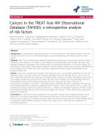
Figure 1 Representative FISH images. (A) Unknown RET rearrangement, (B) RET-KIF5B fusion, (C) RET gene amplifications and (D) low RET gene
copy number gain. (E) Loci for RET FISH probes.
Platt et al. BMC Cancer (2015) 15:171
Page 5 of 10
paired green/gold signal. Tumors were categorized as
low RET gene copy number gain if ≥40% of tumor cells
had 4–6 copies of red/green RET signals. Tumors were
categorized as amplified if >10% of tumor cells had ≥7
red/green signals or signal clusters. In the IHC analysis,
tumor samples with >100 intact tumor cells were
assessed by a system similar to that described previously
[8]. Tumors were categorized as RET expression positive
where the staining signal intensity was ++ or +++ (0 to +++
scale) in >10% of tumor cells. The objective response rate
(ORR) was presented by RET biomarker status and treatment arm with corresponding 95% confidence intervals
(CIs). Prevalence rates were estimated across all patients
with a known result (including those not randomized to
treatment) and were presented as a percentage with corresponding 95% CIs.
Results
Patients
From 4089 patients across the four NSCLC studies,
1291 and 1234 screened patients had tumor samples
available for FISH and IHC analysis, respectively. Evaluable data were obtained for 944 (FISH; Additional file 1:
Table S2) and 1102 (IHC; Additional file 1: Table S3) patients, with seven and eight patients donating FISH and
IHC samples, respectively, not randomized to treatment.
Failure rates in the IHC analysis (10.7%) were largely
due to an inadequate number of tumor cells in the samples, whereas in the FISH analysis, failure rates (26.9%)
were largely due to an inadequate number of tumor cells
or sample quality. The median age of patients was
61 years; approximately two-thirds were white, with the
remainder predominantly of Asian origin. Most patients
(61%) presented with adenocarcinoma. Patient demographics and baseline characteristics for patients with
tumor samples evaluable for FISH or IHC analysis and
clinicopathologic characteristics of patients and their
RET biomarker status are outlined in Additional file 1:
Tables S2–S4, respectively.
Prevalence of RET biomarkers
In this NSCLC patient population, the overall prevalence
of RET rearrangements was 0.7% (95% CI 0.3–1.5)
among patients with a known result. Seven tumor samples were classified as positive for RET rearrangements
(vandetanib treatment, n = 3; comparator treatment, n =
4; Table 1; Figure 1a and b). Five of the seven RET rearrangements were RET-KIF5B and the other two had unknown fusion partners with RET. Single red or green
signals without a corresponding 3′ or 5′ RET signal were
occasionally seen in samples but were not scored as
rearrangements.
RET gene amplifications and low RET gene copy number gains were reported in 26 (2.8%; 95% CI 1.8–4.0)
and 76 (8.1%; 95% CI 6.4–10.0) patients, respectively
(Table 1; Figure 1c and d). RET expression was positive
in samples from 92 (8.3%; 95% CI 6.8–10.1) patients
(Table 1). Tumor cell immunostaining was generally
cytoplasmic and diffuse (Figure 2). The prevalence of
RET rearrangement, RET protein expression, RET amplification or low RET gene copy number gain was similar
for Asian and non-Asian populations (Table 1).
In our study, four out of seven tumors that were positive for RET rearrangement expressed RET when evaluated with IHC. Of the remaining three samples, one
RET-KIF5B and two RET-other rearrangements did not
express any detectable RET protein when evaluated with
IHC (Additional file 1: Table S5). In all IHC-positive
cases, RET was predominantly cytoplasmic and typically
of moderate to weak intensity. In two cases, RET was
Table 1 Frequency of RET biomarkers in vandetanib Phase III NSCLC trial program
Clinical study
RET biomarker, n (%) [95% CI]
RET rearrangement
RET amplification
Low RET gene copy number gain
RET expression
Van
Van
Van
Van
Comp
Comp
Comp
Comp
ZODIAC + ZEAL
1
2**
6
7
14
14
16
24
ZEPHYR
2
1
2
3
19
5
18
10
ZEST
0
1**
4
2
8
14
12
10
Untreated*
–
2
2
2
Non-Asian
Asian
Overall
5/632 (0.8)
19/632 (3.0)
49/632 (7.7)
52/756 (6.9)
[0.3–1.8]
[1.8–4.6]
[5.7–10.0]
[5.2–8.9]
2/305 (0.7)
7/305 (2.3)
27/305 (8.8)
40/346 (11.6)
[0.1–2.3]
[0.9–4.6]
[5.9–12.5]
[8.4–15.4]
7/937 (0.7)
26/937 (2.8)
76/937 (8.1)
92/1102 (8.3)
[0.3–1.5]
[1.8–4.0]
[6.4–10.0]
[6.8–10.1]
Comp, comparator; Van, vandetanib. *One patient randomized to ZODIAC and one randomized to ZEAL did not receive treatment. **One RET rearrangement with
an unknown, non-KIF5B fusion partner was identified in the ZEAL comparator and one was identified in the ZEST comparator arm.
Platt et al. BMC Cancer (2015) 15:171
Page 6 of 10
Figure 2 Representative IHC images positive for RET expression. (A) Tumor biopsies and (B) resections. (C) Negative (weak) staining.
only detected in the focal areas of tumor cells. Weak
staining for RET was also observed in the stroma of
three RET-rearrangement-positive tumors. Of the seven
RET-rearrangement-positive tumors, only one showed
amplification which was also positive when evaluated
with IHC; this case also showed weak RET staining
across all of the stroma present in the sample.
Clinical outcome by positive RET biomarker status
None of the three vandetanib-treated RET-rearrangementpositive patients had an objective response (Table 2).
Radiologic evidence of tumor shrinkage was observed in
two of these patients (ZEPHYR, 23% and 33% shrinkage
of target lesions). However, a response could not be confirmed at the next visit. One patient in ZODIAC who received docetaxel alone had a confirmed objective response
and 32% shrinkage of target lesions at day 85 (Table 3).
The ORRs were similar for the vandetanib and comparator arms for patients positive for RET amplification (8.3%
vs 8.3%), those with low RET gene copy number gain
(9.8% vs 9.1%) or those positive for RET protein expression (15.2% vs 13.6%, Table 2). In conclusion, we considered there were too few RET-rearrangement-positive
patients to draw definitive conclusions regarding efficacy
in this patient population. However, the lack of additional
benefit observed in the higher prevalence biomarkerpositive groups of RET amplification and low RET gene
copy number gain is consistent with these biomarkers
having little predictive utility to identify those patients
who will benefit from vandetanib therapy.
Discussion
In this retrospective study, the overall prevalence of RET
rearrangements within the Phase III vandetanib NSCLC
clinical program was determined as 0.7% among patients
with a known RET rearrangement status. We found consistent frequencies of RET rearrangement in Asian
(0.7%) and non-Asian patients (0.8%). In general, RET
rearrangement prevalence rates may be considered as
low and may vary according to the proportions of
smokers, racial origin and histological subtype in the
study population. Prevalence rates of RET rearrangement
in Asian populations have been reported at 1–2% for
NSCLC [5,11] and lung adenocarcinoma [2,3,7,12], and
were estimated as high as approximately 6% in lung
adenocarcinoma [4]. Our own study contains a high proportion of non-Asian patients (67%, Additional file 1:
Table S2) and smokers/ex-smokers (77%, Additional file
1: Table S2), in contrast to previous reports on RET rearrangement prevalence rates [3,5,7,11,12], in which
study populations were either entirely or largely Asian
and non-smokers.
RET rearrangements have previously been reported in
squamous cell carcinoma [5], lung neuroendocrine
tumor [5] and adenosquamous tumor [11]; however, the
majority occur in adenocarcinomas. This is consistent
Table 2 ORRs (RECIST) in patients positive for RET biomarkers
Clinical study
Objective responses, n/N (%)
RET rearrangement
RET amplification
Low RET gene copy number gain
RET expression
Van
Comp
Van
Comp
Van
Comp
Van
ZODIAC + ZEAL
0/1
1/2
1/6
1/7
2/14
2/14
5/16
Comp
3/24
ZEPHYR
0/2
0/1
0/2
0/3
0/19
0/5
0/18
0/10
ZEST
0/0
0/1
0/4
0/2
2/8
1/14
2/12
3/10
Overall
0/3 (0.0)
1/4 (25.0)
1/12 (8.3)
1/12 (8.3)
4/41 (9.8)
3/33 (9.1)
7/46 (15.2)
6/44 (13.6)
Study
Age
Sex Race
(years)
Smoking
status*
Histology
EGFR status
KRAS
status
Dose/ Exposure
day
RECIST
response
Tumor shrinkage
Reason for
RET
% cells with
discontinuation partner rearrangements
detected
Vandetanib
ZODIAC 68
F
Asian Nonsmoker
Adenocarcinoma Mutation
negative
Negative
100
mg
21 days
Progressive
disease
–
Progressive
disease
KIF5B
50%
ZEPHYR 69
F
White Nonsmoker
Adenocarcinoma Mutation
negative;
amplification
positive
Negative
300
mg
180 days
Stable disease
23% shrinkage of
target lesions
Adverse event
KIF5B
75–100%
ZEPHYR 59
F
Asian Nonsmoker
Adenocarcinoma Mutation
negative;
amplification
positive
Negative
300
mg
57 days
Progressive
disease
33% shrinkage of
target lesions
(progressive disease
in non-target lesions)
Progressive
disease
KIF5B
50–75%
ZODIAC 59
F
White Ex-smoker Adenocarcinoma Mutation
negative
Unknown –
Six cycles
docetaxel
Partial response
(day 85);
progressive
disease (day
210)
32% shrinkage of
target lesions at day
85
Completed six
cycles
KIF5B
75–100%
ZEAL**
58
M
White Ex-smoker Large cell
carcinoma
Mutation
negative
Negative
–
Five cycles Progressive
pemetrexed disease (day
245)
None
Completed five
cycles
Not
known
75–100%
ZEPHYR 57
M
White Ex-smoker Adenocarcinoma Mutation
negative
Negative
–
26 days
placebo
Progressive
None
disease (day 25)
Progressive
disease
KIF5B
75–100%
ZEST**
M
White Ex-smoker Adenocarcinoma Mutation
negative;
amplification
positive
Negative
–
315 days
erlotinib
Progressive
disease (day
166)
Progressive
disease
Not
known
25–50%
Platt et al. BMC Cancer (2015) 15:171
Table 3 Clinicopathologic characteristics of seven patients positive for RET rearrangements
Comparator
70
None
F, female; M, male. *Non-smoker = never smoked >20 g tobacco in lifetime; ex-smoker = stopped smoking ≥1 year ago; occasional smoker = <1 tobacco product per day; habitual smoker = ≥1 tobacco product per
day. **RET rearrangements with unknown, non-KIF5B fusion partners.
Page 7 of 10
Platt et al. BMC Cancer (2015) 15:171
with our findings, which show a higher prevalence of
RET rearrangements in patients with adenocarcinoma
(1.2%, 6/510) compared with those in other histology
types (0.2%, 1/427). Our data are not in agreement with
a number of studies that report a higher frequency of
RET rearrangements in non-smokers compared with
smokers/ex-smokers; in our study, we have observed three
and four RET rearrangements, respectively (Table 3)
[2,5,11]. As in previous studies, we did not observe cooccurrence of RET rearrangements with EGFR and KRAS
mutations. Interestingly, one of the three RET-KIF5B tumors reported by Go et al. [28] in lung adenocarcinomas
negative for KRAS and EGFR mutations and ALK rearrangements was from a smoker. However, all of these observations should be interpreted with caution given the
small numbers.
The techniques used to identify RET rearrangement
genes in previously reported studies were sequencing
[2-4] or reverse transcription-polymerase chain reaction
followed by verification with FISH [11], sequencing
[5,12] or differential expression of 3′ and 5′ RET exons
[7,9,11]. Some of these techniques may underestimate
the frequency of RET rearrangement genes by failing to
detect fusions to partner genes other than KIF5B. We
used a four-probe FISH assay to detect RET rearrangements. This technique is highly sensitive in detecting
chromosomal rearrangements and has the advantage of
detecting other partners or isoforms, though it is not
known whether all these rearrangements are functional.
For example, in a study using a split FISH assay to
evaluate 1528 lung cancers, 22 tumors were detected
with split RET signals, of which 12 were confirmed as fusions with KIF5B and one with CCDC6 [10] and the
remaining nine tumors showed little or no expression of
the RET kinase domain.
Although the prevalence of RET rearrangements in
NSCLC patients is low, RET inhibition may be efficacious
within a subset of patients who carry these genetic aberrations. In this study, there were too few vandetanib-treated
patients with RET rearrangements to form conclusions regarding association with efficacy. A number of studies
have reported increased expression of RET protein in
NSCLC tumor cells, not necessarily associated with RET
rearrangements [2,3,8,11]. This led us to investigate both
IHC and RET copy number gain as possible predictive biomarkers for vandetanib response. No difference was observed in the ORRs between vandetanib and comparator
arms for IHC and copy number analyses.
In our study, we observed RET-KIF5B and other RET
rearrangements in samples that were negative for RET
protein expression. This observation is in line with previous studies of NSCLC samples which have used a
range of anti-RET antibodies (including the Epitomics
EPR2871 antibody we have used here) and differing IHC
Page 8 of 10
techniques [2,3,8,28]. Sasaki et al. [8] reported three
cases of RET translocation (from 371 NSCLC samples),
all of which had weak positive cytoplasmic staining when
evaluated with a 3F8 anti-RET mouse monoclonal antibody (Vector Laboratories, Peterborough, UK). In contrast, when using the EPR2871 antibody used in our
study, weak, moderate and strong staining were reported
for the three RET translocation positive samples; this
suggests that apparent RET expression is dependent on
both the antibody and the local IHC protocol used. Another study has reported weak to strong RET expression
with IHC when using the 3F8 anti-RET antibody; however, only one of the RET IHC positive cases was also
RET-KIF5B positive [3]. Using the EPR2871 antibody,
Kohno et al. reported 48/222 NSCLC cases to express
RET in the absence of a RET fusion; all cases of RETKIF5B were also RET positive with IHC [2]. Go et al.
[28] used three different anti-RET antibodies to screen
53 NSCLC cases for RET protein expression. RET IHC
positive cases were defined as those with >30% of cells
expressing cytoplasmic RET. Three samples that were
RET-fusion positive with whole transcript sequencing
were negative for RET with IHC, whereas RET protein
was identified in four samples, none of which harbored a
RET-KIF5B rearrangement. Taken together, these NSCLC
studies, along with our results, suggest that not all cases
of RET-KIF5B or other RET rearrangements express
RET protein when evaluated with IHC. RET protein appears to be largely cytoplasmic; however, considerable
inter-patient variation and heterogeneity among tumor
cells within individual tumors is observed.
Investigation of RET inhibitors in NSCLC patients
with a documented confirmation of a RET rearrangement
is an active area of research with three clinical studies
currently ongoing (NCT01639508, NCT01823068 and
NCT01813734). Results from a study on the use of vandetanib in RET-rearrangement-positive NSCLC patients
(NCT01823068) should provide further insight into the
role of vandetanib in this patient population. Preliminary data from NCT01639508, a prospective Phase II
trial investigating the use of cabozantinib, a smallmolecule inhibitor of MET, VEGFR2 and RET, has been
published [6]. For the first three patients treated with
cabozantinib, two patients showed confirmed partial
clinical responses and the third patient had prolonged
stable disease approaching 8 months [6]. A case study
in a patient with poorly differentiated lung adenocarcinoma, positive for a RET-KIF5B and refractory for
previous chemotherapy, is also noteworthy. In this
patient, 4 weeks of treatment with vandetanib 300 mg
once daily produced a fluorodeoxyglucose-positron
emission tomography/computed tomography response
[29]. In addition, in a preliminary study in which two
heavily pretreated patients with confirmed RET
Platt et al. BMC Cancer (2015) 15:171
rearrangements were treated with vandetanib, stable
disease was observed following treatment [30].
Conclusions
This study has demonstrated an overall prevalence of
0.7% for RET rearrangements in a large Phase III
NSCLC patient population among patients with a known
determination of RET rearrangement status composed
predominantly of non-Asian patients and smokers. RET
rearrangements were found most frequently, but not exclusively, in adenocarcinomas and occurred in tumors
negative for other driver mutations, in agreement with
previous reports in predominantly Asian populations.
The prevalence of RET rearrangement is too low in this
unselected population to determine whether RET-rearrangement-positive patients can be effectively treated
with RET inhibitors, such as vandetanib. Changes in
RET gene copy number and level of RET protein expression are more frequent aberrations than RET rearrangement in this NSCLC population, but also do not provide
predictive markers for response to vandetanib. Results
from additional studies, specifically in RET-rearrangement-positive NSCLC patients, are needed to determine
whether this patient population can be effectively treated
with RET inhibitors, such as vandetanib. If these studies
support RET rearrangements as a clinically relevant target, then screening of NSCLC patients for RET rearrangements may become part of standard care.
Additional file
Additional file 1: Supporting information for FISH assay validation,
Western blotting/IHC antibody specificity, localization of RET in
tumor tissue sections, patient demographics and baseline
characteristics for patients with tumor samples evaluable for FISH
and/or IHC analysis.
Competing interests
G Bigley, A Dale, S Fan, H Fu, Q Ji, A Platt, J Read, X Su, V Williams, Q Ye,
L Zheng and T Zhang are employed (other than primary affiliation; e.g.,
consulting) by AstraZeneca. L Blockley, E Donald, P Elvin, G Harrod, J Stevens,
J Morten, C Cresswell, A Davies, A Gladwin and C Womack are employed
by and own shares in AstraZeneca. J-S Lee has received research funding
from AstraZeneca. R de Boer has received research funding and honoraria
as a consultant from AstraZeneca. J Vasselli is employed by MedImmune.
R Herbst has no potential conflicts of interest.
Authors’ contributions
AP, PE, JM, ED, AG, CW and QJ were involved in the conception and design
of the study. XS, ED, VW, GB and CW developed the methodology for the
study. All authors obtained data for the study. The analysis and interpretation
of the data was performed by AP, JM, GB, LB, CC, AD, AD, NG, SF, HF, GH, JR,
JS, VW, QY, TZ, QJ, XS, LZ, ED, CW and PE. All authors contributed to the
writing and critical review of the manuscript. All authors read and approved
the final manuscript.
Authors’ information
Dr Womack was employed by AstraZeneca at the time of the study.
Page 9 of 10
Acknowledgments
We dedicate this article to the memory of our colleague Neil Gray, FIBMS.
The authors would like to acknowledge John Williams for facilitating the
delivery of the contracts with external parties to time, quality and cost,
Darren Hodgson for clinical data management, transmission, reporting and
critical review of data analysis files, and Jennifer Bradford for assistance with
analysis and interpretation.
This study was sponsored by AstraZeneca. Medical writing assistance was
provided by Claire Routley from Mudskipper Business Ltd and funded by
AstraZeneca.
Financial support
This study was sponsored by AstraZeneca.
Previously presented at American Society for Clinical Oncology (ASCO) 2013;
J Clin Oncol 31:(suppl; abst 8045).
Author details
1
AstraZeneca, da Vinci Building, Melbourn Science Park, Cambridge Road,
Melbourn, Royston, Hertfordshire SG8 6HB, UK. 2AstraZeneca, Macclesfield,
UK. 3Innovation Cancer Center, AstraZeneca R&D, Shanghai, China.
4
Department of Hematology & Medical Oncology, Western Hospital,
Melbourne, Victoria, Australia. 5Yale Comprehensive Cancer Center, New
Haven, CT, USA. 6National Cancer Center, Goyang, Republic of Korea.
7
AstraZeneca, Wilmington, DE, USA. 8Current address – MedImmune,
Gaithersburg, MD, USA.
Received: 18 March 2014 Accepted: 27 February 2015
References
1. Oxnard GR, Binder A, Janne PA. New targetable oncogenes in non-small-cell
lung cancer. J Clin Oncol. 2013;31:1097–104.
2. Kohno T, Ichikawa H, Totoki Y, Yasuda K, Hiramoto M, Nammo T, et al. KIF5B-RET
fusions in lung adenocarcinoma. Nat Med. 2012;18:375–7.
3. Lipson D, Capelletti M, Yelensky R, Otto G, Parker A, Jarosz M, et al. Identification
of new ALK and RET gene fusions from colorectal and lung cancer biopsies. Nat
Med. 2012;18:382–4.
4. Ju YS, Lee WC, Shin JY, Lee S, Bleazard T, Won JK, et al. A transforming KIF5B
and RET gene fusion in lung adenocarcinoma revealed from whole-genome
and transcriptome sequencing. Genome Res. 2012;22:436–45.
5. Cai W, Su C, Li X, Fan L, Zheng L, Fei K, et al. KIF5B-RET fusions in Chinese
patients with non-small cell lung cancer. Cancer. 2013;119:1486–94.
6. Drilon A, Wang L, Hasanovic A, Suehara Y, Lipson D, Stephens P, et al.
Response to cabozantinib in patients with RET fusion-positive lung
adenocarcinomas. Cancer Discov. 2013;3:630–5.
7. Li F, Feng Y, Fang R, Fang Z, Xia J, Han X, et al. Identification of RET gene
fusion by exon array analyses in “pan-negative” lung cancer from never
smokers. Cell Res. 2012;22:928–31.
8. Sasaki H, Shimizu S, Tani Y, Maekawa M, Okuda K, Yokota K, et al. RET
expression and detection of KIF5B/RET gene rearrangements in Japanese
lung cancer. Cancer Med. 2012;1:68–75.
9. Suehara Y, Arcila M, Wang L, Hasanovic A, Ang D, Ito T, et al. Identification
of KIF5B-RET and GOPC-ROS1 fusions in lung adenocarcinomas through a
comprehensive mRNA-based screen for tyrosine kinase fusions. Clin Cancer
Res. 2012;18:6599–608.
10. Takeuchi K, Soda M, Togashi Y, Suzuki R, Sakata S, Hatano S, et al. RET, ROS1
and ALK fusions in lung cancer. Nat Med. 2012;18:378–81.
11. Wang R, Hu H, Pan Y, Li Y, Ye T, Li C, et al. RET fusions define a unique
molecular and clinicopathologic subtype of non-small-cell lung cancer.
J Clin Oncol. 2012;30:4352–9.
12. Yokota K, Sasaki H, Okuda K, Shimizu S, Shitara M, Hikosaka Y, et al. KIF5B/
RET fusion gene in surgically-treated adenocarcinoma of the lung. Oncol
Rep. 2012;28:1187–92.
13. Lanzi C, Borrello MG, Bongarzone I, Migliazza A, Fusco A, Grieco M, et al.
Identification of the product of two oncogenic rearranged forms of the RET
proto-oncogene in papillary thyroid carcinomas. Oncogene. 1992;7:2189–94.
14. Carlson KM, Dou S, Chi D, Scavarda N, Toshima K, Jackson CE, et al. Single
missense mutation in the tyrosine kinase catalytic domain of the RET
protooncogene is associated with multiple endocrine neoplasia type 2B.
Proc Natl Acad Sci U S A. 1994;91:1579–83.
Platt et al. BMC Cancer (2015) 15:171
Page 10 of 10
15. Donis-Keller H, Dou S, Chi D, Carlson KM, Toshima K, Lairmore TC, et al.
Mutations in the RET proto-oncogene are associated with MEN 2A and
FMTC. Hum Mol Genet. 1993;2:851–6.
16. Mulligan LM, Kwok JB, Healey CS, Elsdon MJ, Eng C, Gardner E, et al. Germ-line
mutations of the RET proto-oncogene in multiple endocrine neoplasia type
2A. Nature. 1993;363:458–60.
17. Elisei R, Cosci B, Romei C, Bottici V, Renzini G, Molinaro E, et al. Prognostic
significance of somatic RET oncogene mutations in sporadic medullary
thyroid cancer: a 10-year follow-up study. J Clin Endocrinol Metab.
2008;93:682–7.
18. Wedge SR, Ogilvie DJ, Dukes M, Kendrew J, Chester R, Jackson JA, et al.
ZD6474 inhibits vascular endothelial growth factor signaling, angiogenesis,
and tumor growth following oral administration. Cancer Res. 2002;62:4645–55.
19. Carlomagno F, Vitagliano D, Guida T, Ciardiello F, Tortora G, Vecchio G, et al.
ZD6474, an orally available inhibitor of KDR tyrosine kinase activity,
efficiently blocks oncogenic RET kinases. Cancer Res. 2002;62:7284–90.
20. Matsubara D, Kanai Y, Ishikawa S, Ohara S, Yoshimoto T, Sakatani T, et al.
Identification of CCDC6-RET fusion in the human lung adenocarcinoma cell
line, LC-2/ad. J Thorac Oncol. 2012;7:1872–6.
21. Herbst RS, Sun Y, Eberhardt WE, Germonpre P, Saijo N, Zhou C, et al. Vandetanib
plus docetaxel versus docetaxel as second-line treatment for patients with
advanced non-small-cell lung cancer (ZODIAC): a double-blind, randomised,
phase 3 trial. Lancet Oncol. 2010;11:619–26.
22. de Boer RH, Arrieta Ó, Yang C-H, Gottfried M, Chan V, Raats J, et al. Vandetanib
plus pemetrexed for the second-line treatment of advanced non-small-cell
lung cancer: a randomized, double-blind Phase III trial. J Clin Oncol.
2011;29:1067–74.
23. Natale RB, Thongprasert S, Greco FA, Thomas M, Tsai C-M, Sunpaweravong
P, et al. Phase III trial of vandetanib compared with erlotinib in patients with
previously treated advanced non-small-cell lung cancer. J Clin Oncol.
2011;29:1059–66.
24. Lee JS, Hirsh V, Park K, Qin S, Blajman CR, Perng R-P, et al. Vandetanib versus
placebo in patients with advanced non-small-cell lung cancer after prior
therapy with an epidermal growth factor receptor tyrosine kinase inhibitor:
a randomized, double-blind Phase III trial (ZEPHYR). J Clin Oncol.
2012;30:1114–21.
25. AstraZeneca. Global Policy: Bioethics. 2011. Available at: http://
www.astrazeneca.com/Responsibility/Code-policies-standards/Our-globalpolicies.
26. MHRA. Good Clinical Practice for Clinical Laboratories. 2009. Available at:
/>www.mhra.gov.uk/home/groups/is-insp/documents/websiteresources/
con051910.pdf.
27. Xie L, Su X, Zhang L, Yin X, Tang L, Zhang X, et al. FGFR2 gene amplification
in gastric cancer predicts sensitivity to the selective FGFR inhibitor
AZD4547. Clin Cancer Res. 2013;19:2572–83.
28. Go H, Jung YJ, Kang HW, Park IK, Kang CH, Lee JW, et al. Diagnostic method
for the detection of KIF5B-RET transformation in lung adenocarcinoma.
Lung Cancer. 2013;82:44–50.
29. Gautschi O, Zander T, Keller FA, Strobel K, Hirschmann A, Aebi S, et al.
A patient with lung adenocarcinoma and RET fusion treated with
vandetanib. J Thorac Oncol. 2013;8:e43–4.
30. Varella-Garcia M, Xue LG, Mahale S, Berge EM, Bennati C, Le AT, et al. RET
rearrangements detected by FISH in “pan-negative” lung adenocarcinoma.
J Clin Oncol. 2013;31:abstr 8024.
Submit your next manuscript to BioMed Central
and take full advantage of:
• Convenient online submission
• Thorough peer review
• No space constraints or color figure charges
• Immediate publication on acceptance
• Inclusion in PubMed, CAS, Scopus and Google Scholar
• Research which is freely available for redistribution
Submit your manuscript at
www.biomedcentral.com/submit