Adult metanephric adenoma presumed to be all benign? A clinical perspective
Bạn đang xem bản rút gọn của tài liệu. Xem và tải ngay bản đầy đủ của tài liệu tại đây (1.84 MB, 8 trang )
Li et al. BMC Cancer (2015) 15:310
DOI 10.1186/s12885-015-1211-3
RESEARCH ARTICLE
Open Access
Adult metanephric adenoma presumed to be all
benign? A clinical perspective
Gang Li1, Yuhong Tang2, Renya Zhang3, Hualin Song1, Shumin Zhang1 and Yuanjie Niu1*
Abstract
Background: In most documented literature, metanephric adenoma (MA) is described as a benign tumour.
Nevertheless, the nature of MA remains unclear and the clinical criteria of different MA subtypes are not well
established. In the present study, we investigated the clinicopathological characteristics of MA, especially those
of the uncommon histological subtypes.
Methods: A cohort study was performed on 18 patients with pathologically proven MA in our institute from
January 2004 to June 2014. The patients’ clinicopathological and radiological data were retrospectively analysed
and evaluated with an emphasis on the corresponding subtypes.
Results: The patient population had a female: male ratio of 1:1 and mean age of 50 years (range, 18–66 years). The
mean tumour size was 3.9 cm (range, 1.4–9.0 cm). There were no pathognomonic radiological features that posed a
challenge for a preoperative diagnosis of MA. Fourteen patients underwent radical nephrectomy, and the other
four underwent partial nephrectomy. Three histological subtypes were observed: classic MA (n = 10), malignant MA
(n = 2), and composite MA with coexistence of different malignant components (n = 6). Despite the presence of
atypical histological features and malignant components among the patients, only one patient developed distant
metastasis (median postoperative follow-up, 56 months; range, 30–86 months).
Conclusions: MAs are a heterogeneous group of neoplasms with different biological characteristics. The correct
identification of this entity and its subtypes would facilitate stratification of optimal management protocols and
accurate assessment of the prognosis.
Keywords: Metanephric adenoma, MA, Benign tumour, MA subtypes, Clinicopathological characteristic
Background
The term ‘metanephric adenoma’ (MA) was originally
described by Bove in 1979 and is known to be associated
with Wilms’ tumour [1]. To date, fewer than 200 cases
of MA have been reported worldwide in the Englishlanguage literature. In most documented literature, MA
is characterised as a rare benign tumour of the kidney
that accounts for approximately 0.2% of adult renal epithelial neoplasms. It generally occurs in adults and has
an excellent prognosis. Nevertheless, the detailed nature
of MA remains unclear. Several reports have suggested
that a small subset of these tumours has atypical histological features or even an exponential growth pattern
[2], and the capacity for MA to become malignant has
* Correspondence:
1
Department of Urology, The second Hospital of Tianjin Medical University,
Tianjin Institute of Urology, Tianjin 300211, China
Full list of author information is available at the end of the article
been reported [3]. However, the clinical criteria of different MA subtypes are not well established. In the present
study, we investigated different MA subtypes and aimed
to establish clinical criteria that will facilitate more accurate therapy planning by using pathological findings as
the gold standard. Limited clinical data on MA are available in the English-language literature. To the best of
our knowledge, this is the largest clinical series to date
focusing on clinical and pathological subtype analysis
of MA.
Methods
Study design and patient selection
This retrospective observational cohort study was approved by the Institutional Review Board of the Second
Hospital of Tianjin Medical University. The study was
approved by all patients and written informed consents
© 2015 Li et al.; licensee BioMed Central. This is an Open Access article distributed under the terms of the Creative Commons
Attribution License ( which permits unrestricted use, distribution, and
reproduction in any medium, provided the original work is properly credited. The Creative Commons Public Domain
Dedication waiver ( applies to the data made available in this article,
unless otherwise stated.
Li et al. BMC Cancer (2015) 15:310
Page 2 of 8
were obtained from all patients to publish their clinical
details and images. The medical records of 18 patients
with pathologically proven MA were retrieved from the
archival files and retrospectively analysed in our institute
from January 2004 to June 2014. All pathologic specimens were acquired after surgery, and none were
diagnosed by biopsy. Preoperative abdominal ultrasound
and computed tomography (CT) examinations were
performed in all cases; magnetic resonance imaging was
performed in only three cases. The patients’ demographic characteristics, clinical presentation, radiological
characteristics (tumour diameter, location, CT value, and
growth and enhancement patterns), histological findings,
and perioperative and follow-up data were recorded.
Details of the patient’s clinicodemographic characteristics and CT findings are listed in Table 1. Abdominal
biphasic CT scans and three-phase contrast-enhanced
CT scans were performed in all cases. Data on calcification, tumour-spreading patterns, lymphadenopathy, and
enhancement patterns (homogeneous, heterogeneous)
were recorded and retrospectively analysed. All patients
were treated surgically; 14 underwent radical nephrectomy, and four underwent partial nephrectomy. The
tumour grade was assigned according to the World
Health Organization grading system. All pathological
diagnoses were determined by at least two urological pathologists. In inconclusive cases, the final diagnosis was
determined after consultation with senior pathologists.
No patients received any adjuvant therapeutic modalities.
The median follow-up period was 56 months (range, 30–
86 months). The therapeutic modalities, pathological
findings, and follow-up data are detailed in Table 1. The
types of surgical interventions, complications, postoperative management, and survival results were all retrospectively analysed.
Statistical analysis
The chi-squared test was used for categorical variables.
All reported nonparametric p-values are two-sided, and
statistical significance was set at p < 0.05. Ratios were
compared between the two groups using T tests. All data
were analysed using SPSS, version 17 (SPSS Inc., Chicago,
IL, USA).
Results
Clinical data and surgical treatment
In nine patients, the tumours were incidentally detected
on imaging studies performed for unrelated clinical presentations. Gross haematuria was found in five patients,
and loin pain was present in four patients. For small
tumours (<4 cm), the choice of surgical approach depended on the patient’s compliance and the attending
urologist’s individual preference. In complicated circumstances, such as tumour localisation in the central part
Table 1 The clinical, CT and pathological characteristics of MA
NO.
Sex/Age (years)
Tumor size (cm)
Unenhanced/enhanced
attenuation(Hu)
Treatment
modality
Pathology
diagnosis
Recurrence
or metastasis
1
F/48
2
37/61
NSS
MA
NO
2
F/65
3
30/87
RN
MA
NO
3
M/62
6.2
9/12
RN
MA
NO
4
M/45
4
21/42
RN
MA, PT
NO
5
F/33
2
20/25
NSS
MA
NO
6
F/64
4.5
30/46
RN
MA
NO
7
F/53
6.5
36/58
RN
MA, PT
NO
8
M/64
6
45/71
RN
MA
NO
9
F/38
5
46/107
RN
MA
NO
10
M/65
2.7
43/51
NSS
MA, AC
NO
11
F/50
9
20/58
RN
MA, CCC,
NO
12
M/50
4.3
25/53
RN
MA
NO
13
F/38
3.5
25/30
NSS
Malignant
NO
14
M/33
3.5
24/58
RN
MA
NO
15
F/47
4.7
33/76
RN
MA,OC
NO
16
M/43
4
26/64
RN
Malignant
M
17
M/51
3
28/34
RN
MA
NO
18
M/18
5.3
32/46
RN
MA, PT
NO
F: female, M: male, RN: radical nephrectomy, NSS: nephron sparing surgery, MA: Metanephric adenomas, CCC:chromophobe cell carcinoma, OC: Oncocytic carcinoma,
PT: Papillary tumor, AC: adenocarcinoma, M: metastasis.
Li et al. BMC Cancer (2015) 15:310
Page 3 of 8
of the kidney or the presence of an entophytic tumour,
partial nephrectomy is very difficult. Surgical parameters
including tumour stage, tumour size, operating time,
warm ischaemia time, and complications were documented. In the partial nephrectomy group, the mean preoperative tumour size was 2.5 cm (range, 1.4–3.5 cm), and
the clinical stage was T1a. All patients in the partial nephrectomy group underwent pedicle clamping, and the
mean (± standard deviation) warm ischaemia time was
26 ± 6 min. All surgical margins were negative. In the
radical nephrectomy group, the mean preoperative
tumour size was 5.2 cm (range, 3.5–9.0 cm), and the clinical stages were T1a (n = 13) and T1b (n = 1). One patient underwent resection of an enlarged lymph node with
a pathologically proven inflammatory reaction. All patients tolerated the surgery well and had an unremarkable
postoperative recovery.
Radiological findings
Ten tumours were found on the right side and eight were
found on the left side. Seven tumours were found in the
upper pole of the kidney, five in the middle pole, and six
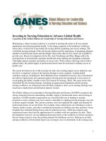
in the lower pole. The most common imaging characteristic on unenhanced abdominal CT was the presence of
homogeneous, well-defined solid renal masses (n = 15,
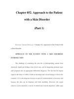
83.3%) (Figure 1); the least common was the presence of
heterogeneous or centrally located low-attenuation masses
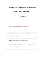
(n = 3, 16.7%) (Figure 2). Contrast-enhanced CT revealed
heterogeneity and varying degrees of enhancement in 16
(88.9%) tumours (Figure 3), while 2 (11.2%) tumours did
not exhibit increased attenuation. Scattered calcification
and an enlarged lymph node were found in only one patient (5.6%).
Pathological findings
Figure 2 CT showing a heterogeneous or centrally located
low-attenuation mass.
tan or yellowish-white colour, and the tumour was an encapsulated, generally well-circumscribed mass (Figure 4).
Microscopically, the MAs comprised variable proportions
of proliferating cells forming small glomeruloid bodies
(Figure 5). The tumour cells had uniformly small and indistinct nucleoli and scanty cytoplasm (Figure 6). Immunohistochemical staining showed that most tumour cells were
positive for WT-1 (Figure 7), CD57 (Figure 8), MIB-1,
Vimentin, and EMA, while CK7 staining showed weak
focal positivity. In two cases, the tumour cells exhibited
epithelial elements, lacked the typical architecture of tubules and glomeruloid bodies, and showed atypia and mitotic activity (Figure 9). The proliferation index of the
Macroscopically, the MAs ranged in size from 1.4 to
9.0 cm (mean, 4.5 cm). The cut surface was a homogenous
Figure 1 CT showing the presence of homogeneity and well-defined
solid renal masses.
Figure 3 Contrast-enhanced CT image revealed heterogeneous and
varying degrees of enhancement.
Li et al. BMC Cancer (2015) 15:310
Page 4 of 8
Figure 6 Tumours cells had uniformly small and indistinct nucleoli,
and scanty cytoplasm.
Figure 4 Macroscopically, MA revealed a homogenous tan or
yellowish-white colour cut surface and the encapsulated tumour
generally formed well-circumscribed mass.
MAs was 3–5% according to the MIB-1 count. The following composite tumours with foci of malignant tumour
cells were found in six patients: papillary tumour (n = 3),
oncocytic carcinoma (n = 1), adenocarcinoma (n = 1), and
chromophobe cell carcinoma (n = 1). The 18 cases were
divided into three subtypes according to the pathological
findings: classic MA (n = 10), malignant MA (n = 2), and
composite MA with coexistence of different malignant
components (n = 6). Pathological examination revealed
Figure 5 Microscopically,the tumour was composed of variable
proportions of cells proliferated with formation of small
glomeruloid bodies.
that six (33.3%) tumours had other carcinoma components
concomitantly and that two (11.1%) were malignant MA,
with a surprisingly high proportion of malignant case. All
of these pathological findings indicated the presence of
MA subtypes and provided useful information. When
stratified by malignant component groups, no significant
difference in prognosis was found (p > 0.05).
Follow-up
All patients were followed up with physical examinations,
laboratory tests, chest X-rays, and renal ultrasound or abdominal CT every 3–6 months and then annually. Clinical
outcomes were estimated from the date of surgery to the
date of death or last follow-up. The median postoperative
follow-up period was 56 months (range, 30–86 months),
and no local recurrence or metastatic lesions were found
with the exception of one patient who developed distant
metastasis pathologically diagnosed as malignant MA.
Figure 7 Immunohistochemical staining revealed most tumor cells
were positive expression of WT-1 (original magnification, ×200).
Li et al. BMC Cancer (2015) 15:310
Figure 8 Immunohistochemical staining of tumor cells were positive
for CD57 (original magnification, ×200).
Discussion
MA was well recognised as a distinct entity in 1988 and
was subsequently considered to be a separate entity [3].
The concept of MA has recently been broadened to include MAs, adenofibromas and stromal tumours. There
is a female preponderance and a peak age of occurrence
in the fifth or sixth decade of life. MA constitutes approximately 0.2% of all adult renal epithelial neoplasms.
The incidence of MA in our institution accounts for <1%
of all renal tumours, similar to previous reports. Approximately 100 cases of MA have been reported in the
English-language literature to date [1]. However, most reports focused mainly on pathology; few reports on the
clinical or radiological features are available.
Histogenetically, MA contains renal epithelial or stromal cells. It is postulated to be a benign counterpart of
Figure 9 Tumour cells were composed of epithelial elements and
lack of typical architecture of tubules and glomeruloid bodies
(original magnification, ×100), atypia and mitotic activity were
present (original magnification, ×200).
Page 5 of 8
Wilms’ tumour and may be derived from remnants of
metanephric blastemal or embryonic renal tissue. MA is
considered to represent the most hyperdifferentiated end
of the nephroblastoma spectrum and might sometimes
coexist with Wilms’ tumour [4]. The genetic profile and
chromosomal abnormalities of MA are distinct from those
of papillary renal cell carcinoma and Wilms’ tumour. The
simultaneous presence of BRAF gene mutation and 2p deletion plays a great role in the pathogenesis of MA [5].
Microscopically, the tumour cells have uniformly small
and indistinct nucleoli with scanty cytoplasm. Variable
proportions of cells proliferate with the formation of
small glomeruloid bodies. Immunohistochemical staining
shows that most tumour cells are positive for MIB-1,
vimentin, EMA, WT-1, and CD57; in contrast, CK7 staining exhibits weak focal positivity. In rare cases, the
tumour cells have epithelial elements, lack the typical
architecture of tubules and glomeruloid bodies, and show
atypia and mitotic activity. Atypical MA needs to be differentiated from Wilms’ tumour, nephrogenic rests, and papillary renal cell carcinoma [6]. MA has morphological
similarities to solid papillary renal cell neoplasms; both
exhibit significant similarities such as well-circumscribed
tumours comprising small tightly packed cells arranged in
solid sheets or ill-defined tubules. Some of the morphological features overlap; thus, the differential diagnosis is
crucial. MA must also be distinguished from metastatic
cancers, particularly those of the thyroid gland and lung.
Despite the overlapping features, careful morphological
evaluation, especially immunohistochemical staining with
CD57, WT1, and CK7, may be useful for differentiation
and accurate diagnosis. Meanwhile, genetic analysis may
facilitate discrimination in difficult cases. The presence of
cytological atypia, mitoses, and anaplastic foci favour the
diagnosis of malignant MA, especially distant metastasis.
Malignant MA tumours such as metanephric adenocarcinoma, mixed MA, and papillary carcinoma have also
been reported [7].
Surprisingly, tumours with typical histological characteristics of MA can present with metastatic disease [2]. Although the natural history of these composite tumours is
unknown, they theoretically exhibit aggressive behaviour
and the potential for metastasis. In the present study, MA
with other concomitant tumour types was determined to
be composite MA. This was based on the existing literature stating that a tumour mainly comprising MA that
exhibits sporadic concurrent tumours should be classified
as a subtype of MA. Composite MA with a co-existing
malignant component such as papillary renal cell carcinoma also has metastatic potential [8,9]. The features of
these composite tumours are emphasised to promote a
better and broader understanding of this uncommon
tumour. Notably, oncocytic carcinoma, renal adenocarcinoma, and chromophobe cell carcinoma mixed with
Li et al. BMC Cancer (2015) 15:310
MA were reported for the first time in the present investigation. Meanwhile, the cells of two tumours had atypical
epithelial elements and mitotic activities, lacked the
typical architecture of tubules, and glomeruloid bodies,
and were pathologically diagnosed as malignant MA; one
tumour was found to be a lung metastasis 46 months
postoperatively. The pathological criteria of malignant
MA are not well established, and rare metastatic MA has
been reported. In contrast to typical MA, malignant MA
comprises hypercellular uniform cells in a solid-acini pattern; the cells are variable in size, the nucleoli are prominent, and some cells show increased numbers of mitoses
with small uniform nuclei. The diagnosis of malignant
MA requires the incorporation of clinical information,
histopathological features, and related immunohistochemical staining markers.
Clinically, MA occurs predominantly in adult women
and rarely in children,the reported age ranged from
15 months to 83 years [10]. Most patients with MA are
asymptomatic or present with nonspecific clinical manifestations such as haematuria, a palpable mass, flank
pain, or chyluria [11]. Polycythaemia, which may be associated with para-neoplastic syndrome, is frequently reported among patients with MA. Most patients in the
present series were asymptomatic, and no special symptoms were noted. The regular performance of physical
examinations has led to a rise in the incidental detection
of asymptomatic renal masses. Additionally, MA may be
multifocal or bilateral [12,13]. Laboratory tests would be
less useful in this setting because no special tumour
markers are noted. Urinalysis and renal and hepatic
function tests were essentially within normal limits in
our series.
Various imaging modalities may be used to characterise
MAs. With respect to echogenicity, MA is a hypovascular
tumour and has most often been described as a hyperechoic mass [14]. However, the tumours in the present
study were hypoechoic, isoechoic, and hyperechoic in
nine, four, and five patients, respectively. Abdominal
three-phase contrast-enhanced CT was performed in all
18 patients, and no obvious correlations between morphologic features and characteristic CT imaging features were
found. No radiological findings were of substantial help in
differentiating MA from malignant renal tumours, especially for small masses. In our series, data on the tumourspreading patterns, lymphadenopathy, and enhancement
patterns were recorded and retrospectively analysed. An
enlarged lymph node was noted in one patient; the node
was pathologically proven to have an inflammatory reaction, similar to a pseudometastatic lesion. The most
common CT imaging characteristic was the presence of
homogeneous and well-defined solid renal masses (n = 15,
83.3%), and the least common was the presence of heterogeneous or centrally located low-attenuation masses
Page 6 of 8
(n = 3, 16.7%). Contrast-enhanced CT revealed hypoattenuating heterogeneous masses with varying degrees of
contrast enhancement in 16 (88.9%) patients, while 2
(11.2%) did not show increased attenuation. On unenhanced CT, one tumour (5.6%) showed scattered calcification with higher attenuation than the renal parenchyma.
MA appears to be more commonly calcified than other
neoplasm [15], which is speculated to be related to the
presence of psammomatous calcification or a high nuclearto-cytoplasmic ratio. Less frequently reported is hypoattenuation or predominantly cystic lesions consistent with
necrosis [16]. The hypovascularity of MA seems to reflect
the histological findings of mainly acinar and tubular patterns with few vessels. The magnetic resonance imaging
features of MA are unspecific; limited cases showed
hypointense or isointense lesions on both T1- and T2weighted magnetic resonance images [17].
Given the rarity of this tumour and lack of pathognomonic clinical and radiographic criteria, pathologic examination is necessary to establish a definitive diagnosis.
Because of the undetermined radiological characteristics
of MA, several reports recommend percutaneous fineneedle aspiration to confirm the diagnosis preoperatively
[18]. However, differentiation of MA from Wilms’ tumour
based on fine-needle aspiration biopsy may be difficult
[19]. MA may be mixed with other neoplasms that may
not be detected by intraoperative biopsy; thus, intraoperative frozen section is not recommended.
From a diagnostic and therapeutic viewpoint, most
renal masses should be regarded as malignant and managed surgically; the exception is small renal masses with
clinically benign behaviour. Accurate preoperative diagnosis could facilitate optimal management. More widespread recognition of this rare tumour and its subtypes
is of great importance for appropriate management of
this disease. Our initial classification of three subtypes of
MA may contribute to the establishment of guidelines
for the management of MA and help in selecting an appropriate surgical method.
Awareness of these subtypes may avoid diagnostic confusion, especially when percutaneous biopsy is indeterminate. When choosing a treatment modality, it might be
possible to propose conservative treatment or active surveillance, especially in patients with contraindications to
surgery [20]. It should be emphasised that these tumours
may not be entirely benign and that their biologic behaviour is uncertain, particularly malignant tumours and
those with malignant components. Thus, careful active
surveillance may be needed even for MAs of <4 cm. Continued growth or metastatic potential may be lethal; in
our opinion, therefore, MA should be routinely resected.
Although nephron-sparing surgery is currently the reference standard treatment for clinically localised T1a renal
tumours, subjective clinical factors such as surgeon biases
Li et al. BMC Cancer (2015) 15:310
and tumour characteristics, including the growth pattern,
more likely influence the decision-making process regarding the most appropriate treatment method. Small renal
tumours, especially exophytic and peripheral tumours, are
ideal candidates for nephron-sparing surgery, either open
or laparoscopic partial nephrectomy [21]. Several efficacious therapeutic procedures, such as cryoablation or radiofrequency ablation, are alternative treatment options
[22]. In our series, 14 patients underwent radical nephrectomy and only four underwent partial nephrectomy. Most
of our patients underwent nephrectomy mainly because of
the difficult preoperative differentiation of their lesions
from malignant renal tumours. Because imaging is unable
to exclude renal cell carcinoma, centrally located or anatomically complex masses should be treated by radical
nephrectomy [23].
Patients with MA treated with partial or total nephrectomy have an excellent prognosis. Only one patient in the
present series developed distant metastasis 46 months
after surgery. Long-term active surveillance is necessary
because of the uncertainty of the biological behaviour and
potentially composite malignant components of MA. Further studies on various subtypes are needed to identify the
possibility or occurrence of metastasis. Metastatic MA
containing foci of papillary carcinoma to local lymph
nodes were reported in one study; surprisingly, however,
the metastatic lesion was an MA, not a papillary carcinoma [7]. Therefore, aggressive intervention is needed for
composite MA with a coexisting malignant component.
There were several limitations in the present study.
First, the retrospective design and involvement of a single
centre might have introduced patient selection bias as well
as treatment bias with respect to surgeon preference. Additionally, the time interval of 10 years may have changes
in surgical techniques in our institute. Second, there was
lack of further molecular analysis of each subtype to
elucidate its histogenesis. Third, certain limitations were
unavoidable considering the relatively small number of
patients and the scarcity of different tumour subtype
variants. Moreover, it was not possible to determine the
percentage of morphological differentiation in the whole
group of specimens. Because only two malignant cases
were included in this study, statistical analysis and determination of significant differences were limited.
Conclusions
We demonstrated multiple variations in MA subtypes,
suggesting that their classification spectrum might be
wider than originally described. These interesting findings urge timely surgical treatment in all patients with
MA. The concept of the disease risk associated with malignant potential has been developed to aid clinicians
when deciding on treatment strategies; therefore, regular
follow-up is recommended.
Page 7 of 8
Competing interests
The authors declare that they have no competing interests.
Authors’ contributions
GL and TY conceived and directed the project. RZ, HS, SZ and YN analyzed data
and wrote the paper. All authors read and approved the final manuscript.
Acknowledgements
This study was supported by the National Natural Science Foundation for
Young Scholars of China (Grant 81302211) and Tianjin Research Program of
Application Foundation and Advanced Technology. (NO: 14CYBJC29800).
Author details
1
Department of Urology, The second Hospital of Tianjin Medical University,
Tianjin Institute of Urology, Tianjin 300211, China. 2Hebei North University,
Laboratory Medicine College, Zhangjiakou 075000, China. 3Department of
Pathology, Affiliated Hospital of Jining Medical University, Jining, China.
Received: 21 November 2014 Accepted: 18 March 2015
References
1. Spaner SJ, Yu Y, Cook AJ, Boag G. Pediatric metanephric adenoma: case
report and review of the literature. Int Urol Nephrol. 2014;46(4):677–80.
2. Renshaw AA, Freyer DR, Hammers YA. Metastatic metanephric adenoma in
a child. Am J Surg Pathol. 2000;24(4):570–4.
3. Grignon DJ, Eble JN. Papillary and metanephric adenomas of the kidney.
Semin Diagn Pathol. 1998;15(1):41–53.
4. Argani P. Metanephric neoplasms: the hyperdifferentiated, benign end of
the Wilms tumor spectrum? Clin Lab Med. 2005;25(2):379–92.
5. Dadone B, Ambrosetti D, Carpentier X, Duranton-Tanneur V, Burel-Vandenbos F,
Amiel J, et al. A renal metanephric adenoma showing both a 2p16e24 deletion
and BRAF V600E mutation: a synergistic role for a tumor suppressor gene on
chromosome 2p and BRAF activation? Cancer Genetics. 2013;206(9–10):347–52.
6. Pins MR, Jones EC, Martul EV, Kamat BR, Umlas J, Renshaw AA. Metanephric
adenoma-like tumors of the kidney: report of 3 malignancies with emphasis
on discriminating features. Arch Pathol Lab Med. 1999;123(5):415–20.
7. Drut R, Drut RM, Ortolani C. Metastatic metanephric adenoma with foci of
papillary carcinoma in a child: a combined histologic,
immunohistochemical, and FISH study. Int J Surg Pathol. 2001;9(3):241–7.
8. Galluzzo ML, Garcia de Davila MT, Vujanic GM. A composite renal tumor:
metanephric adenofibroma, Wilms tumor, and renal cell carcinoma:
a missing link? Pediatr Dev Pathol. 2012;15(1):65–70.
9. Zhu P, Yan F, Yang Z, Meng L, Ao Q. Composite tumor of metanephric
adenoma and Wilms’ tumor of the kidney: a case report and review of the
literature. Oncol Letters. 2013;5(4):1311–4.
10. Hartman DJ, Maclennan GT. Renal metanephric adenoma. J Urol.
2007;178(3 Pt 1):1058.
11. McNeil JC, Corbett ST, Kuruvilla S, Jones EA. Metanephric adenoma in a
five-year-old boy presenting with chyluria: case report and review of
literature. Urology. 2008;72(3):545–7.
12. Amie F, Andre D, Foulet Roge A, Goura E, Chautard D, Colombel P. [Bilateral
renal metanephric adenoma]. Prog Urol. 2004;14(4):534–7. discussion 537.
13. Kohashi K, Oda Y, Nakamori M, Yamamoto H, Tamiya S, Toubo T, et al.
Multifocal metanephric adenoma in childhood. Pathol Int. 2009;59(1):49–52.
14. Fielding JR, Visweswaran A, Silverman SG, Granter SR, Renshaw AA. CT and
ultrasound features of metanephric adenoma in adults with pathologic
correlation. J Comput Assist Tomogr. 1999;23(3):441–4.
15. Bastide C, Rambeaud JJ, Bach AM, Russo P. Metanephric adenoma
of the kidney: clinical and radiological study of nine cases. BJU Int.
2009;103(11):1544–8.
16. Torres Gomez FJ, Torres Olivera FJ, Garcia Escudero A. [Predominantly cystic
renal metanephric adenoma. Case report]. Arch Esp Urol. 2006;59(1):90–3.
17. Araki T, Hata H, Asakawa E, Araki T. MRI of metanephric adenoma. J Comput
Assist Tomogr. 1998;22(1):87–90.
18. Khayyata S, Grignon DJ, Aulicino MR, Al-Abbadi MA. Metanephric adenoma
vs. Wilms’ tumor: a report of 2 cases with diagnosis by fine needle
aspiration and cytologic comparisons. Acta Cytol. 2007;51(3):464–7.
19. Blanco Jr LZ, Schein CO, Patel T, Heagley DE, Cimbaluk DJ, Reddy V, et al.
Fine-needle aspiration of metanephric adenoma of the kidney with clinical,
Li et al. BMC Cancer (2015) 15:310
20.
21.
22.
23.
Page 8 of 8
radiographic and histopathologic correlation: a review. Diagn Cytopathol.
2013;41(8):742–51.
Torricelli FC, Marchini GS, Campos RS, Gil AO. Metanephric Adenoma:
clinical, imaging, and Histological findings. Clinics (Sao Paulo, Brazil).
2011;66(2):359–61.
Kumar S, Mandal AK, Acharya NR, Kakkad N, Singh SK. Laparoscopic
nephron-sparing surgery for metanephric adenoma. Surg Laparosc Endosc
Percutan Tech. 2007;17(6):573–5.
Conzo G, Sciascia V, Palazzo A, Stanzione F, Della Pietra C, Insabato L, et al.
Radiofrequency-assisted partial nephrectomy for metanephric adenoma:
a case report and literature review. Surg Innov. 2013;20(1):55–8.
Ebine T, Ohara R, Momma T, Saito S, Kuramochi S. Metanephric adenoma
treated with laparoscopic nephrectomy. Int J Urol. 2004;11(4):232–4.
Submit your next manuscript to BioMed Central
and take full advantage of:
• Convenient online submission
• Thorough peer review
• No space constraints or color figure charges
• Immediate publication on acceptance
• Inclusion in PubMed, CAS, Scopus and Google Scholar
• Research which is freely available for redistribution
Submit your manuscript at
www.biomedcentral.com/submit