The DNA load of six high-risk human papillomavirus types and its association with cervical lesions
Bạn đang xem bản rút gọn của tài liệu. Xem và tải ngay bản đầy đủ của tài liệu tại đây (581.8 KB, 11 trang )
Del Río-Ospina et al. BMC Cancer (2015) 15:100
DOI 10.1186/s12885-015-1126-z
RESEARCH ARTICLE
Open Access
The DNA load of six high-risk human papillomavirus
types and its association with cervical lesions
Luisa Del Río-Ospina1,2, Sara Cecilia Soto-De León1,3, Milena Camargo1,3, Darwin Andrés Moreno-Pérez1,3,
Ricardo Sánchez1,4, Antonio Pérez-Prados5, Manuel Elkin Patarroyo1,4 and Manuel Alfonso Patarroyo1,2*
Abstract
Background: Analysing human papillomavirus (HPV) viral load is important in determining the risk of developing
cervical cancer (CC); most knowledge to date regarding HPV viral load and cervical lesions has been related to HPV-16.
This study evaluated the association between the viral load of the six most prevalent high-risk viral types in Colombia
and cervical intraepithelial neoplasia (CIN) frequency.
Methods: 114 women without CIN and 59 women having CIN confirmed by colposcopy, all of them positive by
conventional PCR for HPV infection in the initial screening, were included in the study. Samples were tested for six
high-risk HPV types to determine viral copy number by real-time PCR. Crude and adjusted odds ratios (ORa) were estimated
for evaluating the association between each viral type’s DNA load and the risk of cervical lesions occurring.
Results: The highest viral loads were identified for HPV-33 in CIN patients and for HPV-31 in patients without lesions (9.33
HPV copies, 2.95 interquartile range (IQR); 9.41 HPV copies, 2.58 IQR). Lesions were more frequent in HPV-16 patients having a
low viral load (3.53 ORa, 1.16–10.74 95%CI) compared to those having high HPV-16 load (2.62 ORa, 1.08–6.35 95%CI). High
viral load in HPV-31 patients was associated with lower CIN frequency (0.34 ORa, 0.15–0.78 95%CI).
Conclusions: An association between HPV DNA load and CIN frequency was seen to be type-specific and may have
depended on the duration of infection. This analysis has provided information for understanding the effect of HPV DNA load
on cervical lesion development.
Keywords: Cervical intraepithelial neoplasia, HR-HPV, HPV DNA load, RT-PCR
Background
The main factor for developing cervical cancer (CC) lies
in persistent infection by at least one viral type of highrisk human papillomavirus (HR-HPV). Fifteen types of
HR-HPV have been described, 99.7% being associated
with cases of CC and/or cervical intraepithelial neoplasia
(CIN) [1-3]. However, some host and virus related factors modulate such association, i.e. HPV viral load [4,5].
Researchers have thus become interested in HPV viral
load. Its association with infection duration has already
been described [6,7]. Prior studies have determined the
association between viral load and CC severity, progression and development, whilst others have found that the
* Correspondence:
1
Molecular Biology and Immunology Department, Fundación Instituto de
Inmunología de Colombia (FIDIC), Carrera 50#26-20, Bogotá, Colombia
2
School of Medicine and Health Sciences, Universidad del Rosario, Carrera
24#63C-69, Bogotá, Colombia
Full list of author information is available at the end of the article
amount of HPV DNA increases proportionally with lesion severity and can even be detected before cervical lesions develop [8-11]. However, other studies have found
no such association [12-14].
As HPV-16 is the viral type most associated with cases
of CC (50%–70%) [3,5], most knowledge concerning
HPV viral load and CC has been based on HPV-16.
Studies, which have included other HR-HPV types, have
not led to comparable results regarding those obtained
for HPV-16 [15,16].
The real-time polymerase chain reaction (RT-PCR)
has been widely used and described in detecting and
typing HPV, as well as quantifying a broad range of viral
copies and normalising viral load according to the
amount of human DNA, having high reproducibility,
sensitivity, specificity and yield [13,17]. It was thus considered that it would provide a suitable approach for
© 2015 Del Río-Ospina et al.; licensee BioMed Central. This is an Open Access article distributed under the terms of the
Creative Commons Attribution License ( which permits unrestricted use,
distribution, and reproduction in any medium, provided the original work is properly credited. The Creative Commons Public
Domain Dedication waiver ( applies to the data made available in this
article, unless otherwise stated.
Del Río-Ospina et al. BMC Cancer (2015) 15:100
measuring HPV viral load, thereby facilitating investigating
the role of HR-HPV viral load in developing CC [10,12,18].
The present study was thus aimed at using RT-PCR
for determining the association between HPV viral load
and the presence of CIN for six HR-HPV types, which
have been previously reported as having the greatest
prevalence in Colombia [19]. It was thus expected to
contribute towards knowledge regarding the parameters
leading to identifying HPV positive women having a
higher risk of developing cervical lesions.
Methods
Study population and ethical considerations
Women eligible for the present study were voluntarily
attending their cervical screening consultations between
April 2007 and March 2010 in three Colombian regions
(Girardot, Chaparral and Bogotá). Bogotá (the capital of
Colombia) has the highest percentage of inhabitants, being mainly an urban population. Girardot is a city located in the Cundinamarca department which has
focused its economy on the tourist sector due to its climate and infrastructure. The city of Chaparral (Tolima
department) was included in the study as it is located in
Colombia’s coffee-growing region and is also known for
ecotourism. Girardot and Chaparral were grouped together in the “other city” category to improve the quality
of the present study’s statistical analysis.
All the women signed a written informed consent
form and completed a questionnaire regarding their
sociodemographic characteristics, sexual behaviour and
risk factor data before undergoing a gynaecological
examination and providing a cervical smear. Samples
were analysed using the Papanicolaou test and HPV
DNA detection. Colposcopy and biopsy were performed in
accordance with current Colombian screening programme
guidelines, thereby establishing that women having normal,
satisfactory cytology would continue following the 1-1-3
scheme, meaning that they should have a new control in a
year’s time and, if this continued being normal, in three
year’s time. However, colposcopy would be required when
cytology was abnormal and, in case colposcopy was abnormal, samples would then be taken for pathology study, as in
this study, for diagnosing CIN 1 and CIN 2+ [20]. Colposcopy and biopsy were also carried out for women having
normal cytology but who were positive for HPV by conventional PCR, as previous studies have reported an increased
risk of CIN 2+ development in women having normal cytology when they are HPV positive [21]. Due to biopsy not
being taken from women having negative colposcopy,
complete or satisfactory colposcopy (squamocolumnar
junction completely visible), evaluation of the transformation area, having normal vascularisation and squamous,
cylindrical epithelia without alterations were taken as
criteria for guaranteeing the absence of lesions [22].
Page 2 of 11
Colposcopy was chosen as the best method for defining the presence or absence of cervical lesions, as previous studies have found that colposcopy has a good
correlation with histological results [23] and it remains the standard for detecting cervical lesions until
new methods can be applied; in addition, cervical cytology has been reported worldwide as having variable
sensitivity for detecting pre-neoplastic lesions and is
considered a screening method which identifies
women at risk of developing CC who must then be
submitted to definitive diagnostic methods (colposcopy and biopsy) [20,24-26]. Women who had both a
colposcopy result and HPV DNA detected by conventional PCR were thus included. Women were excluded in
whom there was no amplification of the Homo sapiens
hydroxymethylbilane synthase (HMBS) gene (Gene ID:
3145) by RT-PCR and those having an insufficient sample
for analysis (Figure 1).
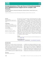
Figure 1 Flowchart of the studied population. *Inclusion criteria:
women who had both a colposcopy result and HPV DNA detected
by conventional PCR. RT-PCR: real-time polymerase chain reaction;
HMBS: hydroxymethylbilane synthase gene; HR-HPV: high-risk human
papillomavirus; CIN: cervical intraepithelial neoplasia; CIN 1: cervical
intraepithelial neoplasia 1; CIN 2+: cervical intraepithelial neoplasia 2
or 3.
Del Río-Ospina et al. BMC Cancer (2015) 15:100
Page 3 of 11
This study was supervised and approved by each institution’s Ethics Committee as follows: Fundación Instituto de Inmunología de Colombia’s Ethics Committee
and the Ethics Committee of the Nuevo Hospital San
Rafael E.S.E, Girardot, the Hospital San Juan Bautista de
Chaparral E.S.E. Bioethics Committee and Hospital de
Engativá (level II) Ethics Committee.
HPV DNA collection, processing and detection by
conventional PCR
Genomic DNA from cervical samples (stored at 4°C, in
95% ethanol) taken from HR-HPV 16, 18, 31, 33, 45 and
58 patients, which had been previously confirmed by
conventional PCR (proving positive for at least one of
the following previously described primers: GP5+/6+,
MY09/11 or pU1M/2R) [27], was extracted using a
Quick Extract DNA Extraction Solution kit (Epicentre,
Madison, WI), according to the manufacturer’s recommendations. The samples were homogenised in 200 μL
lysis buffer and incubated at 65°C for 6 minutes and
then at 92°C for 2 minutes. The samples were then spun
at 13,000 rpm for 10 minutes and the supernatant was
stored at −20°C until use.
Viral load quantification by RT-PCR
The methodology used in this study has already been described in detail in a previous article by our group [28].
Briefly, specific primers for each viral type and for
HMBS were synthesised according to a study published
by Moberg et al. [13]. The probes for each viral type and
HMBS were designed, taking into account the types included in each reaction. Four parallel duplex real-time
PCRs per patient were carried out (Table 1).
The cervical samples processed and identified as being
HPV-positive by conventional PCR were used as template in PCR reactions for each fragment. The amplicons
so obtained were purified with a Wizard PCR preps kit
Table 1 The probes and quenchers used for real-time
polymerase chain reaction
Test
Viral type
Size (bp)
Probe
Quencher
Reaction 1
HPV-16
78
FAM
ZEN/IBFQ
Reaction 2
HPV-18
80
Cy5
IBRQ
HPV-31
78
HEX
ZEN/IBFQ
HPV-33
78
FAM
ZEN/IBFQ
HPV-45
76
Cy5
IBRQ
HPV-58
109
HEX
ZEN/IBFQ
HMBS
76
FAM
ZEN/IBFQ
Reaction 3
Reaction 4
Four parallel duplex real-time PCRs were performed per patient. Probe design
for each viral type and HMBS was adjusted based on the types included in
each reaction.
HPV: human papillomavirus; FAM: 6-carboxyfluorescein; Cy5: FluoroLink mono
reactive dye Cy5; HEX: hexachlorofluoresceine; HMBS: hydroxymethylbilane
synthase; ZEN/IBFQ: ZEN and Iowa Black FQ; IBRQ: Iowa Black RQ.
(Promega), once their quality has been evaluated on
3.25% agarose gel. A TOPO TA cloning kit was used for
ligation, followed by transformation in TOP10 E. coli
cells (Invitrogen). Several clones were incubated in LB
broth and kept overnight (250 rpm at 37°C). Recombinant plasmids were purified using an UltraClean mini
plasmid prep kit (MO BIO laboratories, California, USA)
and sequenced using an automatic ABI PRISM 310 Genetic Analyser (PE Applied Biosystems, California, USA).
Each insert’s integrity was checked by aligning the products with the respective theoretical sequenced fragments
from each gene using Clustal W software [29].
Real-time PCR
Standardised RT-PCR assays with 10-fold serial plasmid
dilutions (1011-106copies) (using known DNA concentration and copy number) gave a standard curve for each
viral type and the HMBS gene. CFX96 Touch RT-PCR
detection system was used for analysis. Samples were
tested for HPV-16, HPV-18, HPV-31, HPV-33, HPV-45
and HPV-58. The human HMBS gene was amplified in
all samples to verify DNA integrity and determine viral
copy number per cell. Four RT-PCR reactions were carried
out per sample: HPV-16, HPV-18 and -31, HPV-33 and -45
and HPV-58 and HMBS. RT-PCR reaction conditions and
protocols have been described previously [28].
Each run was performed in 96-well plates, including 6
standards for each viral type and HMBS, involving 10fold plasmid dilutions (1011–106 copy dynamic detection
range) and a no template control to rule out DNA
contamination.
The viral load was normalised to cellular DNA input
using a previously described formula (Equation 1) [15].
Absolute and normalised viral loads were both log10
transformed.
Normalised viral load formula
HPV DNA loadðHPV copies=cellÞ
Number of HPV copies
¼
ðNumber of HMBS copies=2Þ
ð1Þ
Statistical analysis
Sample size was calculated using the difference of proportions test for high viral load between women having
and without cervical lesions (0.42 and 0.052 respectively)
[8,30]; 0.05 significance, 90% statistical power and a 1:2
ratio between both groups were established. This meant
that at least 23 women with lesions and 46 women without them were required for the study. Based on the
availability of women without CIN, two women without
cervical lesions reported by colposcopy were matched to
each woman with CIN by age (within 5 years) and date
of enrolment. As only a limited amount of women had
Del Río-Ospina et al. BMC Cancer (2015) 15:100
CIN 2+ or high-grade squamous intraepithelial lesions
(CIN 2+, according to The Bethesda System (TBS)), CIN
category was established which included women having
CIN 2+ and women with CIN 1 or low-grade squamous
intraepithelial lesions (CIN 1, according to TBS) [31,32]
to improve the quality of the present study’s statistical
analysis.
Analysis was based on type-specific HPV infection rather than on individual women, taking into account that
multiple infection is common in the Colombian population [19].
Categorical variable differences between groups were
assessed by Chi-squared test or Fisher’s test, as appropriate, using a 0.05 significance level. Median and interquartile ranges (IQR) were used for quantitative variables,
according to the data distribution.
HPV DNA load distribution between women according to colposcopy and biopsy results was analysed by the
Mann–Whitney U test or Kruskal Wallis test, depending
on the number of groups to be compared. Both absolute
HPV DNA load and normalised HPV DNA load were
analysed. Absolute viral load was categorised according
to percentile distribution in both groups of patients as
follows: negative ≤ 0, low 0 < VL ≤105 HPV copies and
high >105 HPV copies (to ensure better quality analysis).
Considering that women with CIN were paired with
women without CIN by age and date of entering the
study, conditional logistic regression was used for assessing the association between the HPV DNA load for
each viral type and cervical lesion frequency according
to colposcopy results. This analysis was not done taking
the presence of biopsy-defined cervical lesions as outcome, as histology results were not available for all patients included in the study. Crude odds ratio (OR) and
adjusted OR with their 95% confidence intervals (CI)
were estimated, taking control variables into account,
such as origin, ethnicity, age on starting to have sexual
relations and the number of infecting HPV types. Hypothesis testing involved a two-tailed test (0.05 significance); STATA 10 was used for all statistical analysis.
Results
180 patients fulfilled the inclusion criteria; 7 of them
were excluded from statistical analysis, as their HMBS
gene could not be amplified. This meant that 114
women were classified as negative for intraepithelial lesions (92.98% having normal cytology) and 59 women
having CIN identified by colposcopy (56 women having
CIN 1 and 3 having CIN 2+) were included in the analysis (Figure 1).
According to the diagnostic algorithm, a biopsy was
taken from 59 women having colposcopy-defined cervical lesions; however, results were only obtained for 45
women as the samples taken for pathology regarding the
Page 4 of 11
remaining 14 women were unsatisfactory or had been
lost. 23.73% (n = 14) of the women had confirmation of
CIN 1 by biopsy (only one woman with CIN 2+ was
found). Two of the CIN 2+ women detected by colposcopy had CIN 1 by biopsy.
Regarding women with CIN, median age was 40 years
old (14 years IQR) and 41.5 years old (13 years IQR) in
women without CIN. Most women participating in the
study came from the city of Girardot (60.69%; n = 105);
76.19% (n = 80) of these women were negative for lesions. 95.95% of the women in the study were mestizos
(n = 166) and the remaining percentage (4.05%) was
made up of indigenous, white and black women. The
distribution of socio-demographic characteristics and
risk factors associated with CC and the detection of
HPV infection was compared between both groups
(those with CIN and those without it), significant differences being found regarding origin (p < 0.05) (Table 2).
Overall, 91.91% (n = 159) of the sample proved positive
for the detection of HPV by RT-PCR, i.e. 93.22% (n = 55)
of women with CIN (92.86% positive from the group
having CIN 1 and 100% positive from the group having
CIN 2+) and 91.23% (n = 104) of women without lesions.
79.24% (n = 126) of all infected women were infected by
more than one viral type; this was observed in 81.82%
(n = 45) of women with CIN and 77.88% (n = 81) of
women negative for lesions. Simultaneous infection was
more frequent concerning 2 high-risk viral types in
women without lesions (n = 29; 27.88%) and 3 types in
women with cervical lesions (n = 19; 34.54%). The most
frequently encountered viral types were HPV-18 and
HPV-16 in multiple infections, in both groups.
The type-specific distribution revealed HPV-18 as being most frequent in both groups (69.49% in women
having CIN and 66.66% in women without CIN),
followed by HPV-16 (57.63%) and HPV-45 (38.98%) in
women having lesions and HPV-16 (45.61%), HPV-31
(45.61%) and HPV-45 (38.60%) in women proving negative for lesions. HPV-33 had the lowest infection frequency in both groups.
Higher high viral load was recorded concerning HPV18, HPV-16 and HPV-33 infection in women with CIN,
whilst high viral load was most frequent in HPV-31,
HPV-45 and HPV-58 infection in women without lesions (Table 3).
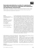
Figures 2 shows absolute (A) and normalised (B) viral
load distribution for each HR-HPV type, comparing both
groups of women. It is worth stating that HPV-31 (in
women without CIN) and HPV-33 (in women having
CIN) were the HR-HPV viral types having the highest
absolute viral load (median = 9.41 (2.58 IQR) HPV copies
for HPV-31 and median = 9.33 (2.94 IQR) HPV copies
for HPV-33) whilst HPV-58 infection had the lowest
absolute viral load in both groups of women. The
Del Río-Ospina et al. BMC Cancer (2015) 15:100
Page 5 of 11
Table 2 The distribution of socio-demographic characteristics and risk factors
Characteristic
Age, years
Origin
Ethnicity
Average monthly income*
Educational level
Marital status
Healthcare scheme affiliation
Smoker
Age at first intercourse, years
Lifetime number of sexual partners
Contraceptive method
Pregnancies
Abortions
STD
Categories
n
%
With CIN (n = 59)
Without CIN (n =114)
n
%
n
%
<30
29
16.76
11
18.64
18
15.79
30–40
54
31.21
21
35.59
33
28.95
>40
90
52.02
27
45.76
63
55.26
Bogotá
65
37.57
32
54.24
33
28.95
Other city
108
62.43
27
45.76
81
71.05
Other
7
4.05
3
5.08
4
3.51
Mestizo
166
95.95
56
94.92
110
96.49
≤ minimum
155
89.06
53
89.83
102
89.47
>minimum
18
10.40
6
10.17
12
10.53
No schooling
1
0.58
1
1.69
0
0.00
Primary
82
47.40
22
37.29
60
52.63
Secondary
74
42.77
28
47.46
46
40.35
Technical
10
5.78
6
10.17
4
3.51
Graduate
6
3.47
2
3.39
4
3.51
Single
17
9.83
4
6.78
13
11.40
Married
20
11.56
7
11.86
13
11.40
Divorced
8
4.62
4
6.78
4
3.51
Living with partner
126
72.83
43
72.88
83
72.81
Widow
2
1.16
1
1.69
1
0.88
Subsidised- linked
159
91.91
52
88.14
107
93.86
Contributory-private
14
8.09
7
11.86
7
6.14
No
146
84.39
49
83.05
97
85.09
Yes
27
15.61
10
16.95
17
14.91
<16
41
23.70
10
16.95
31
27.19
≥16
132
76.30
49
83.05
83
72.81
1
72
41.62
26
44.07
46
40.35
2–3
84
48.55
27
45.76
57
50.00
>3
17
9.83
6
10.17
11
9.65
None
65
37.57
19
32.20
46
40.35
Surgery
52
30.06
15
25.42
22
19.30
Hormonal
19
10.98
18
30.51
34
29.82
Barrier
37
21.39
7
11.86
12
10.53
None
4
2.31
1
1.69
3
2.63
1–2
76
43.93
28
47.46
48
42.11
3–4
74
42.77
27
45.76
47
41.23
>4
19
10.98
3
5.08
16
14.04
None
82
47.40
27
45.76
55
48.25
1
68
39.31
25
42.37
43
37.72
≥2
23
13.29
7
11.86
16
14.04
No
137
79.19
47
79.66
90
78.95
Yes
36
20.81
12
20.34
24
21.05
Values in bold = p < 0.05.
*
The minimum average monthly income (2014 rate) would be roughly US $300.
p = p value; CIN: cervical intraepithelial neoplasia; STD: sexually transmitted disease.
p
0.493
0.001
0.691
0.942
0.094
0.673
0.191
0.726
0.133
0.868
0.697
0.326
0.818
0.913
Del Río-Ospina et al. BMC Cancer (2015) 15:100
Page 6 of 11
Table 3 Type-specific HR-HPV viral load distribution by category
HPV
type
n
%
With CIN (n = 59)
Without CIN(n = 114)
p
Negative
Low viral load
High viral load
Negative
Low viral load
High viral load
n
%
n
%
n
%
n
%
n
%
n
%
HPV-16
86
49.71
25
42.37
12
20.34
22
37.29
62
54.39
13
11.40
39
34.21
0.186
HPV-18
117
67.63
18
30.51
10
16.95
31
52.54
38
33.33
18
15.79
58
50.88
0.928
HPV-31
71
41.04
40
67.80
1
1.69
18
30.51
62
54.39
3
2.63
49
42.98
0.257
HPV-33
14
8.09
54
91.53
0
0.00
5
8.47
105
92.11
1
0.88
8
7.02
0.846
HPV-45
67
38.73
36
61.02
9
15.25
14
23.73
70
61.40
10
8.77
34
29.82
0.366
HPV-58
56
32.37
42
71.19
7
11.86
10
16.95
75
65.79
16
14.04
23
20.18
0.772
159
91.91
4
6.78
8
13.56
47
79.66
10
8.77
12
10.53
92
80.70
0.777
*
HR-HPV
HPV DNA load: categorised as ≤ 0 = negative. 0 < VL ≤ 105 HPV copies = low viral load. >105 HPV copies = high viral load.
*
HR-HPV: high risk-human papillomavirus, infection by at least one high-risk viral type from the 6 analysed here.
HPV: human papillomavirus; CIN: cervical intraepithelial neoplasia; p = p value.
range of values for normalised viral load was lower
than for absolute (up to 108 HPV copies). The highest
absolute viral load was detected for HPV-31 in women
with CIN (1022 HPV copies) and highest normalised
viral load for HPV-33 in women without CIN. No statistically significant differences were observed regarding viral load distribution (absolute and normalised)
for each HR-HPV type in either group of patients.
The three patients having CIN 2+ were positive for
HR-HPV; HPV-18 and HPV-31 were detected in two of
them, whilst the other one was positive for HPV-18,
HPV-16 and HPV-45. Even though women having CIN
2+ had a higher viral load (normalised for HPV-18 and
absolute for HPV-16) than women having CIN 1, the
differences in viral load distribution were not statistically
significant. However, normalised viral load for HPV-31
was greater in women negative for cervical lesion and
having CIN 1 compared to women having CIN 2+ (marginal significance, i.e. p = 0.052).
The distribution of viral load was also analysed for
each HR-HPV type, according to biopsy result. Similar
results were found to those with colposcopy (i.e. higher
absolute viral loads in women having a severer degree of
lesion); and for some types (HPV-31, HPV-33 and HPV58) higher normalised viral loads; however, the differences were not statistically significant due to the amount
of women analysed (Table 4).
Crude and adjusted odds ratios (OR) were calculated for
estimating the magnitude of absolute viral load association
with CIN for each viral type. The conditional logistic regression model revealed that HPV-16 infection was significantly associated with greater frequency regarding cervical
lesions. However, lesions occurred more frequently in the
group of women having low viral load for HPV-16 (0 <
VL ≤ 5.86 HPV copies) than in women having a high load
(>5.86 HPV copies), (3.53 ORa, 1.16–10.74 95%CI; 2.63
ORa, 1.09–6.36 95%CI, respectively). It was also found that
CIN frequency was lower in women having HPV-31 and
high viral load (>5.14 HPV copies; 0.34 ORa, 0.15–0.78
95%CI). No significant associations were obtained for the
other viral types with the presence of CIN (Table 5).
Discussion
This study involved using RT-PCR; this enabled typespecific evaluation of the viral load of the most frequently
occurring oncogenic types in Colombia (HPV-16, -18, -31,
-33, -45 and -58) [19] for determining each type’s association with precursor lesions of CC. As the method has
high sensitivity, specificity and has a broad dynamic range
of viral detection (up to 1022 HPV copies) this provided
the best approach for this study [12,13,16,18,33].
More HPV infections were found in women having
CIN in our sample, amongst whom all women having
CIN 2+ were HPV positive. The foregoing was consistent
with the fact that almost 99.7% of CC cases are associated
with HPV [1]. Previous studies have demonstrated that
HPV prevalence in women having CIN is high, proportionally increasing as lesion severity increases [30,34,35].
The prevalence found here was greater than that reported
in the literature (100% in CIN 2+, 92.86% in CIN 1 and
91.23% in women without CIN). Women were included in
this study who had been previously identified as HPV
positive using conventional PCR; this explained the high
prevalence of HPV when using RT-PCR in women without lesions. However, variable infection prevalence in
women without CIN has been found worldwide (mean =
12.6%) [35,36].
Multiple infection frequency has been variable (16.3%–
55%) in previous reports concerning women having lesions [35]; up to 3.4% infection by multiple types of HRHPV has been described in women without lesions [37].
The present study revealed more multiple infections (in
both the general population and women having CIN and
those without them) regarding previous reports worldwide, but similar to that previously reported in Colombia
[27,38]. However, RT-PCR was used which has high
Del Río-Ospina et al. BMC Cancer (2015) 15:100
Page 7 of 11
Figure 2 Distribution of viral load for 6 HR-HPV types in both groups of patients. A. Absolute viral load. B Normalised viral load. The
dotted line indicates the median; the box represents the interquartile range (IQR). The whiskers extending from the boxes are the upper and
lower limits. Diamond markers represent extreme values. No statistically significant differences were observed regarding DNA load distribution of
each HPV type between both groups of patients (Mann–Whitney U test). CIN: cervical intraepithelial neoplasia.
Table 4 Distribution of 6 HR-HPV types’ viral load regarding biopsy results
Viral type
Negative (n = 28)
CIN 1 (n = 16)
CIN 2+ (n = 1)
% (n)
% (n)
% (n)
Viral load, median (IQR)
Absolute
Normalised
*
Viral load, median (IQR)
Absolute
Normalised*
Viral load, median (IQR)
Absolute
Normalised*
HPV-16
66.67 (22)
6.42 (1.69)
1.79 (0.54)
57.89 (11)
6.77 (3.04)
1.69 (0.64)
0
n/a
n/a
HPV-18
66.67 (22)
6.29 (1.34)
1.84 (0.51)
68.42 (13)
6.61 (2.28)
1.67 (1.79)
100 (1)
7.02 (n/a)
2.07 (n/a)
HPV-31
30.30 (10)
8.51 (1.90)
2.39 (0.38)
31.58 (6)
9.69 (6.00)
3.50 (2.17)
0
n/a
n/a
HPV-33
3.03 (1)
6.75 (n/a)
1.98 (n/a)
10.53 (2)
8.48 (1.70)
2.37 (2.06)
100 (1)
10.57 (n/a)
3.13 (n/a)
HPV-45
51.52 (17)
6.13 (2.95)
1.79 (1.00)
42.11 (8)
6.24 (1.17)
1.61 (0.80)
0
n/a
n/a
HPV-58
21.21 (7)
5.93 (3.89)
2.14 (2.35)
36.84 (7)
6.12 (0.34)
1.75 (0.28)
0
n/a
n/a
HR-HPV**
94.34 (31)
6.37 (1.20)
2.06 (0.63)
94.74 (18)
6.77 (2.97)
2.12 (1.37)
100 (1)
8.80 (n/a)
2.60 (n/a)
Absolute and normalised viral loads were both log10 transformed.
*
HPV copies/cell = number of HPV copies/(number of HMBS copies/2).
**
HR-HPV: high risk-human papillomavirus, infection by at least one high-risk viral type from the 6 analysed here.
HPV: human papillomavirus; CIN: cervical intraepithelial neoplasia; CIN 1: cervical intraepithelial neoplasia 1; CIN 2+: cervical intraepithelial neoplasia 2 or 3; n/a:
not applicable.
Del Río-Ospina et al. BMC Cancer (2015) 15:100
Page 8 of 11
Table 5 Conditional logistic regression model
Adjusted OR*
95%CI
2.19 (0.88–5.43)
3.53
1.16–10.74
1.27 (0.64–2.50)
2.63
1.09–6.36
HPV type
Viral load
With CIN / without CIN
Crude OR (95%CI)
HPV-16
Negative
25/62
Reference
0 < VL ≤ 5.86
12/13
5.86 < VL
22/39
HPV-18
HPV-31
HPV-33
HPV-45
HPV-58
HR-HPV**
Negative
18/38
Reference
0 < VL ≤ 5.95
10/18
1.14 (0.45–2.89)
1.72
0.52–5.69
5.95 < VL
31/58
1.06 (0.52–2.17)
1.77
0.68–4.63
Negative
40/62
Reference
0 < VL ≤ 5.14
1/3
0.52 (0.04–6.29)
0.15
0.01–2.26
5.14 < VL
18/49
0.60 (0.32–1.14)
0.34
0.15–0.78
Negative
54/105
Reference
0 < VL ≤ 4.60
0/1
0.00 (0 - .)
0
0-.
4.60 < VL
5/8
1.43 (0.45–4.50)
1.67
0.44–6.28
Negative
36/70
Reference
0 < VL ≤ 5.98
9/10
1.53 (0.60–3.92)
2.94
0.92–9.44
5.98 < VL
14/34
0.79 (0.38–1.67)
1.13
0.43–2.96
Negative
42/75
Reference
0 < VL ≤ 5.97
7/16
0.83 (0.32–2.11)
0.73
0.23–2.31
0.86
0.35–2.12
5.97 < VL
10/23
0.83 (0.37–1.83)
Negative
4/10
Reference
0 < VL ≤ 5.94
8/12
1.73 (0.40–7.47)
1.01
0.23–4.50
5.94 < VL
47/92
1.18 (0.35–4.00)
1.39
0.25–7.81
Values in bold = p < 0.05.
*
Adjusted for origin, ethnicity, age at first intercourse and number of viral types.
**
HR-HPV: high-risk-human papillomavirus, infection by at least one high-risk viral type from the 6 analysed here (viral load = sum of viral loads of HPV types detected/
number of HPV types detected.
HPV: human papillomavirus; CIN: cervical intraepithelial neoplasia; VL: viral load; OR: odds ratio.
sensitivity and allows small amounts of viral DNA to be
detected, compared to other methods [13,18]. This has
been previously demonstrated by studies carried out involving RT-PCR which have reported high multiple infection
frequency [39,40]. Such differences regarding co-infection
prevalence reported in various studies might have been due
to their design, sample size, the HPV detection methods
used and the population being studied (geographic, demographic and clinical factors) [37].
HPV-18 and HPV-16 occurred most frequently in the
present study, followed by HPV-45 and HPV-58. Differences concerning type-specific prevalence have been reported according to geographic and demographic factors
[3,35]. It is worth noting that the two most common
types found here are responsible for the 70% of cases of
CC [41] and that the HPV genotypes evaluated in this
study have been reported amongst the 8 HR-HPV types
most frequently occurring around the world, in both
women without lesions and women with CC [2,3,35].
Absolute viral load was highest in women having CIN
compared to women without lesions determined by both
colposcopy and biopsy; an increase in the viral load was
observed for HPV-18 and HPV-33 proportional to the
degree of injury. The foregoing was consistent with previous studies which have revealed the effect of viral load
on developing CC. Most HPV-16 studies have found that
viral load has increased in relation to the degree of cervical lesion severity [8-11,15,16,42].
An association between viral load and cervical lesion
frequency (as assessed by colposcopy) was observed in
this study just for HPV-16 and HPV-31. The present
study’s results highlighted the fact that women having
low HPV-16 load (<5.86 HPV copies) had higher cervical
lesion frequency. Such results agreed with those from a
study by Manawapat, Stubenrauch et al., [43] which
showed that women having persistent HPV-16 infection
had lower viral load than those who had a transient infection (4.72 copies/cell cf 20 copies/cell; p = 0.0003). It
has been found recently that low viral load was characteristic of intermittently detected persistent infection
[44]. Reduced viral load has been described in women
having CIN; this has been explained by HPV genome integration associated with down-regulation of viral DNA
synthesis, thereby affecting immune system activation
and thus reducing the probability of infection being
eliminated [43,45-47]. Accordingly, a long period of
Del Río-Ospina et al. BMC Cancer (2015) 15:100
latency accompanied by low viral load would probably
be observed, representing a greater risk for infection persistence and lesion progression [48].
Contrary to our findings regarding HPV-16 viral load,
the present study found that a high HPV-31 load (>5.14
HPV copies) was associated with lower cervical lesion frequency. As mentioned previously regarding HPV-16 results, it has been shown that viral load has been greater in
transitory infections regarding patients having persistent
infection [43]. This agreed with the finding that clearance
of HPV-16 infection has been preceded by a transient viral
load peak or a plateau phase [33]; such high load was
probably necessary for the immunological system to become induced, thereby favouring HPV elimination. According to the above, HPV-31 infections are probably
transitory and such association is mediated by an immune
system response to high viral load which can eliminate the
infection and thus CC precursor lesions do not progress
or such lesions regress spontaneously [47].
Regarding the other viral types (HPV-18, -33, -45
and -58), no association was found between viral load
and cervical lesion frequency; such result was supported by data from other authors [14-16,42,49,50].
However, a study by Moberg, Gustavsson et al., found
that high HPV-16, HPV-31 and HPV-18/45 viral load
increased the risk of developing carcinoma in situ
(CIS) [51].
The pertinent literature gives different cut-off points
when categorising viral load, depending on the quantification technique used (RT-PCR, Hybrid Capture II
(HCII)) [8] and distribution in a particular population
being evaluated [9,51]. A study which evaluated the clinical significance of HPV-16 and -18 viral loads determined that HPV-16 viral load was related to cervical
lesion severity, having a 3.0×10⁶ copies/million cells
threshold, this being highly specific for grade 2 diagnosis
[15]. Taking the foregoing into account, viral load was
categorised in the present study according to percentile
distribution, leaving 106 copies as cut-off point for ensuring analysis quality.
It is worth stressing that this technique managed to
detect a broad range of viral load, even after stratifying
by colposcopy result and viral type. However, this hampered establishing viral load cut-off points to enable
identifying women at greater risk of developing cervical
lesions; previous studies have also experienced such difficulty [12,16,33].
This work’s value lies in it being a study where a reproducible, sensitive and specific technique (i.e. RTPCR) was used for detecting and quantifying viral load
(absolute and normalised) not just for one viral type but
for the 6 most frequently occurring high-risk HPV types
described to date in Colombia. Besides, this is the first
study carried out in Colombia which has included
Page 9 of 11
women from regions having high HPV infection prevalence and which was aimed at evaluating the association
between HPV viral load and cervical lesion frequency.
This study’s results were obtained from a single evaluation of HPV viral load; this means that predicting the
risk of lesion progression and developing CC later on
cannot be ascertained from this. However, it can be
stated that our results were consistent with some findings reported in longitudinal studies [33,43,44,48]. The
infection duration time of the women included in this
study was also unknown; HPV-16 might thus have been
greater in women having CIN and lower in HPV-31
women. Another limitation of this study was the low
number of women having CIN 2+ which hindered generalising the results to all CC precursor lesions. An analysis of HPV viral load dynamics could thus be more
reliable and provide more information for estimating
whether HPV infection will worsen or clear and predicting the development of CC or cervical lesions. Prospective studies on women having HPV infection which
would include type-specific determination (according to
local prevalence) of viral load and women having cervical lesions with different degrees of severity are thus
needed for confirming our results.
Conclusions
A significant association was found in this study, low
HPV-16 and high HPV-31 viral loads were associated
with higher CIN frequency; this might have been related
to infection duration and immune system response.
HPV infection’s effect on developing CC is influenced by
viral load, meaning that measuring load could improve
the predictive value of HPV detection; however, the
scope of quantification depends on the viral type being
detected. These findings support the idea of quantifying
viral load (as a type-specific marker of CC), coupled to
cytology, for improving and strengthening CC screening
programmes. This would lead to identifying HPV positive women at greater risk of developing cervical lesions,
as well as identifying women as yet lacking cervical
anomalies for predicting the beginnings of neoplasia.
Abbreviations
HPV: Human papillomavirus; HR-HPV: High-risk human papillomavirus;
CC: Cervical cancer; CIN: Cervical intraepithelial neoplasia; CIN 1: Cervical
intraepithelial neoplasia 1; CIN 2+: Cervical intraepithelial neoplasia 2 or 3;
HMBS: Hydroxymethylbilane synthase; PCR: Polymerase chain reaction; RTPCR: Real-time polymerase chain reaction; DNA: Deoxyribonucleic acid;
VL: Viral load; STD: Sexually-transmitted diseases; HC II: Hybrid capture II;
FAM: 6-carboxyfluorescein; Cy5: FluoroLink mono reactive dye Cy5;
HEX: hexachlorofluoresceine; ZEN/IBFQ: ZEN and Iowa Black FQ; IBRQ: Iowa
Black RQ; SD: Standard deviation; CI: Confidence interval; IQR: Interquartile
range; n/a: Not applicable; OR: Odds ratio.
Competing interests
All authors declare that they have no competing interests.
Del Río-Ospina et al. BMC Cancer (2015) 15:100
Authors’ contributions
All the authors were involved in developing the study and preparing the
ensuing article. LDRO and SCSDL provided the concept and designed the
study, as well as acquiring, analysing and interpreting the data and writing
the article. MC helped draft the manuscript and assisted with data analysis.
DAMP developed the methodology and was involved in drafting the
manuscript. RS provided statistical analysis, interpreted data and helped in
writing the manuscript. The study was supervised by APP, MEP and MAP
who revised the document and lent their expertise regarding the discussion
of results. All authors have read and approved the final version of the
manuscript.
Acknowledgments
This project was supported by the Basque Development Cooperation
Agency, the Spanish International Development Cooperation Agency (AECID)
(Project 10-CAP1-0197) and the Colombian Science, Technology and
Innovation Department (COLCIENCIAS) (contract # 0709-2013). The sponsors
played no role in study design, data collection and/or analysis, decision to
publish, or preparation of the manuscript. We would like to express our
thanks to Jason Garry for translating and revising this manuscript.
Author details
1
Molecular Biology and Immunology Department, Fundación Instituto de
Inmunología de Colombia (FIDIC), Carrera 50#26-20, Bogotá, Colombia.
2
School of Medicine and Health Sciences, Universidad del Rosario, Carrera
24#63C-69, Bogotá, Colombia. 3Faculty of Natural and Mathematical Sciences,
Universidad del Rosario, Carrera 24#63C-69, Bogotá, Colombia. 4School of
Medicine, Universidad Nacional de Colombia, Carrera 45#26-85, Bogotá,
Colombia. 5Mathematics Department, Universidad Pública de Navarra,
Pamplona, Spain.
Received: 18 December 2014 Accepted: 24 February 2015
References
1. Ault KA. Epidemiology and natural history of human papillomavirus
infections in the female genital tract. Infect Dis Obstet Gynecol. 2006;2006
(Suppl):1–5.
2. Munoz N, Bosch FX, de Sanjose S, Herrero R, Castellsague X, Shah KV, et al.
Epidemiologic classification of human papillomavirus types associated with
cervical cancer. N Engl J Med. 2003;348:518–27.
3. Li N, Franceschi S, Howell-Jones R, Snijders PJ, Clifford GM. Human papillomavirus
type distribution in 30,848 invasive cervical cancers worldwide: Variation
by geographical region, histological type and year of publication. Int
J Cancer. 2011;128:927–35.
4. de Freitas AC, Gurgel AP, Chagas BS, Coimbra EC, Do Amaral CM.
Susceptibility to cervical cancer: an overview. Gynecol Oncol. 2012;126:304–11.
5. Bosch FX, Munoz N. The viral etiology of cervical cancer. Virus Res.
2002;89:183–90.
6. Munoz N, Hernandez-Suarez G, Mendez F, Molano M, Posso H, Moreno V,
et al. Persistence of HPV infection and risk of high-grade cervical intraepithelial
neoplasia in a cohort of Colombian women. Br J Cancer. 2009;100:1184–90.
7. Ramanakumar AV, Goncalves O, Richardson H, Tellier P, Ferenczy A, Coutlee
F, et al. Human papillomavirus (HPV) types 16, 18, 31, 45 DNA loads and
HPV-16 integration in persistent and transient infections in young women.
BMC Infect Dis. 2010;10:326.
8. Hernández-Hernández DM, Ornelas-Bernal L, Guido-Jiménez M, Apresa-Garcia
T, Alvarado-Cabrero I, Salcedo-Vargas M, et al. Association between high-risk
human papillomavirus DNA load and precursor lesions of cervical cancer in
Mexican women. Gynecol Oncol. 2003;90:310–7.
9. Josefsson AM, Magnusson PK, Ylitalo N, Sorensen P, Qwarforth-Tubbin P,
Andersen PK, et al. Viral load of human papilloma virus 16 as a determinant for
development of cervical carcinoma in situ: a nested case–control study. Lancet.
2000;355:2189–93.
10. Moberg M, Gustavsson I, Wilander E, Gyllensten U. High viral loads of
human papillomavirus predict risk of invasive cervical carcinoma. Br
J Cancer. 2005;92:891–4.
11. Ylitalo N, Sorensen P, Josefsson AM, Magnusson PK, Andersen PK, Ponten
J, et al. Consistent high viral load of human papillomavirus 16 and risk of
cervical carcinoma in situ: a nested case–control study. Lancet.
2000;355:2194–8.
Page 10 of 11
12. Andersson S, Safari H, Mints M, Lewensohn-Fuchs I, Gyllensten U, Johansson
B. Type distribution, viral load and integration status of high-risk human
papillomaviruses in pre-stages of cervical cancer (CIN). Br J Cancer.
2005;92:2195–200.
13. Moberg M, Gustavsson I, Gyllensten U. Real-time PCR-based system for
simultaneous quantification of human papillomavirus types associated
with high risk of cervical cancer. J Clin Microbiol.
2003;41:3221–8.
14. Sherman ME, Schiffman M, Cox JT, Atypical Squamous Cells of
Undetermined Significance/Low-Grade Squamous Intraepithelial Lesion
Triage Study G. Effects of age and human papilloma viral load on
colposcopy triage: data from the randomized Atypical Squamous Cells of
Undetermined Significance/Low-Grade Squamous Intraepithelial Lesion
Triage Study (ALTS). J Natl Cancer Inst. 2002;94:102–7.
15. Carcopino X, Henry M, Mancini J, Giusiano S, Boubli L, Olive D, et al.
Significance of HPV 16 and 18 viral load quantitation in women referred for
colposcopy. J Med Virol. 2012;84:306–13.
16. Swan DC, Tucker RA, Tortolero-Luna G, Mitchell MF, Wideroff L, Unger ER,
et al. Human papillomavirus (HPV) DNA copy number is dependent on
grade of cervical disease and HPV type. J Clin Microbiol. 1999;37:1030–4.
17. Jenkins A, Allum AG, Strand L, Aakre RK. Simultaneous detection, typing and
quantitation of oncogenic human papillomavirus by multiplex consensus
real-time PCR. J Virol Methods. 2013;187:345–51.
18. Schmitt M, Depuydt C, Benoy I, Bogers J, Antoine J, Pawlita M, et al. Viral
load of high-risk human papillomaviruses as reliable clinical predictor for
the presence of cervical lesions. Cancer Epidemiol Biomarkers Prev.
2013;22:406–14.
19. Camargo M, Soto-De Leon SC, Sanchez R, Perez-Prados A, Patarroyo ME,
Patarroyo MA. Frequency of human papillomavirus infection, coinfection,
and association with different risk factors in Colombia. Ann Epidemiol.
2011;21:204–13.
20. Ramírez VG, Bustamante MA, Sarmiento CA. Norma Técnica para la
Detección Temprana del Cáncer de Cuello Uterino y Guía de Atención de
Lesiones Preneoplásicas de Cuello Uterino. Colombia: Ministerio de Salud,
Dirección General de Promoción y Prevenció; 2000. p. 1–26.
21. Rijkaart DC, Berkhof J, van Kemenade FJ, Coupe VM, Rozendaal L, Heideman
DA, et al. HPV DNA testing in population-based cervical screening
(VUSA-Screen study): results and implications. Br J Cancer.
2012;106:975–81.
22. Sellors J, Sankaranarayanan R. La colposcopia y el tratamiento de la
neoplasia intraepitelial cervical: Manual para principiantes. Lyon, Francia:
International Agency for Research on Cancer (IARC); 2003. p. 140.
23. Boicea A, Patrascu A, Surlin V, Iliescu D, Schenker M, Chiutu L. Correlations
between colposcopy and histologic results from colposcopically directed
biopsy in cervical precancerous lesions. Rom J Leg Med.
2012;53:735–41.
24. Kitchener HC, Castle PE, Cox JT. Chapter 7: Achievements and limitations of
cervical cytology screening. Vaccine. 2006;24 Suppl 3:S3/63–70.
25. Cronje HS, Cooreman BF, Beyer E, Bam RH, Middlecote BD, Divall PD.
Screening for cervical neoplasia in a developing country utilizing cytology,
cervicography and the acetic acid test. Int J Gynecol Obstet. 2001;72:151–7.
26. Cronje HS, Parham GP, Cooreman BF, de Beer A, Divall P, Bam RH.
A comparison of four screening methods for cervical neoplasia in a
developing country. Am J Obstet Gynecol. 2003;188:395–400.
27. Soto-De Leon S, Camargo M, Sanchez R, Munoz M, Perez-Prados A, Purroy
A, et al. Distribution patterns of infection with multiple types of human
papillomaviruses and their association with risk factors. PLoS One.
2011;6:e14705.
28. Soto-De Leon SC, Del Rio-Ospina L, Camargo M, Sanchez R, Moreno-Perez
DA, Perez-Prados A, et al. Persistence, clearance and reinfection regarding
six high risk human papillomavirus types in Colombian women: a follow-up
study. BMC Infect Dis. 2014;14:395.
29. Thompson JD, Higgins DG, Gibson TJ. CLUSTAL W: improving the sensitivity
of progressive multiple sequence alignment through sequence weighting,
position-specific gap penalties and weight matrix choice. Nucleic Acids Res.
1994;22:4673–80.
30. Wu Y, Chen Y, Li L, Yu G, Zhang Y, He Y. Associations of high-risk HPV types
and viral load with cervical cancer in China. J Clin Virol. 2006;35:264–9.
31. Solomon D, Davey D, Kurman R, Moriarty A, O'Connor D, Prey M, et al. The
2001 Bethesda system: terminology for reporting results of cervical
cytology. JAMA. 2001;2002(287):2114–9.
Del Río-Ospina et al. BMC Cancer (2015) 15:100
32. Broutet N, Dangou JM, Fadhil I, Lazdane G, Luciani S, Mathur A, et al. WHO
guidelines for screening and treatment of precancerous lesions for cervical
cancer prevention. South Africa: World Health Organization; 2013. p. 1–60.
33. Monnier-Benoit S, Dalstein V, Riethmuller D, Lalaoui N, Mougin C, Pretet JL.
Dynamics of HPV16 DNA load reflect the natural history of cervical
HPV-associated lesions. J Clin Virol. 2006;35:270–7.
34. Moore RA, Ogilvie G, Fornika D, Moravan V, Brisson M, Amirabbasi-Beik M,
et al. Prevalence and type distribution of human papillomavirus in 5,000
British Columbia women–implications for vaccination. Cancer Causes Control.
2009;20:1387–96.
35. Guan P, Howell-Jones R, Li N, Bruni L, de Sanjose S, Franceschi S, et al. Human
papillomavirus types in 115,789 HPV-positive women: a meta-analysis
from cervical infection to cancer. Int J Cancer. 2012;131:2349–59.
36. Poljak M, Seme K, Maver PJ, Kocjan BJ, Cuschieri KS, Rogovskaya SI, et al.
Human papillomavirus prevalence and type-distribution, cervical cancer
screening practices and current status of vaccination implementation in
Central and Eastern Europe. Vaccine. 2013;31 Suppl 7:H59–70.
37. Cuschieri KS, Cubie HA, Whitley MW, Seagar AL, Arends MJ, Moore C, et al.
Multiple high risk HPV infections are common in cervical neoplasia and
young women in a cervical screening population. J Clin Pathol. 2004;57:68–72.
38. Garcia DA, Cid-Arregui A, Schmitt M, Castillo M, Briceno I, Aristizabal FA.
Highly sensitive detection and genotyping of HPV by PCR multiplex and
luminex technology in a cohort of colombian women with abnormal
cytology. Open Virol J. 2011;5:70–9.
39. Xi LF, Hughes JP, Edelstein ZR, Kiviat NB, Koutsky LA, Mao C, et al. Human
Papillomavirus (HPV) type 16 and type 18 DNA Loads at Baseline and
Persistence of Type-Specific Infection during a 2-year follow-up. J Infect Dis.
2009;200:1789–97.
40. Schmitt M, Depuydt C, Benoy I, Bogers J, Antoine J, Arbyn M, et al. Multiple
human papillomavirus infections with high viral loads are associated with
cervical lesions but do not differentiate grades of cervical abnormalities.
J Clin Microbiol. 2013;51:1458–64.
41. Ibeanu OA. Molecular pathogenesis of cervical cancer. Cancer Biol Ther.
2011;11:295–306.
42. Zerbini M, Venturoli S, Cricca M, Gallinella G, De Simone P, Costa S, et al.
Distribution and viral load of type specific HPVs in different cervical lesions
as detected by PCR-ELISA. J Clin Pathol. 2001;54:377–80.
43. Manawapat A, Stubenrauch F, Russ R, Munk C, Kjaer SK, Iftner T. Physical
state and viral load as predictive biomarkersfor persistence and progression
of HPV16-positive cervical lesions: results from a population based long-term
prospective cohort study. Am J Cancer Res. 2012;2:192–203.
44. Winer RL, Xi LF, Shen Z, Stern JE, Newman L, Feng Q, et al. Viral load and
short-term natural history of type-specific oncogenic human papillomavirus
infections in a high-risk cohort of midadult women. Int J Cancer.
2014;134:1889–98.
45. Kulmala SM, Syrjanen SM, Gyllensten UB, Shabalova IP, Petrovichev N, Tosi P,
et al. Early integration of high copy HPV16 detectable in women with
normal and low grade cervical cytology and histology. J Clin Pathol.
2006;59:513–7.
46. Nakagawa M, Stites DP, Patel S, Farhat S, Scott M, Hills NK, et al. Persistence
of human papillomavirus type 16 infection is associated with lack of
cytotoxic T lymphocyte response to the E6 antigens. J Infect Dis.
2000;182:595–8.
47. Brenna SM, Syrjanen KJ. Regulation of cell cycles is of key importance in
human papillomavirus (HPV)-associated cervical carcinogenesis. Sao Paulo
Med J. 2003;121:128–32.
48. van Duin M, Snijders PJ, Schrijnemakers HF, Voorhorst FJ, Rozendaal L,
Nobbenhuis MA, et al. Human papillomavirus 16 load in normal and
abnormal cervical scrapes: an indicator of CIN II/III and viral clearance. Int
J Cancer. 2002;98:590–5.
49. Chan PK, Cheung JL, Cheung TH, Lo KW, Yim SF, Siu SS, et al. Profile of viral
load, integration, and E2 gene disruption of HPV58 in normal cervix and
cervical neoplasia. J Infect Dis. 2007;196:868–75.
50. Gravitt PE, Kovacic MB, Herrero R, Schiffman M, Bratti C, Hildesheim A, et al.
High load for most high risk human papillomavirus genotypes is associated
with prevalent cervical cancer precursors but only HPV16 load predicts the
development of incident disease. Int J Cancer. 2007;121:2787–93.
51. Moberg M, Gustavsson I, Gyllensten U. Type-specific associations of human
papillomavirus load with risk of developing cervical carcinoma in situ. Int
J Cancer. 2004;112:854–9.
Page 11 of 11
Submit your next manuscript to BioMed Central
and take full advantage of:
• Convenient online submission
• Thorough peer review
• No space constraints or color figure charges
• Immediate publication on acceptance
• Inclusion in PubMed, CAS, Scopus and Google Scholar
• Research which is freely available for redistribution
Submit your manuscript at
www.biomedcentral.com/submit