DEK is a potential marker for aggressive phenotype and irinotecan-based therapy response in metastatic colorectal cancer
Bạn đang xem bản rút gọn của tài liệu. Xem và tải ngay bản đầy đủ của tài liệu tại đây (2.05 MB, 10 trang )
Martinez-Useros et al. BMC Cancer 2014, 14:965
/>
RESEARCH ARTICLE
Open Access
DEK is a potential marker for aggressive
phenotype and irinotecan-based therapy
response in metastatic colorectal cancer
Javier Martinez-Useros1, Maria Rodriguez-Remirez1, Aurea Borrero-Palacios1, Irene Moreno1, Arancha Cebrian1,
Teresa Gomez del Pulgar1, Laura del Puerto-Nevado1, Ricardo Vega-Bravo2, Alberto Puime-Otin2, Nuria Perez2,
Sandra Zazo2, Clara Senin3, Maria J Fernandez-Aceñero5, Maria S Soengas4, Federico Rojo2
and Jesus Garcia-Foncillas1*
Abstract
Background: DEK is a transcription factor involved in stabilization of heterochromatin and cruciform structures. It
plays an important role in development and progression of different types of cancer. This study aims to analyze the
role of DEK in metastatic colorectal cancer.
Methods: Baseline DEK expression was firstly quantified in 9 colorectal cell lines and normal mucosa by WB.
SiRNA-mediated DEK inhibition was carried out for transient DEK silencing in DLD1 and SW620 to dissect its role
in colorectal cancer aggressiveness. Irinotecan response assays were performed with SN38 over 24 hours and
apoptosis was quantified by flow cytometry. Ex-vivo assay was carried out with 3 fresh tumour tissues taken from
surgical resection and treated with SN38 for 24 hours. DEK expression was determined by immunohistochemistry in
67 formalin-fixed paraffin-embedded tumour samples from metastatic colorectal cancer patients treated with
irinotecan-based therapy as first-line treatment.
Results: The DEK oncogene is overexpressed in all colorectal cancer cell lines. Knock-down of DEK on DLD1
and SW620 cell lines decreased cell migration and increased irinotecan-induced apoptosis. In addition, low DEK
expression level predicted irinotecan-based chemotherapy response in metastatic colorectal cancer patients with
KRAS wild-type.
Conclusions: These data suggest DEK overexpression as a crucial event for the emergence of an aggressive
phenotype in colorectal cancer and its potential role as biomarker for irinotecan response in those patients with
KRAS wild-type status.
Keywords: DEK, Irinotecan, Aggressive phenotype, Metastatic colorectal cancer, KRAS
Background
Colorectal cancer (CRC) is one of the most common
gastrointestinal malignant tumors in the world and it
has one of the highest rates of morbidity and mortality
worldwide. There are about 1.36 million new-onset patients around the world each year, and 0.7 million CRC
patients died of it in 2012 [1]. The 5-year survival rate
* Correspondence:
1
Translational Oncology Division, OncoHealth Institute, Health Research
Institute - University Hospital “Fundación Jiménez Díaz”-UAM, Av. Reyes
Católicos 2, 28040 Madrid, Spain
Full list of author information is available at the end of the article
for colorectal cancer is approximately 55% because of its
invasion and metastasis.
The first-line treatment of metastatic colorectal cancer
(mCRC) is based on fluoropyrimidines (5-fluorouracil/
folinic acid) given in combination with the prodrugs oxaliplatin [2-4] and/or irinotecan [5-9].
The active metabolite of irinotecan, SN38, inhibits topoisomerase I and prevents DNA from unwinding [10].
Topoisomerase I expression has correlated with irinotecan response in several studies [11,12] but this procedure is not currently performed as part of the selection of
therapy for mCRC.
© 2014 Martinez-Useros et al.; licensee BioMed Central. This is an Open Access article distributed under the terms of the
Creative Commons Attribution License ( which permits unrestricted use,
distribution, and reproduction in any medium, provided the original work is properly credited. The Creative Commons Public
Domain Dedication waiver ( applies to the data made available in this
article, unless otherwise stated.
Martinez-Useros et al. BMC Cancer 2014, 14:965
/>
DEK was identified as a fusion protein with the CAN
nucleoporin due to the translocation t(6;9) in a subtype
of acute myeloid leukaemia [13]. It was later described
as a transcription factor overexpressed in multiple neoplasms including bladder cancer [14], breast cancer [15],
glioblastoma [16], hepatocellular carcinoma [17], melanoma [18], retinoblastoma [19,20], colorectal cancer [21,22]
and other types of cancer, such as oral, ovarian, or uterine
cervical cancer [21,23-25].
It has been reported that DEK promoter is regulated
by E2F1 [21], and its activation leads to transcription of
DEK mRNA. Functionally, DEK is involved in the DNA
repair machinery through interaction with PARP-1 [26],
suppresses cellular senescence, apoptosis, differentiation,
and promotes transformation in vitro and in vivo [27-29].
Furthermore, DEK has been suggested as a potential marker for bladder cancer [14], an independent predictor for
prognosis in colorectal cancer patients (stages I-III) [22]
and a specific marker to neoadjuvant chemotherapy for
breast cancer [30].
In this study, we analyze the oncogenic role of DEK in
CRC cell lines. As well as, we propose its potential use
as a marker of irinotecan-based chemotherapy response
in metastatic colorectal cancer patients.
This new function of DEK settles this oncogene as a
potential marker for clinical practice, as only 20% to
30% of patients with mCRC respond to irinotecan-based
therapy in first-line treatment. The applicability of DEK
as a tool for improved decision-making in routine diagnostic assessment requires further validation.
Methods
Page 2 of 10
of the 67 patients included in the study are summarized in
Table 1.
Clinical samples used in the study were kindly supplied
from the BioBank of the Fundacion Jimenez DiazUniversidad Autonoma de Madrid (RD09/0076/00101 Spain). This study has been evaluated by The Ethics
Table 1 Clinical features of metastatic colorectal cancer
patients treated with irinotecan-based therapy
Characteristics
Patients (N = 67)
Median age-years (range)
62 (33–79)
Sex
Male
47 (70%)
Female
20 (30%)
Median CEA (range, ng/mL)
16 (0–2066)
Performance status WHO
0
29 (43%)
1
35 (52%)
2
3 (5%)
Site of primary tumor
Colon
35 (52%)
Rectum
32 (48%)
METASTASIS
Liver
31 (47%)
Liver & other
19 (28%)
Other
15 (22%)
N.A.
2 (3%)
KRAS
Cell lines
Wild-type
35 (52%)
Nine human-derived CRC cell lines obtained from the
American Type Culture Collection (SW620 (CCL-227)
and LOVO (CCL-229) from metastatic foci origin; DLD1
(CCL-221), SW480 (CCL-228), RKO (CRL-2577), WIDR
(CCL-218), LS513 (CRL-2134), HCT15 (CCL-225), and
HCT116 (CCL-247) from primary tumor origin) were cultured with RPMI (Gibco) supplemented with 10% FBS
(Gibco), penicillin (100 U/mL)/streptomycin (100 U/mL)
(Invitrogen, Life Technologies). Two human colon mucosa from frozen tissue were used as controls.
Mutated
26 (39%)
N.A.
6 (9%)
BRAF
Wild-type
60 (90%)
Mutated
7 (10%)
Biologic treatment
Bevacizumab
23 (34%)
Cetuximab
7 (11%)
None
37 (55%)
TOPO I expression level
Patient samples
A total of 67 mCRC patients who received FOLFIRI regimen as first-line treatment were collected for the study.
KRAS mutation status was determined with Cobas® KRAS
Mutation Test (Roche Diagnostics) that offers broad mutation coverage of KRAS codons 12, 13 and 61. We found
35 patients with KRASwt, 26 patients with KRASmut status
and 6 could not be determined because the quality of
DNA was not enough. The clinical-pathological features
High
21 (31%)
Low
20 (30%)
N.A.
26 (39%)
DEK expression level
High
21 (31%)
Low
46 (69%)
N.A.: not available. Other refers to lung, lymph node and/or
peritoneal metastasis.
Martinez-Useros et al. BMC Cancer 2014, 14:965
/>
Committee of Clinical Research of Fundacion Jimenez
Diaz (act number 17/14).
Page 3 of 10
on a FACSCanto II flow cytometer (BD Biosciences) and
analyzed with FACSDiva software (BD Biosciences). All experiments were performed in triplicate.
Ex-vivo assay
Ex-vivo assays were designed to predict the sensitivity or
resistance of a set of tumors to irinotecan. To perform
these assays, three tumor samples from 3 different patients were taken after surgical resection. Each sample
was divided in two pieces and transferred onto a 12-well
plate and cultured in DMEM (Gibco) supplemented with
10% FBS, penicillin (100 U/mL)/streptomycin (100 U/mL).
One of the tumor pieces was treated with SN38 (5 nM)
(Sigma-Aldrich), whereas the other half remained untreated. After 24 hours, the tissues were processed for IHC.
Western blot
Total protein from CRC cell lines and normal mucosa
was extracted with RIPA buffer supplemented with protease inhibitor cocktail (Roche). Samples were fractionated
by SDS–polyacrylamide gel electrophoresis, transferred to
nitrocellulose membranes (Biorad), and proteins were
detected using specific antibodies for DEK (610948, BD
Biosciences), cleaved-Caspase-3 (9664, Cell Signaling) and
actin (a1978, Sigma-Aldrich). Horseradish peroxidaselinked sheep anti-mouse (NA931V) antibodies (GEHealthcare) were used as the secondary antibodies.
Blots were developed with the Amersham ECL Prime
Western Blotting Detection Reagent (GE-Healthcare).
Wound healing and Boyden chamber migration assay
Cell motility after DEK downregulation was estimated
by wound healing assays. Cells were grown as a monolayer
and an artificial homogenous wound was created with a
sterile plastic 10 μL micropipette tip. The growth of cells
in the wound was measured at 6, 12, and 24 hours.
Migration assays were performed in cell culture inserts with 8-μm pores in 24-well plates (Transwells, BD
Biosciences). DLD1 and SW620 cells were seeded at a
density of 5×10⁴ cells per insert in 300 μl RPMI. The recipient wells received 750 μl RPMI supplemented with
20% FBS. The migration was determinated after 24 h.
Afterwards, cells were fixed and stained with toluidine
blue (Sigma-Aldrich). The non-migrated cells on the
upper side of the membrane were removed with a cotton
swab. On each membrane, the cells of 10 randomly selected fields (10X objective) were counted, and the mean
number of cells per visual field was determined. The migration index was determined as migrated cells ratio relative to siRNA control transfected cells. Three independent
experiments were done and all experiments were performed in triplicate wells.
DEK silencing
Immunohistochemistry
Three different siRNAs for DEK were used (Silencer Select Pre-designed siRNA s15457, s15458, and s15459)
(Ambion, Life Technologies). Gene silencing was performed with 3.5 million cells from two different CRC
cell lines, DLD1 and SW620, by transfecting 600 pmol
of each siRNA or the Silencer Negative Control-1 siRNA
(Ambion, Life Technologies) using Lipofectamine 2000
reagent (Invitrogen, Life Technologies).
Immunohistochemical staining was conducted in formalinfixed paraffin-embedded (FFPE) tumor sections. Biopsies
were cut and incubated with PT-Link (Dako) for 20 min at
95°C in a high pH buffered solution (EnVision Dako kit).
To block endogenous peroxidase holders were incubated
with peroxide (EnVision Flex peroxidase-blocking reagent).
Biopsies were stained for 20 min with a 1:50 dilution
of DEK antibody (610948, BD Biosciences), 1:100 of
cleaved-Caspase-3 (9664, Cell Signaling), 1:150 of Ki-67
(clone SP6, Master Diagnostica) or 1:500 of Topoisomerase I (NBP1-95632, Novus Biologicals) followed by
incubation with the appropriate anti-Ig horseradish
peroxidase-conjugated polymer (EnVision, Dako) to detect antigen-antibody. Sections were then visualized with
3,3’-diaminobenzidine as a chromogen for 5 min and
counterstained with haematoxylin.
Immunoreactivity was scored semiquantitatively for
both the intensity and the proportion of cell staining. A
HistoScore (HScore) was calculated as the percentage of
cells positively stained with low, medium or high staining intensity. The final score was determined after applying a weighting factor to each estimate. The following
formula was used: HScore = (low%) × 1 + (medium%) ×
2 + (high%) × 3 and the results ranged from 0 to 300.
Cell viability, apoptosis, and cell cycle
Cell viability was determined using the 3-(4,5-dimethylthiazol-2yl)-5-(3-carboxymethoxyphenyl)-2-(4-sulfophenyl)2H-tetrazolium (MTS) reduction assay (Promega).
Apoptosis and cell cycle were analyzed after DEK silencing and treatment for 24 hours with the known IC50 dose
of active principle of irinotecan (SN38, 50 nM) [31], oxaliplatin (LOHP, 1 μM) [32] and 5-fluorouracil (5FU, 1 μM)
[33]. Apoptosis was assessed using the Annexin-V-FITC
Apoptosis Detection Kit (BD Biosciences) according to
the manufacturer’s protocol. For cell cycle analysis, cells
were collected by centrifugation, fixed with pre-cooled
70% ethanol for 2 h, incubated with 0.5 mg/mL RNase
(Sigma-Aldrich) at 37°C for 30 min, and stained with propidium bromide (BD Biosciences). Fluorescence was detected
Martinez-Useros et al. BMC Cancer 2014, 14:965
/>
Statistical analysis
Mann–Whitney test was used to compare differences between groups.
Demographic and baseline characteristics of mCRC
patients included in the study were summarized by descriptive statistics.
Statistical association between DEK expression and
progression-free survival was assessed. Patients were divided into expression groups (tertiles: low, medium,
high) based on DEK levels. The third tertile was established as the cut-off point, leaving low- and high-risk patient groups. In the case of topoisomerase I, patients
were stratified in low- or high-risk groups using the median as cut-off point. Survival curves were estimated
using the Kaplan-Meier method and significant survival
differences between groups were determined by the logrank test.
Univariate and multivariate Cox proportional-hazards
analyses were used to assess the association between DEK
expression and patient survival. In the multivariate analysis only those variables that were statistically significant
in the univariate analysis were considered. A P value <0.05
indicated statistical significance. All statistics were performed with the IBM SPSS statistics 20.0.
Results
DEK downregulation significantly decreased cell viability
and migration
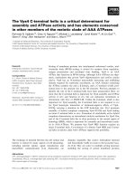
DEK protein levels were analyzed in a panel of 9 humanderived CRC cell lines and compared with the expression
in 2 non-tumor mucosa tissues. All tested cell lines
showed high DEK expression levels compared to mucosa
tissues (Figure 1). It is highlighting that two out of the
three cell lines showing the highest DEK expression levels
are those with metastatic origin (SW620 and LOVO).
To assess whether DEK is involved in aggressive phenotype, we selected DLD1 cell line derived from the primary tumor and SW620 derived from a metastatic focus,
due to the different origin and different DEK expression
pattern.
To downregulate DEK expression, 3 different siRNA sequences were used to transfect DLD1 and SW620 cells.
Figure 1 DEK is overexpressed in CRC. Western blot analysis of a
panel of human derived colorectal cancer cell lines showed higher
DEK expression than human non-tumor mucosa tissues (NT1, NT2).
Page 4 of 10
Proteins were extracted at 24, 48, and 72 hours after transfection. DEK downregulation was confirmed at protein
level by showing a decreased expression from 48 to
72 hours in both cell lines (Figure 2A).
We observed that when DEK was silenced, cell viability significantly decreased in both cell lines (P < 0.001)
(Figure 2B).
We then aimed to analyze whether downregulation of
DEK affects migration of DLD1 and SW620 cell lines.
When wound healing assays were performed, a delay of
12 to 24 hours was observed in DEK silenced cells compared to control (Figure 2C). In addition, we observed a
significant reduction in migration ability of both cell
lines, being higher on DLD1 (P < 0.001) than SW620
(P = 0.023) (Figure 2D).
Low DEK expression sensitized to SN38
Silenced DEK cell lines were cultured in the presence of
oxaliplatin active principle, LOHP, irinotecan active
principle, SN38, and 5FU. 72 hours after DEK downregulation, and 24 hours after treatments, cell cycle and
apoptosis were assessed. No significant effect was observed in the cell cycle analysis (data not shown).
DEK downregulation was not enough to produce significant annexin-V induction. However when it was combined with SN38, annexin-V levels significantly increased
in both cell lines (P < 0.05) (Figure 3). This effect was
not observed when DEK silencing was combined with
5FU treatment (Figure 3) or LOHP treatment (data not
shown).
This relation between DEK expression and irinotecan
therapy was confirmed in three tumor samples cultured
ex-vivo with SN38 for 24 hours. Following SN38 treatment, tissues were stained for DEK, Ki-67, and cleavedCaspase-3. DEK expression was similar between untreated
and treated samples indicating that SN38 did not affect its
expression. In one of the three patients a substantial reduction in Ki-67 and an increase in cleaved-Caspase-3 expression were observed. This sample corresponds with the
one showing the lowest DEK expression levels. The other
two tumor samples showed higher DEK expression levels
and we did not find differences in any of three analyzed
markers (Figure 4A). These results could suggest that
DEK level is related to irinotecan response.
To assess more deeply the role of DEK level in the induction of the apoptosis, DLD1 and SW620 cells were
transfected with siDEK (siDEKsec57) and cleaved Caspase
3 was detected after 72 hours by Western Blot. After DEK
downregulation both cell lines increased considerably
cleaved Caspase 3 level. Interestingly, cell line with the
lowest DEK level, DLD1, showed the highest induction
after silencing (Figure 4B). This result suggests the involvement of DEK in apoptosis.
Martinez-Useros et al. BMC Cancer 2014, 14:965
/>
Page 5 of 10
Figure 2 DEK downregulation decreases cell viability, migration and invasion. A) Three different siRNAs of DEK (siDEKsec57, sec58 and
sec59) were used to downregulate DEK protein expression in DLD1 and SW620 cell lines. We verified DEK expression levels by western blot at 24,
48, and 72 hours after transfection. B) MTS assay showed that both DLD1 and SW620 cell lines decreased cell viability at 72 hours after DEK
downregulation (P < 0.001). C) Microscope images of wound healing assay showed a reduced cell migration after DEK downregulation in both
cell lines. Images are representative from one experiment and were taken at 6, 12, and 24 hours after scratching. Arrows represent distance
between cell migration heads. D) Cell invasion assays performed in Boyden chamber showed that DEK silencing decreased invasion ability of
both cell lines. All assays were performed with 2 different siRNA sequences (siDEKsec57 and 58).
DEK is a potential predictive marker of survival in KRASwt
mCRC patients
Based on the previous results suggesting an association
between DEK expression and irinotecan sensitivity, we
hypothesized that DEK expression levels could be related
to the response to irinotecan-based chemotherapy in
mCRC. For this purpose, 67 samples from mCRC patients
receiving irinotecan-based chemotherapy were selected.
Representative images of different DEK expression levels
are shown in Figure 5A.
Association between DEK expression levels and progression-free survival after first-line irinotecan-based
treatment was analyzed using the Kaplan-Meier method.
Survival analysis demonstrated a trend to shorter progression-free survival for patients with higher DEK levels
(data not shown). When patients were stratified by KRAS
mutation status, no correlation between DEK expression
and progression-free survival of KRASmut patients was
observed (data not shown). However, a significant association with the outcome of patients KRASwt was found
(P = 0.03, data not shown). The third tertile was established as the best cut-off point, leaving low- and high-risk
patient groups (P = 0.01, Figure 5B).
Cox regression analysis showed that KRASwt patients
with high DEK expression showed increased risk of progression [HR 2.82 (95% CI 1.24-6.45), P = 0.01], that
remained significant after multivariate analysis [HR 2.4
(95% CI 1.04-5.58), P = 0.04] (Table 2).
These results suggest that DEK expression could be a
potential predictive marker of sentitivity in KRASwt
mCRC patients receiving irinotecan-based therapy as
single first-line treatment.
Martinez-Useros et al. BMC Cancer 2014, 14:965
/>
Page 6 of 10
Figure 3 Irinotecan response is increased when DEK is silenced. Diagram shows percentage of apoptotic cells stained with Annexin-V after
treatments. DLD1 and SW620 cell lines were DEK knocked-down and treated with SN38 or 5FU separately and compared with control cells
(control siRNA and untreated). Combination of siDEK and SN38 increased apoptosis (P < 0.05) compared to control or DEK silenced in both cell
lines. In addition, combination increased apoptosis (P < 0.05) compared to single SN38 treatment in DLD1 cell line. No significant differences were
observed after 5FU treatment. Results are expressed as the average of downregulation with 2 different siRNA sequences (siDEKsec57 and 58)
in triplicate.
Discussion
DEK is a non-histone nuclear protein that performs
a transcriptional activity involved in carcinogenesis at
multiples levels. In addition, DEK is able to bind cruciform structures and superhelical DNA over linear DNA
and introduces positive supercoils [34-36]. Moreover,
other studies have shown how nuclear DEK is also able
to perform a transcriptional repression of NF-κB pathway through transcriptional repression of cIAP2 and IL-8
in response to TNFα treatment [37]. However, the effect
of DEK is not only focused on inflammation but also on
neoplasms development and aggressive phenotype maintenance. DEK overexpression has been observed in different tumors [14-21,23-25,38].
Our results show DEK downregulation in primary
and metastatic CRC human cell lines reduces the migration ability and cell viability, both involved in maintain aggressive phenotype, according to previous studies
[27-29,38].
Irinotecan is a common drug used in clinical practice to
treat CRC patients. It is activated by glucuronidation to
SN38 and it prevents DNA from unwinding by inhibition
of topoisomerase I. After a combination of SN38 treatment and silenced DEK, we observed a significant increase
in annexin-V positive cells compared to those treated only
with SN38 or DEK knock-down. It is important to note
that this effect was not observed after 5FU or LOHP treatments alone or in combination with DEK knock-down.
This suggests that low DEK expression sensitizes to SN38.
Regarding to apoptosis process, we observed how DEK
downregulation activated Caspase 3. This result correlated with ex-vivo assay where an association between
low DEK expression and irinotecan sensitivity by induction of cleaved Caspase 3 was found. Lin et al. have been
recently reported that silencing of DEK resulted in a decrease in cell proliferation and apoptosis induction revealed by an increase in cleaved Caspase 3 and 9 [38].
These results agreed our data and highlight the involvement of DEK in the proliferation of CRC and its potential role as therapeutic target alone or in combination
with irinotecan.
Topoisomerase I expression was determined in most
of the samples since it has correlated with irinotecan response in several studies [11,12] but no association was
Martinez-Useros et al. BMC Cancer 2014, 14:965
/>
Page 7 of 10
Figure 4 Low DEK level sensitizes to irinotecan and induces cleaved Caspase 3 expression. A) Ex vivo assay results for the tumor samples
with the lowest (T1) and the highest (T2) DEK expression levels. SN38 treatment did not affect DEK expression, but a substantial reduction in Ki-67
and an increase in cleaved-Caspase 3 expression were observed in T1. No changes on Ki-67 or cleaved-Caspase 3 appeared in T2. B) Representative
Western Blot of DEK, cleaved Caspase 3 and Actin expression after 72 hours of DEK downregulation in SW620 and DLD1 cells with siDEKsec57 (left
panel). Densitometric data of Western Blot expressed as ratio of DEK/Actin and cleaved Caspase 3/Actin expression (right panel).
found between topoisomerase I levels and progressionfree survival in this set of patients. Our immunohistochemistry results showed that those mCRC patients with
higher levels of DEK expression presented a tendency of
shorter progression-free survival after first-line treatment
with irinotecan-based chemotherapy. When we stratified
Martinez-Useros et al. BMC Cancer 2014, 14:965
/>
Page 8 of 10
Figure 5 DEK expression is related to irinotecan response and correlates with poor outcome in KRASwt patients. A) Representative IHC
images for low, medium or high staining intensities of DEK expression levels. B) Kaplan-Meier plot shows a significant association between high
DEK level and lower progression-free survival after irinotecan-based treatment in KRASwt patients (P = 0.010).
patients according to KRAS status, we found that KRASwt
patients with DEK higher expression had poor outcome
independently of topoisomerase I levels. Therefore, these
data suggest that DEK is a potential marker of poor prognosis of mCRC patients with KRASwt status.
The reports that involve DEK as a nuclear protein related to cell metabolism by changing supercoiled DNA
and maintaining heterochromatin structure along DNA
transcription [36] could explain tumor aggressive behaviour and chemoresistance properties. Moreover, we suggest DEK overexpression allows this aggressive phenotype
by stabilizing DNA in CRC cells which agrees with higher
DEK levels on analyzed metastatic cells lines. We propose
that tumor cells induce their apoptosis cascade when two
Table 2 Univariate and multivariate Cox analysis results
UNIVARIATE PFS
MULTIVARIATE PFS
95% CI
95% CI
HR
Lower
Upper
P
HR
Lower
Upper
P
AGE
0,966
0,936
0,996
0,025
0,971
0,941
1,002
0,068
CEA
1,002
0,998
1,005
0,333
METASTASIS
0,398
Liver
1,000
Liver & other
1,786
0,571
5,585
Other
1,872
0,701
4,995
BRAF
0,828
wild-type
1,000
mutated
1,119
0,410
3,055
BIOLOGIC TREATMENT
0,763
None
1,000
Yes
0,880
0,383
2,022
TOPO I
0,974
Low
1,000
High
1,017
0,364
2,845
DEK
0,014
Low
1,000
High
2,825
0,040
1,000
1,238
Other refers to lung, lymph node and/or peritoneal metastasis.
6,449
2,408
1,039
5,579
Martinez-Useros et al. BMC Cancer 2014, 14:965
/>
events occur. On one hand, DNA transcription is altered
when DEK protein in downregulated and on the other
hand, DNA replication is stopped due to topoisomerase I
inhibition by irinotecan treatment. For all this, we propose
that chemoresistance could be explained by the DNA stabilization properties of DEK.
Page 9 of 10
4.
5.
Conclusions
The overall data presented here clearly point to a new
role of DEK oncogene as a clear factor for the maintenance of the aggressive phenotype in metastatic colorectal
cancer and as a potential marker of irinotecan-based
therapy response for KRASwt patients.
Abbreviations
CRC: Colorectal cancer; 5FU: 5-fluorouracil; EGFR: Epidermal growth factor
receptor; FFPE: Formalin-fixed paraffin-embedded; FOLFIRI: Folinic acid5FU-irinotecan; IHC: Immunohistochemistry; MCRC: Metastatic colorectal
cancer; PARP-1: Poly-ADP-ribose polymerase 1; siRNA: Small interference RNA
sequences; Topo I: Topoisomerase I.
6.
7.
8.
Competing interests
The authors declare that they have no competing interests.
Authors’ contributions
JM-U and JG-F. designed research; JM-U., MR-R, AB-P, IM, SZ, RV-B, AP-O, NP,
CS and L dP-N performed research; JM-U, AC, TG delP, FR, MSS, MJF-A and
JG-F contributed to analytic tools; JM-U, and JG-F analysed data; and JM-U
wrote the paper. All authors read and approved the final manuscript.
Acknowledgements
We thank Dr. Carlos Pastor from Surgery Department from Fundación
Jiménez-Díaz Hospital (Madrid, Spain) for providing us human samples, and
Dr. Juan Valcárcel from Centre of Genomic Regulation (Barcelona, Spain) for
his appreciated leadership of the CONSOLIDER-Consortium.
This work has been carried out with the support of the RNA-Reg.
CONSOLIDER-Consortium (CSD2009-00080) and by grants: RD12/0036/0051,
RD09/0076/0101, PI12/01552 from Spanish Institute of Health Carlos III (ISCIII)
and S2010/BMD2344.
9.
10.
11.
12.
Author details
1
Translational Oncology Division, OncoHealth Institute, Health Research
Institute - University Hospital “Fundación Jiménez Díaz”-UAM, Av. Reyes
Católicos 2, 28040 Madrid, Spain. 2Department of Pathology, University
Hospital “Fundación Jiménez Díaz”-UAM, Madrid, Spain. 3Department of
Oncology, Vigo Hospital, Vigo, Spain. 4Melanoma Research Group, Spanish
National Cancer Research Centre, Madrid, Spain. 5Department of Pathology,
Clinico San Carlos University Hospital, Madrid, Spain.
13.
14.
Received: 14 October 2014 Accepted: 11 December 2014
Published: 16 December 2014
15.
References
1. Ferlay JSI, Ervik M, Dikshit R, Eser S, Mathers C, Rebelo M, Parkin DM, Forman
D, Bray F: Cancer incidence and mortality worldwide: IARC Sources,
methods and major patterns in GLOBOCAN 2012. Int J Cancer 2014, 136
(5):E359-386.
2. Diaz-Rubio E, Tabernero J, Gomez-Espana A, Massuti B, Sastre J, Chaves M,
Abad A, Carrato A, Queralt B, Reina JJ, Maurel J, González-Flores E, Aparicio
J, Rivera F, Losa F, Aranda E: Phase III study of capecitabine plus oxaliplatin compared with continuous-infusion fluorouracil plus
oxaliplatin as first-line therapy in metastatic colorectal cancer: final
report of the Spanish Cooperative Group for the Treatment of Digestive
Tumors Trial. J Clin Oncol 2007, 25(27):4224–4230.
3. Cassidy J, Clarke S, Diaz-Rubio E, Scheithauer W, Figer A, Wong R, Koski S, Lichinitser M, Yang TS, Rivera F, Couture F, Sirzén F, Saltz L: Randomized phase III study
of capecitabine plus oxaliplatin compared with fluorouracil/folinic acid plus
16.
17.
18.
oxaliplatin as first-line therapy for metastatic colorectal cancer. J Clin Oncol
2008, 26(12):2006–2012.
Porschen R, Arkenau HT, Kubicka S, Greil R, Seufferlein T, Freier W,
Kretzschmar A, Graeven U, Grothey A, Hinke A, Schmiegel W, Schmoll HJ;
AIO Colorectal Study Group: Phase III study of capecitabine plus
oxaliplatin compared with fluorouracil and leucovorin plus oxaliplatin in
metastatic colorectal cancer: a final report of the AIO Colorectal Study
Group. J Clin Oncol 2007, 25(27):4217–4223.
Souglakos J, Androulakis N, Syrigos K, Polyzos A, Ziras N, Athanasiadis A,
Kakolyris S, Tsousis S, Kouroussis Ch, Vamvakas L, Kalykaki A, Samonis G,
Mavroudis D, Georgoulias V: FOLFOXIRI (folinic acid, 5-fluorouracil, oxaliplatin
and irinotecan) vs FOLFIRI (folinic acid, 5-fluorouracil and irinotecan) as
first-line treatment in metastatic colorectal cancer (MCC): a multicentre
randomised phase III trial from the Hellenic Oncology Research Group
(HORG). Br J Cancer 2006, 94(6):798–805.
Jordan K, Kellner O, Kegel T, Schmoll HJ, Grothey A: Phase II trial of
capecitabine/irinotecan and capecitabine/oxaliplatin in advanced
gastrointestinal cancers. Clin Colorectal Cancer 2004, 4(1):46–50.
Koopman M, Antonini NF, Douma J, Wals J, Honkoop AH, Erdkamp FL, de Jong
RS, Rodenburg CJ, Vreugdenhil G, Loosveld OJ, van Bochove A, Sinnige HA,
Creemers GJ, Tesselaar ME, Slee PH, Werter MJ, Mol L, Dalesio O, Punt CJ:
Sequential versus combination chemotherapy with capecitabine,
irinotecan, and oxaliplatin in advanced colorectal cancer (CAIRO): a phase
III randomised controlled trial. Lancet 2007, 370(9582):135–142.
Zarate R, Rodriguez J, Bandres E, Patino-Garcia A, Ponz-Sarvise M, Viudez A,
Ramirez N, Bitarte N, Chopitea A, Gacia-Foncillas J: Oxaliplatin, irinotecan
and capecitabine as first-line therapy in metastatic colorectal cancer
(mCRC): a dose-finding study and pharmacogenomic analysis. Br J Cancer
2010, 102(6):987–994.
Muro K, Boku N, Shimada Y, Tsuji A, Sameshima S, Baba H, Satoh T, Denda T, Ina
K, Nishina T, Yamaguchi K, Takiuchi H, Esaki T, Tokunaga S, Kuwano H, Komatsu Y,
Watanabe M, Hyodo I, Morita S, Sugihara K: Irinotecan plus S-1 (IRIS) versus fluorouracil and folinic acid plus irinotecan (FOLFIRI) as second-line chemotherapy for metastatic colorectal cancer: a randomised phase 2/3 non-inferiority
study (FIRIS study). Lancet Oncol 2010, 11(9):853–860.
Jansen WJ, Hulscher TM, van Ark-Otte J, Giaccone G, Pinedo HM, Boven E:
CPT-11 sensitivity in relation to the expression of P170-glycoprotein and
multidrug resistance-associated protein. Br J Cancer 1998, 77(3):359–365.
Kostopoulos I, Karavasilis V, Karina M, Bobos M, Xiros N, Pentheroudakis G,
Kafiri G, Papakostas P, Vrettou E, Fountzilas G: Topoisomerase I but not
thymidylate synthase is associated with improved outcome in patients
with resected colorectal cancer treated with irinotecan containing
adjuvant chemotherapy. BMC Cancer 2009, 9:339.
Braun MS, Richman SD, Quirke P, Daly C, Adlard JW, Elliott F, Barrett JH,
Selby P, Meade AM, Stephens RJ, Parmar MK, Seymour MT.: Predictive
biomarkers of chemotherapy efficacy in colorectal cancer: results from
the UK MRC FOCUS trial. J Clin Oncol 2008, 26(16):2690–2698.
von Lindern M, Breems D, van Baal S, Adriaansen H, Grosveld G:
Characterization of the translocation breakpoint sequences of two
DEK-CAN fusion genes present in t(6;9) acute myeloid leukemia and a
SET-CAN fusion gene found in a case of acute undifferentiated leukemia.
Genes Chromosomes Cancer 1992, 5(3):227–234.
Datta A, Adelson ME, Mogilevkin Y, Mordechai E, Sidi AA, Trama JP:
Oncoprotein DEK as a tissue and urinary biomarker for bladder cancer.
BMC Cancer 2011, 11:234.
Privette Vinnedge LM, McClaine R, Wagh PK, Wikenheiser-Brokamp KA, Waltz
SE, Wells SI: The human DEK oncogene stimulates beta-catenin signaling,
invasion and mammosphere formation in breast cancer. Oncogene 2011,
30(24):2741–2752.
Kroes RA, Jastrow A, McLone MG, Yamamoto H, Colley P, Kersey DS, Yong
VW, Mkrdichian E, Cerullo L, Leestma J, Moskal JR: The identification of
novel therapeutic targets for the treatment of malignant brain tumors.
Cancer Lett 2000, 156(2):191–198.
Kondoh N, Wakatsuki T, Ryo A, Hada A, Aihara T, Horiuchi S, Goseki N,
Matsubara O, Takenaka K, Shichita M, Tanaka K, Shuda M, Yamamoto M:
Identification and characterization of genes associated with human
hepatocellular carcinogenesis. Cancer Res 1999, 59(19):4990–4996.
Khodadoust MS, Verhaegen M, Kappes F, Riveiro-Falkenbach E, Cigudosa JC,
Kim DS, Chinnaiyan AM, Markovitz DM, Soengas MS: Melanoma proliferation
and chemoresistance controlled by the DEK oncogene. Cancer Res 2009,
69(16):6405–6413.
Martinez-Useros et al. BMC Cancer 2014, 14:965
/>
19. Grasemann C, Gratias S, Stephan H, Schuler A, Schramm A, Klein-Hitpass L,
Rieder H, Schneider S, Kappes F, Eggert A, Lohmann DR: Gains and overexpression identify DEK and E2F3 as targets of chromosome 6p gains
in retinoblastoma. Oncogene 2005, 24(42):6441–6449.
20. Paderova J, Orlic-Milacic M, Yoshimoto M, da Cunha Santos G, Gallie B,
Squire JA: Novel 6p rearrangements and recurrent translocation
breakpoints in retinoblastoma cell lines identified by spectral karyotyping
and mBAND analyses. Cancer Genet Cytogenet 2007, 179(2):102–111.
21. Carro MS, Spiga FM, Quarto M, Di Ninni V, Volorio S, Alcalay M, Muller H:
DEK Expression is controlled by E2F and deregulated in diverse tumor
types. Cell Cycle 2006, 5(11):1202–1207.
22. Lin L, Piao J, Gao W, Piao Y, Jin G, Ma Y, Li J, Lin Z: DEK over expression as
an independent biomarker for poor prognosis in colorectal cancer.
BMC Cancer 2013, 13:366.
23. Han S, Xuan Y, Liu S, Zhang M, Jin D, Jin R, Lin Z: Clinicopathological
significance of DEK overexpression in serous ovarian tumors. Pathol Int
2009, 59(7):443–447.
24. Wu Q, Li Z, Lin H, Han L, Liu S, Lin Z: DEK overexpression in uterine
cervical cancers. Pathol Int 2008, 58(6):378–382.
25. Nagpal JK, Das BR: Identification of differentially expressed genes in
tobacco chewing-mediated oral cancer by differential displaypolymerase chain reaction. Eur J Clin Invest 2007, 37(8):658–664.
26. Gamble MJ, Fisher RP: SET and PARP1 remove DEK from chromatin to
permit access by the transcription machinery. Nat Struct Mol Biol 2007,
14(6):548–555.
27. Wise-Draper TM, Allen HV, Thobe MN, Jones EE, Habash KB, Munger K, Wells SI: The
human DEK proto-oncogene is a senescence inhibitor and an upregulated
target of high-risk human papillomavirus E7. J Virol 2005, 79(22):14309–14317.
28. Wise-Draper TM, Allen HV, Jones EE, Habash KB, Matsuo H, Wells SI:
Apoptosis inhibition by the human DEK oncoprotein involves
interference with p53 functions. Mol Cell Biol 2006, 26(20):7506–7519.
29. Kim D, Kim J, Kang SS, Jin EJ: Transforming growth factor-beta3-induced
Smad signaling regulates actin reorganization during chondrogenesis of
chick leg bud mesenchymal cells. J Cell Biochem 2009, 107(4):622–629.
30. Witkiewicz AK, Balaji U, Knudsen E: Systematically defining single gene
determinants of response to neoadjuvant chemotherapy reveals specific
biomarkers. Clin Cancer Res 2014, 20(18):4837–4848.
31. Souza V, Dong YB, Zhou HS, Zacharias W, McMasters KM: SW-620 cells
treated with topoisomerase I inhibitor SN-38: gene expression profiling.
J Transl Med 2005, 3:44.
32. Cristobal I, Rincon R, Manso R, Madoz-Gurpide J, Carames C, Del PuertoNevado L, Rojo F, Garcia-Foncillas J: Hyperphosphorylation of PP2A in
colorectal cancer and the potential therapeutic value showed by its
forskolin-induced dephosphorylation and activation. Biochim Biophys Acta
2014, 1842(9):1823–1829.
33. Tan WL, Bhattacharya B, Loh M, Balasubramanian I, Akram M, Dong D,
Wong L, Thakkar B, Salto-Tellez M, Soo RA, Fichtner I, Iacopetta B, Soong R:
Low cytosine triphosphate synthase 2 expression renders resistance to
5-fluorouracil in colorectal cancer. Cancer Biol Ther 2011, 11(6):599–608.
34. Waldmann T, Baack M, Richter N, Gruss C: Structure-specific binding of the
proto-oncogene protein DEK to DNA. Nucleic Acids Res 2003, 31(23):7003–7010.
35. Waldmann T, Scholten I, Kappes F, Hu HG, Knippers R: The DEK protein–an
abundant and ubiquitous constituent of mammalian chromatin. Gene
2004, 343(1):1–9.
36. Alexiadis V, Waldmann T, Andersen J, Mann M, Knippers R, Gruss C: The
protein encoded by the proto-oncogene DEK changes the topology of
chromatin and reduces the efficiency of DNA replication in a chromatinspecific manner. Genes Dev 2000, 14(11):1308–1312.
37. Sammons M, Wan SS, Vogel NL, Mientjes EJ, Grosveld G, Ashburner BP:
Negative regulation of the RelA/p65 transactivation function by the
product of the DEK proto-oncogene. J Biol Chem 2006, 281(37):26802–26812.
38. Lin L, Piao J, Ma Y, Jin T, Quan C, Kong J, Li Y, Lin Z: Mechanisms
underlying cancer growth and apoptosis by DEK overexpression in
colorectal cancer. PLoS One 2014, 9(10):e111260.
Page 10 of 10
Submit your next manuscript to BioMed Central
and take full advantage of:
• Convenient online submission
• Thorough peer review
• No space constraints or color figure charges
doi:10.1186/1471-2407-14-965
Cite this article as: Martinez-Useros et al.: DEK is a potential marker for
aggressive phenotype and irinotecan-based therapy response in
metastatic colorectal cancer. BMC Cancer 2014 14:965.
• Immediate publication on acceptance
• Inclusion in PubMed, CAS, Scopus and Google Scholar
• Research which is freely available for redistribution
Submit your manuscript at
www.biomedcentral.com/submit