Radiosensitization with combined use of olaparib and PI-103 in triple-negative breast cancer
Bạn đang xem bản rút gọn của tài liệu. Xem và tải ngay bản đầy đủ của tài liệu tại đây (1.15 MB, 9 trang )
Jang et al. BMC Cancer (2015) 15:89
DOI 10.1186/s12885-015-1090-7
RESEARCH ARTICLE
Open Access
Radiosensitization with combined use of olaparib
and PI-103 in triple-negative breast cancer
Na Young Jang1,6†, Dan Hyo Kim2†, Bong Jun Cho2†, Eun Jung Choi2, Jong-Soo Lee3, Hong-Gyun Wu4,
Eui Kyu Chie4 and In Ah Kim2,4,5*
Abstract
Background: Triple-negative breast cancer (TNBC) shows aggressive clinical behavior, but the treatment options are
limited due to lack of a specific target. TNBC shares many clinical and pathological similarities with BRCA-deficient
breast cancer, for which poly(ADP-ribose) polymerase (PARP) inhibitor is effective, but PARP inhibitor alone failed to show
clinical effects in patients with sporadic TNBC. Radiation induces DNA double-strand breaks, and the phosphoinositide
3-kinase (PI3K) signaling pathway has been known to regulate steady-state levels of homologous recombination. A recent
preclinical study showed that PI3K inhibition impairs BRCA1/2 expression and sensitizes BRCA-proficient TNBC to PARP
inhibition. Therefore, we assessed the radiosensitizing effect, and the underlying mechanism of combination treatment
with PARP inhibitor olaparib and PI3K inhibitor PI-103 in BRCA-proficient TNBC cells.
Methods: MDA-MB-435S cells were divided into four treatment groups, irradiation (IR) alone, olaparib plus IR, PI-103 plus
IR, and olaparib plus PI-103 plus IR. Cells were exposed to the drugs for 2 hours prior to irradiation, and the cell survival
curve was obtained using a clonogenic assay. Western blotting and immunofluorescent detection of γH2AX foci were
performed. Xenograft and bioluminescence imaging were carried out to assess in vivo radiosensitivity.
Results: Combined use of olaparib and PI-103 enhanced radiation-induced death of MDA-MB-435S (sensitizer
enhancement ratio[SER]0.05,1.7) and MDA-MB-231-BR (SER0.05,2.1) cells and significantly reduced tumor volume in a
xenograft models (P < 0.001). Treatment with PI-103 showed persistent γH2AX foci, indicating delayed repair of DNA
strand breaks. PI-103 alone increased levels of poly(ADP-ribose) and phosphorylated extracellular signal-regulated
kinase, and downregulated BRCA1.
Conclusions: Combined use of olaparib and PI-103 enhanced radiation-induced cell death in BRCA-proficient
MDA-MB-435S and MDA-MB-231-BR cells and xenografts. TNBC patients have high incidences of locoregional relapse
and distant metastasis, and radiation therapy targets both locoregional control and treatment of distant recurrences
such as brain metastasis or other oligometastasis. Targeting of the PI3K signaling pathway combined with PARP
inhibition maybe a feasible approach to enhance effects of radiation in BRCA-proficient TNBC.
Keywords: Triple-negative breast cancer, Radiotherapy, Olaparib, PI-103, PARP inhibitor, PI3K inhibitor
Background
Triple-negative breast cancer (TNBC) is defined as a
tumor that does not express the estrogen receptor (ER),
progesterone receptor, or human epidermal growth factor
receptor 2 (HER2). Aggressive clinical behaviors, such as
early distant metastasis and lack of specific treatment
* Correspondence:
†
Equal contributors
2
Medical Science Research Institute, Seoul National University Bundang
Hospital, Seongnam, Korea
4
Department of Radiation Oncology, Seoul National University, Seoul, Korea
Full list of author information is available at the end of the article
targets, i.e. ER or HER2, have been obstacles to the treatment of TNBC [1]. Cytotoxic chemotherapy or combination with a targeted agent, such as bevacizumab or
cetuximab, has been used, but the results were disappointing [2]. Meanwhile, the introduction of poly(ADP-ribose)
polymerase (PARP) inhibitors were expected to provide a
promising new therapeutic strategy for TNBC.
The PARP family of enzymes is involved in DNA repair, cell proliferation and death, and genomic stability
[3-5]. PARP1 is the most abundant PARP and is involved
in base-excision repair (BER) [6]. When a DNA strand
© 2015 Jang et al.; licensee BioMed Central. This is an Open Access article distributed under the terms of the Creative
Commons Attribution License ( which permits unrestricted use, distribution, and
reproduction in any medium, provided the original work is properly credited. The Creative Commons Public Domain
Dedication waiver ( applies to the data made available in this article,
unless otherwise stated.
Jang et al. BMC Cancer (2015) 15:89
break occurs, PARP1 rapidly binds to the break site and induces auto-poly(ADP-ribosyl)ation. Auto-poly(ADP-ribosyl)ation creates a negative charge, which recruits the
enzymes required for BER [6,7]. Synthetic lethality occurs
when two otherwise nonlethal mutations together result in
an inviable cell [8]. When DNA damage occurs, cells with
mutations in either PARP or BRCA, which is involved in
homologous recombination (HR), can survive, whereas cells
with mutations in both cannot [9].
This strategy is very effective for treatment of breast
cancer patients with BRCA mutations, but the problem is
that incidence of BRCA-related breast cancer is less than
10% [10]. Recently, some investigators have used the term
“BRCA-ness” of TNBC, because BRCA-deficient breast
cancer and sporadic basal-like or TNBC share many clinical and pathological similarities [11,12]. Exploiting these
similarities, clinical trials have tested the effectiveness of
PARP inhibitors on BRCA-proficient TNBC patients.
However, PARP inhibitor alone failed to show clinical effects in patients with TNBC [13]. Subsequent studies are
ongoing to test the efficacy of the combined use of PARP
inhibitor and cytotoxic chemotherapy or radiotherapy.
Radiation induces DNA double-strand breaks (DSBs),
and the phosphoinositide 3-kinase (PI3K) signaling pathway
has been known to regulate steady-state levels of HR [14].
Furthermore, Ibrahim et al. published interesting research
results showing that PI3K inhibition impairs BRCA1/2 expression and sensitizes BRCA-proficient TNBC to PARP
inhibition [15]. Inhibition of the PI3K signaling pathway
induces feedback upregulation of extracellular signalregulated kinase (ERK) and subsequent increased activation
of ETS1 as an ERK-related transcription factor. ETS1 suppresses BRCA1/2 expression and impairs HR, thereby sensitizing the cells to the PARP inhibitor. In addition,
preclinical studies showed increased radiosensitivity with
use of the PARP inhibitor in replicating cells [16].
Taken together, these findings show that radiation, PI3K
inhibitors, and PARP inhibitors may enhance each other’s
tumor cell killing effects, and we postulated that the combined use of a PARP inhibitor and PI3K inhibitor would
sensitize cells to radiation. Therefore, we assessed the
radiosensitizing effect of combined treatment of BRCAproficient TNBC cells with olaparib and PI-103and investigated the underlying mechanism of action.
Page 2 of 9
Pharmacologic inhibitors
Olaparib was obtained from Selleck Chemicals (Houston,
TX, USA) and PI-103 (pyridinylfuranopyrimidine inhibitor) was obtained from Calbiochem (Billerica, MA, USA).
Drugs were diluted in dimethylsulfoxide (DMSO).
Short interfering RNA (siRNA) transfection
BRCA1 siRNA was obtained from BioNeer (Alameda, CA,
USA). Cells were plated in six-well plates and transfected
with 100 nM BRCA1 siRNA using Lipofectamine® RNAiMAX transfection reagent (Life Technologies, Grand
Island, NY, USA) according to the manufacturer’s protocol.
Clonogenic assay
The clonogenic assay was carried out according to a previously described protocol [17]. Appropriate numbers of
cells were plated across the different treatment groups
for each radiation dose. Treatment groups were as follows; irradiation (IR) alone, olaparib and IR, PI-103 and
IR, and olaparib and PI-103 and IR. Cells were treated
with olaparib (1 μM) and PI-103 (0.4 μM) 2 hours before
IR, as described in previous studies [18,19]. A specified
number of cells were seeded in six-well plates and irradiated with 6 MV x-ray from a linear accelerator (Varian
Medical Systems, Palo Alto, CA, USA) at a dose rate of
2.46 Gy/min. After 22 hours incubation, medium was replaced with drug-free, FBS-containing medium, and cells
were incubated for 14 to 21 days to allow colony formation. Colonies were fixed in methanol and stained with
0.5% crystal violet, and the number of colonies containing at least 50 cells was determined and the surviving
fraction was calculated. Survival data was fitted to a
linear-quadratic model using Kaleidagraph version 3.51
(Synergy Software, Reading, PA, USA). Each point on
the survival curve represents the mean surviving fraction
(SF) from at least three dishes. The sensitizer enhancement ratio 0.05 (SER0.05) was defined as the ratio of the
isoeffective dose at SF 0.05 in the absence of inhibitors
to that in the presence of inhibitors. The average SF
relative to the radiation-alone group (SFO) at each radiation dose was calculated. Expected SF for the two-drug
combination (SFE) was calculated as the product of the
SFOs of the individual single-drug groups. The synergistic index (SI) was calculated as SFE/SFO, and SI >1.00 indicates a synergistic effect [20].
Methods
Cell culture
Western blotting
Triple-negative, BRCA-proficient breast cancer cell lines
MDA-MB-435S and MDA-MB-231-BR (American Type
Culture Collection, Rockville, MD, USA) were cultured
in RPMI 1640 medium (Gibco; Invitrogen, Carlsbad,
CA, USA) supplemented with 10% fetal bovine serum
(Gibco; Invitrogen) at 37°C in an atmosphere of 95% air
and 5% CO2.
Cells were washed, harvested by scraping, and resuspended in lysis buffer (iNtRON Biotechnology, Seongnam,
Korea). Proteins were solubilized by sonication, and equal
amounts of protein were separated by SDS-PAGE and
electroblotted onto polyvinylidenedifluoride membranes
(EMD Millipore, Billerica, MA, USA). Membranes were
blocked in phosphate-buffered saline (PBS) containing
Jang et al. BMC Cancer (2015) 15:89
0.1% Tween 20 and 5%nonfat powdered milk, and probed
with primary antibody directed against p-AKT (Ser473),
p-ERK (Tyr202/204), Rad51 (all from Cell Signaling
Technology, Danvers, MA, USA),BRCA1, p-DNAprotein kinase (PK; Ser2056), PAR (all from Abcam,
Cambridge, UK), and β-actin (Santa Cruz Biotechnology,
Santa Cruz, CA, USA). Membranes were washed and incubated with secondary antibody consisting of peroxidaseconjugated goat anti-rabbit or anti-mouse IgG(Jackson
ImmunoResearch Laboratories, West Grove, PA, USA) at a
dilution of1:10,000 for 1 hour. Membrane washing and
western blotting was performed using an ECL kit (iNtRON
Biotechnology, Seongnam, Korea).
Total RNA extraction and reverse transcription
Total cellular RNA was isolated using an RNeasy Mini
Kit (Qiagen, Carlsbad, CA, USA), and cDNA was
made using M-MLV reverse transcriptase (M1705,
Promega, Madison, WI, USA) according to manufacturers’ instruction.
Quantitative real-time polymerase chain reaction (PCR)
Quantitative real-time reverse transcription (RT) PCR
was performed using SYBR Premix Ex Taq (Takara Bio
Inc., Otsu, Shiga, Japan). The following primer was used:
Rad51 (forward 5′-gcataaatgccaacgatgtg-3′, reverse 5′atgatctctgaccgcctttg-3′). For quantification, all values were
normalized to β-actin using the ΔΔCt method with data
from 3–5 independent experiments. Data were analyzed
using a TP800 (Takara Bio Inc., Otsu, Shiga, Japan).
Page 3 of 9
Cell labeling and implantation
MDA-MB-435S cells were transfected with a pGL4 luciferase reporter vector (Promega, Madison, WI, USA) according to the manufacturer’s protocol. Nude mice were
anesthetized and immobilized, and transfected MDA-MB435S cells were subcutaneously implanted. One week after
implantation, intraperitoneal administration of olaparib
and PI-103 of 10 mg/kg was initiated and carried out three
times weekly for 2 weeks. After drug treatment, mice were
irradiated three times weekly with 3 Gy per fraction. Mice
were then observed for 2 weeks.
Bioluminescence imaging (BLI)
BLI was carried out using the IVIS Lumina II BLI system
(Caliper, Hopkinton, MA, USA) according to the manufacturer’s protocol. One week after tumor cell implantation, baseline imaging was performed and, 2 weeks
after IR, follow-up imaging was carried out. Mice were
anesthetized and D-luciferin was injected intraperitoneally. Imaging was carried out 5 minutes after luciferin
injection and repeated every few minutes to determine
the maximum luminescence intensity in photons/second. After image acquisition, a region of interest (ROI)
was circumscribed for each tumor and a corresponding
tumor-free ROI was circumscribed to generate a
background-corrected bioluminescence flux value. The
maximum background-corrected value for that tumor
during the 30-minute imaging session was used as the
maximum bioluminescent value.
Statistical analysis
Immunohistochemical analysis of γH2AX
Cells were cultured and treated on chamber slides.
Seventeen hours after irradiation, cover slips were rinsed,
and cells were fixed in 4% paraformaldehyde and permeablized in methanol for 20 minutes. Cells were subsequently washed and blocked in PBS containing 2% bovine
serum albumin for 1 hour. Primary antibody against
γH2AX (Cell Signaling Technology) was applied to the
cells and incubated overnight. Secondary AlexaFluor 488conjugated donkey anti-goat antibody (Molecular Probes,
Eugene, OR, USA) was applied and incubated for 1 hour.
Nuclei were counterstained with 4′,6-diamidino-2-phenylindole (DAPI) by incubation of cells in 1 μg/mL DAPI for
5 minutes. Slides were examined on a ZeissAxio Scope.A1
Imager fluorescence microscope. Images were captured
using AxioCamMRc5 and AxioVision v.4.4 acquisition
software (Carl Zeiss, Jena, Germany).
In vivo tumor model
Animal experimentation was performed according to a
protocol approved by the Institutional Animal Care and
Use Committee of Seoul National University Bundang
Hospital (No. BA1103-078/015-01).
Statistical significance was assessed by Student t-test and
one way analysis of variance (ANOVA) using SPSS ver.
12.0 (SPSS Inc., Chicago, IL, USA).
Results
Combined use of olaparib and PI-103 enhanced
radiation-induced death of TNBC cells
To evaluate in vitro radiation sensitization effect of olaparib and PI-103, we performed clonogenic assay. Pretreatment with the combination of olaparib and PI-103
resulted in a significant increase in radiation-induced
death of both MDA-MB-435S (SER0.05 1.7) and MDAMB-231-BR cells (SER0.052.1) (Figure 1A and B).
Colony formation in the combined treatment group
without radiation was also decreased compared to the
control group (P = 0.03), but was not significantly different from the single-drug groups (P > 0.05). Data was
adjusted by control (without radiation) and fitted to a
linear quadratic model.
Radiation-induced cell death with 8 Gy was significantly enhanced in the combination treatment group
compared to the single-drug groups for both MDA-MB435S cells (P =0.005 for PI-103 and P < 0.001 for
Jang et al. BMC Cancer (2015) 15:89
Page 4 of 9
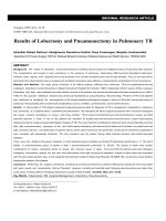
Figure 1 Effect of olaparib and PI-103 on radiosensitivity of MDA-MB-435S cells. MDA-MB-435S (A), MDA-MB-231 (B), and MDA-MB-435S
cells transfected with BRCA1 siRNA (C) were treated with the indicated drugs prior to receiving the indicated dose of radiation. Western blotting
showed decreased levels of BRCA1 in cells transfected with BRCA1 siRNA (D) compared with untransfected control cells (NC).
olaparib) and MDA-MB-231-BR cells (P < 0.001 for PI103 and P =0.01 for olaparib) based on t-test. The SI
of combination treatment at each radiation dose
was >1.00 (1.07, 1.27, 1.20, and 1.81 at 2, 4, 6, and
8 Gy, respectively).
To compare these results with results of the same
treatments in BRCA-deficient cells, MDA-MB-435S
cells were transfected with BRCA1 siRNA. We thought
naturally occurring BRCA mutated cells could have
many different molecular characteristics other than
BRCA. In order to exclude confounding factors we
chose siRNA transfection in the same cell lines instead
of using naturally occurring BRCA mutated cell lines.
As shown in Figure 1C, there appeared to be a radiosensitizing effect with olaparib treatment in the
BRCA1-knockdown cells (SER0.05 1.24) compared with
the control. However, addition of PI-103 to the olaparib treatment did not result in further enhancement
of the radiation-induced cell death.
Combined use of olaparib and PI-103 enhanced in vivo
radiation-induced cell death
After confirming the in vitro radiosensitizing effect of
combination treatment with olaparib and PI-103, we investigated the in vivo effect. MDA-MB-435S cells transfected with a luciferase reporter vector were implanted
in nude mice, which were treated with olaparib and PI103 alone or together, with or without radiation. Two
weeks after commencement of the treatment, the size of
tumors was examined in vivo using the IVIS Lumina II
BLI system. As shown in Figure 2, a marked decrease in
tumor volume was induced with combined use of olaparib and PI-103, compared to radiation alone (P < 0.001
by t-test). Means of ROI values were significantly different between the groups (P < 0.001 by one way ANOVA).
PI-103 induced persistent γH2AX foci
According to previous studies, radiation induces DNA
damage and PARP/PI3K inhibition impairs DNA damage
Jang et al. BMC Cancer (2015) 15:89
Page 5 of 9
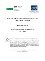
Figure 2 In vivo radiosensitizing effect of combined use of olaparib and PI-103. Bioluminescence imaging of nude mice implanted with
MDA-MB-435S cells transfected with pGL4 luciferase reporter vector and treated with indicated drugs alone (upper left) or prior to irradiation (IR)
(upper right) and quantitation (lower panels). A marked decrease in tumor volume was induced with combined use of olaparib and PI-103,
compared to radiation alone (P < 0.001 by t-test). Means of ROI values were significantly different between groups (P < 0.001). Y-axes show
bioluminescence in photons/second. P < 0.001, ***.
repair [14-16]. We detected γH2AX foci to evaluate the
capacity of repair of radiation induced DNA damage in
each treatment groups.
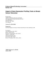
Pretreatment with PI-103, alone and together with olaparib, followed by irradiation caused marked prolongation
of γH2AX foci formation, indicating delayed DNA repair,
whereas pretreatment with olaparib alone followed by irradiation showed relatively few foci 17 hours after treatment (Figure 3).
Based on the results observed with PI-103 pretreatment,
we evaluated the molecules involved in DNA repair. Pretreatment with PI-103 was associated with decreased pDNA-PK and Rad51 (Figure 4C). Quantitative real-time
RT PCR data showed decreased mRNA expression of
Rad51 with treatment with PI-103 and olaparib (p = 0.001
by t-test, Figure 4D).
PI-103 induced upregulation of ERK and downregulation
of BRCA1
Decreased levels of p-AKT and PAR induced by treatment with PI-103 and olaparib, respectively, showed that
the drugs were working well (Figure 4A,B). To investigate
the possible mechanisms of radiosensitization, we analyzed
changes in the candidate proteins with reference to the
study of Ibrahim et al. [15]. It is a well-known phenomenon
that inhibition of the PI3K signaling pathway induces activation of the ERK pathway [21,22]. As expected, treatment
with PI-103 induced elevation of p-ERK level. Pretreatment
Jang et al. BMC Cancer (2015) 15:89
Page 6 of 9
Figure 3 Immunofluorescence-based detection of γH2AX. γH2AX was detected in MDA-MB-435S cells treated with the indicated drugs prior
to irradiation (IR). Nuclei were counterstained with DAPI.
with PI-103 was associated with activation of ERK and
downregulation of BRCA1, whereas the increased level of
PAR observed with PI-103 treatment disappeared with the
addition of olaparib (Figure 4A,B).
Discussion
TNBC is known to have aggressive clinical behaviors
and high mortality rates [1]. A high incidence and early
development of distant metastasis to areas, such as brain
or lung, in TNBC or basal-like breast cancer cases have
been reported in the literature, and the median duration
of survival after distant metastasis was significantly
shorter than that of other subtypes [23]. In addition, the
basal-like subtype was associated with an increased risk
of local and regional relapse after surgery, based on
multivariate analysis [24]. Furthermore, lack of specific
treatment targets, such as ER or HER2, presents a major
problem for the treatment of TNBC patients. To reduce
locoregional relapse and to treat brain metastasis or
other oligometastasis, the effect of radiation on TNBC
needs to be enhanced.
PARP inhibitor has shown a significant clinical benefit
in BRCA-related TNBC patients [25]. However, in
clinical situations, carriers of BRCA mutation account
for only a part of TNBC or basal-like breast cancer patients, so we also need to focus on the treatment of
BRCA-proficient TNBC. As BRCA-mutated breast cancer
and sporadic basal-like or TNBC share many clinical and
pathological similarities [11,12], use of PARP inhibitors
was expected to usher in a new era in the treatment of
TNBC. However, clinical outcomes of treatment with
PARP inhibitor alone were disappointing with sporadic
TNBC. To enhance the efficacy, studies on the combined
use of PARP inhibitor and other treatments are ongoing.
Herein, we assessed the radiosensitizing effect of combined treatment with olaparib and PI-103in BRCAproficient TNBC cell lines derived from metastatic sites.
Survival curves generated using the clonogenic assay
showed increased radiation-induced cell death with the
combined treatment of olaparib and PI-103 in both MDAMB-435S (Figure 1A) and MDA-MB-231-BR (Figure 1B)
cells. The MDA-MB-231-BR cell line is a subclone of
MDA-MB-231 that selectively metastasizes to the brain,
demonstrating that this combined targeting strategy may
be applied to enhance the effects of radiation on brain metastasis in TNBC patients.
Jang et al. BMC Cancer (2015) 15:89
Page 7 of 9
Figure 4 Western analysis of drug- and radiation-treated cells. The indicated proteins were detected by western analysis of MDA-MB-435S cells
treated with the indicated drugs prior to irradiation (IR) (A, B, C). β-actin served as internal control. Relative Rad51 mRNA expression level was measured by
quantitative real-time reverse transcription polymerase chain reaction in MDA-MB-435S cells treated with the indicated drugs (D). P < 0.001, **.
Therapeutic radiation induces DNA damage with single strand breaks (SSBs) and DSBs. PARP1 detects SSBs,
binds to the DNA break sites, and is then auto-poly
(ADP-ribosyl)ated and recruits the enzymes required to
form the BER multi-protein complex [6,7]. DSBs are
repaired by nonhomologous end joining (NHEJ) and
HR. NHEJ repairs most radiation-induced DSBs, does
not require a template, and may occur during any stage
of the cell cycle. In contrast, HR is an error-prone
process, requires a sister chromatid as a template, and
thus can only occur during the S and G2 phases [16,26].
Under normal conditions, SSBs are efficiently repaired
and do not lead to significant cell death. However, unrepaired SSBs or delayed repair of SSBs during the DNA
replication process can induce DSBs, which must be
repaired by HR. Therefore, PARP inhibition is an effective treatment strategy for tumors with HR deficiency,
such as those with a BRCA mutation. As expected,
siRNA-mediated knockdown of BRCA1 caused a high
degree of radiosensitization in itself, and addition of olaparib further enhanced the effects of radiation, as shown
in Figure 1C.
Radiation induces DNA damage, and PARP inhibitor
suppresses DNA repair. Thus, radiation and PARP inhibitor would have synergistic effects in killing tumor
cells, and many preclinical studies have shown increased
radiosensitization with the use of PARP inhibitor on replicating cells [16]. Generally, tumors have a higher proportion of replicating cells than the surrounding normal
tissues, making this strategy very attractive in the field of
radiation oncology. Consistent with other studies, we
also observed in vitro and in vivo radiosensitizing effects
of PARP inhibition.
In addition, targeting of the PI3K signaling pathway is a
well-known strategy to enhance radiation sensitivity, based
on the finding that PI3K controls DNA DSB repair [14].
PI-103 is a potent inhibitor of class I PI3Ks/mTOR/DNAPK. Pretreatment with PI-103 could impair DNA repair
via inhibition of the PI3K signaling pathway and DNAPK. As expected, PI-103 delayed DNA repair and was
associated with decreased RAD51 and p-DNA-PK in
the MDA-MB-435S cells tested, as shown in the current
study (Figure 4C). Under impaired conditions of DNA
DSB repair caused by PI-103, cells may become more
Jang et al. BMC Cancer (2015) 15:89
dependent on BER. Consequently, pretreatment with PI103 induced increased PAR levels (i.e., increased PARP activity) in this study (Figure 4), as seen in other studies
[15,27]. This increased activation of PARP was completely
blocked by adding olaparib, and the combined use of PI103 and olaparib showed increased effects of radiation
treatment in both in vitro and in vivo models.
It is well known that inhibition of the PI3K signaling
pathway induces compensatory activation of the ERK
pathway [21,22], and results also revealed elevated p-ERK
after treatment with PI-103 (Figure 4A). Abnormally high
ERK activity was generally thought to be associated with
cancer cell survival and progression [28], but the recent
study of Ibrahim et al. showed the interesting result that
PI3K inhibition impaired BRCA1/2 expression via elevated
ERK and sensitized BRCA-proficient TNBC to PARP inhibition [15]. They measured BRCA1/2 mRNA levels in
several BRCA-proficient TNBC cell lines (MDA-MB-468,
MDA-MB-231, HCC1143, and HCC70) treated with the
pan-PI3K inhibitor NVP-BKM120 by quantitative realtime PCR, and decreased BRCA1/2 mRNA level occurred
in all of the tested cell lines. Inhibition of the PI3K signaling pathway induces feedback upregulation of ERK and
subsequent increased activation of the ERK-related transcription factor ETS1. ETS1 is a negative regulator of
BRCA1/2 expression, and elevated ETS1 suppresses
BRCA1/2 expression. Consequently, the impaired HR sensitizes the cells to PARP inhibitors. As in Ibrahim’s study,
it was also revealed that PI-103 induces downregulation of
the PI3K pathway, upregulation of p-ERK, and decreases BRCA1 levels (Figure 4A).Further, adding PI103 to olaparib did not result in further enhancement of
radiation-induced cell death compared to olaparib alone
in MDA-MB-435Scells transfected with BRCA1 siRNA
(Figure 1C), supporting the hypothesis that PI3K inhibition results in HR impairment via BRCA downregulation.
Conclusions
In summary, the combined use of olaparib and PI-103 enhanced radiation-induced cell death in BRCA-proficient
MDA-MB-435S and MDA-MB-231-BR cell lines and xenografts. TNBC patients have high incidences of locoregional relapse and distant metastasis, and radiation therapy is
involved not only in locoregional control, but also in the
treatment of distant recurrences, such as brain metastasis
or other oligometastasis. Targeting of the PI3K signaling
pathway combined with PARP inhibition may be a feasible approach to enhance effects of radiation on BRCAproficient TNBC.
Abbreviations
TNBC: Triple negative breast cancer; PARP: Poly(ADP-ribose) polymerase;
PI3K: Phosphoinositide 3-kinase; IR: Irradiation; ER: Estrogen receptor;
HER2: Human epidermal growth factor 2; BER: Base excision repair;
HR: Homologous recombination; DSB: Double-strand break; ERK: Extracellular
Page 8 of 9
signal-regulated kinase; SER: Sensitizer enhancement ratio; SF: Surviving
fraction; SI: Synergistic index; RT: Reverse transcription; PCR: Polymerase chain
reaction; BLI: Bioluminescent imaging; SSB: Single-strand break;
NHEJ: Nonhomologous end joining.
Competing interests
The authors declare that they have no competing interests.
Authors’ contributions
NYJ was involved in the data analysis and interpretation, and writing of the
manuscript. DHK, BJC, EJC carried out the clonogenic assay and western
blotting. BJC and EJC participated in the immunofluorescence and the
animal studies. JSL was involved in RNA extraction and quantitative real-time
RT PCR. HGW and EKC were involved in revising the manuscript. IAK was the
primary contributor to the study concept and design, supervised the study,
interpreted the data, and revised the manuscript for important intellectual
content. All authors read and approved the final manuscript.
Acknowledgments
Work supported by grant (#2012-0004867 & #2013R1A1A2074531) from
National Research Foundation, Korean Ministry of Future Creative Science
to Kim IA.
Author details
1
Department of Radiation Oncology, Graduate School of Medicine, Seoul
National University, Seoul, Korea. 2Medical Science Research Institute, Seoul
National University Bundang Hospital, Seongnam, Korea. 3Department of Life
Science, College of Natural Sciences, Ajou University, Suwon, Korea.
4
Department of Radiation Oncology, Seoul National University, Seoul, Korea.
5
Cancer Research Institute, Seoul National University, Seoul, Korea.
6
Department of Radiation Oncology, Veterans Health Service Medical Center,
Seoul, Korea.
Received: 23 October 2014 Accepted: 19 February 2015
References
1. Dent R, Trudeau M, Pritchard KI, Hanna WM, Kahn HK, Sawka CA, et al.
Triple-negative breast cancer: clinical features and patterns of recurrence.
Clin Cancer Res. 2007;13:4429–34.
2. Hudis CA, Gianni L. Triple-negative breast cancer: an unmet medical need.
Oncologist. 2011;16 Suppl 1:1–11.
3. Pieper AA, Verma A, Zhang J, Snyder SH. Poly (ADP-ribose) polymerase,
nitric oxide and cell death. Trends Pharmacol Sci. 1999;20:171–81.
4. Shall S, de Murcia G. Poly(ADP-ribose) polymerase-1: what have we learned
from the deficient mouse model? Mutat Res. 2000;460:1–15.
5. Ziegler M, Oei SL. A cellular survival switch: poly(ADP-ribosyl)ation
stimulates DNA repair and silences transcription. BioEssays. 2001;23:543–8.
6. Dantzer F, Schreiber V, Niedergang C, Trucco C, Flatter E, De La Rubia G,
et al. Involvement of poly(ADP-ribose) polymerase in base excision repair.
Biochimie. 1999;81:69–75.
7. Fortini P, Pascucci B, Parlanti E, D’Errico M, Simonelli V, Dogliotti E. The base
excision repair: mechanisms and its relevance for cancer susceptibility.
Biochimie. 2003;85:1053–71.
8. Tucker CL, Fields S. Lethal combinations. Nat Genet. 2003;35:204–5.
9. Comen EA, Robson M. Inhibition of poly(ADP)-ribose polymerase as a
therapeutic strategy for breast cancer. Oncology. 2010;24:55–62.
10. Gage M, Wattendorf D, Henry LR. Translational advances regarding
hereditary breast cancer syndromes. J Surg Oncol. 2012;105:444–51.
11. Anders CK, Winer EP, Ford JM, Dent R, Silver DP, Sledge GW, et al. Poly
(ADP-Ribose) polymerase inhibition: “targeted” therapy for triple-negative
breast cancer. Clin Cancer Res. 2010;16:4702–10.
12. Turner N, Tutt A, Ashworth A. Hallmarks of ‘BRCAness’ in sporadic cancers.
Nat Rev Cancer. 2004;4:814–9.
13. Gelmon KA, Hirte HW, Robidoux A, Tonkin KS, Tischkowitz M, Swenerton K,
et al. Can we define tumors that will respond to PARP inhibitors- A phase II
correlative study of olaparib in advanced serous ovarian cancer and
triple-negative breast cancer. J Clin Oncol. 2010;28:abstr 3002.
14. Kumar A, Fernandez-Capetillo O, Carrera AC. Nuclear phosphoinositide 3-kinase
beta controls double-strand break DNA repair. Proc Natl Acad Sci U S A.
2010;107:7491–6.
Jang et al. BMC Cancer (2015) 15:89
Page 9 of 9
15. Ibrahim YH, Garcia-Garcia C, Serra V, He L, Torres-Lockhart K, Prat A, et al.
PI3K inhibition impairs BRCA1/2 expression and sensitizes BRCA-proficient
triple-negative breast cancer to PARP inhibition. Cancer Discovery.
2012;2:1036–47.
16. Chalmers AJ, Lakshman M, Chan N, Bristow RG. Poly(ADP-ribose)
polymerase inhibition as a model for synthetic lethality in developing
radiation oncology targets. Semin Radiat Oncol. 2010;20:274–81.
17. Kim IA, Shin JH, Kim IH, Kim JH, Kim JS, Wu HG, et al. Histone deacetylase
inhibitor-mediated radiosensitization of human cancer cells: class differences
and the potential influence of p53. Clin Cancer Res. 2006;12:940–9.
18. Choi EJ, Ryu YK, Kim SY, Wu HG, Kim JS, Kim IH, et al. Targeting epidermal
growth factor receptor-associated signaling pathways in non-small cell lung
cancer cells: implication in radiation response. Mol Cancer Res. 2010;8:1027–36.
19. Senra JM, Telfer BA, Cherry KE, McCrudden CM, Hirst DG, O’Connor MJ, et al.
Inhibition of PARP-1 by olaparib (AZD2281) increases the radiosensitivity of
a lung tumor xenograft. Mol Cancer Ther. 2011;10:1949–58.
20. Elias L, Crissman HA. Interferon effects upon the adenocarcinoma 38 and
HL-60 cell lines: antiproliferative responses and synergistic interactions with
halogenated pyrimidine antimetabolites. Cancer Res. 1988;48:4868–73.
21. Chandarlapaty S, Sawai A, Scaltriti M, Rodrik-Outmezguine V, Grbovic-Huezo
O, Serra V, et al. AKT inhibition relieves feedback suppression of receptor
tyrosine kinase expression and activity. Cancer Cell. 2011;19:58–71.
22. Serra V, Scaltriti M, Prudkin L, Eichhorn PJ, Ibrahim YH, Chandarlapaty S,
et al. PI3K inhibition results in enhanced HER signaling and acquired ERK
dependency in HER2-overexpressing breast cancer. Oncogene.
2011;30:2547–57.
23. Kennecke H, Yerushalmi R, Woods R, Cheang MC, Voduc D, Speers CH, et al.
Metastatic behavior of breast cancer subtypes. J Clin Oncol. 2010;28:3271–7.
24. Voduc KD, Cheang MC, Tyldesley S, Gelmon K, Nielsen TO, Kennecke H.
Breast cancer subtypes and the risk of local and regional relapse. J Clin
Oncol. 2010;28:1684–91.
25. Tutt A, Robson M, Garber JE, Domchek SM, Audeh MW, Weitzel JN, et al.
Oral poly(ADP-ribose) polymerase inhibitor olaparib in patients with BRCA1
or BRCA2 mutations and advanced breast cancer: a proof-of-concept trial.
Lancet. 2010;376:235–44.
26. Jackson SP. Sensing and repairing DNA double-strand breaks. Carcinogenesis.
2002;23:687–96.
27. Juvekar A, Burga LN, Hu H, Lunsford EP, Ibrahim YH, Balmana J, et al.
Combining a PI3K inhibitor with a PARP inhibitor provides an effective
therapy for BRCA1-related breast cancer. Cancer Discovery. 2012;2:1048–63.
28. Dhillon AS, Hagan S, Rath O, Kolch W. MAP kinase signalling pathways in
cancer. Oncogene. 2007;26:3279–90.
Submit your next manuscript to BioMed Central
and take full advantage of:
• Convenient online submission
• Thorough peer review
• No space constraints or color figure charges
• Immediate publication on acceptance
• Inclusion in PubMed, CAS, Scopus and Google Scholar
• Research which is freely available for redistribution
Submit your manuscript at
www.biomedcentral.com/submit