Gastric cancer-associated enhancement of von Willebrand factor is regulated by vascular endothelial growth factor and related to disease severity
Bạn đang xem bản rút gọn của tài liệu. Xem và tải ngay bản đầy đủ của tài liệu tại đây (1.2 MB, 11 trang )
Yang et al. BMC Cancer (2015) 15:80
DOI 10.1186/s12885-015-1083-6
RESEARCH ARTICLE
Open Access
Gastric cancer-associated enhancement of von
Willebrand factor is regulated by vascular
endothelial growth factor and related to disease
severity
Xia Yang1*, Hai-jian Sun1, Zhi-rong Li1, Hao Zhang1, Wei-jun Yang2, Bing Ni1 and Yu-zhang Wu1*
Abstract
Background: von Willebrand factor (vWF) is a potent regulator of angiogenesis, tumor growth, and metastasis. Yet,
the expression pattern of vWF in human gastric cancer (GC) tissues and its relation to clinicopathological features of
these cases remains unknown.
Methods: Tumor and 5-cm adjacent non-tumoral parenchyma specimens were collected from 99 patients with GC
(early stages I/II and late stages III/IV), and normal specimens were collected from 32 healthy controls (reference
group). Plasma vWF antigen (vWF:Ag) and vWF activity were assessed by ELISA. The role of vascular endothelial
growth factor (VEGF) in differential vWF expression was investigated using cultured human umbilical vein endothelial
cells (HUVECs). vWF and VEGF protein and mRNA expression levels were investigated by qRT-PCR, western
blotting and immunohistochemistry (IHC) respectively. The correlation of IHC-detected vWF expression with
patient clinicopathological characteristics was analyzed.
Results: Compared to the reference group, the patients with late GC showed significantly higher levels of vWF:
Ag (72% (21-115) vs. 101% (40-136)) and vWF activity (62% (20-112) vs. 117% (33-169)) (both P < 0.001). The GC
tumor tissues also showed higher vWF mRNA and protein levels than the adjacent non-tumoral parenchyma.
Patients at late GC stage had significantly higher median number of vWF-positive cells than patients at early GC
stage (P < 0.05). VEGF induced vWF mRNA and protein expression in HUVECs in dose- and time-dependent manners.
Patients with late GC stage also had significantly higher serum VEGF than patients at early GC stage (23 ± 26 vs. 10 ±
12 pg/mL, P < 0.01). Most of the undifferentiated GC tumor tissues at late disease stage exhibited strong VEGF and
VEGFR2 protein staining, which co-localized with the vWF protein staining pattern.
Conclusions: GC-related plasma vWF:Ag and vWF activity levels become substantially elevated in the late stage of
disease. The higher mRNA and protein expression of vWF in GC tumor stroma may be regulated by the VEGF-VEGFR2
signaling pathway in vitro and may contribute to GC progression in vivo.
Keywords: Von Willebrand factor, Gastric cancer, VEGF, Clinicopathological characteristics
* Correspondence: ;
1
Institute of Immunology, Third Military Medical University, 30 Gaotanyan
Street, Shapingba District, Chongqing 400038, PR China
Full list of author information is available at the end of the article
© 2015 Yang et al.; licensee BioMed Central. This is an Open Access article distributed under the terms of the Creative
Commons Attribution License ( which permits unrestricted use, distribution, and
reproduction in any medium, provided the original work is properly credited. The Creative Commons Public Domain
Dedication waiver ( applies to the data made available in this article,
unless otherwise stated.
Yang et al. BMC Cancer (2015) 15:80
Background
Gastric cancer (GC) is the second leading cause of cancer death worldwide, and the annual rate of new cases is
increasing by about 1 million [1]. Over half of the reported new GC cases are from developing countries,
with China accounting for a large portion of those [2].
As one of the most lethal malignant diseases, a strong
correlation exists between GC and aberrant hemostasis.
Concomitant thromboembolism conditions observed in
GC patients include disseminated intravascular coagulation or acute disseminated intravascular coagulation [3],
hemolytic-uremic syndrome [4], Budd-Chiari syndrome
[5], portal vein thrombosis, intravascular coagulation,
thrombotic microangiopathy, thrombotic thrombocytopenic purpura, immune thrombocytopenia, obliterative
endarteritis, pulmonary thromboembolism, nonbacterial
thrombotic endocarditis, and acquired factor deficiency
[6]. Research on the GC-hemostasis association has revealed that the increased expression of tissue factor (TF)
promotes the pathogenic conditions of coagulation,
tumor growth, and angiogenesis [7].
von Willebrand factor (vWF), the macromolecular
plasma glycoprotein named for its contribution to the
hereditary bleeding disorder known as von Willebrand
disease (vWD), functions as a key regulator of primary
hemostasis. As such, vWF also represents a potential
etiological factor throughout the myriad spectrum of
vascular disorders, and has been implicated in thrombotic thrombocytopenic purpura clotting disorder, coronary heart disease [8], ischemia stroke [9], cerebral
sinus and venous thrombosis [10], atrial fibrillation [11],
hypertension [12], and sickle cell disease [13]. vWF is
produced exclusively by endothelial cells and megakaryocytes. Following cleavage of the precursor prepro-vWF
form, the mature vWF is stored in Weibel-Palade bodies
until its release is stimulated by various secretagogues or
pathological stimuli, including inflammatory factors. The
circulating vWF exists in an ultra-large form (ULvWF)
composed of several hundred vWF monomers which are
more likely to bind platelets and collagen and therefore
to promote clotting [14].
The integral link between tumorigenesis and angiogenesis supports a potential role for vWF in cancer. Indeed,
studies of tumorigenic properties in a vWF-null mouse
with lung cancer revealed a potential protective role for
vWF against metastasis [15]. In a study of the human
tissue microenvironment in non-small cell lung cancer
demonstrated that the disintegrin and metalloproteinase
28 (ADAM28) can promote metastasis by binding to
and cleaving vWF in carcinoma cells [16]. Moreover, a
study of vWF expression in endothelial cells showed that
short interfering RNA-mediated inhibition of vWF
in vitro promoted angiogenesis and vascular endothelial
growth factor (VEGF)-dependent proliferation and
Page 2 of 11
migration [17]. However, another human study of patients with colorectal cancer observed higher numbers
of vWF-positive microvessels and a striking absence of
macrophages in the tumor tissues, and suggested a positive association between these findings and poor clinical
outcome [18]. While a subsequent study of tumor angiogenesis characterized vWF staining as an effective clinical maker of microvessel density, suggesting its clinical
utility as a prognostic marker of cancer progression or
patient survival [19], its roles in GC have not yet been
fully characterized.
The present study was designed to assess the expression of vWF using ex vivo analysis of human specimens
of GC and adjacent non-tumor parenchymal tissues and
to investigate the potential molecular mechanism of
GC-related differential expression of vWF using in vitro
analysis of human umbilical vein endothelial cells
(HUVECs) exposed to VEGF.
Methods
Patients and tissue specimens
All study procedures involving human patients and specimens were carried out with pre-approval by the Institutional Ethics Board of Chongqing Cancer Hospital. All
study participants provided written informed consent
prior to enrollment.
Ninety-nine patients with GC were recruited from the
Department of Gastroenterological Surgery at Chongqing
Cancer Hospital between 2008 and 2012. The study group
consisted of 33 men and 66 females, with an average age
of 57.1 ± 11.4 (range: 28-86 years). No patient had received
neoadjuvant chemotherapy. GC specimens and biopsies of
normal gastric mucosa (5 cm away from the tumor
margin) were collected from all patients. The results of
pathological analysis, including histological subtype and
tumor-node-metastasis (TNM) stage, are shown in
Table 1. Disease stage was classified as early (stages I
and II) or late (stages III and IV). Blood samples were
drawn from each patient, mixed with sodium citrate
(0.129 mol/L) at a 9:1 volume ratio, and centrifuged
(2,500 g for 15 min at 4°C); the resultant serum samples were stored at -80°C until use.
Assays to measure concentrations of serum inflammation
cytokines
Serum from patients with GC were subjected to flow cytometric analysis to quantitatively assess the profiles of
secreted inflammatory cytokines (including interleukin-8
(IL-8), interleukin-1β (IL-1β), interleukin-6 (IL-6),
interleukin-10 (IL-10), tumor necrosis factor-alpha
(TNF-α), and interleukin-12p70 (IL-12p70)) using a
Cytometric Bead Array (CBA) Human Inflammatory
Cytokines Kit (BD-Bioscience, San Diego, CA, USA) and
the BD FACSAria flow cytometer equipped with FCAP
Yang et al. BMC Cancer (2015) 15:80
Page 3 of 11
Table 1 Clinical characteristics of 99 patients with gastric
cancer
Characteristics
No. (%)
Age, years
Median
57.1 ± 11.4
Range
28-86
Sex
Male
66 (66.7)
Female
33 (33.3)
Tumor location
Lower stomach
50 (50.5)
Middle stomach
14 (14.1)
Upper stomach
22 (22.2)
Whole stomach
13 (13.1)
Tumor (T) stage
T1
6 (6.0)
T2
16 (16.2)
T3
67 (67.7)
T4
10 (10.1)
Lymphatic vessel invasion
With
70 (70.7)
Without
29 (29.3)
Pathological lymph node (N) status
N0
25 (25.2)
N1
37 (37.4)
N2
34 (34.3)
N3
3 (3.0)
Distant metastasis (M) status
M0
94 (94.9)
M1
5 (5.1)
TNM stage
I
16 (16.2)
II
14 (14.1)
III
55 (55.6)
IV
14 (14.1)
Histological type
Differentiated
30 (30.3)
Undifferentiated
69 (69.7)
Array analytical software (Becton, Dickinson and Company,
Franklin Lakes, NJ, USA).
Assays of vWF activity, vWF antigen (vWF:Ag)
concentration, and serum VEGF concentration
The plasma control group consisted of 32 healthy subjects
(15 females and 17 males) aged 21-63 years (average age:
42.2 ± 13.3). Plasma samples from the control group
and the group of patients with GC were prepared by
centrifuging anticoagulated blood (in 3.8 g/dL sodium
citrate) specimens at 2,000 g for 15 min at 4°C, and stored
in aliquots at -80°C until analysis. The plasma vWF activity
was detected using a commercially available direct enzymelinked immunosorbent assay (ELISA) kit (IMUBIND;
American Diagnostica Inc., Stamford, CT, USA). The
plasma vWF:Ag was quantified by sandwich ELISA using
the rabbit anti-human vWF polyclonal antibody (Dako,
Kyoto, Japan). Serum concentrations of VEGF were analyzed using a commercially available direct ELISA kit
(NeoBioscience Technology Co. Ltd, Beijing, China).
Cell culture
HUVECs were cultured at 37°C (humidified 5% CO2
atmosphere) in M-199 culture medium containing 10%
fetal bovine serum (FBS), 50 μg/mL endothelial cell
growth supplement (Sigma, St Louis, MO, USA), 90 μg/
mL heparin (Gibco, Invitrogen, Carlsbad, CA, USA), 50
U/mL penicillin, and 50 U/mL streptomycin (Gibco, Invitrogen). After reaching confluence, the medium was
replaced with an FBS-free medium and cells were incubated for an additional 2 h to achieve synchronization.
The cells were then stimulated by exposure to recombinant human VEGF165 (Peprotech, Rocky Hill, NJ, USA) at
various concentrations (10, 50 or 100 ng/mL in water) for
various times (5, 20, 40, 80 or 120 min). Unstimulated
synchronized HUVECs (0 ng/mL in water) served as
controls.
RNA isolation and real-time quantitative reverse
transcription (qRT)-PCR
The mRNA expression of vWF was evaluated in GC
tissues, normal tissues, and HUVECs using qRT-PCR.
Briefly, total RNA was extracted using the Trizol Reagent (Invitrogen) and reverse transcribed (1 μg aliquot)
using PrimeScriptTM Reverse Transcriptase Kit (Takara
Bio Inc., Dalian, China). The resultant cDNA (2 μL) was
applied as template for qPCR amplification with the
SYBR Premix ExTaq PCR Kit reagents (Takara Bio Inc.,
Dalian, China) and the following gene-specific primer
pairs respectively (1 μL each; sense and antisense): vWF:
5'-TAAGTCTGAAGTAGAGGTGG-3' and 5'-AGAGCA
GCAGGAGCACTGGT-3'; 18 s rRNA: 5'-CAGCCACCC
GAGATTGAGCA-3' and 5'-TAGTAGCGACGGGCGG
TGTG-3'. The reactions were performed on a Mx3000P
real-time PCR system (Agilent Technologies Inc., Santa
Clara, CA, USA) with the following thermal cycling parameters: one cycle of denaturation at 95°C for 5 min
and 45 cycles of amplification consisting of denaturation
at 95°C for 20 sec, annealing and extension at 60°C for
40 sec. Each sample was analyzed in triplicate. The relative levels of gene expression were calculated by the
2-ΔΔCt method. Results are expressed as the ratio of
vWF mRNA to the geometric average of 18 s rRNA.
Yang et al. BMC Cancer (2015) 15:80
Page 4 of 11
Western blot analysis
Statistical analysis
The protein expression of vWF and β-actin was evaluated in GC tissues, normal gastric tissues, and HUVECs
by western blotting. Briefly, total protein was extracted
by RIPA (Beyotime Biotechnology, Shanghai, China) and
the concentration was determined by a BCA protein
assay kit (Beyotime Biotechnology, Shanghai, China).
Equal amounts of protein (20 μg) were resolved by SDSPAGE and transferred onto PVDF membranes (Millipore,
Billerica, MA, USA) [20]. After non-specific binding sites
were blocked by a 2 h incubation with 5% milk at room
temperature, the membranes were exposed to primary
rabbit anti-vWF antibodies (1:800 dilutions; ab6994,
Abcam, Cambridge, UK) at 4°C for overnight and anti-βactin antibodies (1:2000 dilutions; NBL02, NeoBioscience)
for 2 h at room temperature. Membranes were then
washed with TBS with 0.1% Tween-20 and exposed to the
appropriate horseradish peroxidase-conjugated secondary
antibodies for 2 h at room temperature. The bands were
visualized by using Digital Imaging System (Carestream
Image Station 4000MM, Carestream Health, Inc) with
ECL substrate (Beyotime Biotechnology, Shanghai, China).
All statistical analyses were carried out with the SPSS
v13.0 software (SPSS Inc., Chicago, IL, USA). Intergroup differences were evaluated by the Student's t-test,
with the threshold of statistical significance represented
by a P-value of <0.05. The correlation analysis between
vWF, VEGF, VEGFR2 and clinicopathologic variables of
GC was evaluated by Wilcoxon rank sum test or
Kruskal-Wallis H test.
Immunohistochemistry (IHC)
The human tissue specimens were formalin-fixed,
paraffin-embedded, and sectioned (4 μm thickness). For
IHC, the sections were deparaffinized thoroughly by
xylene and then rehydrated through an alcohol gradient. Antigen retrieval was carried out by immersing the
samples in pre-heated (90°C) EDTA (pH 8.0) for VEGF
detection or citrated buffer for all other antigens’ detection, and heated (by microwave) at 95°C for 20 min.
After cooling to room temperature, the samples were
thoroughly washed with PBS and exposed to 5% H2O2
in 50% methanol at room temperature for 1 h to block
endogenous peroxidase activities and goat serum at 4°C
for 30 min to block non-specific binding sites. Then,
the samples were exposed to the primary antibodies
rabbit anti-vWF (1:400; ab6994, Abcam), rabbit antiCD31 (1:100; ab28364, Abcam), mouse anti-VEGF and
anti-FVIII (1:1; Maixin-Bio, Fuzhou, China), and rabbit
anti-VEGFR2 (1:2; Zhongshan Golden Bridge Biotechnology, Beijing, China) at 4°C overnight in a humidity
box. After a triplicate PBS wash, the immunostaining
was visualized by DAB and hematoxylin. Negative controls were generated using the same procedure but with
the primary antibodies of mouse anti-IgG1 (Dako) and
normal goat IgG (Santa Cruz Biotechnology Inc., Santa
Cruz, CA, USA).
The mean amount of positive-staining cells in each
sample was determined by averaging the numbers from
five separate high-power microscopic field (HPF) regions
(×200; BX51 microscope, Olympus, Tokyo, Japan).
Results
GC tissues show substantially elevated levels of vWF:Ag
and vWF activity in plasma
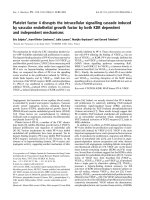
Compared to the healthy controls, patients with GC
showed higher levels of the secreted cytokines IL-6, IL-8
and TNF-α (all P < 0.05) (Figure 1A); the levels of IL-1β,
IL-10 and IL-12p70 were not significantly different between the two groups. Compared to the healthy controls
(median: 72% [range: 21-115]), the patients with GC
showed significantly enhanced plasma vWF:Ag levels
(P < 0.05 for all patients with GC) (Figure 1B). Moreover,
the GC-related increase in plasma vWF:Ag levels was associated with disease severity, with patients with late disease stage showing higher levels than patients with early
disease stage (101% [40-136] and 82% [8-118] vs. healthy
controls, P < 0.001).
A similar trend was seen in the plasma vWF activity
levels, where the levels were significantly enhanced in
patients with GC (vs. healthy controls: 62% [20-112],
P < 0.01) and followed the disease severity (late disease
stage: 117% [33-169] and early disease stage: 75% [22-145]
vs. healthy controls, P < 0.001) (Figure 1C).
Gastrointestinal stromal tumors show increased
expression levels of vWF
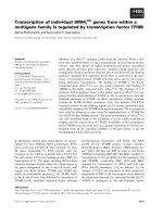
In the patients with GC, the level of vWF expression
was significantly higher in the tumor tissues than in the
adjacent normal tissues, at both the mRNA (Figure 2A)
and protein (Figure 2B) levels. In addition, IHC detected
remarkably higher levels of vWF protein expression concentrated in the tumor stroma region (Figure 2C). Interestingly, the expression of FVIII protein was expressed
in microvascular of tumor stroma region, and consistent
with the expression of vWF protein (Figure 2D).
Patients with GC have elevated serum levels of VEGF and
VEGF treatment induces vWF mRNA and protein
expression in the HUVEC endothelial cell line
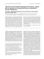
Enhanced serum VEGF was detected in the patients with
GC upon hospital admission; in comparison, the healthy
controls had undetectable levels of VEGF in serum (data
not shown). When the GC-related enhanced levels of
serum VEGF were evaluated in accordance of disease
state, it was found that patients with late disease had
Yang et al. BMC Cancer (2015) 15:80
Page 5 of 11
A
4
0.0
0.6
0.4
0.2
IL-12p70 ( pg / mL)
IL-10 ( pg / mL)
0.5
2
NS
0.3
0.2
0.1
0.0
on
tro
l
I / gro
III II s up
/ I ta
V ge
st
ag
e
0.0
C
C
on
tro
l
g
I / rou
p
I
III I st
/ I ag
V
e
st
ag
e
l
tro
on
4
0
C
C
C
B
100
50
**
*
*
150
100
50
/I
V
st
ag
st
ag
e
III
ou
p
gr
l
on
tro
C
/I
V
st
ag
e
st
ag
e
III
II
I/
ro
up
lg
Co
nt
ro
e
0
0
II
VWF:Ag (100%)
*
200
*
VWF activity (100%)
**
150
I/
on
tro
l
I / gro
III II s up
/ I tag
V
st e
ag
e
0
1.0
0.8
C
0
NS
on
tro
l
I / gro
III II s up
/ I ta
V ge
st
ag
e
5
0.4
1.0
on
tro
l
I / gro
III II s up
/ I ta
V ge
st
ag
e
1
10
6
NS
1.5
*
*
C
2
15
8
IL-8 ( pg / mL)
*
*
*
I / gro
III II s up
/ I ta
V ge
st
ag
e
*
IL-6 ( pg / mL)
3
20
Figure 1 Patients with GC have elevated levels of IL-6, IL-8, TNF-α and vWF in plasma. (A) Inflammatory cytokines measured included IL-1β,
IL-6, IL-8, IL-10, TNF-α and IL-12p70. (B) VWF:Ag and (C) VWF activity levels in control group and patients with early disease (I/II stages) and late
disease (III/IV stage). Data are expressed as percentages of the respective vWF parameter measured in the healthy control group. Horizontal lines
represent medians. *p < 0.05 and **p < 0.01 by Student's t-test. NS, non-significant.
significantly higher levels than those with early disease
(23 ± 26 pg/mL vs. 10 ± 12 pg/mL, P < 0.01) (Figure 3).
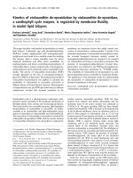
To investigate the potential impact of up-regulated
VEGF on vWF expression, HUVECs were treated with different doses and times of VEGF and the changes in vWF
gene and protein expression were examined. The highest
level of vWF protein expression occurred upon exposure
to the highest concentration of VEGF (100 ng/mL), showing a dose-dependent response trend for VEGF effects on
vWF protein (Figure 4A). Similarly, the highest level of
vWF protein occurred after the 40 minute exposure time,
suggesting a time-dependent response trend for VEGF
effects on vWF protein (Figure 4B). Similar dose- and
time-dependent responses to VEGF were observed for
vWF at the mRNA level (Figure 4C, 4D).
Intratumoral distribution of vWF, VEGF and VEGFR2
expression and the relationship with GC
clinicopathological features
The IHC staining patterns of vWF, VEGF, and VEGFR2
and corresponding GC clinicopathological features are
presented in Table 2. vWF immunostaining was highest
Yang et al. BMC Cancer (2015) 15:80
Page 6 of 11
A
B
P < 0.001
vWF-to-18s mRNA ratio
(relative expression)
6000
P < 0.001
T
Case 1
Case 2 Case 3 Case 4
T
N
T
N
N
T
N
T
N
T
N
vWF
N
4000
Relative vWF levels
-actin
2000
1.2
0.8
0.4
0
/I
V
T
Case 1 Case 2
III
I/
II
st
ag
e
st
ag
e
0
1.6
T
N
Case 3
T
N
Case 4
C
T
N
T
N
D
Figure 2 GC tumor specimens have elevated levels of vWF expression. (A) qRT-PCR detected levels of vWF mRNA. Data are presented as
relative Ct values from the GC tumor samples and patient-matched adjacent normal tissue samples (n = 32). (B) Western blot detected levels of
vWF protein in GC tumor samples and patient-matched adjacent normal tissue samples (upper panel). The relative expression level of vWF protein
is shown, normalized to the β-actin loading control (lower panel). IHC detected levels of (C) vWF and (D) FVIII protein (brown: positive cells) in a
representative GC tumor sample and the patient-matched adjacent normal tissue sample. Magnification: ×200. Bar =100 μm. T, tumor sample; N,
normal sample.
around the tumor nests, where microvessel density
(MVD) was highest as well. Compared to patientmatched adjacent non-tumor tissues (Figure 2C), the
tumor tissues from patients with early stage disease
showed slightly increased MVD (Figure 5A, B) while
those from patients with late stage disease showed
markedly increased MVD (Figure 5C). The number of
cells showing vWF-positive staining was significantly
higher in the patients with late disease stage disease
than in those with early stage disease (P < 0.05). No
relationship was found between the level of vWFpositive staining and patient sex, age, presence of
lymph node metastasis or extent of tumor differentiation
(all P > 0.05).
Page 7 of 11
150
**
**
**
100
50
e
ag
V
st
III
/I
II
I/
lg
tro
on
C
st
ag
e
up
0
ro
Serum level of VEGF (pg / mL)
Yang et al. BMC Cancer (2015) 15:80
Figure 3 Patients with GC had elevated levels of VEGF in
plasma. Serum levels of VEGF were measured in patients with GC
with early disease (I/II stages) and late disease (III/IV stages) by ELISA.
**p < 0.01 by Student's t-test.
VEGF and VEGFR2 cytoplasmic immunostaining was
detected in all cancer cells in tumor tissues (Figure 5D-I).
The late stage disease and undifferentiated tumor tissues
from patients with late disease stage exhibited higher
levels of VEGF and VEGFR2 (Figure 5F and I) than those
from patients with early disease stage (Figure 5E and H).
In addition, the quantity of cells showing VEGF-positive
and VEGFR2-positive staining was significantly higher in
those patients (P < 0.05) (Table 2). A higher number of
vWF-positive cells was associated with a higher number of
VEGF-positive and VEGFR2-positive cells in the patients
with late disease stage.
Discussion
Extensive research efforts have been put forth to help
elucidate the dynamic and critical roles of vWF in
hemostatic and thrombotic processes; however, much
less research into its roles in GC pathogenesis has been
conducted and much fewer data have been reported.
The study described herein represents the first clinical
report of the GC-related vWF expression pattern and its
clinicopathological significance for humans. The data
from this study not only provide novel insights into the
likely role of vWF in GC pathogenesis, but also highlight
the potential clinical significance of serum vWF and
tumor-related mRNA and protein expression as markers
of disease stage and prognosis.
Specifically, patients with GC were shown to have enhanced levels of vWF:Ag and vWF activity in plasma
and a strong correlation was observed between these
two variables and disease severity. These findings are
similar to previous data from patients with colorectal
cancer, who showed elevated plasma vWF that correlated with metastatic potential [21]. Interestingly, a previous study of lung cancer showed that ADAM28 can
downregulate vWF and cleave proapoptotic VWF in carcinoma cells, thereby increasing lung metastasis [16].
Data from mouse models (vWF-null) and cultured endothelial cells have supported the potential of a protective
role for vWF against metastasis [15,17].
Considering that vWF may act as a key factor in resistance to metastasis and also as an inhibitor of angiogenesis,
vWF may be a useful progrognostic marker; however, data
from other studies have indicated that it may not be a general marker for all cancer types. Studies of non-small cell
lung cancer patients and breast cancer patients found no
substantial alterations in vWF:Ag levels compared to
reference controls [22,23], and a clinical trial of human patients with colorectal cancer found significantly elevated
levels of plasma vWF but was unable to clearly define the
related role in cancer progression [24]. It is possible that
perturbed plasma vWF:Ag levels may be more indicative
of organ-specific processes, general risk factors, or pathogenic states associated with comorbidities. Indeed, elevated plasma vWF:Ag levels have been reported in cases
of acute liver injury/failure, alcoholic hepatitis, liver cirrhosis, and sickle cell disease [13,25-27]. Conway et al.
also showed that elevated vWF:Ag levels were independently associated with advanced age, prior cerebral ischemia, recent heart failure, diabetes, and non-valvular atrial
fibrillation [28,29]. Ongoing investigations in our laboratory have indicated that patients with liver cirrhosis show
even higher levels of elevated vWF:Ag and vWF activity
than the patients with GC reported herein (data not
shown). Thus, pathogenesis-related elevations in plasma
vWF may be related to endothelial dysfunction. Since the
collective data have yet to provide a precise profile of elevated serum vWF, it cannot be recommended as a clinical
marker of GC.
Similar to the elevated vWF protein expression profile
observed in human GC tissues, the current study also
observed elevated vWF mRNA expression. Furthermore,
the elevated expression was most robust in the tumors’
Yang et al. BMC Cancer (2015) 15:80
Page 8 of 11
A
Dose Response
vWF
-actin
VEGF
(ng/ml)
0
10
50 100
Time Course
B
5
minute
20
40
80
120
vWF
-actin
vWF-to-18s mRNA ratio
(fold changes relative expression)
C
0
10 100 10 100 10 100 10 100 10 100
Dose
Response
24
**
16
8
** **
0
VEGF
(ng/ml)
D
120
vWF-to-18s mRNA ratio
(fold changes relative expression)
VEGF
(ng/ml)
Time
Course
**
80
**
40
*
0
0 10 50 100
minute
0
5
**
20
40 80 120
Figure 4 VEGF treatment induces vWF expression in and secretion from HUVEC cell lines in a dose- and time-dependent manner.
HUVECs were exposed to 0, 10, 50 or 100 ng/mL of VEGF for 1 h and examined by western blot (A) and qRT-PCR (C). The concentration of
100 ng/mL induced the highest level of vWF protein and mRNA expression. HUVECs were exposed to the various doses of VEGF for the indicated
times and examined by western blot (B) and qRT-PCR (D). The exposure time of 40 minutes stimulated the highest level of vWF protein expression but
80 minutes stimulated the highest level of vWF mRNA expression. *p < 0.05 and **p < 0.01 by Student's t-test.
stromal regions and in late disease stage. In a previous
study of colon carcinoma specimens, almost all (5/6)
were found to possess higher vWF mRNA levels than
their patient-matched normal tissues [30]. vWF IHC
staining represents an effective maker of MVD, and as
such has been suggested that as a useful prognostic
marker for colorectal, ovarian and prostate cancers’ progression and/or patient survival [18,31,32]. In particular,
the vWF IHC staining in ovarian solid carcinoma was
shown to be associated with poor survival [33]. In another study based on the HUVEC cell line, it was shown
that the VEGF-VEGFR2 pathway was able to induce the
Yang et al. BMC Cancer (2015) 15:80
Page 9 of 11
Table 2 Immunohistochemical staining of the vWF, VEGF, or VEGFR2 and clinicopathological characteristics in patients
with gastric cancer
Variables
vWF-positive
VEGF-positive
≤125.75
>125.75
Female
15
18
Male
31
35
≥57
23
30
<57
21
25
No
10
15
Yes
32
42
I + II
20
10
III + IV
20
49
Differentiated
13
17
Undifferentiated
30
39
p-value
≤68.12
17
16
0.619
32
34
25
28
0.256
24
22
14
11
0.059
35
39
16
14
20
49
18
12
25
44
>68.12
VEGFR2-positive
p-value
≤114.56
14
19
0.851
30
36
29
24
0.736
22
24
12
13
0.692
40
34
15
15
0.016*
19
50
16
14
0.043*
23
46
>114.56
p-value
Sex
0.307
Age, years
0.674
Lymph node metastasis
0.716
TNM stage
0.019*
0.013*
Histological type
0.173
0.023*
*p < 0.05; statistical significant values indicated by bold font.
Figure 5 IHC staining patterns of vWF, VEGF, and VEGFR2 in GC tissues. The following representative tumors are presented: (A, D, G)
well-differentiated with TNM stage II; (B, E, H) undifferentiated tumor with TNM stage II; (C, F, I) undifferentiated tumor with TNM stage IIIb, IIIa
and IV respectively. vWF staining is shown in (A-C). VEGF staining is shown in (D-F). VEGFR2 staining is shown in (G-I). Positive cells are stained
brown. Magnification: ×200. Bar = 100 μm.
Yang et al. BMC Cancer (2015) 15:80
release of full-length vWF, and that this process involved
cAMP/protein kinase A (PKA) signaling [34]. While the
results from the present study are in agreement with
these previous findings the precise functional mechanism of vWF in tumorigenesis and tumor progression remain far from being completely understood.
Our in vitro-based data revealed a possible functional
network involving VEGF signaling and vWF expression
in human GC, which our ex vivo experiments indicated
was also related to severity of disease state. Moreover,
the IHC-observed co-localization of VEGF and VEGFR2
molecules with GC-elevated vWF proteins further supported the theory that these factors may represent a
mechanism of GC pathogenesis (and possible target of
future molecular therapies). A recent study indicated
that vWF may play a protective role by promoting resistance to tumor cell metastasis and dissemination in vivo
[35], lending further support to the promising potential
of vWF manipulation while highlighting the fact that
our understanding of the molecular basis for achieving
such a therapeutic effect remains largely incomplete.
The data in the current study serves to justify the continuance of such vWF-focused studies, especially in GC.
Conclusions
In conclusion, plasma vWF:Ag and vWF activity levels
are substantially elevated in patients with GC, especially
in those who have reached the late stage of the disease
condition. The particularly robust enhancement of vWF
protein and mRNA expression in stromal regions of GC
tumors, along with the physical proximity and functional
relationship to the VEGF-VEGFR2 molecules and signaling pathway, suggest a potential pathogenic mechanism
of GC and targets of future molecular therapies.
Competing interests
The authors declare that they have no competing interests.
Authors’ contributions
XY and YZW designed the study, and analyzed and interpreted the data. XY,
HJS, ZRL, HZ and WJY carried out the experiments. BN contributed to data
analysis and interpretation. All authors made substantial contributions
towards drafting the manuscript, reviewed the final manuscript for
intellectual content, and authorized the submission. All authors read and
approved the final manuscript.
Acknowledgements
This research was supported by grants from the National High Technology
Research and Development Program of China (863 program) (No. 2012AA02A407),
the National Basic Research Program of China (973 program) (No. 2013CB531500)
and the Program for Changjiang Scholars and Innovative Research Team at the
University of China (PCSIRT 10521). We would like to thank Dr. Jennifer C van
Velkinburgh (van Velkinburgh Initiative for Collaboratory BioMedical Research,
Santa Fe, NM, USA) for helpful discussions and for polishing the manuscript.
Author details
1
Institute of Immunology, Third Military Medical University, 30 Gaotanyan
Street, Shapingba District, Chongqing 400038, PR China. 2Department of
General Surgery, First People’s Hospital of Guiyang, Guiyang 550002, PR
China.
Page 10 of 11
Received: 10 September 2014 Accepted: 12 February 2015
References
1. Dikken JL, van de Velde CJ, Coit DG, Shah MA, Verheij M, Cats A. Treatment
of resectable gastric cancer. Therap Adv Gastroenterol. 2012;5(1):49–69.
2. Yang X, Qian F, He HY, Liu KJ, Lan YZ, Ni B, et al. Effect of thymosin alpha-1
on subpopulations of Th1, Th2, Th17, and regulatory T cells (Tregs) in vitro.
Braz J Med Biol Res. 2012;45(1):25–32.
3. Huang TC, Yeh KH, Cheng AL, Hsu CH. Weekly 24-hour infusional 5-fluorouracil
as initial treatment for advanced gastric cancer with acute disseminated
intravascular coagulation. Anticancer Res. 2008;28(2B):1293–7.
4. Lesesne JB, Rothschild N, Erickson B, Korec S, Sisk R, Keller J, et al.
Cancer-associated hemolytic-uremic syndrome: analysis of 85 cases from a
national registry. J Clin Oncol. 1989;7(6):781–9.
5. Kakeji Y, Hashizume M, Kitano S, Hasuo K, Sugimachi K. Successful treatment
of Budd-Chiari syndrome with early gastric cancer. Int Surg. 1991;76(1):55–7.
6. Wojtukiewicz MZ, Sierko E, Zacharski LR, Zimnoch L, Kudryk B, Kisiel W.
Tissue factor-dependent coagulation activation and impaired fibrinolysis in
situ in gastric cancer. Semin Thromb Hemost. 2003;29(3):291–300.
7. van den Berg YW, Osanto S, Reitsma PH, Versteeg HH. The relationship
between tissue factor and cancer progression: insights from bench and
bedside. Blood. 2012;119(4):924–32.
8. Morange PE, Simon C, Alessi MC, Luc G, Arveiler D, Ferrieres J, et al.
Endothelial cell markers and the risk of coronary heart disease: the
Prospective Epidemiological Study of Myocardial Infarction (PRIME) study.
Circulation. 2004;109(11):1343–8.
9. Bongers TN, de Maat MP, van Goor ML, Bhagwanbali V, van Vliet HH,
Gomez Garcia EB, et al. High von Willebrand factor levels increase the risk of
first ischemic stroke: influence of ADAMTS13, inflammation, and genetic
variability. Stroke. 2006;37(11):2672–7.
10. Bugnicourt JM, Roussel B, Tramier B, Lamy C, Godefroy O. Cerebral venous
thrombosis and plasma concentrations of factor VIII and von Willebrand factor:
a case control study. J Neurol Neurosurg Psychiatry. 2007;78(7):699–701.
11. Roldan V, Marin F, Muina B, Torregrosa JM, Hernandez-Romero D, Valdes M,
et al. Plasma von Willebrand factor levels are an independent risk factor for
adverse events including mortality and major bleeding in anticoagulated
atrial fibrillation patients. J Am Coll Cardiol. 2011;57(25):2496–504.
12. Khaleghi M, Singletary LA, Kondragunta V, Bailey KR, Turner ST, Mosley Jr TH,
et al. Haemostatic markers are associated with measures of vascular disease
in adults with hypertension. J Hum Hypertens. 2009;23(8):530–7.
13. Chen J, Hobbs WE, Le J, Lenting PJ, de Groot PG, Lopez JA. The rate of
hemolysis in sickle cell disease correlates with the quantity of active von
Willebrand factor in the plasma. Blood. 2011;117(13):3680–3.
14. Luo GP, Ni B, Yang X, Wu YZ. von Willebrand factor: more than a regulator
of hemostasis and thrombosis. Acta Haematol. 2012;128(3):158–69.
15. Terraube V, Pendu R, Baruch D, Gebbink MF, Meyer D, Lenting PJ, et al.
Increased metastatic potential of tumor cells in von Willebrand factor-deficient
mice. J Thromb Haemost. 2006;4(3):519–26.
16. Mochizuki S, Soejima K, Shimoda M, Abe H, Sasaki A, Okano HJ, et al. Effect
of ADAM28 on carcinoma cell metastasis by cleavage of von Willebrand
factor. J Natl Cancer Inst. 2012;104(12):906–22.
17. Starke RD, Ferraro F, Paschalaki KE, Dryden NH, McKinnon TA, Sutton RE,
et al. Endothelial von Willebrand factor regulates angiogenesis. Blood.
2011;117(3):1071–80.
18. Lackner C, Jukic Z, Tsybrovskyy O, Jatzko G, Wette V, Hoefler G, et al.
Prognostic relevance of tumour-associated macrophages and von
Willebrand factor-positive microvessels in colorectal cancer. Virchows Arch.
2004;445(2):160–7.
19. Mahzouni P, Mohammadizadeh F, Mougouei K, Moghaddam NA, Chehrei A,
Mesbah A. Determining the relationship between "microvessel density" and
different grades of astrocytoma based on immunohistochemistry for "factor
VIII-related antigen" (von Willebrand factor) expression in tumor microvessels.
Indian J Pathol Microbiol. 2010;53(4):605–10.
20. Wei ZF, Tong B, Xia YF, Lu Q, Chou GX, Wang ZT, et al. Norisoboldine
suppresses osteoclast differentiation through preventing the accumulation
of TRAF6-TAK1 complexes and activation of MAPKs/NF-kappaB/c-Fos/
NFATc1 Pathways. PLoS One. 2013;8(3):e59171.
21. Wang WS, Lin JK, Lin TC, Chiou TJ, Liu JH, Yen CC, et al. Plasma von
Willebrand factor level as a prognostic indicator of patients with metastatic
colorectal carcinoma. World J Gastroenterol. 2005;11(14):2166–70.
Yang et al. BMC Cancer (2015) 15:80
Page 11 of 11
22. Martini F, Ferroni P, Guadagni F, Basili S, Spila A, D'Alessandro R, et al.
Plasma von Willebrand factor antigen levels in non-small cell lung cancer
patients. Anticancer Res. 2005;25(1B):403–7.
23. Toth B, Nieuwland R, Liebhardt S, Ditsch N, Steinig K, Stieber P, et al.
Circulating microparticles in breast cancer patients: a comparative analysis
with established biomarkers. Anticancer Res. 2008;28(2A):1107–12.
24. Schellerer VS, Mueller-Bergh L, Merkel S, Zimmermann R, Weiss D, Schlabrakowski
A, et al. The clinical value of von Willebrand factor in colorectal carcinomas. Am J
Transl Res. 2011;3(5):445–53.
25. Matsuyama T, Uemura M, Ishikawa M, Matsumoto M, Ishizashi H, Kato S,
et al. Increased von Willebrand factor over decreased ADAMTS13
activity may contribute to the development of liver disturbance and
multiorgan failure in patients with alcoholic hepatitis. Alcohol Clin Exp
Res. 2007;31(1):S 27–35.
26. Lisman T, Bongers TN, Adelmeijer J, Janssen HL, de Maat MP, de Groot PG,
et al. Elevated levels of von Willebrand Factor in cirrhosis support platelet
adhesion despite reduced functional capacity. Hepatology. 2006;44(1):53–61.
27. Hugenholtz GC, Adelmeijer J, Meijers JC, Porte RJ, Stravitz RT, Lisman T. An
unbalance between von Willebrand factor and ADAMTS13 in acute liver
failure: implications for hemostasis and clinical outcome. Hepatology.
2013;58(2):752–61.
28. Conway DS, Pearce LA, Chin BS, Hart RG, Lip GY. Plasma von Willebrand
factor and soluble p-selectin as indices of endothelial damage and platelet
activation in 1321 patients with nonvalvular atrial fibrillation: relationship to
stroke risk factors. Circulation. 2002;106(15):1962–7.
29. Conway DS, Pearce LA, Chin BS, Hart RG, Lip GY. Prognostic value of plasma
von Willebrand factor and soluble P-selectin as indices of endothelial
damage and platelet activation in 994 patients with nonvalvular atrial
fibrillation. Circulation. 2003;107(25):3141–5.
30. Zanetta L, Marcus SG, Vasile J, Dobryansky M, Cohen H, Eng K, et al.
Expression of Von Willebrand factor, an endothelial cell marker, is
up-regulated by angiogenesis factors: a potential method for objective
assessment of tumor angiogenesis. Int J Cancer. 2000;85(2):281–8.
31. Lo CW, Chen MW, Hsiao M, Wang S, Chen CA, Hsiao SM, et al. IL-6
trans-signaling in formation and progression of malignant ascites in ovarian
cancer. Cancer Res. 2011;71(2):424–34.
32. Yoshioka N, Wang L, Kishimoto K, Tsuji T, Hu GF. A therapeutic target for
prostate cancer based on angiogenin-stimulated angiogenesis and cancer
cell proliferation. Proc Natl Acad Sci. 2006;103(39):14519–24.
33. Koh SC, Khalil R, Lim FK, Ilancheran A, Choolani M. The association between
fibrinogen, von Willebrand Factor, antithrombin III, and D-dimer levels and
survival outcome by 36 months from ovarian cancer. Clin Appl Thromb
Hemost. 2006;12(1):3–8.
34. Xiong Y, Huo Y, Chen C, Zeng H, Lu X, Wei C, et al. Vascular endothelial growth
factor (VEGF) receptor-2 tyrosine 1175 signaling controls VEGF-induced von
Willebrand factor release from endothelial cells via phospholipase
C-gamma 1- and protein kinase A-dependent pathways. J Biol Chem.
2009;284(35):23217–24.
35. Ripoll GV, Alonso DF. Implication of von Willebrand factor as a regulator of
tumor cell metastasis: potential perioperative use of desmopressin and
novel peptide analogs. Acta Haematol. 2013;129(4):223–4.
Submit your next manuscript to BioMed Central
and take full advantage of:
• Convenient online submission
• Thorough peer review
• No space constraints or color figure charges
• Immediate publication on acceptance
• Inclusion in PubMed, CAS, Scopus and Google Scholar
• Research which is freely available for redistribution
Submit your manuscript at
www.biomedcentral.com/submit