Is stereotactic radiosurgery a rational treatment option for brain metastases from small cell lung cancer? A retrospective analysis of 70 consecutive patients
Bạn đang xem bản rút gọn của tài liệu. Xem và tải ngay bản đầy đủ của tài liệu tại đây (442.39 KB, 8 trang )
Yomo and Hayashi BMC Cancer (2015) 15:95
DOI 10.1186/s12885-015-1103-6
RESEARCH ARTICLE
Open Access
Is stereotactic radiosurgery a rational treatment
option for brain metastases from small cell
lung cancer? A retrospective analysis of 70
consecutive patients
Shoji Yomo1,2* and Motohiro Hayashi2
Abstract
Background: Because of the high likelihood of multiple brain metastases (BM) from small cell lung cancer (SCLC),
the role of focal treatment using stereotactic radiosurgery (SRS) has yet to be determined. We aimed to evaluate
the efficacy and limitations of upfront and salvage SRS for patients with BM from SCLC.
Methods: This was a retrospective and observational study analyzing 70 consecutive patients with BM from SCLC
who received SRS. The median age was 68 years, and the median Karnofsky performance status (KPS) was 90.
Forty-six (66%) and 24 (34%) patients underwent SRS as the upfront and salvage treatment after prophylactic or
therapeutic whole brain radiotherapy (WBRT), respectively. Overall survival (OS), neurological death-free survival,
remote and local tumor recurrence rates were analyzed.
Results: None of our patients were lost to follow-up and the median follow-up was 7.8 months. One-and 2-year OS
rates were 43% and 15%, respectively. The median OS time was 7.8 months. One-and 2-year neurological death-free
survival rates were 94% and 84%, respectively. In total, 219/292 tumors (75%) in 60 patients (86 %) with sufficient
radiological follow-up data were evaluated. Six-and 12-month rates of remote BM relapse were 25% and 47%,
respectively. Six-and 12-month rates of local control failure were 4% and 23%, respectively. Repeat SRS, salvage
WBRT and microsurgery were subsequently required in 30, 8 and one patient, respectively. Symptomatic radiation
injury, treated conservatively, developed in 3 patients.
Conclusions: The present study suggested SRS to be a potentially effective and minimally invasive treatment
option for BM from SCLC either alone or after failed WBRT. Although repeat salvage treatment was needed in
nearly half of patients to achieve control of distant BM, such continuation of radiotherapeutic management might
contribute to reducing the rate of neurological death.
Keywords: Brain metastases, Small cell lung cancer, Stereotactic radiosurgery, Whole brain radiotherapy
Background
Lung cancer is the most common source of brain metastasis (BM). Given that the cumulative incidence of BM from
small cell lung cancer (SCLC) at 2 years is approximately
50% [1], prophylactic cranial irradiation (PCI) combined
with systemic chemotherapy, which moderately prolongs
overall survival (OS) by reducing the incidence of delayed
* Correspondence:
1
Division of Radiation Oncology, Aizawa Comprehensive Cancer Center,
Aizawa Hospital, 2-5-1, Honjo, Matsumoto, Nagano 390-0814, Japan
2
Saitama Gamma Knife Center, San-ai Hospital, Saitama, Japan
BM, has long been accepted as the standard of care for
most patients [2-5]. Recurrence or progression of intracranial disease after such an intensive treatment regimen is,
however, not uncommon despite the radiosensitive nature
of SCLC [6]. The prognosis of patients with recurrent BM
generally remains dismal.
Stereotactic radiosurgery (SRS) has emerged as the preferred treatment modality, either alone or in combination
with other modalities. Recently, in selected patients, whole
brain radiotherapy (WBRT) has been omitted from the
initial management for BM with the aim of reducing the
© 2015 Yomo and Hayashi; licensee BioMed Central. This is an Open Access article distributed under the terms of the Creative
Commons Attribution License ( which permits unrestricted use, distribution, and
reproduction in any medium, provided the original work is properly credited. The Creative Commons Public Domain
Dedication waiver ( applies to the data made available in this article,
unless otherwise stated.
Yomo and Hayashi BMC Cancer (2015) 15:95
Page 2 of 8
potential risk of delayed neurological toxicity [7,8]. Given
the propensity for dissemination of SCLC, SRS does not
appear to be a rational approach to this malignancy. To
date, there have been only a few, relatively small, studies
of SRS for SCLC with or without prior WBRT (Table 1)
[9-13]. Thus, the role of focal treatment by means of SRS
for BM from SCLC remains to be elucidated.
We retrospectively investigated the efficacy and limitations of our SRS-oriented treatment strategy for patients
with newly diagnosed and recurrent BM from SCLC.
Methods
Patient population
The present study was conducted in compliance with
the Declaration of Helsinki (6th revision, 2008), and fulfilled all of the requirements for patient anonymity. The
San-ai Hospital Institutional Review Board approved this
retrospective clinical study in January 2014. Between
January 2009 and October 2013, 70 consecutive patients
with BM originating from histologically proven primary
SCLC underwent Gamma Knife SRS in our institution.
Fifty-five patients were male and 15 were female. The
median age was 68 years (range: 44–85 years). The median Karnofsky performance status (KPS) at the time of
SRS was 90 (range: 30–100). Before SRS, 7 patients had
undergone microsurgical resection for BM and one had
received third ventriculostomy for obstructive hydrocephalus. Prior WBRT had been conducted at the referring regional hospitals, prophylactically in 7 patients and
in a therapeutic setting in 16. One patient had undergone hypofractionated radiotherapy for a large tumor located in the posterior cranial fossa. All patients with
prior WBRT had documented intracranial failure (either
new lesions or progression of preexisting metastases).
The median interval between primary diagnosis and SRS
was 11.4 months (range: 0.1–150 months). Patient characteristics are summarized in Table 2.
Radiosurgical indications and techniques
All patients included in the present study had been diagnosed and their primary tumors treated at the referring
regional hospitals, whose own cancer boards had provisionally determined the appropriateness of SRS. The patients were then referred to our institution to receive SRS
for BM. The SRS protocol used in this study was based on
the standard care established at our institution. In the upfront setting, patients with up to ten BM principally received SRS. When abnormal enhancement of cranial
nerves, the ventricular ependymal layer and/or the cortical
surface or more than 10 BM were documented by high
resolution magnetic resonance (MR) imaging at the time
of initial SRS, WBRT was recommended. In the salvage
setting, the treatment protocol in the author’s institution
has no set limit on the number of BM. Providing that
WBRT had either already been performed or refused by
the patient, SRS was applied for multiple BM, even in
cases with more than 10 lesions, when the patient’s systemic condition was such that SRS intervention would be
tolerable and fully informed consent for treatment had
been obtained. Surgical resection was, in principle, indicated for large tumors (≥10 mL) with a mass effect unresponsive to corticosteroid therapy. If surgery did not seem
feasible due to a poor prognosis or advanced systemic disease, 2-session SRS was indicated for carefully selected
large tumors (≥10 mL) [14].
SRS was performed using the Leksell G stereotactic
frame (Elekta Instruments, Stockholm, Sweden). The frame
was placed on the patient’s head under local anesthesia
supplemented with mild sedation. Three-dimensional volumetric gadolinium-enhanced T1-weighted MR images,
2 mm in thickness T2-weighted MR images and contrastenhanced computed tomography covering the whole brain
were routinely used for dose planning with Leksell Gamma
Plan software (Elekta Instruments). When performing
salvage SRS after prior WBRT, the targets were limited to
recurrent or newly emerging lesions. Stable lesions continued to be monitored unless regrowth was documented.
Prescribed doses were selected in principle according to
the dose protocol of the JLGK 0901 study [15], though a
margin of approximately 1 to 2 mm was added to the visible lesion in consideration of the infiltrative nature of
SCLC [16]. The technical details of 2-session SRS were
Table 1 Outcomes of patients undergoing SRS for BM from SCLC
First author & year
Treatment
modality
No. of
Patients
No. receiving prior
WBRT (%)
MST after SRS
(months)
Local tumor
control
Remote brain
recurrence
Wegner 2011 [9]
GK
44
30 (68)
9
90%/1 year
61%/7 months
Jo 2011 [12]
GK
50
38 (76)
*6.3
70.3%/5.6 months
29.7% (crude)
Harris 2012 [13]
GK
51
34 (67)
5.9
57%/1 year
58%/1 year
Olson 2012 [10]
CK
27
19 (70)
3
76.5/1 year
60%/3.5 months
Nakazaki 2013 [11]
GK
44
44 (100)
5.8
95.8%/4 months
50%/6 months
Present study 2014
GK
70
24 (34)
7.8
77%/1 year
47%/1 year
SRS stereotactic radiosurgery, BM brain metastasis, SCLC small cell lung cancer, WBRT whole brain radiotherapy, MST median survival time, GK gamma knife, CK
cyberknife, *mean value.
Yomo and Hayashi BMC Cancer (2015) 15:95
Page 3 of 8
Table 2 Summary of clinical data from 70 consecutive
patients
Characteristics
Overall (n=70)
Sex (male/female)
55/15
Age (years), median (range)
68 (44–85)
KPS, median (range)
90 (30–100)
Active extra-CNS disease
45 (64%)
Prior WBRT
24 (34%)
Post-SRS chemotherapy
50 (71%)
Time from primary diagnosis to initial SRS (months),
median (range)
11.4 (0.1–150)
Cumulative PTV on initial SRS (mL), median (range)
4.4 (0.5–50.3)
No. of intracranial lesions on initial SRS, median (range) 2 (1–21)
KPS Karnofsky performance status, CNS central nervous system, WBRT whole
brain radiotherapy, SRS stereotactic radiosurgery, PTV planning target volume.
previously described in detail [14]. All treatments were
performed with the Leksell Gamma Knife Model C or
Perfexion.
Post-SRS management and follow-up evaluation
Clinical follow-up data as well as contrast-enhanced MR
images were obtained every one to three months. If
metachronous remote metastases were identified, they
were, in principle, managed with repeat SRS. When miliary metastases (numerous tiny enhanced lesions) and/or
leptomeningeal carcinomatosis was newly documented,
WBRT was then considered unless it had been used previously. Local control failure was defined as an at least
20% increase in the diameter of the targeted lesions, taking as a reference the pre-SRS diameter, irrespective of
whether the lesion was a true recurrence or delayed radiation injury. Delayed radiation injury was differentiated
from tumor recurrence using serial MR imaging [17]
and, in selected cases, 11C-methionine positron emission
tomography. Additional SRS was possible provided that
the volume of the local tumor recurrence was small
enough for single-dose SRS. Surgical removal was indicated when neurological signs became refractory to
conservative management, regardless of whether the
radiological diagnosis was local tumor progression or radiation necrosis. Any adverse events attributable to SRS
procedures were evaluated based on the National Cancer
Institute Common Terminology Criteria for Adverse
Events (NCI-CTCAE; ver.3.0). Before closing the research database for analysis, the authors updated the
follow-up data of patients who had not visited our outpatient department for more than two months. Inquiries
about the date and mode of death were made by directly
corresponding with the referring physician and/or the
family of the deceased patient, with written permission
obtained at the time of undertaking SRS from all patients and/or their relatives, allowing the use of personal
data for clinical research. Neurological death was defined
as death attributable to central nervous system (CNS)
metastases including tumor recurrence and carcinomatous meningitis.
Statistical analysis
The overall survival (OS) rate was calculated by the
Kaplan-Meier product limit method. The neurological
and non-neurological death rates were calculated employing Gray’s test [18], wherein each event was
regarded as a competing risk for another event. For the
estimation of local control failure rates and distant BM
recurrence, Gray’s test was similarly used, with subsequent WBRT for remote recurrence and the patient’s
death being regarded as competing events, respectively.
All of the above analyses were based on the interval
from the date of initial SRS treatment until the date of
each event. The Cox and Fine-Gray proportional hazards
models [19] were employed to investigate prognostic factors for OS and neurological death-free survival, and for
local tumor control, respectively. Potential prognostic
factors were selected with reference to other SRS series
[9-13]. The survival results were tested employing two
prognostic scoring systems validated for SCLC (Diagnosis-specific graded prognosis assessment (DS-GPA) and
Rades’s survival score). The statistical processing software package “R” version 3.0.1 (The R Foundation for
Statistical Computing, Vienna, Austria) was used for all
statistical analyses. A P-value of < 0.05 was considered to
indicate a statistically significant difference.
Results
SRS was conducted as an initial treatment in 46 patients
(66%) and as salvage in 24 (34%). Forty-five patients
(64%) had active systemic disease and/or extra-CNS metastases and 50 patients (71%) were still receiving systemic chemotherapy at the time of the initial SRS. In
total, 292 tumors were being treated at the time of the
initial SRS. The median planning target volume (PTV)
was 0.60 mL (range: 0.04–22.3 mL). The median number
of BM at SRS was 2 (range: 1–21 tumors) and the median cumulative PTV was 4.4 mL (range: 0.5–50 mL).
Prescribed doses ranged from 12 Gy to 22 Gy (median:
20 Gy). Seven patients with large tumors were allocated
to 2-session SRS.
Full clinical results were available for all 70 patients as
none were lost to follow-up. The median follow-up time
after SRS was 7.8 months (range: 0.6–56 months). At
the time of assessment, 8 patients (11%) were alive and
62 (89%) had died. The causes of death were intracranial
local progression in 3 cases, meningeal carcinomatosis
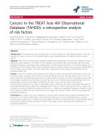
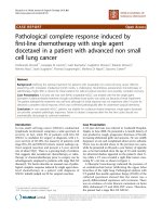
in 9 and progression of the primary lesion in 50. The 1and 2-year OS rates after SRS were 43% and 15%, respectively (Figure 1). The median OS time was 7.8
Yomo and Hayashi BMC Cancer (2015) 15:95
Page 4 of 8
Figure 1 Survival results for patients with BM from SCLC treated
with SRS. The solid line represents overall survival (OS) probability. The
median survival time (MST) was 7.8 months (95% CI: 6.2–12.6). One-and
2-year OS rates after SRS were 43% and 15%, respectively. The dotted
line represents the neurological death-free survival (NS) probability
adjusted for competing events. The 1-and 2-year NS rates after SRS
were 94 and 84%, respectively. Note that the distance between
these two lines, NS and OS, represents the cumulative incidence of
non-neurological death.
months (95% CI: 6.2–12.6). The proportional hazards
model for OS identified high KPS (HR: 0.493, 95% confidence interval (CI): 0.279–0.871, P=0.015) and solitary
metastasis (HR: 0.419, 95% confidence interval (CI):
0.205–0.857, P=0.017) as favorable prognostic factors
independently predicting OS rates (Table 3). One-and 2year neurological death-free survival probabilities adjusted for competing events (non-neurological death)
were 94% and 84%, respectively (Figure 1). The proportional hazards model suggested high KPS and no prior
WBRT to be associated with lower risk of neurological
death (Table 3), though neither reached statistical significance. The survival results were tested with validated prognostic scoring systems (Table 4). The DS-GPA showed
significant differences in median survival time (MST):
DS-GPA 3–4 points: 12.4 months (95% CI: 4.2-not reached),
1.5–2.5 points: 7.8 months (95% CI: 4.8–18.0), ≤1.0 points:
6.7 months (95% CI: 4.7–12.6) (P=0.036, log-rank test)
(Table 4). A survival scoring system specifically for patients with BM from SCLC, as proposed by Rades et al.
[20], also allowed stratification by 6-month patient survival rates: 15 points: 78% (95% CI: 51–91), 9–12 points:
66% (95% CI: 49–79), 5–8 points: 43% (95% CI: 18–66)
(P=0.006, log-rank test) (Table 4).
Only the 219/292 tumors (75%) in 60 patients (86%)
who had sufficient radiological follow-up data were analyzed herein because the other 10 patients died from
extra-CNS progression without follow-up MR imaging.
Remote metachronous BM were observed in 33 patients
(55%). The 6-month and 1-year remote BM recurrence
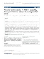
rates (per patient) after SRS were 25% and 47%, respectively (Figure 2A). The 6-month and 1-year local tumor
control failure rates (per lesion) were 4% and 23%, respectively (Figure 2B). Twenty-three metastases were
eventually diagnosed as local recurrence or delayed radiation injury at a median time of 8.2 months after SRS
(range: 4.6–17 months). The proportional hazards model
demonstrated low marginal dose (HR: 4.24 95% CI:
1.21–14.8, P=0.024) and prior WBRT (HR: 7.11 95% CI:
2.80–18.0, P < 0.001) to be factors predicting a higher
local tumor control failure rate (Table 5). Two-session
SRS conducted for large tumors achieved a durable tumor
volume reduction coupled with symptom relief in 6 of 7
cases. One male patient with a large brainstem metastasis
experienced local control failure, which eventually resulted
in neurological death 12 months after SRS.
Thirty patients (43%) required repeat SRS for remote
or local BM recurrence. The total number of SRS sessions ranged up to 5 (median: 1) and the total number
of BM treated per patient ranged up to 72 (median: 5).
Eight patients (17%) without prior WBRT underwent
salvage WBRT at a median time of 9.8 months after SRS
(range: 2.8–22.6 months) because of subsequent development of miliary BM and/or leptomeningeal dissemination. Microsurgical resection was necessary for local
tumor recurrence in one patient at 15 months after SRS.
Table 3 Analysis of factors predicting patient survival after SRS (Proportional hazards model)
Covariate
OS
NS
P value
Hazard ratio (95% CI)
P value
Hazard ratio (95% CI)
Young (≤65 years)
0.838
1.06 (0.612–1.83)
0.290
0.490 (0.131–1.83)
High KPS (≥90)
0.015
0.493 (0.279–0.871)
0.055
0.236 (0.054–1.03)
Controlled extra-CNS disease
0.365
0.709 (0.337–1.49)
0.150
2.44 (0.715–8.33)
Prior WBRT
0.629
0.868 (0.487–1.54)
0.071
3.81 (0.891–16.3)
Post-SRS chemotherapy
0.077
0.564 (0.299–1.06)
0.350
2.39 (0.383–14.9)
Single BM
0.017
0.419 (0.205–0.857)
0.340
1.69 (0.572–5.00)
SRS stereotactic radiosurgery, OS overall survival, NS neurological death-free survival, CI confidence interval, KPS Karnofsky performance status, WBRT whole brain
radiotherapy, BM brain metastases.
Yomo and Hayashi BMC Cancer (2015) 15:95
Page 5 of 8
Table 4 Survival of patients with BM from SCLC stratified
with prognostic classification systems
Survival results
(No. of patients)
Overall MST in months
P value
7.8 (70)
DS-GPA (MST in months)
0.036
0–1.0
6.7 (35)
1.5–2.5
7.8 (27)
3.0–4.0
12.4 (8)
Rades’s survival score (6-month
survival rate)
0.006
5–8
43% (14)
9–12
66% (38)
15
78% (18)
BM brain metastases, SCLC small cell lung cancer, MST median survival time,
DS-GPA diagnosis specific-graded prognosis assessment.
None of the adverse effects observed in this series
exceeded NCI-CTCAE grade 3 toxicity. Three patients
required oral steroids coupled with hyperbaric oxygen
therapy for delayed radiation injury (NCI-CTCAE Grade
3 toxicity) and eventually showed clinical and radiological stabilization.
Discussion
Advances in the development of systemic treatments, together with judicious use of surgical resection, WBRT
and SRS, have led to increases in the number of longterm survivors and the MST. The long-term control of
CNS disease has become increasingly important not only
for overall disease control but also for the patient’s quality of life. The risk of developing BM in SCLC is higher
than with other histologies. Seute et al. reported the cumulative risk of BM at 2 years after the diagnosis to be
49% to 65% in SCLC [1]. Thus, PCI has long been advocated to reduce the incidence of BM development [2-5].
The survival advantage in previous randomized trials
supporting PCI as the standard of care is widely recognized as level 1 evidence. This approach may, however,
at least theoretically increase the potential risk of leukoencephalopathy in patients without any known intracranial disease, but with a 50% probability that at some
point CNS disease will appear [21,22]. In addition, intracranial disease control failure will continue to occur despite the relatively radiosensitive nature of SCLC [3,4,23].
Certainly, WBRT only treats existing disease and there is
no evidence indicating that PCI prevents new BM from
developing in patients with active systemic disease.
SRS for BM from SCLC has been relegated to use
mainly after failed WBRT probably due to lack of evidence of the efficacy of SRS for this malignancy [9-13].
However, recent refinements in diagnostic and therapeutic modalities may impact the modern management
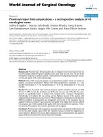
Figure 2 Cumulative incidences of distant intracranial recurrence
(A) and local tumor control failure (B). The 6-and 12-month distant
intracranial recurrence rates were 25% and 47%, respectively. The 6-and
12-month local tumor control failure rates were 4% and 23%, respectively.
Table 5 Analysis of factors predicting local tumor control
failure (Proportional hazards model)
Covariate
P value
Prior WBRT
< .001
Hazard ratio (95% CI)
7.11 (2.80–18.0)
Large target volume (>2 mL)
0.085
0.865 (0.193–3.88)
Tumor causing focal deficit
0.97
1.05 (0.104–5.14)
Low marginal dose (<20Gy)
0.024
4.24 (1.21–14.8)
CI confidence interval, WBRT whole brain radiotherapy.
Yomo and Hayashi BMC Cancer (2015) 15:95
of BM. High-resolution neuroimaging such as 3dimensional volumetric imaging and the 3-tesla unit
might become routinely available for visualizing lesions
that used to be undetectable with older imaging modalities [24,25]. Recent technological breakthroughs in the
SRS apparatus [26] have made it possible to safely treat
20, or even more, BM, provided that the lesions are
small, in a one-day session. The delivery of highly focused radiation with a sharp dose fall-off is theoretically expected to reduce delayed neurotoxicity, and this
feature makes it applicable both in the upfront and the
salvage setting after recurrence or progression after
prophylactic or therapeutic WBRT. A recent Japanese
multi-institutional prospective study including 1194 patients (76% with lung cancer) suggested that the upfront SRS strategy is reasonable for patients with up to
10 lesions [15]. However, a critical argument can be
made that the pathology of SCLC is unsuitable for SRS
because of the disseminated nature of this malignancy.
Thus, in our view, the efficacy and limitations of a focal
therapeutic approach for BM from SCLC have yet to be
determined.
The survival results after SRS in the present study are
comparable to those of previous studies [9-11,13]
(Table 1). What makes the present study different from
the previous series is the ratio of upfront to salvage
intervention. Upfront treatment accounts for almost
two-thirds of our cohort, while salvage treatments were
most numerous in previous series. We had anticipated
before this investigation that the survival results would
be worse in patients undergoing salvage treatment than
in those receiving upfront treatment, but there was, in
fact, no significant difference between these two groups
(Table 3). We speculate that this might, at least in part,
be attributable to patients receiving SRS as salvage having been self-selected to do well by virtue of having had
time to develop recurrent BM and not dying of their
systemic disease. Patient survival could be stratified
employing validated prognostic grading systems. The
DS-GPA index is one of the most relevant diagnostic
tools for predicting the survival of patients with newly
diagnosed BM [27]. In the original DS-GPA study, where
the majority of patients (82.6%) received WBRT as the
sole treatment, the survival of those with newly diagnosed BM from SCLC was 4.9 months, which was worse
than those for patients with tumors at other primary
sites. If confined to DS-GPA scores not exceeding 1.0,
the MST was as short as 2.8 months. Considering that
half the patients had DS-GPA scores of 1 or less in our
cohort, the survival outcomes after SRS appear to be acceptable. Rades’s survival scoring system [20] also predicted the survival rates in our cohort, with the survival
rates in the present study being higher in the lower score
classes than in the original dataset. With regard to
Page 6 of 8
prognostic factors, high KPS and solitary BM were associated with improved patient survival in multivariate
analyses (Table 3). Both variables were actually incorporated into the above survival scoring systems and these
findings were also reproduced in prior studies focusing
mainly on salvage treatment [9,13]. Identifying prognostic factors for longer survival in patients with BM would
be critically important for assigning patients to the optimal treatment modality. This observation suggests that
selected subsets of patients can be expected to experience prolonged survival, though the expected survival of
patients with BM from SCLC may be limited in the majority of cases.
In the curve of local tumor control failure, an irregular
elevation was observed around 8 months after SRS
(Figure 2B). We speculate that the following factors may
account for this observation. In a male patient who had
received WBRT for multiple BM, multiple recurrent tumors initially responded well to SRS but the enhancement subsequently enlarged in most of these lesions.
They were eventually diminished again by salvage reSRS. Considering that salvage was successful, these lesions should be regarded as true local recurrence. The
reason for the higher rate of local tumor control failure
in patients with prior WBRT demonstrated herein remains unknown. However, it might be attributable to selective regrowth of radio-resistant tumor cells or to the
surrounding brain tissue being predisposed to radiation
injury. Thus, we recommend a high marginal dose (≥20
Gy), when possible, being given even for recurrent BM
after WBRT, by referring to the results of multivariate
analysis for local tumor control (Table 5).
Nearly half of our patients eventually experienced
metachronous recurrence outside the treated area after
the initial SRS. Subsequent SRS was needed in as many
as 30 patients (43%), mostly because of remote BM recurrence. These patients were successfully managed
with minimal toxicity. Only eight patients without prior
WBRT eventually underwent salvage WBRT because of
miliary metastases or leptomeningeal dissemination.
Considering that remote recurrence frequently developed, meticulous clinical and neuroimaging follow-up
and salvage SRS in a timely manner should be considered essential for assuring the relevance of SRS
management. Such a continued radiotherapeutic management protocol might contribute to reducing the
neurological death rate, though OS results after SRS
were comparable to those of previous studies (Figure 1).
This finding is not consistent with the previous study
by Harris et al. showing the rate of neurological death
to be as high as 53% [13]. In our country, nation-wide
availability of advanced diagnostic imaging facilities and
radiosurgical equipment as well as the public healthcare
system may, fortunately, be making it possible to provide
Yomo and Hayashi BMC Cancer (2015) 15:95
cancer patients with easy access to necessary advanced
medical services [28].
The present results must be interpreted with caution.
Although the treatment results in our cohort suggested
survival similar to that obtained with WBRT in properly
stratified populations, a patient selection bias inherent to
the retrospective approach is unavoidable. One of the critical issues in the present study is that the reason for PCI
having been omitted could not be specified for all cases. It
must be appreciated that we cannot address the potential
role of SRS in comparison to WBRT because this was a
small retrospective observational study. The survival advantage in previous randomized trials supporting PCI as
the standard of care also cannot be ignored. The evidence
for the clinical efficacy of SRS for BM from SCLC remains
insufficient and more evidence-based information and
additional research are needed to confirm the therapeutic
benefits of SRS. We consider the present retrospective
study to have been necessary as a means of hypothesis
generation for future investigations.
Conclusions
To our knowledge, this is the largest retrospective study
investigating the efficacy of SRS for BM in patients with
SCLC. Our results suggest SRS to be a potentially effective and minimally invasive treatment option for BM
from SCLC either alone or after failed WBRT. Continued radiotherapeutic management might contribute to
reducing the neurological death rate, though OS results
after SRS were comparable to those of previous studies.
SRS provided durable local tumor control, but repeat
salvage treatment was needed in nearly half of patients
to achieve control of distant BM.
Abbreviations
BM: Brain metastases; SCLC: Small cell lung cancer; SRS: Stereotactic
radiosurgery; KPS: Karnofsky performance status; WBRT: Whole brain
radiotherapy; PCI: Prophylactic cranial irradiation; OS: Overall survival;
MR: Magnetic resonance; NCI-CTCAE: National cancer institute common
terminology criteria for adverse events; CNS: Central nervous system;
DS-GPA: Diagnosis-specific graded prognostic assessment; PTV: Planning
target volume; HR: Hazard ratio; CI: Confidence interval; MST: Median
survival time.
Competing interests
The authors declare that they have no competing interests.
Authors’ contributions
SY performed the radiosurgical management of these patients and prepared
the manuscript. MH critically reviewed the manuscript for important intellectual
content. Both authors have read and approved the final manuscript.
Acknowledgements
The authors certify that no funding was received to conduct this study
and/or for preparation of this manuscript. We are grateful to Bierta Barfod,
M.D., M.P.H. for her help with the preparation of this manuscript.
Received: 23 September 2014 Accepted: 20 February 2015
Page 7 of 8
References
1. Seute T, Leffers P, ten Velde GP, Twijnstra A. Neurologic disorders in 432
consecutive patients with small cell lung carcinoma. Cancer. 2004;100(4):801–6.
2. Schild SE, Foster NR, Meyers JP, Ross HJ, Stella PJ, Garces YI, et al.
Prophylactic cranial irradiation in small-cell lung cancer: findings from a
North Central Cancer Treatment Group Pooled Analysis. Ann Oncol.
2012;23(11):2919–24.
3. Auperin A, Arriagada R, Pignon JP, Le Pechoux C, Gregor A, Stephens RJ,
et al. Prophylactic cranial irradiation for patients with small-cell lung cancer
in complete remission. Prophylactic Cranial Irradiation Overview
Collaborative Group. N Engl J Med. 1999;341(7):476–84.
4. Slotman B, Faivre-Finn C, Kramer G, Rankin E, Snee M, Hatton M, et al.
Prophylactic cranial irradiation in extensive small-cell lung cancer. N Engl J
Med. 2007;357(7):664–72.
5. Meert AP, Paesmans M, Berghmans T, Martin B, Mascaux C, Vallot F, et al.
Prophylactic cranial irradiation in small cell lung cancer: a systematic review
of the literature with meta-analysis. BMC Cancer. 2001;1:5.
6. Ramlov A, Tietze A, Khalil AA, Knap MM. Prophylactic cranial irradiation in
patients with small cell lung cancer. A retrospective study of recurrence,
survival and morbidity. Lung Cancer. 2012;77(3):561–6.
7. Serizawa T, Ono J, Iichi T, Matsuda S, Sato M, Odaki M, et al. Gamma knife
radiosurgery for metastatic brain tumors from lung cancer: a comparison
between small cell and non-small cell carcinoma. J Neurosurg.
2002;97(5 Suppl):484–8.
8. Sneed PK, Suh JH, Goetsch SJ, Sanghavi SN, Chappell R, Buatti JM, et al. A
multi-institutional review of radiosurgery alone vs. radiosurgery with whole
brain radiotherapy as the initial management of brain metastases. Int J
Radiat Oncol Biol Phys. 2002;53(3):519–26.
9. Wegner RE, Olson AC, Kondziolka D, Niranjan A, Lundsford LD, Flickinger JC.
Stereotactic radiosurgery for patients with brain metastases from small cell
lung cancer. Int J Radiat Oncol Biol Phys. 2011;81(3):e21–7.
10. Olson AC, Wegner RE, Rwigema JC, Heron DE, Burton SA, Mintz AH. Clinical
outcomes of reirradiation of brain metastases from small cell lung cancer
with Cyberknife stereotactic radiosurgery. J Cancer Res Ther. 2012;8(3):411–6.
11. Nakazaki K, Higuchi Y, Nagano O, Serizawa T. Efficacy and limitations of salvage
gamma knife radiosurgery for brain metastases of small-cell lung cancer after
whole-brain radiotherapy. Acta Neurochir. 2013;155(1):107–13. discussion 113–104.
12. Jo KW, Kong DS, Lim Do H, Ahn YC, Nam DH, Lee JI. The role of
radiosurgery in patients with brain metastasis from small cell lung
carcinoma. J Korean Neurosurg Soc. 2011;50(2):99–102.
13. Harris S, Chan MD, Lovato JF, Ellis TL, Tatter SB, Bourland JD, et al. Gamma
knife stereotactic radiosurgery as salvage therapy after failure of whole-brain
radiotherapy in patients with small-cell lung cancer. Int J Radiat Oncol Biol
Phys. 2012;83(1):e53–9.
14. Yomo S, Hayashi M, Nicholson C. A prospective pilot study of two-session
Gamma Knife surgery for large metastatic brain tumors. J Neurooncol.
2012;109(1):159–65.
15. Yamamoto M, Serizawa T, Shuto T, Akabane A, Higuchi Y, Kawagishi J, et al.
Stereotactic radiosurgery for patients with multiple brain metastases
(JLGK0901): a multi-institutional prospective observational study.
Lancet Oncol. 2014;15(4):387–95.
16. Baumert BG, Rutten I, Dehing-Oberije C, Twijnstra A, Dirx MJ, DebougnouxHuppertz RM, et al. A pathology-based substrate for target definition in
radiosurgery of brain metastases. Int J Radiat Oncol Biol Phys. 2006;66(1):187–94.
17. Kano H, Kondziolka D, Lobato-Polo J, Zorro O, Flickinger JC, Lunsford LD. T1/T2
matching to differentiate tumor growth from radiation effects after stereotactic
radiosurgery. Neurosurgery. 2010;66(3):486–91. discussion 491–482.
18. Gray RJ. A class of K-sample tests for comparing the cumulative incidence
of a competing risk. Ann Stat. 1988;16(3):1141–54.
19. Fine JP, Gray RJ. A proportional hazards model for the subdistribution of a
competing risk. J Am Stat Assoc. 1999;94(446):496–509.
20. Rades D, Dziggel L, Segedin B, Oblak I, Nagy V, Marita A, et al. A new
survival score for patients with brain metastases from non-small cell lung
cancer. Strahlenther Onkol. 2013;189(9):777–81.
21. McDuff SG, Taich ZJ, Lawson JD, Sanghvi P, Wong ET, Barker 2nd FG, et al.
Neurocognitive assessment following whole brain radiation therapy and
radiosurgery for patients with cerebral metastases. J Neurol Neurosurg
Psychiatry. 2013;84(12):1384–91.
22. D’Ambrosio DJ, Cohen RB, Glass J, Konski A, Buyyounouski MK, Feigenberg
SJ. Unexpected dementia following prophylactic cranial irradiation for small
cell lung cancer: case report. J Neurooncol. 2007;85(1):77–9.
Yomo and Hayashi BMC Cancer (2015) 15:95
Page 8 of 8
23. Carmichael J, Crane JM, Bunn PA, Glatstein E, Ihde DC. Results of
therapeutic cranial irradiation in small cell lung cancer. Int J Radiat Oncol
Biol Phys. 1988;14(3):455–9.
24. Kato Y, Higano S, Tamura H, Mugikura S, Umetsu A, Murata T, et al.
Usefulness of contrast-enhanced T1-weighted sampling perfection with
application-optimized contrasts by using different flip angle evolutions in
detection of small brain metastasis at 3 T MR imaging: comparison with
magnetization-prepared rapid acquisition of gradient echo imaging.
AJNR Am J Neuroradiol. 2009;30(5):923–9.
25. Park J, Kim J, Yoo E, Lee H, Chang JH, Kim EY. Detection of small metastatic
brain tumors: comparison of 3D contrast-enhanced whole-brain black-blood
imaging and MP-RAGE imaging. Invest Radiol. 2012;47(2):136–41.
26. Regis J, Tamura M, Guillot C, Yomo S, Muraciolle X, Nagaje M, et al.
Radiosurgery with the world’s first fully robotized Leksell Gamma Knife
PerfeXion in clinical use: a 200-patient prospective, randomized, controlled
comparison with the Gamma Knife 4C. Neurosurgery. 2009;64(2):346–55.
discussion 355–346.
27. Sperduto PW, Chao ST, Sneed PK, Luo X, Suh J, Roberge D, et al. Diagnosisspecific prognostic factors, indexes, and treatment outcomes for patients
with newly diagnosed brain metastases: a multi-institutional analysis of
4,259 patients. Int J Radiat Oncol Biol Phys. 2010;77(3):655–61.
28. Teshima T, Numasaki H, Nishio M, Ikeda H, Sekiguchi K, Kamikonya N, et al.
Japanese structure survey of radiation oncology in 2009 based on
institutional stratification of the Patterns of Care Study. J Radiat Res.
2012;53(5):710–21.
Submit your next manuscript to BioMed Central
and take full advantage of:
• Convenient online submission
• Thorough peer review
• No space constraints or color figure charges
• Immediate publication on acceptance
• Inclusion in PubMed, CAS, Scopus and Google Scholar
• Research which is freely available for redistribution
Submit your manuscript at
www.biomedcentral.com/submit