Tumour suppressor gene methylation and cervical cell folate concentration are determinants of high-risk human papillomavirus persistence: A nested case control study
Bạn đang xem bản rút gọn của tài liệu. Xem và tải ngay bản đầy đủ của tài liệu tại đây (1.49 MB, 9 trang )
Flatley et al. BMC Cancer 2014, 14:803
/>
RESEARCH ARTICLE
Open Access
Tumour suppressor gene methylation and cervical
cell folate concentration are determinants of
high-risk human papillomavirus persistence:
a nested case control study
Janet E Flatley1, Alexandra Sargent2, Henry C Kitchener3, Jean M Russell4 and Hilary J Powers5*
Abstract
Background: Persistent infection with one or more high-risk human papillomavirus [HR-HPV] types increases the risk of
intraepithelial neoplasia and cervical cancer. A nested case–control study was conducted to investigate the importance
of cervical cell folate concentration and tumour suppressor gene methylation as risk factors for HR-HPV persistence.
Methods: Cervical cell samples from 955 women with HR-HPV infection and normal, borderline or mild dyskaryosis were
retrieved from the archive of a population-based screening trial. Women were classified as cases or controls, reflecting
the presence or absence [respectively] of any HR-HPV infection at a follow-up clinic at least 6 months from baseline.
Cervical cell folate concentration and promoter methylation of five tumour suppressor genes were measured in
independent samples from cases and controls.
Results: A higher cervical cell folate concentration [P = 0.015] was an independent predictor of infection at follow-up,
together with infection with HPV-16 or infection with multiple HR-HPV types. Methylation of the tumour suppressor
gene DAPK was associated with a 2.64-fold [95% CI, 1.35-5.17] increased likelihood of HPV infection whilst CDH1
methylation was associated with a 0.53-fold [95% CI, 0.331-0.844] likelihood of HR-HPV infection at follow-up.
When considering women with normal or abnormal cytology, the predictive effect of higher cervical cell folate
was only seen in women with mild cytology [P = 0.021]; similarly the effect of DAPK methylation was seen in
women with mild or borderline cytology [P < 0.05].
Conclusions: Higher cervical cell folate concentration and promoter methylation of the tumour suppressor gene,
DAPK, in women with cervical cell dyskaryosis, are associated with increased risk of HR-HPV persistence.
Keywords: Folate, DAPK methylation, HPV persistence, Cervical cancer
Background
Infection with one or more high-risk human papillomavirus [HR-HPV] types increases the risk of the occurrence and progression of cervical intraepithelial
neoplastic [CIN] lesions and invasive cervical cancer [1].
Whilst the majority of infections are transient and resolve
of their own accord, a recent meta-analysis gave a summary estimate for HR-HPV persistence [HR-HPV positive at 2 or more consecutive time-points] of about 40%,
* Correspondence:
5
Human Nutrition Unit, Department of Oncology, Faculty of Medicine,
Dentistry and Health, University of Sheffield, Sheffield S10 2TN, UK
Full list of author information is available at the end of the article
for both nontype-specific and type-specific HR-HPV [2].
Women with persistent infection with HR-HPV have a
greater risk of developing cervical cancer; the risk is
greater for those with type-specific persistence [3]. A
number of factors are thought to influence the likelihood
of HR-HPV persistence, including immunocompetence,
use of oral contraceptives, smoking, parity, genotype and
diet, although results are not wholly consistent [2,4-6].
There is evidence that folate status influences the natural history of HPV infection. A prospective follow-up
study that monitored 345 women over a 24-month
period showed that women with low folate status were at
a higher risk of acquiring a HR-HPV infection and of
© 2014 Flatley et al.; licensee BioMed Central Ltd. This is an Open Access article distributed under the terms of the Creative
Commons Attribution License ( which permits unrestricted use, distribution, and
reproduction in any medium, provided the original work is properly credited. The Creative Commons Public Domain
Dedication waiver ( applies to the data made available in this article,
unless otherwise stated.
Flatley et al. BMC Cancer 2014, 14:803
/>
repeat infection with HR-HPV, than women with higher
folate status [7]. Furthermore, in our previous cross sectional study of 308 women with different cervical cytology, women with HR-HPV infection had a lower red
blood cell folate concentration than women free of infection [P <0.05] [8]. In the same study it was shown that
women diagnosed with CIN grades 1, 2, or 3, or cancer
had a significantly lower red cell folate status than those
with normal cervical histology, independent of HPV
status [P < 0.05] [8].
The usefulness of tumour suppressor gene hypermethylation as a prognostic biomarker is under intense investigation in many different cancers, including cervical
cancer and its precursor lesions. Several groups, including our own, have reported that high-grade cervical cell
abnormality or invasive cervical cancer is associated with
an increased likelihood of promoter methylation of selected tumour suppressor genes compared with normal
cells [8-10]. Folate, as a methyl donor, is considered to
be an important determinant of normal DNA methylation, although direct evidence that folate status influences gene specific methylation in a predictable and
consistent manner in humans is lacking.
The approach to cervical cancer prevention up until
now has involved primary screening for cytological abnormalities followed by direct referral to colposcopy for
women with persistent borderline changes, mild, moderate and severe dyskaryosis. HPV triage is now being implemented across England, which involves HPV testing
in the event of cytology reported as borderline or mild
dyskaryosis. Women who are HPV positive are referred
to colposcopy and HPV negative women are referred back
to routine recall. At colposcopy women with < CIN1 are
not treated and referred back to routine recall whilst
women with CIN1 have a repeat cytology and HPV test at
6 months [11]. Predictive markers of HPV persistence may
be clinically useful and inform patient management.
We conducted a case–control study, nested in a
randomised trial [ARTISTIC; A Randomised Trial In
Screening To Improve Cytology] conducted within the
routine NHS Cervical Screening Programme in Greater
Manchester [12], to assess the predictive power of cervical
cell folate status and tumour suppressor gene methylation
in determining HR-HPV clearance.
Methods
Study design and sample retrieval
A nested case–control study was conducted within the
ARTISTIC study. Ethics approval was obtained from
Multicentre Research Ethics Committee, North West,
UK [MREC 00/8/30]. The ARTISTIC study was primarily concerned with examining the potential value of HPV
detection in cervical cell samples, to enhance screening
for cervical cancer risk [13]. Women were randomised
Page 2 of 9
to the HPV result being revealed or being concealed. An
archive of over 20,000 liquid-based cervical cell samples
collected from women aged 20–64 years old at baseline
and at follow-up appointments was established [12].
Samples from all women with normal, borderline or mild
cell dyskaryosis, representing the range from normal to
early changes in cervical cytology, were selected from the
ARTISTIC archive for this nested case–control study. This
group comprised 955 women. Samples [n = 482] were selected from women who were HR-HPV positive at baseline and positive for any HR-HPV at a follow-up clinic at
least 6 months later; these women were assigned ‘case’
status. Samples [n = 473] from women diagnosed as HRHPV positive at baseline, but HR-HPV negative at a
follow-up clinic at least 6 months later were assigned ‘control’ status. For the purpose of this study, HPV persistence
was defined as infection with any HR-HPV type at followup. In the ARTISTIC study HPV genotyping was carried
out using the Roche reverse line blot assay [13]. Samples
were collected from the Manchester archive in batches
and transported, frozen, to Sheffield, for storage at −80°C.
Samples were randomly assigned to gene methylation
or folate measurement. Previous experience had indicated that a single cervical cell sample from one woman
would generally provide insufficient material for the reliable measurement of cervical cell folate concentration.
The pooling of biological samples is an accepted means
of overcoming analytical limitations imposed by small
sample volumes [14,15]. It had been anticipated that at
least two samples would need to be pooled for the measurement of cervical cell folate, therefore 556 samples
were selected for folate measurements and 399 samples
were selected for gene methylation measurements.
Cervical cell folate
In total, 556 cervical cell samples [cases n = 283; controls
n = 273] were analysed for the concentration of total folates. Three samples were pooled from those with normal
cytology, 2 samples were pooled from those with borderline or mild cytology. Pooling was random within cytology
groups, for both cases and controls. Cervical cell samples,
each suspended in 250 μl PBS, were pooled, and cell lysis
performed using the Precellys®24 homogenizer using the
CK14 ceramic bead kit [Bertin Technologies]. Folate concentration was measured using the Access folate competitive binding assay; the protein concentration of the
supernatant was analysed using a protein assay [Bio-Rad]
and folate concentration was expressed as ng/mg protein.
A total of 217 measurements of tissue folate were made,
reflecting an average pooling of 2.5 samples.
DNA hypermethylation
Of 399 samples analysed for gene-specific methylation, 199
were cases and 200 were controls. Promoter methylation
Flatley et al. BMC Cancer 2014, 14:803
/>
of some tumour suppressor genes has been shown to be
greater in cervical cell dysplasia, and cervical cancer, than
in normal cervical tissue and we have previously postulated
that a gene-specific hypermethylation profile might be used
as a predictive biomarker of cervical cancer risk [8]. Additionally, methylation of the HPV genome can influence
virus activity in the host cell [16]. Five tumour suppressor
genes were selected for study on the basis of consistent evidence for hypermethylation in cervical dysplasia or cancer
[17]. These were, DAPK, CDH1, MGMT, MLH1 and p16.
Genomic DNA was isolated from cervical cells using the
QIAmp mini DNA kit [Qiagen] according to manufacturer’s instructions and quantified using the Nanodrop
ND-1000. For DNA methylation analysis, 2 μg of genomic
DNA were modified with a sodium bisulphite treatment as
previously described [8]. Quantitative methylation-specific
PCR [QMS-PCR] was used to determine the CpG methylation status of the five tumour suppressor genes, and
of β-actin as an internal reference gene, using the ABI
StepOnePlus™ system. Primer sets and TaqMan probes
for ACTB, DAPK, CDH1, MLH1 [18], MGMT [19] and
p16 [20] were obtained from Sigma-Genosys and Applied
Biosystems respectively. Placental DNA [Sigma], methylated using CpG methyltransferase [M.SssI] [New England
Biolabs] and sodium bisulphite treated, was used as a
positive methylated control in each assay. A negative noDNA-template control was also included in every run.
The assay was performed in a reaction volume of 20 μl in
96 well plates. The final reaction volume was composed
of 1 X TaqMan® Fast Universal PCR master mix, no
AmpErase® UNG [Applied Biosystems], 4 pmol of each
primer, 2 pmol TaqMan probe, 10 ng of template and
water. PCR was performed under the following conditions: denaturing at 95°C for 20 s followed by 40 cycles of
95°C for 1 s and 60°C for 20 s.
The PCR efficiencies of the target and reference gene
(ACTB) were checked and found to be within 10% of
one another and considered valid for the delta Ct calculation. The Ct threshold was determined automatically
by the ABI software. Each sample was analysed in triplicate and the result was only considered valid if 2 out
of 3 triplicates, and the positive control, showed amplification above the set Ct threshold. PMR was calculated as previously described [21]: [target gene:ACTB
sample /target gene:ACTB ratio in control methylated
DNA] x 100.
We had anticipated low levels of DNA methylation, if
present at all, in cell samples with low level cytological
abnormality [8]. On this basis we classified the detection
of any methylation as being methylated so if PMR > 0
the sample was classed as being methylated.
Ct values for ACTB for the methylation control sample were compared across sample runs within assays
for individual target genes and across assays for the 5
Page 3 of 9
different target genes as a measure of assay precision.
Coefficients of variation (CV%) were as follows: DAPK
2.9%, CDH1 2.8%, MGMT 2.6%, MLH1 2.9%, P16 2.8%.
Statistical analysis
Mann Whitney U test was used to compare age between
women in case and control groups and between folate
and methylation subsets within case and control groups.
A comparison of the prevalence of individual HPV types
between cases and control groups was made using the
Chi-squared test. Cervical cell folate concentration was
compared between cases and controls, and between
women with normal and abnormal cytology, using the
Mann Whitney U test. Logistic regression analysis was
used on the folate subset to examine the importance of
age, HR-HPV strain, and folate concentration as independent predictors of HR-HPV clearance and on the
gene methylation subset to examine the importance of
age, HR-HPV type and tumour suppressor gene methylation as independent predictors of HR-HPV clearance.
The Benjamini-Hochburg correction was used to correct
for overfitting in multivariate testing. P < 0.05 was taken
to indicate statistical significance.
Results
The main focus of interest was factors which discriminated women who were observed to carry an HR-HPV
infection at a follow-up clinic [cases] from those who
did not [controls].
The total cohort
Women in the case group had a mean [SD] age of 30.05
[8.92] years, those in the control group had a mean [SD]
age of 31.56 [9.49] years [Table 1]. Table 1 also shows
mean ages of women in the folate and methylation subsets. There were no differences in age between cases and
controls, for the total sample or for the two subsets, or
between folate and methylation subsets within cases or
controls.
Table 2 shows the prevalence of infection with specific
high-risk HPV types for the whole cohort, according to
case or control status and cytology. The most prevalent
infection was HPV-16, with a prevalence of 25%. Of
Table 1 Age of women in the study, according to case or
control status
Cases
Controls
n
age [y]
n
age [y]
Total cohort
482
30.05 ± 8.92
473
31.56 ± 9.49
Folate cohort
283
30.51 ± 9.46
273
30.10 ± 8.46
Methylation cohort
199
29.39 ± 8.08
200
33.56 ± 10.43
Values are presented as means ± SD, for women in the total cohort, and the
folate and methylation sub-groups, according to case or control status.
Flatley et al. BMC Cancer 2014, 14:803
/>
Page 4 of 9
Table 2 Baseline HPV prevalence [%] for total cohort according to case–control status and cytology
HPV type
Cases
Controls
Normal
Borderline
Mild
All
Normal
Borderline
Mild
All
16
29.3
23.0
32.3
28.6a
20.5
23.1
25.3
22.0
18
14.8
19.2
13.13
15.4
13.1
13.3
6.1
11.6
31
11.7
17.3
14.14
13.3
9.5
11.0
2.0
8.3
33
5.0
11.0
8.08
6.9
5.7
12.1
2.0
6.1
35
4.2
6.1
4.04
4.6
3.5
6.6
6.1
4.7
39
9.8
11.0
10.10
10.2
9.2
13.2
13.1
10.8
45
8.1
2.7
9.09
7.1
8.5
5.5
5.1
7.2
51
11.0
12.1
15.15
12.0
9.9
7.7
28.3
13.3
52
18.4
13.0
18.18
17.2a
9.9
22.0
9.1
12.1
56
4.2
5.3
12.12
6.0
8.8
12.1
12.1
10.2b
58
5.0
11.2
17.17
8.7
5.7
3.3
3.0
4.7
59
10.6
6.1
4.04
8.3
7.1
5.5
5.1
6.3
68
3.9
2.6
6.06
3.9
4.6
1.1
5.1
4.0
73
3.5
2.0
2.02
2.9
2.5
4.4
4.0
3.2
82
0.40
1.1
0
0.4
2.8
0.0
0.00
1.7
N
283
100
99
482
283
91
99
473
a
significantly greater than controls, P < 0.02; bsignificantly greater than cases, P < 0.05.
those HPV types showing an overall prevalence greater
than 10%, HPV-16 and −52 were more prevalent in cases
than controls [P < 0.02]; strain 56 was less prevalent in
cases than controls [P < 0.05]. 50% of women were infected with more than one HPV type.
Folate subset
The folate subset was comparable with the whole cohort
in terms of age and HPV infection profile. Cervical cell
folate concentration was higher in women with normal
cytology [median 3.41, 25th centile 1.94, 75th centile
6.72 ng/mg protein] than those with borderline or mild
cervical cell abnormality [median 2.68 ng/mg protein,
25th centile 1.22, 75th centile 6.11] [P = 0.002] [Table 3].
The table also shows cervical cell folate concentration
according to case and control status, for each cytology
group. In women with the most abnormal cytology [mild],
the cervical cell folate concentration was significantly
higher in cases [median 3.88 25th centile 2.16, 75th centile
6.94] than controls [median 2.33, 25th centile 1.02, 75th
centile 3.55] [P = 0.004].
Logistic regression analysis of determinants of HRHPV infection at follow-up was initially carried out on
the whole folate subset; age, cervical cell folate concentration, infection with HPV-16, −18, 52, and infection
with multiple HPV types, were entered into a step-down
model. In the whole sub-set, higher cervical cell folate concentration [P = 0.015], infection with HPV-16 [P = 0.038],
or infection with more than one HR-HPV type [P = 0.038],
were each independently and significantly predictive of
a HR-HPV infection at follow-up. When however the
analysis was conducted in different cytology groups,
other determinants were identified. In women with normal cytology at baseline, the age, the presence of HPV-16
infection and the presence of multiple infections were all
significantly predictive of being a case rather than a control [Table 4]. Effects are multiplicative, meaning that, for
example if we compare two women with normal cytology, one woman who is aged 20 and has no HPV-16
infection, the other woman who is 30 and has multiple
infections including HPV-16, this latter woman has a
415% increased risk of having an HR-HPV infection at
follow-up. Among women with mild abnormalities at
baseline [Table 3], a lower age, a higher cervical cell folate
concentration, and infection with HPV-52 were all significantly predictive of being a case rather than a control.
For women with this level of cervical cell abnormality,
for every ng folate/mg protein increase in cervical cell
folate concentration, a woman would be 14.4% more
likely to have an HR-HPV infection persist. No significant determinants of HR-HPV persistence emerged for
women with a diagnosis of borderline abnormality. When
the analysis was repeated for the borderline and mild cytology groups combined, the final model showed that
lower age [P = 0.027], [OR 0.959, CI 0.923, 0.996] and
higher folate concentration [P = 0.013], [OR 1.099, CI,
1.107-1.187], were significantly predictive of HR-HPV infection persisting.
Flatley et al. BMC Cancer 2014, 14:803
/>
Page 5 of 9
Table 3 Cervical cell folate concentrations [ng/mg protein] according to case or control status, and cytology
Cases
Controls
All
Normal cytology
Borderline abnormality
Mild abnormality
Mild + Borderline
All
n
61
25
25
50
111
median
3.23
2.28
3.88**
3.11
3.21
interquartile range
1.88-7.08
0.73-6.11
2.16-6.94
1.00-6.77
1.80-7.03
n
61
20
25
45
106
median
3.60
2.97
2.33
2.50
3.03
interquartile range
2.30-6.00
2.12-4.89
1.02-3.55
1.47-4.10
1.83-5.42
n
122
45
50
95
217
median
3.41
2.68
2.72
2.68*
3.07
interquartile range
1.94-6.72
0.92-6.11
1.36-5.82
1.22-6.11
1.81-6.21
A total of 556 cervical cell samples were used for folate measurements, 2–3 samples were pooled for these measurements, n values refer to the number of actual
measurements made.
*Significantly lower than in women with normal cytology [P = 0.002].
**Significantly higher than in controls [P = 0.004].
Methylation subset
Tumour suppressor gene methylation was examined in
a subset of 399 women. This subset was comparable to
the whole cohort in terms of age and frequency of infection with specific HPV strains. Prevalence of specific HRHPV strains was comparable with the folate subset; 25% of
the women were infected with HPV-16 and 10 - 16% of
women carried an infection with HPV-18, −31, −39, −51
or −52. Table 5 shows the methylation data for this subset.
CDH1 had a relatively high frequency of methylation
[up to 56%], particularly in the samples with mild and
borderline cytology. The frequency of DAPK methylation
was low in women with normal cytology, both for cases
and controls, compared with women with borderline or
mild cytology. The methylation of MGMT gene was similar [10-14%] for all levels of cytology and cases and
Table 4 Determinants of HR-HPV persistence for women
with normal or mild cervical abnormality, for the folate
sub-set; results of a multivariate analysis
Variable
Odds ratio [95% CI]
P value
Folate [ng/mg]
1.065 [1.012; 1.120]
0.015
HPV-16 infection
1.509 [1.022; 2.228]
0.038
Multiple HR-HPV infections
1.440 [1.026; 2.019]
0.036
Age
1.031 [1.007; 1.055]
0.012
HPV-16 infection
1.915 [1.176; 3.119]
0.009
Multiple HR-HPV infections
1.570 [1.020; 2.416]
0.040
Age
0.925 [0.872; 0.982]
0.010
Folate [ng/mg]
1.144 [1.020; 1.282]
0.021
HPV-52 infections
4.924 [1.310; 18.513]
0.018
Total sample
Normal cervical cytology
Mild cervical cytology
HPV persistence refers to any HR-HPV infection at follow-up.
controls groups. A low frequency of methylation for p16
and MLH1 was detected across all cytological groups in
both cases and controls. Logistic regression analysis for the
methylation subset as a whole showed that infection with
HPV-18 [P = 0.028], −52 [P = 0.007] or −58 [P <0.001] and
promoter methylation of DAPK or CDH1 [P < 0.01] were
predictive of HR-HPV infection at follow-up. Methylation of DAPK was associated with a 2.64-fold [95% CI,
1.35-5.17] increased likelihood of HR-HPV infection at
follow-up whilst CDH1 methylation was associated with
a 0.53-fold [95% CI, 0.331-0.844] likelihood of HR-HPV
infection at follow-up. Data were also examined according to whether women had normal or abnormal cytology
[borderline and mild combined] at baseline. In women
with borderline or mild cervical cell abnormality at baseline, but not in women with normal cervical cells, DAPK
had a significantly higher frequency of methylation in
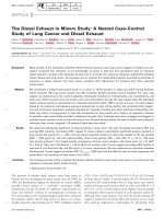
cases than controls [Figure 1] [P < 0.05].
Discussion
Persistent infection with high-risk HPV increases the
risk of cervical cancer. In this study, cervical cell folate
concentration, tumour suppressor gene methylation, and
particular HR-HPV types, were all shown to be associated with an increased likelihood of persistent HR-HPV
infection, defined as infection with any HR-HPV type at
follow-up. HPV persistence has been defined and estimated in a number of ways; a recent meta-analysis providing data on more than 100,000 women worldwide,
found that 73% of studies defined persistence as HPV
positivity at a minimum of two time points [2].
Consistent with the literature, HPV-16 was the most
prevalent infection [22]. HPV-16 infection and multiple
HR-HPV infections were found to be significant independent determinants of persistent HR-HPV infection,
which is consistent with findings from a study of typespecific HPV persistence in Finnish women [23].
Flatley et al. BMC Cancer 2014, 14:803
/>
Page 6 of 9
Table 5 Frequency of gene specific methylation according to case or control status and cytology group
Frequency of sample methylation [%]
DAPK
Case
Mild
Borderline
Normal
Total
CDH1
Control
Case
26.5 [13/49]
14 [7/50]
28 [14/50]
14 [7/50]
MGMT
MLH1
Control
Case
Control
32.7 [16/49]
40 [20/50]
12.2 [6/49]
10 [5/50]
42 [21/50]
56 [28/50]
14 [7/50]
10 [5/50]
Case
P16
Control
Case
Control
0 [0/49]
0 [0/50]
2.0 [1/49]
0 [0/50]
0 [0/50]
0 [0/50]
0 [0/50]
0 [0/50]
6 [6/100]
6 [6/100]
26 [26/100]
33 [33/100]
10 [10/100]
10 [10/100]
1 [1/100]
0 [0/100]
5 [5/100]
7 [7/100]
23.6a [47/199]
17 [34/200]
31.7 [63/199]
40.5b [81/200]
11.6 [23/199]
5.5 [11/200]
0.05 [1/199]
0 [0/200]
3 [6/199]
3.5 [7/200]
Promoter methylation of selected tumour suppressor genes, expressed as a percentage of women in each group. Values are shown for cases and controls,
according to cytology group.
a
Significantly greater than controls, P = 0.001; bSignificantly greater than cases, P = 0.016.
Cervical cell folate concentration was higher in women
with normal cytology than women with borderline or
mild cytology, which is consistent with previous findings
in which folate was measured in red blood cells [8] or
serum [24]. Multivariate modelling, taking account of
HR-HPV-type, infection with multiple HR-HPV types,
and a woman’s age, showed higher cervical cell folate
concentration to be associated with an increased likelihood of HR-HPV persistence. When the analysis was
conducted in each of the three cytology groups separately, this effect was only evident in women who had
mild cervical cell abnormalities.
Few other studies on folate status in cervical tissue
have been published. The concentrations of folate in cervical cells collected by liquid-based cytology in this study
were comparable to those measured in cervical biopsies
by Fowler et al., who reported concentrations of 2.75 to
4.39 ng/mg protein [25]. In their small cross-sectional
study, Fowler et al. reported a higher mean concentration of cervical cell folate in women who tested HRHPV positive [4.05 ng/mg] than in women who tested
HR-HPV negative [3.13 ng/mg], but the difference did
not reach statistical significance. In contrast, Flatley et al.
[8] found a lower red blood cell folate concentration in
women carrying an HR-HPV infection compared with
those free of infection and Piyathilake et al. [7] showed
that a higher circulating folate concentration was associated with a greater likelihood of clearing an HR-HPV
infection. Unlike Piyathilake et al. (7) we were able to
examine a possible role of folate in HPV persistence taking
cytology into account as well as HR-HPV type and infection with multiple HPV types. This is important because
the complex literature around folate and cancer suggests
very strongly that the presence of cell abnormality influences the association. Generally, studies have suggested
that a better folate status might protect against certain
Figure 1 Frequency of DAPK methylation according to case or control status and cytology. Gene promoter methylation determined in
cervical cells from 199 cases and 200 controls. Comparison between cases and controls in women with normal cytology [norm] or with
borderline or mild [mild + bord] cellular abnormality. *P < 0.05 significantly greater in cases than controls.
Flatley et al. BMC Cancer 2014, 14:803
/>
cancers but that where an underlying neoplasm exists, supplemental folate might accelerate carcinogenesis [26,27].
Finding from this study differ from the Piyathilake
study (7) in which folate was measured in the blood. It
is not clear whether the concentration of folate in the
blood is strongly correlated with that in cervical tissue,
particularly in non-normal tissue. Some cancer cells are
known to upregulate folate receptors [28], which facilitates folate uptake and could fuel enhanced cell proliferation. Although cervical cancer cells (HeLa cells) do
express a high density of the folic acid receptor [29], it is
not known whether this represents an upregulation from
the normal cell. An immunohistochemical study of folate
receptor expression in cervical tissue showed no difference between normal cells and low-grade abnormalities
and a reduced expression in higher-grade abnormalities
and cancer [30]. In our study, the higher concentration
of folate in the cases would be expected to increase host
cell proliferation and this would facilitate viral replication. The downstream effect on persistence is not clear;
enhanced viral replication might lead to greater reinfection of adjacent sites, but it might also lead to adaptive immune responses. A recent elegant study of HPV
integration in human keratinocytes showed that folate
deficiency impaired the cells’ ability to make HPV-16 virion particles, and this was associated with enhanced integration into the host DNA [31]. In our study, cells
with higher folate concentration may have been able to
produce more virus particles than cells with lower folate
concentration, and therefore might have been more permissive of re-infection.
The temporal relationship might be between a lower
folate concentration and low-grade cell abnormalities, as
observed in the ‘baseline’ measurements is not known.
Neither can we be certain what the temporal relationship between a persistent HPV infection and cell folate
concentration might be. Although we have discussed
how a higher folate concentration might increase the
likelihood of HR-HPV persistence, given the understanding that HPV infection can lead to changes in DNA
methylation and thereby alter expression of genes [32],
we cannot rule out the possibility that persistent HPV
infection and cell changes have a synergistic influence
on the expression of the folate receptor and folate uptake into cells.
In the multivariate analysis, the association between
age and risk of HPV persistence was different, depending
on the underlying cytology. In the total sample, age was
not associated with risk of HPV persistence. We did not
have access to demographic data on alcohol and tobacco
use for women whose samples were used in this study; it
is possible that there was a difference between younger
and older women which could have confounded the association between age and HPV persistence. Cigarette
Page 7 of 9
smoking and alcohol consumption are both thought to
influence HPV infection and the risk of cervical cancer
and both show an age association. Studies have examined HR-HPV persistence by age but the meta-analysis
of HR-HPV persistence by Rositch et al. [2] reports no
consistent trend.
Promoter methylation of the tumour suppressor gene,
death-associated protein kinase, DAPK, was also associated with an increased likelihood of HR-HPV persistence.
DAPK has a known role as a promoter of programmed
cell death and DAPK promoter methylation has been
reported for several cancers including cervical cancer
[33,34]. Promoter methylation of this gene is associated
with gene silencing [35]. A link with HR-HPV persistence
has not been reported previously but there is a plausible
mechanism for a causal relationship. Viruses have evolved
different strategies to avoid the host immune response to
infection. The inhibition of apoptosis is important to viral
pathogenesis. Should DAPK be silenced through promoter
methylation it no longer promotes cell death via the normal apoptotic pathway of an HR-HPV-infected cell and
the host cell may survive and differentiate. Under such
circumstances the HR-HPV virus would have longer to
replicate, increasing copy number and the likelihood of
infecting other cells. A mechanistic link between CDH1
methylation and HR-HPV persistence is less easy to explain. Promoter methylation of this gene can lead to gene
silencing [9]. CDH1 is a member of the cadherin family,
loss of expression would be expected to reduce cell-cell
contact, leading to an increase in cell motility and invasion, hallmarks of metastasis. Whilst cadherin expression
is important to bacterial adherence and internalisation,
this group of proteins has not been implicated in cellular
uptake of viruses, although cellular uptake by endocytosis
is common to both bacteria and viruses [36].
The temporal relationship between gene methylation,
cervical cell folate concentration and infection with HRHPV in baseline samples is not clear. HR-HPV infection
can induce change in gene methylation [37]. This would
require recruitment of the host cell methylation apparatus
and utilisation of intracellular folate, as methyl donor. Folate status of the cell may influence DNA methylation
through effects on DNA methyltransferases [DNMTs].
DNMT downregulation in response to folate depletion
has been reported for human colon cancer cells in vitro
[38] and unpublished data from our laboratory show that
methyl donor depletion of cervical cancer cells in vitro
leads to downregulation of DNA methyltransferases. By
inference, higher cellular folate might increase DNMT expression, and facilitate DAPK methylation. This provides a
putative link between higher cell folate status and DAPK
methylation in HR-HPV infection. The low frequency of
DAPK methylation in women with normal cytology is
compatible with our previous findings [8] and other
Flatley et al. BMC Cancer 2014, 14:803
/>
studies showing that DAPK methylation occurs on the
pathway of HR-HPV-induced cell transformation [10] may
explain the lack of association with HR-HPV persistence in
women with normal cytology.
It would have been preferable to have had access to
sufficient cervical cell material to allow the measurement
of tissue folate concentration and gene methylation on
the same samples, and to avoid sample pooling for folate
measurements. This must be considered a limitation of
the study but is unlikely to be resolved without access to
biopsy material. It would also have been useful to have
been able to include information about smoking and alcohol use into the multivariate analyses, as these are factors
are thought to influence the process of viral infection and
clearance.
Conclusions
Persistent infection with HR-HPV causes cervical cancer,
and therefore factors which influence the natural history
of HR-HPV infection may be important modulators of
cervical cancer risk, but the mechanisms that favour
HPV persistence are not understood. We have shown that
a higher concentration of folate in cervical cells, and promoter methylation of the tumour suppressor gene DAPK,
in women with cervical cell dyskaryosis, are associated with
increased risk of HR-HPV persistence.
We hypothesize that HR-HPV infection induces DAPK
methylation in dyskaryotic cells, supported by a high
intracellular folate, and that DAPK methylation leads to
dysregulation of apoptosis and promotes HR-HPV persistence. There is a need for in vitro studies to examine
these hypotheses and so shed further light on mechanisms of viral persistence. An understanding of such
mechanisms may have predictive value and inform patient management.
Abbreviations
HR-HPV: High-risk human papillomavirus; CIN: Cervical intraepithelial
neoplasia; ARTISTIC: A Randomised Trial In Screening To Improve Cytology;
DAPK: Death–associated protein kinase; CDH1: Cadherin-1; MLH1: MutL
homolog1, colon cancer, nonpolyposis type2 (E. coli); MGMT: 0-6methylguanine-DNA-methyl transferase; p16: Cyclin-dependent kinase
inhibitor 2A; ACTB: Beta-actin; PMR: Percent methylation reference.
Competing interests
The authors declare that they have no competing interests.
Authors’ contributions
JEF, HK and HJP conceived and designed the study; JEF conducted all the
molecular and biochemical analyses; JR carried out the statistical analysis of
data; AS was responsible for sample and data retrieval; JEF and HJP drafted
the manuscript, all authors contributed to the manuscript and read and
approved the final version.
Acknowledgements
This study was supported by The World Cancer Research Fund International
[Grant 2009/30].
Page 8 of 9
Author details
1
Human Nutrition Unit, Department of Oncology, Faculty of Medicine,
Dentistry and Health, University of Sheffield, Sheffield S10 2TN, UK.
2
Department of Clinical Virology, Central Manchester University Hospitals,
Manchester M139WL, UK. 3Gynecological Oncology Group, Cancer Studies,
Faculty of Human and Medical Sciences, University of Manchester,
Manchester M13 9BL, UK. 4Corporate Information and Computing Services,
University of Sheffield, Sheffield S10 2FN, UK. 5Human Nutrition Unit,
Department of Oncology, Faculty of Medicine, Dentistry and Health,
University of Sheffield, Sheffield S10 2TN, UK.
Received: 11 September 2013 Accepted: 23 September 2014
Published: 3 November 2014
References
1. Bosch FX, Lorincz A, Munoz N, Meijer CJ, Shah KV: The causal relation
between human papillomavirus and cervical cancer. J Clin Pathol 2002,
55:244–265.
2. Rositch AF, Koshiol J, Hudgens MG, Razzaghi H, Backes DM, Pimenta JM,
Franco EL, Poole C, Smith JS: Patterns of persistent genital human
papillomavirus infection among women worldwide: A literature review
and meta-analysis. Int J Cancer 2013, 133(6):1271–1285.
3. Baseman JG, Koutsky LA: The epidemiology of human papillomavirus
infections. J Clin Virol 2005, 32(Suppl 1):S16–S24.
4. Stern PL: Immune control of human papillomavirus [HPV] associated
anogenital disease and potential for vaccination. J Clin Virol 2005,
32:S72–S81. Review.
5. Winer RL, Hughes JP, Feng Q, Xi LF, Lee SK, O'Reilly SF, Kiviat NB, Koutsky LA:
Prevalence and risk factors for oncogenic human papillomavirus infections
in high-risk mid-adult women. Sex Transm Dis 2012, 39(11):848–856.
6. Goodman MT, Shvetsov YB, McDuffie K, Wilkens LR, Zhu X, Franke AA,
Bertram CC, Kessel B, Bernice M, Sunoo C, Ning L, Easa D, Killeen J,
Kamemoto L, Hernandez BY: Hawai cohort study of serum micronutrient
concentrations and clearance of incident oncogenic human
papillomavirus infection in the cervix. Cancer Res 2007, 67(12):5987–5996.
7. Piyathilake CJ, Henao OL, Macaluso M, Cornwell PE, Meleth S, Heimburger DC,
Partridge EE: Folate is associated with the natural history of high-risk human
papillomaviruses. Cancer Res 2004, 64:8788–8793.
8. Flatley JE, McNeir K, Balasubramani L, Tidy J, Stuart EL, Young TA, Powers HJ:
Folate status and aberrant DNA methylation are associated with HPV
infection and cervical pathogenesis. Cancer Epidem Biom Prev 2009,
18:2782–2789.
9. Narayan G, Arias-Pulido H, Koul S, Vargas H, Zhang FF, Villella J, Schneider A,
Terry MB, Mansukhani M, Rao PH, Murty VV: Frequent promoter methylation
of CDH1, DAPK, RARB, and HIC1 genes in carcinoma of the cervix uteri: its
relationship to clinical outcome. Mol Cancer 2003, 2:24.
10. Henken FE, Wilting SM, Overmeer RM, van Rietschoten JGI, Nygren AOH,
Errami A, Schouten JP, Meijer CJ, Snijders PJ, Steenbergen RD: Sequential
gene promoter methylation during HPV-induced cervical carcinogenesis.
Brit J Cancer 2007, 97:1457–1464.
11. Kyrgiou M, Koliopoulos G, Martin-Hirsch P, Kehoe S, Flannelly G, Mitrou S,
Arbyn M, Prendiville W, Paraskevaidis E: Management of minor cervical
cytological abnormalities: a systematic review and a meta-analysis of the
literature. Cancer Treat Rev 2007, 33:514–520.
12. Kitchener HC, Almonte M, Wheeler P, Desai M, Gilham C, Bailey A, Sargent A,
Peto J: HPV testing in routine cervical screening: cross sectional data from
the ARTISTIC trial. Br J Cancer 2006, 95:56–61.
13. Kitchener HC, Almonte M, Thomson C, Wheeler P, Sargent A, Stoykova B,
Gilham C, Baysson H, Roberts C, Dowie R, Desai M, Mather J, Bailey A,
Turner A, Moss S, Peto J: HPV testing in combination with liquidbased cytology in primary cervical screening [ARTISTIC]: a randomised
controlled trial. Lancet Oncol 2009, 10(7):672–682.
14. Peto R: The marked differences between carotenoids and retinoids:
Methodological implications for biochemical epidemiology. Cancer Surv
1983, 2:317–340.
15. Wahrendorf J, Hanck AB, Munoz N, Vuilleumier JP, Walker AM: Vitamin
measurements in pooled blood samples. Amer J Epidem 1986,
123:544–550.
16. Oka N, Kajita M, Nishimura R, Ohbayashi C, Sudo T: L1 gene methylation in
high-risk human papillomaviruses for the prognosis of cervical intraepithelial
neoplasia. Int J Gynecol Cancer 2013, 23(2):235–243.
Flatley et al. BMC Cancer 2014, 14:803
/>
17. Wentzensen N, Sherman ME, Schiffman M, Wang SS: Utility of methylation
markers in cervical cancer early detection: appraisal of the state-of-thescience. Gynecol Oncol 2009, 112:293–299.
18. Wisman GB, Nijhuis ER, Hoque MO, Reesink-Peters N, Koning AJ, Volders HH,
Buikema HJ, Boezen HM, Hollema H, Schuuring E, Sidransky D, van der Zee AG:
Assessment of gene promoter hypermethylation for detection of cervical
neoplasia. Int J Cancer 2006, 119:1908–1914.
19. Uccella S, Cerutti R, Placidi C, Marchet S, Carnevali I, Bernasconi B,
Proserpio I, Pinotti G, Tibiletti MG, Furlan D, Capella C: MGMT methylation in
diffuse large B-cell lymphoma: validation of quantitative methylation-specific
PCR and comparison with MGMT protein expression. J Clin Pathol 2009,
62:715–723.
20. Harden SV, Tokumaru Y, Westra WH, Goodman S, Ahrendt SA, Yang SC,
Sidransky D: Gene promoter hypermethylation in tumors and lymph
nodes of stage I lung cancer patients. Clin Cancer Res 2003, 9:1370–1375.
21. Trinh BN, Long TI, Laird PW: DNA methylation analysis by MethyLight
technology. Methods 2001, 25:456–462.
22. Ylitalo N, Sorensen P, Josefsson AM, Magnusson PK, Andersen PK, Ponten J,
Adami HO, Gyllensten UB, Melbye M: Consistent viral load of human
papillomavirus-16 and risk of cervical carcinoma in situ: a nested case–
control study. Lancet 2000, 355:2194–2198.
23. Louvanto K, Rintala MA, Syrjanen KJ, Grenman SE, Syrjanen SM: Genotypespecific persistence of genital human papillomavirus [HPV] infections in
women followed for 6 years in the Finnish family HPV study. J Infect Dis
2010, 202(3):436–444.
24. Abike F, Engin AB, Dunder I, Tapisiz OL, Aslan C, Kutluay L: Human
papillomavirus persistence and neopterin, folate and homocysteine
levels in cervical dysplasia. Gynecol Oncol 2011, 284:209–214.
25. Fowler BM, Giuliano AR, Piyathilake C, Nour M, Hatch K: Hypomethylation
in cervical tissue: is there a correlation with folate status? Cancer Epidem
Biom Prev 1998, 7:901–906.
26. Mason JB: Unravelling the complex relationship between folate and
cancer risk. BioFactors 2011, 37(4):253–600.
27. Wien TN, Pike E, Wisløff T, Staff A, Smeland S, Klemp M: Cancer risk with
folic acid supplements: a systematic review and meta-analysis. BMJ Open
2012, 2(1):e000653.
28. Nunez MI, Behrens C, Wood DM, Lin H, Suraokar M, Kadara H, Hoffstetter W,
Kalhor N, Lee JJ, Franklin W, Stewart DJ, Wistuba II: High expression of
folate receptor alpha in lung cancer correlates with adenocarcinoma
histology and mutation. J Thorac Oncol 2012, 7(5):833–840.
29. Castillo JJ, Sventsen WE, Rozlosnik N, Estobar P, Martinez F, Castillo-Leon J:
Detection of cancer cells using a peptide nanotube-folic acid modified
graphens electrode. Analyst 2013, 138:1026–1031.
30. Pillai MR, Chacko P, Kesari LA, Jayaprakash PG, Jayaram HN, Antony AC:
Expression of folate receptors and heterogeneous nuclear
ribonucleoprotein E1 in women with human papillomavirus
mediated transformation of cervical tissue to cancer. J Clin Pathol
2003, 56(8):569–574.
31. Xiao S, Tang YS, Khan RA, Zhang Y, Kusumanchi P, Stabler SP, Jayaram HN,
Antony AC: Influence of physiologic folate deficiency on human
papillomavirus type 16 [HPV16]-harboring human keratinocytes in vitro
and in vivo. J Biol Chem 2012, 287(15):12559–12577.
32. Jimenez-Wences H, Peralta-Zaragoza O, Fernandez-Tilapa G: Human
papillomavirus, DNA methylation and microRNA expression in cervical
cancer. Oncol Rep 2014, 31(6):2467–2476.
33. Yang HJ, Liu VW, Wang Y, Tsang PC, Ngan HY: Differential DNA methylation
profiles in gynaecological cancers and correlation with clinic-pathological
data. BMC Cancer 2006, 23:212.
34. Kim JH, Choi YD, Lee JS, Lee JH, Nam JH, Choi C: Assessment of DNA
methylation for the detection of cervical neoplasia in liquid-based
cytology specimens. Gynecol Oncol 2010, 116(1):99–104.
35. Raval A, Tanner SM, Byrf JC, Angerman EB, Perko JD, Chen SS, Hackanson B,
Grever MR, Lucas DM, Matkovic JJ, Lin TS, Kipps TJ, Murray F, Weisenburger D,
Sanger W, Lynch J, Watson P, Jansen M, Yoshinaga Y, Rosenquist R, de Jong PJ,
Coggill P, Beck S, Lynch H, de la Chapelle A, Plass C: Down-regulation
of death-associated protein kinase 1 [DAPK1] in chronic lymphocytic
leukemia. Cell 2007, 129(5):879–890.
36. Hauck CR: Cell adhesion receptors-signalling capacity and exploitation by
bacterial pathogens. Med Microbiol 2002, 191(2):55–62.
37. Leonard SM, Wei W, Collins SI, Pereira M, Diyaf A, Constandinou-Williams C,
Young LS, Roberts S, Woodman CB: Oncogenic human papillomavirus
Page 9 of 9
imposes an instructive pattern of DNA methylation change which parallels
the natural history of cervical HPV infection in young women. Carcinogenesis
2012, 33(7):1286–1293.
38. Stempak JM, Sohn KJ, Chiang EP, Shane B, Kim YI: Cell and stage
transformation -specific effects of folate deficiency on methionine cycle
intermediates and DNA methylation in an in vitro model. Carcinogenesis
2005, 26(5):981–990.
doi:10.1186/1471-2407-14-803
Cite this article as: Flatley et al.: Tumour suppressor gene methylation
and cervical cell folate concentration are determinants of high-risk human
papillomavirus persistence: a nested case control study. BMC Cancer
2014 14:803.
Submit your next manuscript to BioMed Central
and take full advantage of:
• Convenient online submission
• Thorough peer review
• No space constraints or color figure charges
• Immediate publication on acceptance
• Inclusion in PubMed, CAS, Scopus and Google Scholar
• Research which is freely available for redistribution
Submit your manuscript at
www.biomedcentral.com/submit