Biomarkers of thyroid function and autoimmunity for predicting high-risk groups of thyroid cancer: A nested case-control study
Bạn đang xem bản rút gọn của tài liệu. Xem và tải ngay bản đầy đủ của tài liệu tại đây (332.23 KB, 10 trang )
Cho et al. BMC Cancer 2014, 14:873
/>
RESEARCH ARTICLE
Open Access
Biomarkers of thyroid function and autoimmunity
for predicting high-risk groups of thyroid cancer:
a nested case–control study
Young Ae Cho1†, Sun-Young Kong2,3†, Aesun Shin1,4, Jeonghee Lee1, Eun Kyung Lee5, You Jin Lee5
and Jeongseon Kim1*
Abstract
Background: A remarkable increase in the number of thyroid cancer cases has been reported in recent years;
however, the markers to predict high-risk groups have not been fully established.
Methods: We conducted a case–control study (257 cases and 257 controls) that was nested in the Cancer Screenee
Cohort Study between August 2002 and December 2010; the mean follow-up time for this study was 3.1 ± 2.2 years.
The levels of total triiodothyronine (TT3), free thyroxine (FT4), thyroid-stimulating hormone (TSH), thyroglobulin (Tg),
anti-thyroperoxidase antibody (TPOAb), and anti-thyroglobulin antibody (TgAb) were measured using samples
with pre-diagnostic status. Logistic regression models were used to examine the association between thyroid
function/autoimmunity and thyroid cancer risk.
Results: When the markers were categorized by the tertile distributions of the control group, the highest tertile
of FT4 (OR = 1.73, 95% CI = 1.11 − 2.69) and the middle tertile of TSH (OR = 1.77, 95% CI = 1.14 − 2.74) were
associated with an increased risk of thyroid cancer by multivariate analyses. In addition, an elevated risk for
thyroid cancer was found in subjects with TPOAb levels above 30 IU/mL (OR = 8.47, 95% CI = 5.39 − 13.33 for
30–60 IU/mL and OR = 4.48, 95% CI = 2.59 − 7.76 for ≥60 IU/mL). Stratified analyses indicated that some of these
associations differed by sex, BMI, smoking status, and the duration of follow-up.
Conclusions: This study demonstrated that the levels of biomarkers of thyroid function/autoimmunity, particularly the
presence of TPOAb, might be used as diagnostic markers for predicting thyroid cancer risk. Our findings suggest that
careful monitoring of thyroid biomarkers may be helpful for identifying Korean populations at high-risk for thyroid
cancer.
Keywords: Thyroid cancer, Biomarkers, Thyroid function, Autoimmunity, TPOAb
Background
Thyroid cancer is the most frequent cancer among
endocrine tumors, and its incidence has been greatly
increasing in many countries [1]. In particular, the incidence of thyroid cancer in Korea has increased rapidly
and has become one of the highest in the world [2].
Although the increased incidence rate of thyroid cancer is
partly attributed to the increased detection of subclinical
* Correspondence:
†
Equal contributors
1
Division of Cancer Epidemiology and Prevention, Molecular Epidemiology
Branch, Research Institute, National Cancer Center, 323 Ilsan-ro, Ilsandong-gu,
Goyang-si 410-769, Gyeonggi-do, Korea
Full list of author information is available at the end of the article
cancer resulting from advanced diagnostic technologies
[3], studies have reported a true increase in thyroid cancer
incidence due to changes in lifestyle or environmental
factors (e.g., iodine intake, exposure to radiation) [4,5].
Recently, an effort has been made to predict the risk
of thyroid cancer using the markers of thyroid function/
autoimmunity [6-9]. Although the findings were inconsistent, several studies found biomarkers that predicted thyroid cancer. Some studies have reported that higher levels
of thyroid-stimulating hormone (TSH) are associated with
an increased risk of thyroid malignancy [6,7], possibly
because of its role in affecting thyroid cell differentiation
© 2014 Cho et al.; licensee BioMed Central Ltd. This is an Open Access article distributed under the terms of the Creative
Commons Attribution License ( which permits unrestricted use, distribution, and
reproduction in any medium, provided the original work is properly credited. The Creative Commons Public Domain
Dedication waiver ( applies to the data made available in this article,
unless otherwise stated.
Cho et al. BMC Cancer 2014, 14:873
/>
and proliferation or in stimulating angiogenesis [10]. Other
studies have suggested that thyroid autoantibodies could
be used as predictors of thyroid cancer risk based on the
association between thyroid autoimmune disease and thyroid cancer [9]. However, most studies have investigated
these associations retrospectively, which has the potential
for selection and referral biases.
In this study, we aimed to investigate whether blood
markers representing thyroid function and autoimmunity
could predict thyroid malignancy. We designed a nested
case–control study, which was affected little by bias, to
validate blood markers for thyroid malignancy.
Methods
Study population
We conducted a nested case–control study on participants in the ongoing Cancer Screenee Cohort Study
(CSCS) between August 2002 and December 2010,
which had a mean time of follow-up of 3.1 ± 2.2 years.
The CSCS is a prospective cohort study consisting of
participants of the Cancer Screening Program at the
National Cancer Center in South Korea. Participants
were aged 30 years or older, underwent health-screening
examinations, and were screened for selected cancers.
All of the participants were asked to complete a selfadministered questionnaire at the baseline evaluation.
The data collected in the baseline evaluation included
socio-demographic characteristics, personal and family
medical history, lifestyle factors, and reproductive factors. A total of 22,085 subjects provided written informed consent and provided a blood sample for study
participation.
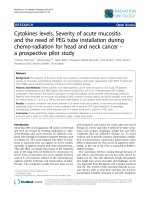
Ascertainment of cases and selection of controls
Potential cases diagnosed with thyroid cancer (ICD10
code C73) were ascertained by linkage to the Korea
Central Cancer Registry (KCCR) database, which was
used to identify the incidence of cancer in Korea.
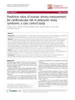
Among 258 thyroid cancer patients, 257 patients were
selected after excluding those who were dead. Among
the potential controls (n = 21,827) who were not diagnosed with thyroid cancer, 3,740 participants were excluded because of the following reasons: death, missing
questionnaire data, history of other cancers, any thyroid disease, thyroid surgery, or thyroid-related medicine. For each case, one control among the remaining
18,807 participants who was matched by entry age (same
age) and sex was selected. In total, 257 incident cases
and 257 controls were used for the final biomarker analysis (Figure 1). The participants were followed up from
the date of blood collection until December 31, 2010.
The study procedure was approved by the institutional
review board of the National Cancer Center (NCCNCS
13–698).
Page 2 of 10
Laboratory procedures
Blood samples were collected at the baseline evaluation
and stored at −80°C until analysis. The serum concentrations of the following six biomarkers were measured for
both cases and controls: total triiodothyronine (TT3),
thyroid-stimulating hormone (TSH), free thyroxine (FT4),
thyroglobulin (Tg), anti-thyroglobulin antibody (TgAb), and
anti-thyroperoxidase antibody (TPOAb). We selected these
biomarkers of thyroid function/autoimmunity based on
their associations with thyroid cancer that had been
reported in previous studies [6-9,11].
The serum concentrations of TT3, TSH, Tg, FT4, TgAb,
and TPOAb were measured using an electrochemiluminescence immunoassay (ELCLIA; Molecular Analytics E170,
Roche kit, Roche, Mannheim, Germany), which had
reference (normal) ranges of 0.82 − 2.0 ng/mL for TT3,
0.27 − 4.20 μIU/mL for TSH, 0.93 − 1.70 ng/dL for
FT4, and 1.4 − 78.0 ng/mL for Tg. TgAb was defined as
negative if ≤115.0 IU/mL, and TPOAb was defined as
negative if ≤34.0 IU/mL. The detection limits were
20 IU/mL for TgAb and 30 IU/mL for TPOAb.
Statistical methods
The general characteristics of the study participants and
the risk factors for thyroid cancer were compared using
t-tests for continuous variables and chi-square tests for
categorical variables. To evaluate the association between
serum biomarkers and thyroid cancer risk, serum levels of
TT3, FT4, TSH, and Tg were categorized into three
groups based on those of the control group. The antibody
titers for TgAb and TPOAb were also categorized into tertiles: the lowest tertile (under detection limit; 20 IU/mL
for TgAb and 30 IU/mL for TPOAb), the middle tertile
(over detection limit − < 60 IU/mL), and the highest tertile
(>60 IU/mL). Then, we performed unconditional and
conditional logistic regressions and calculated odds ratios (ORs) and 95% confidence intervals (CIs) using
univariate and multivariate analyses. The lowest levels
of each biomarker were used as references. The multivariate unconditional logistic regression models were adjusted
for age, sex, body mass index (BMI) (<23, 23 − < 25,
and ≥25 kg/m2), and cigarette smoking (nonsmoker,
former smoker, and current smoker). To analyze the
association between Tg levels and cancer risk, we excluded
subjects who were positive for TgAb because the presence
of TgAb hampers the usefulness of serum Tg as a
tumor marker [12]. To explore potential modifying
factors, analyses stratified according to sex, BMI (<23
and ≥23 kg/m2), and smoking status (nonsmoker and
former/current smoker) were conducted; these factors
showed different distributions between cases and controls
in this study and have been reported to affect thyroid
cancer risk [13,14]. We also conducted an analysis
stratified by the duration of follow-up. To examine the
Cho et al. BMC Cancer 2014, 14:873
/>
Page 3 of 10
Participants of Cancer Screenee Cohort at NCC
from August, 2002-December, 2010 (n=22,085)G
Followed-up
Until December 31st, 2010
Potential Cases with
Thyroid Cancer
(n=258)G
Potential Controls
(n=21,827)G
11,834 Controls were excluded
1 Cases were excluded
• Death (n=1)
Cases with
Thyroid Cancer
(n=257)G
•
•
•
•
•
•
Death (n=149)
Missing questionnaire (n=1,334)
History of Cancer (n=1,264)
Thyroid disease (n=977)
Thyroid surgery (n=37)
Thyroid-related medicine (n=28)
Matched Controls
(n=257)G
Figure 1 Flowchart of the sampling process of the nested case-control samples.
role of TPOAb in the association between thyroid cancer
risk and other biomarkers, we also conducted analyses
stratified by the presence of TPOAb. Because unconditional regression produced more stable results for the
different subanalyses [15], only the results from the
unconditional analyses are presented in the tables. We
verified that both the conditional and unconditional
approaches gave approximately the same results for
the entire dataset.
All statistical analyses were performed using SAS 9.1
software (SAS Institute Inc., Cary, NC). A two-sided
P-value of less than 0.05 was regarded as statistically
significant.
Results
This study included 257 cases and 257 controls, of
whom 70% were women and 30% were men. We examined the differences in the general characteristics of the
study subjects according to thyroid cancer status (Table 1).
The mean age for cases and controls was 49.4 ± 8.9 years.
The cases were more likely to have a higher BMI than the
controls (P = 0.019); however, no differences with respect
to other variables were observed between the cases and
controls.
Table 2 presents the association between the biomarkers
of thyroid function/autoimmunity and thyroid cancer risk.
When the markers were categorized by the tertile distributions of the control group, the highest tertile of FT4
(OR = 1.73, 95% CI = 1.11 − 2.69) showed an increased
risk of thyroid cancer, while the middle tertile of TSH
(OR = 1.77, 95% CI = 1.14 − 2.74) was associated with
thyroid cancer risk. In addition, TPOAb levels greater
than 30 IU/mL (OR = 8.47, 95% CI = 5.39 − 13.33 for
30 − < 60 IU/mL and OR = 4.48, 95% CI = 2.59 − 7.76
for ≥60 IU/mL) were strongly associated with risk of
thyroid cancer when compared with those whose
TPOAb levels were less than 30 IU/mL.
The associations of some markers with thyroid cancer
risk appeared to be different when the data were stratified
by sex, BMI, smoking status, or the duration of follow-up
(Table 3). The association between FT4 levels and thyroid
cancer risk was only significant among women or those
with a BMI <23 kg/m2. The elevated risk for the middle
tertile of TSH was only significant among men, those with
a BMI ≥23 kg/m2, or former/current smokers. The levels
of TT3, FT4, and TSH were associated with thyroid cancer risk only when the duration of follow-up was shorter
than 3 years. However, in all of the analyses, the presence
of TPOAb strongly elevated the risk of thyroid cancer.
Additionally, we examined whether other known risk
factors showed different distributions according to the
presence of TPOAb, but no differences were observed
(Additional file 1: Table S1).
Finally, we examined the role of TPOAb in the association between the other biomarkers (TT3, FT4, TSH,
Tg, and TgAb) and thyroid cancer risk (Table 4). The
Cho et al. BMC Cancer 2014, 14:873
/>
Page 4 of 10
Table 1 General characteristics of the study subjects
Controls (n = 257)
Cases (n = 257)
P-value
49.4 ± 8.9
49.4 ± 8.9
0.994
<23
125(48.6)
95(37.0)
0.019
23 − <25
68(26.5)
75(29.2)
≥ 25
64(24.9)
87(33.9)
93(41.0)
108(46.4)
0.245
5(2.2)
6(2.6)
0.794
21(8.8)
23(9.8)
0.774
Age (years), means
2
BMI (kg/m )
a
Family history of cancer (yes)
Family history of thyroid cancer (yes)a
Educational level
Elementary school or less
Middle school
19(7.9)
16(6.8)
High school
95(39.6)
84(35.9)
College or more
105(43.8)
115(47.4)
Monthly household incomeb
<200
35(16.6)
24(11.8)
200 − <400
56(26.5)
72(35.3)
>400
120(56.9)
108(52.9)
0.102
Marital status
Married
218(90.8)
215(90.0)
Unmarried
8(3.3)
5(2.1)
Divorced/Widowed
14(5.8)
19(8.0)
Nonsmoker
147(64.8)
158(69.3)
Former smoker
33(14.5)
37(16.3)
Current smoker
47(20.7)
33(14.5)
0.480
Smoking status
0.215
Alcohol consumption
Nondrinker
87(36.4)
100(41.5)
Former drinker
7(2.9)
12(5.0)
Current drinker
145(60.7)
129(53.5)
0.208
Age at menarche (years)c
≤13
35(22.0)
31(20.5)
14
34(21.4)
34(22.5)
15
37(23.3)
28(18.5)
≥16
53(33.3)
58(38.4)
83(47.7)
79(43.9)
0.472
<46
16(21.1)
12(17.7)
0.185
46 − <49
14(18.4)
8(11.8)
49 − <52
14(18.4)
23(33.8)
≥52
32(42.1)
25(36.8)
Natural
57(71.3)
51(64.6)
Surgery, Other
23(28.8)
28(35.4)
c
Menopause (yes)
0.680
Age at menopause (years)c
Type of menopause
c
0.366
Cho et al. BMC Cancer 2014, 14:873
/>
Page 5 of 10
Table 1 General characteristics of the study subjects (Continued)
Postmenopausal hormone use (ever)c
28(36.8)
20(30.8)
0.448
Parity (yes)c
157(96.3)
159(98.2)
0.315
a
First-degree relative.
Unit is 10,000 Korean won.
c
Only in women.
b
association between FT4 and thyroid cancer risk was
stronger among those with TPOAb levels <30 IU/mL
(OR = 2.12, 95% CI = 1.06 − 4.24).
Discussion
This study prospectively investigated the association between biomarkers of thyroid function/autoimmunity and
thyroid cancer risk and found that differences in the
levels of thyroid biomarkers, particularly TPOAb, could
predict the incidence of thyroid cancer.
Several studies have examined the association between
thyroid function and thyroid cancer risk [6-8,16,17]. A
large population-based cohort study from Taiwan [8] has
investigated the incidence of cancer in patients with
hyperthyroidism and found that patients with hyperthyroidism were at an increased risk for thyroid cancer.
This group also reported that the duration of hyperthyroidism was related to increased risk of thyroid cancer.
In the present study, the levels of thyroid hormones
were normal in most of the study participants. However,
relatively higher levels of FT4 showed a positive association with thyroid cancer risk. Because the association
between thyroid hormones and thyroid cancer risk has
not been sufficiently studied, the underlying mechanisms
Table 2 The association between the biomarkers of thyroid function/autoimmunity and thyroid cancer risk
Controls (n = 257)
Cases (n = 257)
Crude OR (95% CI)
Adjusted OR (95% CI)c
<1.2
88(34.2)
79(30.7)
1.0(ref)
1.0(ref)
1.2 − <1.4
104(40.5)
109(42.4)
1.17(0.78 − 1.75)
1.18(0.78 − 1.79)
≥1.4
65(25.3)
69(26.9)
1.18(0.75 − 1.86)
1.21(0.75 − 1.94)
85(33.1)
68(26.5)
1.0(ref)
1.0(ref)
TT3 (ng/mL)
FT4 (ng/dL)
<1.25
1.25 − <1.39
85(33.1)
75(29.2)
1.10(0.71 − 1.72)
1.05(0.66 − 1.65)
≥1.39
87(33.9)
114(44.4)
1.64(1.07 − 2.57)*
1.73(1.11 − 2.69)*
84(33.1)
59(23.2)
1.0(ref)
1.0(ref)
TSH (μIU/mL)
<1.36
1.36 − <2.5
86(33.9)
111(43.7)
1.81(1.18 − 2.79)
≥2.5
84(33.1)
84(33.1)
1.40(0.90 − 2.19)
1.37(0.87 − 2.16)
77(33.8)
67(29.7)
1.0(ref)
1.0(ref)
*
1.77(1.14 − 2.74)*
a
Tg (ng/mL)
0 − <3.9
3.9 − <7
74(32.5)
64(28.3)
0.98(0.61 − 1.56)
0.97(0.60 − 1.55)
≥7
77(33.8)
95(42.0)
1.40(0.90 − 2.17)
1.40(0.89 − 2.19)
212(82.5)
203(79.0)
1.0(ref)
1.0(ref)
TgAb (IU/mL)b
<20
20 − <60
17(6.6)
25(9.7)
1.54(0.81 − 2.93)
1.58(0.82 − 3.06)
≥60
28(10.9)
29(11.3)
1.08(0.62 − 1.88)
1.13(0.64 − 2.00)
192(74.1)
77(30.0)
1.0(ref)
131(51.0)
8.60(5.49 − 13.45)
8.47(5.39 − 13.33)*
49(19.1)
4.53(2.64 − 7.76)
4.48(2.59 − 7.76)*
TPOAb (IU/mL)b
<30
30 − <60
≥60
38(14.8)
27(10.5)
1.0(ref)
*
*
Abbreviations: CI, Confidence interval; OR, Odds ratio; TT3, Total triiodothyronine; FT4, Free thyroxine; TSH, Thyroid-stimulating hormone; Tg, Thyroglobulin; TgAb,
Anti-thyroglobulin antibody; TPOAb, Anti-thyroperoxidase antibody.
a
Analyzed only for TgAb-negative subjects; bThe detection limits used were 20 IU/mL for TgAb and 30 IU/mL for TPOAb; cAdjusted for age, sex, BMI, and smoking.
*
P <0.05.
BMI (kg/m2)
Sex
Smoking
Follow-up duration
Men
Women
<23
≥23
Non-smoker
Former/Current smoker
< 3 years
≥3 years
77/77
180/180
125/95
132/162
147/158
80/70
97/171
160/86
1.0(ref)
1.0(ref)
1.0(ref)
1.0(ref)
1.0(ref)
1.0(ref)
1.0(ref)
1.2 − <1.4
1.66(0.68 − 4.04)
1.08(0.67 − 1.75)
1.16(0.63 − 2.15)
1.18(0.67 − 2.08)
0.92(0.55 − 1.54)
≥1.4
1.56(0.61 − 4.00)
1.22(0.69 − 2.14)
0.93(0.43 − 2.00)
1.50(0.81 − 2.78)
0.80(0.43 − 1.51)
1.0(ref)
1.0(ref)
1.0(ref)
1.0(ref)
1.0(ref)
Controls/Cases
TT3 (ng/mL)
<1.2
1.0(ref)
2.07(0.85 − 5.02)
1.81(1.03 − 3.20)
*
0.93(0.47 − 1.83)
1.97(0.80 − 4.89)
2.51(1.21 − 5.22)*
1.10(0.53 − 2.25)
1.0(ref)
1.0(ref)
1.0(ref)
FT4 (ng/dL)
<1.25
1.25 − <1.39
0.65(0.23 − 1.80)
1.19(0.71 − 2.00)
1.37(0.62 − 2.89)
0.91(0.51 − 1.64)
0.82(0.46 − 1.46)
1.05(0.42 − 2.66)
1.58(0.84 − 2.97)
0.73(0.36 − 1.46)
≥1.39
1.44(0.53 − 3.88)
1.76(1.06 − 2.92)*
2.52(1.28 − 4.96)*
1.43(0.78 − 2.60)
1.46(0.83 − 2.56)
1.85(0.77 − 4.41)
3.01(1.57 − 5.77)*
1.10(0.57 − 2.10)
1.0(ref)
1.0(ref)
1.0(ref)
Cho et al. BMC Cancer 2014, 14:873
/>
Table 3 The association between thyroid function/autoimmunity biomarkers and thyroid cancer risk, stratified by sex, BMI, smoking status, and the duration
of follow-upa
TSH (μIU/mL)
<1.36
1.0(ref)
1.0(ref)
1.0(ref)
1.36 − <2.5
2.31(1.06 − 5.02)
*
1.0(ref)
1.0(ref)
1.16(0.59 − 2.29)
2.31(1.30 − 4.13)
*
1.58(0.92 − 2.72)
≥2.5
1.97(0.76 − 5.08)
1.16(0.68 − 1.97)
0.96(0.49 − 1.89)
1.82(0.98 − 3.37)
1.0(ref)
1.0(ref)
1.0(ref)
3.9 − <7
≥7
1.09(0.46 − 2.57)
0.93(0.52 − 1.66)
2.98(1.23 − 7.17)*
1.08(0.63 − 1.83)
1.0(ref)
1.0(ref)
1.71(0.95 − 3.09)
2.44(1.14 − 5.24)
*
2.07(1.11 − 3.88)
*
1.57(0.80 − 3.06)
1.23(0.68 − 2.22)
1.33(0.53 − 3.38)
1.36(0.72 − 2.59)
1.41(0.70 − 2.83)
1.0(ref)
1.0(ref)
1.0(ref)
1.0(ref)
1.0(ref)
1.09(0.52 − 2.30)
0.95(0.52 − 1.77)
0.77(0.41 − 1.43)
1.16(0.48 − 2.79)
1.75(0.87 − 3.52)
0.65(0.32 − 1.33)
1.09(0.55 − 2.14)
1.89(1.02 − 3.49)*
0.98(0.55 − 1.75)
2.52(1.05 − 6.03)*
1.80(0.96 − 3.36)
1.06(0.54 − 2.10)
1.0(ref)
1.0(ref)
1.0(ref)
1.0(ref)
1.0(ref)
1.0(ref)
Tg (ng/mL)b
0 − <3.9
c
TgAb (IU/mL)
<20
20 − <60
1.77(0.31 − 10.20)
1.56(0.77 − 3.19)
1.92(0.77 − 4.79)
1.27(0.50 − 3.26)
1.54(0.71 − 3.33)
1.33(0.28 − 6.26)
1.25(0.55 − 2.83)
1.17(0.35 − 3.89)
≥60
0.25(0.03 − 2.22)
1.31(0.71 − 2.41)
1.16(0.49 − 2.77)
1.11(0.52 − 2.37)
1.14(0.53 − 2.47)
1.07(0.30 − 3.79)
1.67(0.71 − 3.92)
0.77(0.32 − 1.87)
TPOAb (IU/mL)c
<30
1.0(ref)
1.0(ref)
1.0(ref)
1.0(ref)
1.0(ref)
1.0(ref)
1.0(ref)
1.0(ref)
30 − <60
5.59(2.57 − 12.16)
10.67(6.03 − 18.88)
8.77(4.30 − 17.86)
8.40(4.62 − 15.24)
10.96(5.85 − 20.53)
4.97(2.31 − 10.69)
6.10(3.10 − 11.67)
14.75(2.36 − 29.56)*
≥60
3.89(1.05 − 14.41)
4.60(2.50 − 8.46)
4.43(1.95 − 10.04)
4.41(2.10 − 9.26)
3.52(1.78 − 6.96)
6.35(1.80 − 22.41)
4.54(2.10 − 9.84)
4.32(1.77 − 10.52)*
*
*
*
*
*
*
*
*
*
*
*
*
*
*
Page 6 of 10
Abbreviations: BMI, Body mass index; CI, Confidence interval; OR, Odds ratio; TT3, Total triiodothyronine; FT4, Free thyroxine; TSH, Thyroid-stimulating hormone; Tg, thyroglobulin; TgAb, Anti-thyroglobulin antibody;
TPOAb, Anti-thyroperoxidase antibody.
a
Data were analyzed using multivariate logistic regression models which were adjusted for age, sex, BMI, and smoking; bAnalyzed only for TgAb-negative subjects; cThe detection limits used were 20 IU/mL for TgAb
and 30 IU/mL for TPOAb.
*
P <0.05.
Cho et al. BMC Cancer 2014, 14:873
/>
Page 7 of 10
Table 4 The association between thyroid function/autoimmunity biomarkers and thyroid cancer risk, stratified by the
presence of TPOAb
TPOAb (IU/mL)
≥30
<30
Controls/cases
Adjusted OR (95% CI)c
Controls/cases
Adjusted OR (95% CI)c
67/29
1.0(ref)
21/50
1.0(ref)
TT3 (ng/mL)
<1.2
1.2 − <1.4
74/32
1.03(0.55 − 1.91)
30/77
1.05(0.53 − 2.07)
≥1.4
51/16
0.71(0.34 − 1.49)
14/53
1.71(0.76 − 3.85)
66/18
1.0(ref)
19/50
1.0(ref)
FT4 (ng/dL)
<1.25
1.25 − <1.39
62/21
1.14(0.55 − 2.40)
23/54
0.87(0.42 − 1.80)
≥1.39
64/38
2.12(1.06 − 4.24)*
23/76
1.37(0.66 − 2.83)
66/16
1.0(ref)
18/43
1.0(ref)
1.36 − <2.5
≥2.5
TSH (μIU/mL)
<1.36
63/32
2.00(1.00 − 4.01)
*
23/79
1.48(0.73 − 3.00)
63/28
1.82(0.89 − 3.72)
21/56
1.14(0.54 − 2.40)
58/22
1.0(ref)
19/45
1.0(ref)
Tg (ng/mL)a
0 − <3.9
3.9 − <7
59/18
0.70(0.34 − 1.44)
15/46
1.28(0.57 − 2.86)
≥7
63/32
1.21(0.63 − 2.32)
14/63
1.95(0.87 − 4.38)
<20
170/69
1.0(ref)
42/134
1.0(ref)
≥20
22/8
0.95(0.40 − 2.26)
23/46
0.59(0.31 − 1.14)
TgAb (U/mL)b
Abbreviations: CI, Confidence interval; OR, Odds ratio; TT3, Total triiodothyronine; FT4, Free thyroxine; TSH, Thyroid-stimulating hormone; Tg, Thyroglobulin; TgAb,
Anti-thyroglobulin antibody; TPOAb, Anti-thyroperoxidase antibody.
a
Analyzed only for TgAb-negative subjects; bThe detection limits used were 20 IU/mL for TgAb, and we combined these groups into two groups because of the
small sample sizes; cAdjusted for age, sex, BMI, and smoking.
*
P <0.05.
still remain unclear. Pellegriti et al. reported that circulating TSH receptor-stimulating antibodies (TSHR-Abs)
were present in all patients with Graves’ disease [18],
implying an association between TSHR-Abs and elevated
levels of thyroid hormones. TSHR-Abs are known to
stimulate the same intracellular signal pathways as TSH,
which has mitogenic and antiapoptotic effects on thyroid
follicular cells and thus may play a role in thyroid cancer
initiation [19].
The positive association between TSH levels and thyroid cancer risk has been reported in some studies
[6,7,16,17], implying that high TSH levels may play a
key role in the initiation of thyroid carcinogenesis. TSH
has a proliferative effect on thyroid cell growth that is
most likely mediated by TSH receptors on tumor cells
[17]. However, some studies did not find an association
between TSH levels and thyroid cancer risk [20]. Our
study demonstrated that the highest tertile of TSH levels
did not show any association with thyroid cancer, but the
medium tertile of TSH levels seemed to slightly increase
thyroid cancer risk.
Tg is produced by normal thyroid tissue and neoplastic
follicular cells; therefore, serum Tg measurements can
be used as specific and sensitive tumor markers of differentiated thyroid cancer in clinical practice [21]. The level
of serum Tg is known to aid in the detection of residual,
recurrent, or metastatic disease rather than in determining the incidence of thyroid cancer [22], but the role of
Tg in the initiation of thyroid cancer remains unclear.
However, this study has found that Tg is positively associated with thyroid cancer risk only among lean people,
men, or smokers.
A high prevalence of thyroid cancer in those with
autoimmune thyroid diseases [23-25] and systemic autoimmune diseases [26] may imply the possible association
between thyroid autoimmunity and cancer risk. Kim et al.
[24] found an elevated risk of papillary thyroid cancer in
Korean patients with Hashimoto’s thyroiditis with elevated
levels of TPOAb. Antonelli et al. [26] reported the higher
prevalence of papillary thyroid cancer in systemic lupus
erythematosus patients, particularly in patients with thyroid autoimmunity. The results of these studies suggest
Cho et al. BMC Cancer 2014, 14:873
/>
that the risk of thyroid cancer is strongly associated with
elevated levels of TPOAb. TPO is a membrane protein
that catalyzes thyroid hormone synthesis; thus, the presence of TPOAb in the blood may reflect an alteration in
the immune system and lymphocytic infiltration in the
thyroid [27]. TPOAb may destroy thyroid tissue as well as
cytokines produced by infiltrating inflammatory cells,
which may contribute to inflammation-induced carcinogenesis [25,28,29]. Furthermore, the presence of TPOAb
could be associated with thyroid function. In a study using
the NHANES III survey from the United States, Hollowell
et al. reported an association between TPOAb and overt
thyroid dysfunction [30]. We also observed that the presence of TPOAb may affect the association between FT4
levels and the risk of thyroid cancer.
Several factors may modify the association between
thyroid abnormalities/thyroid autoimmunity and thyroid
cancer risk. First, the effect of FT4 and TSH on thyroid
cancer risk was affected by obesity status in the present
study. In addition, previous studies have reported a
positive association between obesity and thyroid cancer
[4,14]. It has also been proposed that obesity may affect
the secretion of certain hormones such as insulin and
sex steroids, which may act on the thyroid to stimulate
cell proliferation and suppress apoptosis [31]. Second,
this study also found that the levels of TSH and Tg were
associated with thyroid cancer risk only among smokers.
Smoking is known to have a negative association with
the risk of thyroid cancer [13,32], possibly by exerting
anti-estrogenic effects or by affecting the immune system
through nicotinic anti-inflammatory pathways [33-35].
Additionally, smoking is known to decrease the levels of
TSH and the positivity of thyroid autoantibodies [36],
which were reported to be positively associated with
thyroid cancer risk. Third, the association of these biomarkers with thyroid cancer was different in men and
women. The higher levels of TPOAb in women and the
higher prevalence of smokers in men may partly explain
the observed differences in the incidence of thyroid cancer based on sex [32]. A negative association between
TPOAb and smoking was also reported [37].
The present study has strengths in its study design in
terms of ascertaining thyroid cancer patients within a
prospective cohort. This study design allowed for determining the potential role of pre-diagnostic serum levels
of biomarkers on thyroid cancer risk. In addition, the
controls were derived from the same cohort as the cases;
thus, the potential selection bias that can occur with a
conventional case–control study was minimized. However,
the findings from the present study should be interpreted
with caution because of several limitations. First, the
duration of follow-up in this study was relatively short.
However, the association between thyroid cancer and
the levels of TPOAb was not modified by duration of
Page 8 of 10
follow-up in this study. Second, we lacked detailed information on the specifics of the thyroid cancer, e.g.,
tumor stage and histological type, because case ascertainment was performed by data linkage with the cancer registry; therefore, we could not include these variables in our
analyses. Third, the sample size was relatively small, especially for a stratified analysis. Finally, the study population
consisted of participants in a cancer-screening program;
thus, these individuals may pay more attention to their
health status and may not be representative of the general
Korean population.
Conclusions
We found that the levels of biomarkers of thyroid function
and autoimmunity could provide additional information
for predicting thyroid malignancy. Particularly, the presence of TPOAb seems to be a strong predictor of thyroid
cancer. Interestingly, most participants who showed positive associations between these biomarkers and thyroid
cancer risk were in the normal ranges of these markers
and may not have had any symptoms of thyroid disease.
Therefore, we cautiously suggest that careful monitoring
of these biomarkers, even within the normal range, may
be helpful for identifying those at high risk for thyroid
cancer and for enhancing the likelihood of early detection
in Koreans.
Additional file
Additional file 1: Table S1. General characteristics of the study
subjects according to the presence of TPOAb.
Abbreviations
BMI: Body mass index; CIs: Confidence intervals; CSCS: Cancer screenee
cohort study; ELCLIA: Electrochemiluminescence immunoassay; FT4: Free
thyroxine; KCCR: The Korea central cancer registry; ORs: Odds ratios;
Tg: Thyroglobulin; TgAb: Anti-thyroglobulin; TPOAb: Anti-thyroperoxidase;
TSH: Thyroid-stimulating hormone; TSHR-Abs: TSH receptor-stimulating antibodies; TT3: Triiodothyronine.
Competing interests
The authors declare that they have no competing interests.
Authors’ contributions
YAC carried out the statistical analysis and interpretation of the data and
drafted the manuscript. SK participated in interpretation of the data and
manuscript preparation. AS participated in the study design, data acquisition,
and quality control of the data. JL participated in the study design and
quality control of the data. EKL and YJL participated in interpretation of the
data. JK contributed to the study concept and design, data acquisition, and
quality control of the data. All authors participated in the revision of the
manuscript and approved the final version.
Acknowledgements
This research was supported by a grant from National Research Foundation
of Korea (NRF-2012R1A1A2044332). The study sponsor had no role in the
study design, in the collection analysis and interpretation of the data, in the
writing of the manuscript, or in the decision to submit the manuscript for
publication.
Cho et al. BMC Cancer 2014, 14:873
/>
Author details
1
Division of Cancer Epidemiology and Prevention, Molecular Epidemiology
Branch, Research Institute, National Cancer Center, 323 Ilsan-ro, Ilsandong-gu,
Goyang-si 410-769, Gyeonggi-do, Korea. 2Division of Cancer Epidemiology
and Prevention, Translational Epidemiology Branch, Research Institute,
National Cancer Center, Goyang-si 410-769, Gyeonggi-do, Korea.
3
Department of Laboratory Medicine, Center for Diagnostic Oncology,
Hospital, National Cancer Center, Goyang-si 410-769, Gyeonggi-do, Korea.
4
Department of Preventive Medicine, College of Medicine, Seoul National
University, Seoul 110-799, Korea. 5Center for Thyroid Cancer, National Cancer
Center, Goyang-si 410-769, Gyeonggi-do, Korea.
Received: 9 May 2014 Accepted: 13 November 2014
Published: 24 November 2014
References
1. Pellegriti G, Frasca F, Regalbuto C, Squatrito S, Vigneri R: Worldwide
increasing incidence of thyroid cancer: update on epidemiology and risk
factors. J Cancer Epidemiol 2013, 2013:965212.
2. Jung KW, Won YJ, Kong HJ, Oh CM, Seo HG, Lee JS: Prediction of cancer
incidence and mortality in Korea, 2013. Cancer Res Treat 2013,
45:15–21.
3. Verkooijen HM, Fioretta G, Pache JC, Franceschi S, Raymond L, Schubert H,
Bouchardy C: Diagnostic changes as a reason for the increase in papillary
thyroid cancer incidence in Geneva, Switzerland. Cancer Causes Control
2003, 14:13–17.
4. Peterson E, De P, Nuttall R: BMI, diet and female reproductive factors
as risks for thyroid cancer: a systematic review. PLoS One 2012,
7:e29177.
5. Navarro Silvera SA, Miller AB, Rohan TE: Risk factors for thyroid cancer: a
prospective cohort study. Int J Cancer 2005, 116:433–438.
6. Haymart MR, Repplinger DJ, Leverson GE, Elson DF, Sippel RS, Jaume JC, Chen
H: Higher serum thyroid stimulating hormone level in thyroid nodule
patients is associated with greater risks of differentiated thyroid cancer and
advanced tumor stage. J Clin Endocrinol Metab 2008, 93:809–814.
7. Boelaert K, Horacek J, Holder RL, Watkinson JC, Sheppard MC, Franklyn JA:
Serum thyrotropin concentration as a novel predictor of malignancy in
thyroid nodules investigated by fine-needle aspiration. J Clin Endocrinol
Metab 2006, 91:4295–4301.
8. Yeh NC, Chou CW, Weng SF, Yang CY, Yen FC, Lee SY, Wang JJ, Tien KJ:
Hyperthyroidism and thyroid cancer risk: a population-based cohort
study. Exp Clin Endocrinol Diabetes 2013, 121:402–406.
9. Kim ES, Lim DJ, Baek KH, Lee JM, Kim MK, Kwon HS, Song KH, Kang MI, Cha
BY, Lee KW, Son HY: Thyroglobulin antibody is associated with increased
cancer risk in thyroid nodules. Thyroid 2010, 20:885–891.
10. Moeller LC, Führer D: Thyroid hormone, thyroid hormone receptors, and
cancer: a clinical perspective. Endocr Relat Cancer 2013, 20:R19–R29.
11. Sherman SI: Thyroid carcinoma. Lancet 2003, 361:501–511.
12. Kitahara CM, Linet MS, Beane Freeman LE, Check DP, Church TR, Park Y, Purdue
MP, Schairer C, Berrington de González A: Cigarette smoking, alcohol intake,
and thyroid cancer risk: a pooled analysis of five prospective studies in the
United States. Cancer Causes Control 2012, 23:1615–1624.
13. Kitahara CM, Platz EA, Freeman LE, Hsing AW, Linet MS, Park Y, Schairer
C, Schatzkin A, Shikany JM, Berrington de González A: Obesity and
thyroid cancer risk among U.S. men and women: a pooled analysis of
five prospective studies. Cancer Epidemiol Biomarkers Prev 2011,
20:464–472.
14. Agudo A, Sala N, Pera G, Capellá G, Berenguer A, García N, Palli D, Boeing H,
Del Giudice G, Saieva C, Carneiro F, Berrino F, Sacerdote C, Tumino R,
Panico S, Berglund G, Simán H, Stenling R, Hallmans G, Martínez C, Bilbao R,
Barricarte A, Navarro C, Quirós JR, Allen N, Key T, Bingham S, Khaw KT,
Linseisen J, Nagel G, et al: Polymorphisms in metabolic genes related to
tobacco smoke and the risk of gastric cancer in the European
prospective investigation into cancer and nutrition. Cancer Epidemiol
Biomarkers Prev 2006, 15:2427–2434.
15. Kim HK, Yoon JH, Kim SJ, Cho JS, Kweon SS, Kang HC: Higher TSH level is a
risk factor for differentiated thyroid cancer. Clin Endocrinol (Oxf ) 2013,
78:472–477.
16. Fiore E, Vitti P: Serum TSH and risk of papillary thyroid cancer in nodular
thyroid disease. J Clin Endocrinol Metab 2012, 97:1134–1145.
Page 9 of 10
17. Pellegriti G, Mannarino C, Russo M, Terranova R, Marturano I, Vigneri R,
Belfiore A: Increased mortality in patients with differentiated thyroid
cancer associated with Graves’ disease. J Clin Endocrinol Metab 2013,
98:1014–1021.
18. Morshed SA, Latif R, Davies TF: Characterization of thyrotropin receptor
antibody-induced signaling cascades. Endocrinology 2009, 150:519–529.
19. Hrafnkelsson J, Tulinius H, Kjeld M, Sigvaldason H, Jónasson JG: Serum
thyroglobulin as a risk factor for thyroid carcinoma. Acta Oncol 2000,
39:973–977.
20. Shivaraj G, Prakash BD, Sonal V, Shruthi K, Vinayak H, Avinash M: Thyroid
function tests: a review. Eur Rev Med Pharmacol Sci 2009, 13:341–349.
21. Park do J, Lim JA, Kim TH, Choi HS, Ahn HY, Lee EK, Kim KW, Park YJ, Yi KH,
Cho BY: Serum thyroglobulin level measured after thyroxine withdrawal
is useful to predict further recurrence in whole body scan-negative
papillary thyroid cancer patients after reoperation. Endocr J 2012,
59:1021–1030.
22. Zhang L, Li H, Ji QH, Zhu YX, Wang ZY, Wang Y, Huang CP, Shen Q, Li DS,
Wu Y: The clinical features of papillary thyroid cancer in Hashimoto’s
thyroiditis patients from an area with a high prevalence of Hashimoto’s
disease. BMC Cancer 2012, 12:610.
23. Kim KW, Park YJ, Kim EH, Park SY, Park DJ, Ahn SH, Park do J, Jang HC, Cho
BY: Elevated risk of papillary thyroid cancer in Korean patients with
Hashimoto’s thyroiditis. Head Neck 2011, 33:691–695.
24. Azizi G, Malchoff CD: Autoimmune thyroid disease: a risk factor for
thyroid cancer. Endocr Pract 2011, 17:201–209.
25. Fiore E, Rago T, Scutari M, Ugolini C, Proietti A, Di Coscio G, Provenzale
MA, Berti P, Grasso L, Mariotti S, Pinchera A, Vitti P: Papillary thyroid
cancer, although strongly associated with lymphocytic infiltration on
histology, is only weakly predicted by serum thyroid auto-antibodies
in patients with nodular thyroid diseases. J Endocrinol Invest 2009,
32:344–351.
26. Antonelli A, Mosca M, Fallahi P, Neri R, Ferrari SM, D’Ascanio A, Ghiri E,
Carli L, Miccoli P, Bombardieri S: Thyroid cancer in systemic lupus
erythematosus: a case–control study. J Clin Endocrinol Metab 2010,
95:314–318.
27. Yoshida H, Amino N, Yagawa K, Uemura K, Satoh M, Miyai K, Kumahara Y:
Association of serum antithyroid antibodies with lymphocytic infiltration
of the thyroid gland: studies of seventy autopsied cases. J Clin Endocrinol
Metab 1978, 46:859–862.
28. Feldt-Rasmussen U, Rasmussen AK: Autoimmunity in differentiated thyroid
cancer: significance and related clinical problems. Hormones 2010,
9:109–117.
29. Balkwill F: Cancer and the chemokine network. Nat Rev Cancer 2004,
4:540–550.
30. Hollowell JG, Staehling NW, Flanders WD, Hannon WH, Gunter EW, Spencer
CA, Braverman LE: Serum TSH, T(4), and thyroid antibodies in the United
States population (1988 to 1994): National Health and Nutrition
Examination Survey (NHANES III). J Clin Endocrinol Metab 2002,
87:489–499.
31. Roberts DL, Dive C, Renehan AG: Biological mechanisms linking
obesity and cancer risk: new perspectives. Annu Rev Med 2010,
61:301–316.
32. Mack WJ, Preston-Martin S, Dal Maso L, Galanti R, Xiang M, Franceschi S,
Hallquist A, Jin F, Kolonel L, La Vecchia C, Levi F, Linos A, Lund E, McTiernan
A, Mabuchi K, Negri E, Wingren G, Ron E: A pooled analysis of case–control
studies of thyroid cancer: cigarette smoking and consumption of
alcohol, coffee, and tea. Cancer Causes Control 2003, 14:773–785.
33. Brand JS, Chan MF, Dowsett M, Folkerd E, Wareham NJ, Luben RN, van
der Schouw YT, Khaw KT: Cigarette smoking and endogenous sex
hormones in postmenopausal women. J Clin Endocrinol Metab 2011,
96:3184–3192.
34. Shiels MS, Rohrmann S, Menke A, Selvin E, Crespo CJ, Rifai N, Dobs A,
Feinleib M, Guallar E, Platz EA: Association of cigarette smoking, alcohol
consumption, and physical activity with sex steroid hormone levels in
US men. Cancer Causes Control 2009, 20:877–886.
35. Scott DA, Martin M: Exploitation of the nicotinic anti-inflammatory
pathway for the treatment of epithelial inflammatory diseases. World J
Gastroenterol 2006, 12:7451–7459.
36. Belin RM, Astor BC, Powe NR, Ladenson PW: Smoke exposure is associated
with a lower prevalence of serum thyroid autoantibodies and
thyrotropin concentration elevation and a higher prevalence of mild
Cho et al. BMC Cancer 2014, 14:873
/>
Page 10 of 10
thyrotropin concentration suppression in the third National Health and
Nutrition Examination Survey (NHANES III). J Clin Endocrinol Metab 2004,
89:6077–6086.
37. Pedersen IB, Laurberg P, Knudsen N, Jørgensen T, Perrild H, Ovesen L,
Rasmussen LB: Smoking is negatively associated with the presence of
thyroglobulin autoantibody and to a lesser degree with thyroid
peroxidase autoantibody in serum: a population study. Eur J Endocrinol
2008, 158:367–373.
doi:10.1186/1471-2407-14-873
Cite this article as: Cho et al.: Biomarkers of thyroid function and
autoimmunity for predicting high-risk groups of thyroid cancer: a
nested case–control study. BMC Cancer 2014 14:873.
Submit your next manuscript to BioMed Central
and take full advantage of:
• Convenient online submission
• Thorough peer review
• No space constraints or color figure charges
• Immediate publication on acceptance
• Inclusion in PubMed, CAS, Scopus and Google Scholar
• Research which is freely available for redistribution
Submit your manuscript at
www.biomedcentral.com/submit