Functional deficiency of NBN, the Nijmegen breakage syndrome protein, in a p.R215W mutant breast cancer cell line
Bạn đang xem bản rút gọn của tài liệu. Xem và tải ngay bản đầy đủ của tài liệu tại đây (2.1 MB, 10 trang )
Schröder-Heurich et al. BMC Cancer 2014, 14:434
/>
RESEARCH ARTICLE
Open Access
Functional deficiency of NBN, the Nijmegen
breakage syndrome protein, in a p.R215W mutant
breast cancer cell line
Bianca Schröder-Heurich1, Natalia Bogdanova1,2, Britta Wieland1, Xiaoxi Xie1, Monika Noskowicz1,
Tjoung-Won Park-Simon1, Peter Hillemanns1, Hans Christiansen2 and Thilo Dörk1*
Abstract
Background: Mutations in NBN, the gene for Nijmegen Breakage Syndrome (NBS), are thought to predispose women
to developing breast cancer, but a breast cancer cell line containing mutations in NBN has not yet been described. The
p.R215W missense mutation occurs at sub-polymorphic frequencies in several populations. We aimed to investigate its
functional impact in breast cancer cells from a carrier of this NBN mutation.
Methods: Breast cancer cell lines were screened by immunoblotting for NBN protein levels, and the NBN coding
region was sequenced for mutation analysis. Radiosensitivity assays and functional studies were performed through
immunocytochemistry and immunoblotting, and flow cytometry was employed to assess cell cycle progression.
Impedance measurements were used to study the consequences of PARP1 inhibition. Statistical comparisons between
cell lines were performed using t-tests.
Results: HCC1395 breast cancer cells exhibited reduced NBN protein levels. Direct sequencing identified the NBN p.
R215W mutation in the hemizygous state, in addition to a truncation in BRCA1. Mutations in both genes were already
present in the heterozygous state in the patient’s germline. HCC1395 cells were highly radiosensitive, susceptible to
apoptosis and were deficient in the formation of NBN foci. There was also evidence for some impairment in the
formation of γH2AX, MDC1, and 53BP1 foci after irradiation; these foci appeared smaller and irregular compared with
repair foci in wild-type cells, although ATM signalling was largely unaffected. In line with their deficiency in NBN and
BRCA1, HCC1395 cells were particularly sensitive to PARP1 inhibition.
Conclusion: Our results indicate that the p.R215W mutation in the HCC1395 breast cancer cell line impairs NBN
function, making this cell line a potentially useful cellular model for studying defective NBN protein within a mutant
BRCA1 background.
Keywords: Breast carcinoma, DNA damage repair, DNA double strand break repair disorder, Ionising radiation sensitivity,
MRN complex
Background
Breast cancer is a genetically heterogeneous disease, and
several predisposing genes are involved in DNA double
strand break repair [1-3]. One of these is NBN, the gene for
Nijmegen Breakage Syndrome (NBS) [4-6]. NBS is an autosomal recessive chromosomal instability disorder characterised by microcephaly, stunted growth, immunodeficiency,
* Correspondence:
1
Clinics of Obstetrics and Gynaecology, Hannover Medical School,
Carl-Neuberg Straße 1, D-30625 Hannover, Germany
Full list of author information is available at the end of the article
a high cancer predisposition and a marked sensitivity
towards ionising radiation [7]. The syndrome is most
common in Eastern Europe due to a Slavic founder mutation, c.657del5, in the NBN gene [4]. This gene encodes a 754 amino-acid protein named NBN, p95 or
nibrin, that interacts with the MRE11 and RAD50 proteins in sensing DNA damage and aids to recruit the
ataxia-telangiectasia mutated kinase, ATM, to the sites
of DNA double-strand breaks [5,8]. It further interacts
at the DNA damage sites with phosphorylated histone
H2AX (γH2AX) through its tandem breast cancer carboxy-
© 2014 Schröder-Heurich et al.; licensee BioMed Central Ltd. This is an Open Access article distributed under the terms of the
Creative Commons Attribution License ( which permits unrestricted use,
distribution, and reproduction in any medium, provided the original work is properly credited.
Schröder-Heurich et al. BMC Cancer 2014, 14:434
/>
terminal (BRCT) domain [9,10]. Loss of NBN function
leads to radioresistant DNA synthesis and deficiencies
in proper DNA double-strand break repair [11,12].
While biallelic mutations in the NBN gene give rise to
NBS, monoallelic mutations have been found to predispose
the heterozygous carriers within NBS families towards malignancies [13]. Furthermore, an increased frequency of the
most common NBN mutation c.657del5 has been observed
in Eastern European breast cancer patients compared with
healthy controls [14-19]. A missense mutation, p.R215W,
in the tandem BRCT domain has been suggested as a more
wide-spread candidate breast cancer susceptibility allele
[15,19]. Compound heterozygosity of the p.R215W substitution with the c.657del5 mutation has been reported in
NBS patients with severe disease [20]. In vitro mutagenesis
studies indicated that p.R215W might be a functional mutation that impairs the association of NBN with γH2AX
[21]. However, further cellular models for this missense
mutation would be useful to fully clarify its role in breast
cancer.
In the present study we report on the identification of
a breast cancer cell line, HCC1395, that harbors p.
R215W in the hemizygous state, and we investigate the
functional competence of the mutant NBN protein in
this cell line.
Page 2 of 10
direct sequencing using BigDye v1.1 terminator chemistry
and a 3100 Avant capillary sequencer (Life Technologies).
Sequencing data were analyzed with the Sequencing
Analysis 5.1.1 software.
Colony formation assay
Cells were seeded in six-well plates and, after 24 hours
incubation, were irradiated with doses between 0–6 Gy.
Radiation doses between 0.1-1 Gy or between 2–6 Gy
were applied in different experiments. Plating efficiency
was consistently lower for HCC1395 cells than for
MCF10A cells (about 58% for MCF10A compared with
about 2% in HCC1395). Medium was gently changed
every two days. After 5–7 days incubation for MCF10A
and 10–12 days incubation for HCC1395, colonies were
fixed with 3% (w/v) PFA, 2% (w/v) sucrose in PBS for
10 min, stained with 0.5% (w/v) crystal violet and
counted by microscopy. Surviving colonies were counted
as positive above a threshold of 50 cells. The survival
fraction (SF) of irradiated cells was expressed as a percentage of colonies per seeded cells after normalisation
by the plating efficiency of unirradiated cells. Each experiment was performed at least 3 times with both cell
lines.
Lysate preparation and immunoblotting
Methods
Cell culture
Cell lines were obtained from the American Type Culture
Collection (ATCC) in 2010. Human breast cancer epithelial cell lines HCC1395 and HCC1937 were cultured in
RPMI 1640 with 10% fetal calf serum, 500 U/ml penicillin, 0.5 mg/ml streptomycin and 2 mM L-Glutamine.
Lymphoblastoid cells HCC1395 BL were cultured in
RPMI1640 with 15% fetal calf serum and supplements
as above. Human normal breast epithelial MCF10A
cells were cultured in MEBM, supplemented with MEGM
Single Quots according to the manufactures instruction
(Lonza). All cells were grown at 37°C in a humidified
atmosphere supplemented with 5% CO2. Ionizing radiation (IR) with doses between 0.1 – 6 Gy was applied to
the cells using a Mevatron MD-2 accelerator (Siemens,
Munich, Germany). Olaparib was purchased from LC
Laboratories (Woburn, MA, USA), dissolved in DMSO
and stored at −20°C before usage.
Genetic analysis
Genomic DNA was extracted from the cultured breast
cancer epithelial cells using proteinase K digestion and
phenol-chloroform extraction. The coding region of
NBN and selected regions of BRCA1 (exon 20), BRCA2
(exon 11) and TP53 (exon 5) gene were amplified by
PCR using the primer pairs given in Additional file 1:
Table S1, and purified PCR products were subjected to
For preparation of protein extracts, cells were lysed in
cell extraction buffer (50 mM Tris pH 7,4, 150 mM
NaCl, 2 mM EGTA, 2 mM EDTA, 25 mM NaF, 0.1 mM
Na3VO4, 0.1 mM PMSF, 2 mg/ml Leupeptin, 2 mg/ml
Aprotinin, 0.2% Triton X-100, 0.3% NonidetP-40) for
30 min on ice. Protein extracts were cleared through
centrifugation at 16100 rcf for 15 min, and supernatants
were separated through SDS-PAGE and transferred to
nitrocellulose membranes. Antibodies to NBN, SMC1pS966, KAP1-pS824 were obtained from Novus Biologicals
(rabbit polyclonal); anti CHEK2-pS19 from Cell Signaling
(rabbit polyclonal); anti RAD50 (mouse monoclonal) from
Abcam; anti MRE11 (mouse monoclonal 12D7) from
GeneTex; PARP1 and cleaved PARP1 (rabbit polyclonal)
from Cell Signaling, and anti β-Actin (mouse monoclonal) from Sigma. Anti-mouse and anti-rabbit horseradish peroxidases labelled secondary antibodies were
purchased from GE Healthcare. Visualization of immunoreactive bands was performed by using ECL (Thermo
Scientific/Pierce), and their intensity was determined
using ImageJ quantitation software.
Immunocytochemistry
Cells grown on cover glasses in six-well plates were fixed
with 3% (w/v) PFA, 2% (w/v) Sucrose in PBS for 10 min.
Cells were permeabilized with 0.2% (v/v) Triton X-100 in
PBS. Antibodies against NBS1 (Novus Biologicals), MDC1
(Abcam), Histone H2A.x Phospho (S139) (Millipore) and
Schröder-Heurich et al. BMC Cancer 2014, 14:434
/>
53BP1 (Bethyl Laboratories) were incubated in 2% (w/
v) normal goat serum (Dianova) for 2 hrs. After PBS
washing, cells were incubated with FITC-conjugated
anti-mouse IgG antibody (Dianova), Alexa Fluor antimouse IgG 488 or Alexa Fluor anti-rabbit IgG 546
(Invitrogen) for 1.5 hrs. DNA was counterstained with
DAPI (Invitrogen) and cells were mounted with ProLong® Gold (Invitrogen). Foci were counted under a
Leica DMI6000B microscope, and results from four independent experiments were statistically analysed using
GraphPad Prism 4 with a t-test. P values below α < 0.05
were considered significant. For a more detailed inspection of the size and area of foci, images were taken as
z-stacks by using a Leica TCS SP2 confocal microscope
(40× or 63× magnification) and image acquisition was
carried out using CorelPhotoPaintX4 Software and
evaluated using ImageJ software. The number of pixels
was determined as a proxy for the foci area.
Flow cytometry
For DNA content analysis cells were irradiated with 6 Gy,
were fixed in 70% ethanol after the indicated time-points
(24, 48, 72, 96 hours) and were stained with propidium
iodide over night at 4°C. Data acquisition and analysis
were performed with a FACS Calibur (BD) flow cytometer
and Summit V5.1 software (Beckman Coulter). Results
from three independent experiments were analysed using
GraphPad Prism 4.
Impedance measurements
For impedance measurements we used the xCELLigence
System (Roche) that provides a highly sensitive method
to measure the cell index as a proxy of cell adhesion,
proliferation, cell death and morphological alterations.
Cell lines were subconfluent, and each cell line has been
optimized in previous titration experiments so that cell
type-specific optimal conditions for the seeding rate and
confluence were obtained for the real-time measurement
in these experiments. Cells were seeded at 5000 cells per
well in quadruplicates in E-Plate VIEW 96 (Roche) and
left untreated for 24 hrs within the RTCA SP station positioned in a 37°C incubator with 5% CO2 supply. For
monitoring compound mediated cytotoxicity, cells were
then treated with DMSO only or with 0.2 μM, 1 μM or
5 μM olaparib and incubated for an additional 72 hrs.
The Cell Index was plotted and analysed using RTCA
software 1.2.1 (Roche).
Results
We screened by immunoblotting six breast cancer cell
lines that are frequently used in molecular cancer research (HCC38, HCC1395, HCC1599, HCC1806, HCC1937,
MDA-MB231), for possible deficiencies in the proteins
of the MRE11-RAD50-NBN (MRN) complex. One cell
Page 3 of 10
line, HCC1395, exhibited significantly reduced levels in
NBN that were not seen in the wildtype breast epithelial cell line MCF10A or in the other tested breast cancer lines (Figure 1A). NBN was observed only at 30-40%
of wildtype levels in HCC1395 cells, but the protein appeared to be phosphorylated correctly following damage
as noted by an electrophoretic mobility shift after irradiation with 6 Gy (Figure 1A).
To identify the molecular basis for the reduced NBN
level, the whole NBN coding region was sequenced and
the missense mutation p.R215W was uncovered in the
hemizygous state (Figure 1B). This result was unexpected
since previous genome-wide sequencing studies had not
found a NBN mutation in HCC1395 cells [22-24]. We
therefore investigated lymphoblastoid cells from the same
breast cancer patient of whom the HCC1395 cell line had
been established. Direct sequencing revealed the p.R215W
mutation in the heterozygous state in the lymphoblastoid
cells (Figure 1B) indicating that it is a germline mutation
and has undergone loss of heterozygosity in the breast
tumour cells. The same was confirmed for a mutation in
BRCA1, p.R1751X, that also was heterozygous in the lymphoblastoid cells and hemizygous in the HCC1395 cell
line [24] (Additional file 2: Figure S1). BRCA1 interacts
with the MRN complex, however, BRCA1 deficiency could
not account for the reduced levels of NBN, since the
BRCA1 mutant HCC1937 cell line has normal levels of
NBN (Figure 1A).
Because NBN deficient cells from NBS patients are
highly radiosensitive, we tested whether the p.R215W
missense mutation was also associated with an increased
sensitivity towards ionising radiation in HCC1395 cells.
As shown in Figure 2A and Additional file 3: Figure S2,
the HCC1395 cell line was highly radiosensitive in a colony formation assay and could be distinguished from the
wildtype response in MCF10A cells even at moderate
doses of radiation down to 100 mGy (Figure 2A). This cellular radiosensitivity could not be associated with overt
cell cycle abnormalities, such as early G2 accumulation, as
judged by flow cytometric analysis of HCC1395 cells 24–
72 hrs after irradiation (Additional file 4: Figure S3). However, increased susceptibility to apoptosis was indicated by
a markedly increased level of PARP1 cleavage in immunoblots from irradiated HCC1395 cells. PARP1 expression
was elevated in HCC1395 cells, and the ratio of cleaved
PARP1 versus total PARP1 was about 10% in unirradiated
and 30% in irradiated HCC1395 cells whereas cleaved
PARP was poorly detectable in MCF10A cells (Figure 2B).
Because the cellular radiation response is mediated
through a cooperation of NBN with ATM, we investigated the radiation-induced kinase activity of ATM in
HCC1395 cells. There was little difference between the
NBN mutant cells and MCF10A in the radiationinduced phosphorylation of SMC1, CHEK2 or KAP1,
Schröder-Heurich et al. BMC Cancer 2014, 14:434
/>
Page 4 of 10
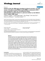
Figure 1 Identification of a NBN-deficient breast cancer cell line. A. Immunoblot analysis of MRN complex proteins in a wildtype mammary
epithelial cell line (MCF10A) and the breast cancer cell lines HCC1395 and HCC1937, each before and after irradiation (6 Gy, 30 min). HCC1395
cells showed some 70% reduction of NBN while MRE11 and RAD50 immunoreactivity remained largely unchanged. Note the mobility shift in
NBN after irradiation that can still be observed in HCC1395 cells at the reduced protein level. B. Identification by direct sequencing of the p.
R215W (c.643C > T) mutation in genomic DNA from HCC1395 (breast cancer cell line, bottom) and HCC1395 BL (lymphoblastoid cell line, middle)
in comparison to wildtype (MCF10A, top). The reverse strand is shown, with the position of the mutation indicated by an arrow. The p.R215W
mutation was found in a heterozygous state in HCC1395 BL, and in the apparently homozygous state in HCC1395 (loss of heterozygosity).
respectively (Figure 2C). This was independent of total
protein levels as exemplified for KAP1 in Additional
file 5: Figure S4. While HCC1395 cells showed a somewhat higher basic level of phosphorylation in the unirradiated state, the fact that irradiated cells reached the
same level of phosphorylation as wildtype cells indicated a largely unaffected ATM kinase activity towards
these major substrates within the dose range studied
(0.5 – 6 Gy; Figure 2C).
In order to investigate the molecular basis of radiation
sensitivity in HCC1395 cells in more detail, we analysed
radiation induced foci by immunocytochemical analyses
of three repair proteins γH2AX, 53BP1 and MDC1. In
HCC1395 cells, foci for these three proteins were generally
less intense and appeared smaller than in MCF10A cells.
In a quantitative approach of counting foci with conventional fluorescence microscopy on MCF10A, HCC1395
and HCC1937 under the same conditions, the percentages
Schröder-Heurich et al. BMC Cancer 2014, 14:434
/>
Page 5 of 10
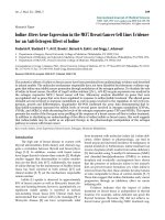
Figure 2 Assessment of cellular radiosensitivity in the colony formation assay. A. Cellular radiosensitivity of p.R215W mutant cells (HCC1395)
compared with wild type breast epithelial cells (MCF10A) as measured by the colony formation assay after irradiation at doses of 0, 0.1, 0.25, 0.5 or 1Gy.
The surviving fraction is presented as the mean value with SEM from at least 3 independent experiments. B. Induction of PARP1 cleavage after irradiation
with 2 Gy in p.R215W mutant cells (HCC1395) compared with wild type breast epithelial cells (MCF10A) as measured by immunoblotting of cleaved
PARP1 (89 kDa) and total PARP1 (116 kDa). C. Immunoblot analysis of radiation-induced ATM signalling in HCC1395 cells compared with
MCF10A. Cells were untreated or irradiated with 0.5, 1, 2, 4 or 6 Gy as indicated. Protein extracts were prepared 30 min after irradiation and
were analysed through Western blotting for their immunoreactivity towards the phosphorylated forms of SMC1 (pSer966, top panel), KAP1
(p824, middle panel), and CHEK2 (pSer19, bottom panel). β-actin served as the loading control in each experiment.
of γH2AX and MDC1 foci were significantly reduced in
HCC1395 cells compared to wildtype MCF10A at 30 min
after irradiation with either 1.5 or 6 Gy, whereas they were
at least as high as wildtype in HCC1937 cells in which
they also persisted at 24 hours after irradiation (Figure 3A).
A closer inspection using confocal laser microscopy revealed a smaller and a more fuzzy appearance of foci in
HCC1395 cells compared with MCF10A. A significant reduction in the size of γH2AX foci was noted as determined by measuring the γH2AX foci area in HCC1395
(Figures 3B-E). When staining against NBN itself, wildtype
MCF10A cells showed multiple discrete NBN foci after irradiation whereas no such NBN foci were detected in
HCC1395 cells, indicating a strongly reduced capability of
p.R215W NBN to accumulate in radiation-induced foci
(Figure 4).
Because NBN may facilitate homologous recombinational repair [25,26], and PARP1 activity has been reported
to be aberrantly high in NBS cells [27], we speculated that
PARP1 inhibition may be effective in HCC1395 cells. We
therefore treated HCC1395 cells with different doses of
olaparib, a known PARP1 inhibitor used in clinical trials,
and determined its effect on cellular viability and proliferation using real time monitoring of cellular impedance.
Olaparib significantly decreased the cell index at doses
as low as 0.2 μM. At this concentration, no such effect
of olaparib was observed in MCF10A (Additional file 6:
Figure S5), nor in HCC1937, suggesting a particular
sensitivity of HCC1395 cells with their combined mutations in BRCA1 and NBN towards PARP1 inhibition.
Discussion
In a search for breast cancer cell lines with deficiencies in
the MRN-ATM pathway, and thus defective DNA double
strand break response, we here describe the triplenegative breast cancer cell line, HCC1395, as being
mutated in the NBN gene. HCC1395 is a known breast
cancer cell line and has been included in previous
genome-wide sequencing studies [22-24] but the NBN
mutation p.R215W had not been uncovered. At the time
of submission, database mining of available cancer cell
lines in the COSMIC database ( />cosmic/) failed to identify any other line with the p.
R215W mutation, and also did not reveal any other breast
cancer cell line with a coding NBN mutation, so that
HCC1395 may provide a unique source for studying the
molecular consequences of NBN deficiency. The observation that p.R215W had already been present as a germline mutation in the heterozygous patient and underwent
loss of heterozygosity in the tumour would be in line with
the view that it represents a driver rather than a passenger
mutation during tumorigenesis, and is consistent with the
previously reported loss of heterozygosity in breast carcinomas harboring the NBN*657del5 mutation [14].
One consequence of the p.R215W mutation appears
to be a somewhat reduced stability of the NBN protein
Schröder-Heurich et al. BMC Cancer 2014, 14:434
/>
Figure 3 (See legend on next page.)
Page 6 of 10
Schröder-Heurich et al. BMC Cancer 2014, 14:434
/>
Page 7 of 10
(See figure on previous page.)
Figure 3 Immunocytochemical analysis of irradiation-induced repair foci. A. Evaluation of immunocytochemical evaluation of γH2AX, 53P1
and MDC1 foci after irradiation with either 1.5 Gy (upper panel) or 6 Gy (bottom panel) using conventional fluorescence microscopy. HCC1395
cells were compared with MCF10A cells as a wildtype control and with HCC1937 as a BRCA1 mutant control. Under these assay conditions,
the numbers of γH2AX and MDC1 foci appeared significantly reduced in p.R215W mutant cells (HCC1395) at 30 min after irradiation, with
53BP1 foci being slightly reduced. No such reduction was observed in HCC1937 cells. HCC1937 exhibited increased residual levels of foci at
24 hours and 48 hours after irradiation. Data represented as mean & SEM, *p < 0.05, **p < 0.01, ***p < 0.001, n = 4. B, C. Radiation-induced
MDC1 foci (B), γH2AX and 53BP1 foci (C). Representative images are shown for the immunocytochemical analysis of MDC1 (green, Figure B),
γH2AX (green, Figure C) and 53BP1 (red, Figure C) foci at 30 min after irradiation with 6 Gy with confocal microscopy revealing discrete and
more diffuse foci formation in HCC1395 compared to wildtype cells (MCF10A). Scale bar = 5 μm. D. Enlarged area of single nuclei from B, C for
quantitative evaluation. Note the smaller area and fuzzy appearance of foci in the HCC1395 cell line. E. Analysis of foci area in HCC1395 and
wildtype cells (MCF10A) for γH2AX, 53P1 and MDC1. For each, areas of 60–90 foci were analysed. Data are represented as mean & SEM. *p < 0.05.
which was expressed at a lower level than would be expected for a hemizygous situation. This did not affect
the levels of MRE11A or RAD50, consistent with the report that BRCT mutations in NBN do not interfere with
MRN complex formation in vitro [28]. The reduced level
of p.R215W mutant protein is also consistent with previous results from lymphoblastoid cells of p.R215W/
c.657del5 compound heterozygous NBS patients which
similarly showed markedly reduced nibrin levels [19,20].
Nevertheless, the residual NBN protein was easily detectable and, at the reduced level, appeared to undergo a
rapid mobility shift after irradiation as expected for normally activated NBN. An unimpaired phosphorylation
shift of NBN after irradiation has similarly been found in
the compound heterozygous NBS patient cells [20] and
may indicate that the p.R215W substitution does not
strongly disturb the ATM-mediated phosphorylation of
NBN at the more distant serine-278 and serine-343 sites.
Furthermore, although NBN has been shown to be required for full ATM activation after irradiation [11,29]
and particularly for its phosphorylation of SMC1 [30,31]
the p.R215W mutant did not seem to strongly disturb
the ATM-mediated phosphorylation of three different
substrates including SMC1 after irradiation in HCC1395
cells, suggesting that it still functions in the intra-Sphase checkpoint. This appears to be inconsistent with a
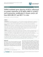
Figure 4 NBN foci formation. Double immunostaining of γH2AX
(green) and NBN (red) foci in HCC1395 and wildtype cells (MCF10A).
Cells were fixed 3 h after irradiation with 1.5 Gy. DNA was counterstained
with DAPI. Note the reduced appearance and brightness of yH2AX foci
and the absence of NBN foci in the HCC1395 cell line. Scale bar = 5 μm.
recent study that reports impairment of ATM-mediated
phosphorylation through p.R215W in retrovirally transduced fibroblasts [32] but is in agreement with the normal behaviour of other BRCT mutants [28] and is
supported by the flow cytometric analyses of HCC1395
cells which did not uncover marked cell cycle abnormalities, such as a rapid G2 accumulation due to radioresistant DNA synthesis, after up to 72 hours. In line with
this, previous work had indicated that the BRCT domain
of NBN is not required for inhibition of DNA synthesis
[11] or for cell cycle checkpoint regulation which rather
may be mediated by the FHA domain that is preserved
in the p.R215W mutant [33]. The retained activation of
the cohesion protein, SMC1, may help to suppress
chromatid-type aberrations [34], and such aberrations
are indeed lacking in p.R215W/c.657del5 compound heterozygous NBS patients [20].
The BRCT domain, initially described in BRCA1, is a
phosphoprotein binding module that is common in several DNA damage responsive proteins [35,36] and, in
case of NBN, has been implicated in the binding of
phosphorylated H2AX [9]. Phosphorylation of H2AX at
Ser139 is among the first events of the repair of double
strand breaks [37]. The gross accumulation of γH2AX
into foci, like those of MDC1 and 53BP1, appeared to be
incomplete in HCC1395 cells although smaller foci
could well be detected. It is possible that, after an initial
phosphorylation of these repair proteins, p.R215W mutant NBN fails to properly localize to γH2AX and to recruit or stabilize ATM which may be required for a
second wave of phosphorylation and focus extension.
Our results support a previous study, using a targeted
mutagenesis approach [21], which showed that mutation
R215W prevented the binding of NBN to γH2AX following radiation, and suggests a functional relevance for
this mutation in protein-protein interactions. This view
is further supported by molecular modelling of the tandem BRCT domain of NBN in which the R215 residue,
similar to the R1989 residue in MDC1, appears to direct
the relative orientation of both BRCT domains and
thereby regulate γH2AX recognition [21]. In a study of
additional single mutations of either the FHA or BRCT
Schröder-Heurich et al. BMC Cancer 2014, 14:434
/>
domains, only a small fraction of mutant NBN accumulated along DNA damage tracks labelled by γH2AX after
irradiation [33], and given that only one BRCT domain
had been mutated, it seems plausible that p.R215W may
have a stronger effect than the p.K160R mutant used in
that study.
The results obtained in HCC1395 might have more
general implications for breast cancer as they strongly
indicate functional and clinical relevance for a missense
mutation that is not uncommon among Europeans. p.
R215W has not been studied in the homozygous state,
thus far, because no homozygous individual has been
identified despite the modestly high prevalence of around
1/200 in some populations [19,20]. As suggested by Seemanova and co-workers, homozygosity for the p.R215W
mutation may lead to early embryonic lethality, albeit the
data here clearly indicate that some residual expression
and function is associated with this mutation. Of note, it
appears from Figure 2C as well as based on Figure 2B that
the hemizygous HCC1395 line may be suffering considerable damage even without additional endogenous exposure (higher levels of pSMC1, and Chk2 phosphorylation
and PARP cleavage in 0 Gy irradiated samples) and this
finding may be related to its poor growth overall. These
features are also commonly observed for classical NBN
mutant lines. Heterozygosity for p.R215W, as observed for
the patient BL1395, has been previously associated with
breast cancer in some, though not all, study populations
[15,19,38], and it is possible that p.R215W plays a predisposing role also for other malignancies [39]. At present,
the functional impact of the p.R215W NBN defect in
breast cancer susceptibility is still controversial, but evidence is accumulating that this mutation might predispose
individuals to disease.
It is also possible that p.R215W exerts such a predisposing role as a modifier of penetrance for additional
mutations such as in BRCA1. In fact, the HCC1395 cell
line was also found to harbor a truncating BRCA1 mutation. This mutation, however, is unlikely to explain the
reduced NBN levels or impaired formation of repair foci
considering that BRCA1 is not upstream of NBN and
was also mutated in the HCC1937 cell line which was
normal in NBN and proficient in the formation of repair
foci. Although we cannot formally exclude the possibility
that a BRCA1 mutation augments the effect of NBN p.
R215W on NBN levels and radiation-induced foci formation, the functional BRCA1 deficiency does not cause
these effects as is also evidenced by the observations of
normal NBN levels and foci formation in other cell lines
such as HCC38 or HCC1806 which are functionally deficient in BRCA1 [40,41]. However, the presence of a
BRCA1 or BRCA2 mutation has been shown to affect
sensitivity towards ionising radiation [42] as well as towards PARP1 inhibition [43,44], so that these endpoints
Page 8 of 10
were not specific for the NBN*R215W mutant. Increased
sensitivity to PARP1 inhibition has previously been reported for NBN deficient cells, and mutations in other
proteins that affect HR, besides BRCA1/2, may also
show some PARP1 inhibitor sensitivity [45,46]. On the
other hand, increased sensitivity to PARP1 inhibition is a
known feature of BRCA1 and BRCA2 mutant cells, and
since we found HCC1395 cells to harbour a BRCA1
truncation in the hemizygous and a BRCA2 mutation in
the heterozygous state, it is likely that the high olaparib
sensitivity is a combinatorial result and we could not
fully determine in the present study to what extent these
observations were due to functional deficiency in NBN,
BRCA1, BRCA2 or perhaps other deficiencies. These results emphasize the need to fully explore the mutational
genotypes of cell lines as well as of primary tumours before final conclusions can be drawn. The slow proliferation of HCC1395 cells and their higher genomic
instability and higher apoptotic activity under basal conditions as indicated in Figures 2B, C and Additional file
6: Figure S5 may further impact on these endpoints and,
given the heterogeneous background of breast tumours,
additional studies of p.R215W carriers will be needed to
fully elucidate this aspect and clarify whether they could
perhaps benefit from treatment with PARP1 inhibitors.
Conclusion
We report for the first time the identification and
characterization of a breast cancer cell line containing
a NBN mutation that affects its function, providing a
potentially useful cellular model for studying defective
NBN protein within a mutant BRCA1 background. The
data indicate that NBN*p.R215W is an unstable protein
that fails to be recruited into foci after irradiation and
also impairs the propagation of γH2AX repair foci. Our
results furnish evidence for the functional impact of
the p.R215W substitution, a suspected cancer susceptibility allele in Europeans.
Additional files
Additional file 1: Table S1. Primer pairs used for amplification and
sequencing the coding region of NBN and selected regions of BRCA1
(exon 20), BRCA2 (exon 11) and TP53 (exon 5).
Additional file 2: Figure S1. Validation by direct sequencing of
additional mutations in BRCA1, BRCA2 and TP53 in HCC1395 breast cancer
cells. Direct sequencing of selected regions of BRCA1, BRCA2 and TP53 in
HCC1395 BL lymphoblastoid cells (top) and HCC1395 breast cancer cells
(bottom) to validate reported mutations and confirm the identity of
HCC1395 cells. Mutated positions in BRCA1 (left panel), BRCA2 (middle
panel) and TP53 (right panel) are indicated by an arrow and the mutated
codon is boxed. Like the NBN mutation p.R215W, the BRCA1 mutation p.
Y1751X appears heterozygous in HCC1395 BL lymphoblasts but homoallelic
in HCC1395 breast cancer cells, whereas TP53 and BRCA2 mutations were
somatic events (with BRCA2 mutated only in the heterozygous state) in
HCC1395 breast cancer cells.
Schröder-Heurich et al. BMC Cancer 2014, 14:434
/>
Additional file 3: Figure S2. Assessment of cellular radiosensitivity in
the colony formation assay (higher dose experiments). Cellular radiosensitivity
of p.R215W mutant cells (HCC1395) compared with wild type breast epithelial
cells (MCF10A) as measured by the colony formation assay after irradiation at
doses of 2, 4, or 6 Gy. The surviving fraction is presented as the mean value
with SEM from at least 3 independent experiments.
Additional file 4: Figure S3. Cell cycle analysis by flow cytometry in
HCC1395 breast cancer cells. Flow cytometric analyses of S-, G1 and G2
cell population in wildtype cells (MCF10A) (A), HCC1395 (B) and HCC1937
(C) after irradiation with 6 Gy and different time points (24 hrs, 48 hrs,
72 hrs, 96 hrs). Data represented as Mean & SEM. Data for 24 hrs, 48 hrs
and 72 hrs: n = 3; data for 96 hrs: n = 1.
Additional file 5: Figure S4. Immunoblot analysis of total and
phosphorylated KAP1 in MCF10A and HCC1395 cells treated with
different doses of irradiation. Immunoblot analysis of radiation-induced
ATM signalling in HCC1395 cells compared with MCF10A. Cells were
untreated or irradiated with 0.5, 1, 2, 4 or 6 Gy as indicated. Protein extracts
were prepared 30 min after irradiation and were analysed through Western
blotting for their immunoreactivity towards the phosphorylated form of
KAP1 (p824, upper panel) and total KAP1 (bottom panel), respectively.
β-actin served as the loading control.
Additional file 6: Figure S5. Response to PARP1 inhibition in X-Celligence
impedance measurements. X-Celligence impedance measurements of
the p.R215W mutant cell line (HCC1395) were performed over three days
after addition of the PARP1 inhibitor olaparib at different concentrations
(0.2 μM, 1 μM, 5 μM; increasing concentrations from top to bottom) and
compared with the DMSO only control. Top: HCC1395 NBN p.R215W
mutant cell line; bottom: MCF10A cell line for comparison. Data are
presented as mean values & SEM from quadruplicates.
Page 9 of 10
2.
3.
4.
5.
6.
7.
8.
9.
10.
11.
Abbreviations
NBS: Nijmegen breakage syndrome; MRN: MRE11-RAD50-NBN;
BRCT: Breast-cancer carboxy-terminal domain; DMSO: Dimethylsulfoxide;
PFA: Paraformaldehyde; PBS: Phosphate-buffered saline; EGTA: Ethylene
glycol tetraacetic acid; EDTA: Ethylenediamine tetraacetic acid;
PMSF: Phenylmethanesulfonylfluoride.
Competing interests
All authors declare to have no financial or non-financial competing interests.
Authors’ contributions
BSH, NB and TD participated in the conception and design of the study.
Data acquisition and analyses were performed by BSH, NB, XX, MN and BW.
Data acquisition was supported by PH, TWPS and HC. The manuscript was
drafted by BSH, NB, MN, XX, BW and TD. All authors read and approved the
final manuscript.
Acknowledgments
We cordially thank Julia Menzel, Martin Werner, Jörg Frühauf and Johann
Hinrich Karstens for their support at the Department of Radiation Oncology
and Matthias Ballmeier for his help in flow cytometry. We are indebted to
the Claudia von Schilling Foundation for Breast Cancer Research for their
continuous support. N.B. is recipient of a Hannelore Munke stipend, and X.X.
is recipient of a stipend from the Chinese government.
Author details
1
Clinics of Obstetrics and Gynaecology, Hannover Medical School,
Carl-Neuberg Straße 1, D-30625 Hannover, Germany. 2Clinics of Radiation
Oncology, Hannover Medical School, Carl-Neuberg Straße 1, D-30625
Hannover, Germany.
12.
13.
14.
15.
16.
17.
18.
19.
Received: 12 February 2013 Accepted: 26 May 2014
Published: 13 June 2014
20.
References
1. Turnbull C, Rahman N: Genetic predisposition to breast cancer: past,
present, and future. Annu Rev Genomics Hum Genet 2008, 9:321–345.
Ghoussaini M, Pharoah PD, Easton DF: Inherited genetic susceptibility to
breast cancer: the beginning of the end or the end of the beginning?
Am J Pathol 2013, 183:1038–1051.
Bogdanova N, Helbig S, Dörk T: Hereditary breast cancer: ever more
pieces to the polygenic puzzle. Hered Cancer Clin Pract 2013, 11:12.
Varon R, Vissinga C, Platzer M, Cerosaletti KM, Chrzanowska KH, Saar K,
Beckmann G, Seemanová E, Cooper PR, Nowak NJ, Stumm M, Weemaes CM,
Gatti RA, Wilson RK, Digweed M, Rosenthal A, Sperling K, Concannon P,
Reis A: Nibrin, a novel DNA double-strand break repair protein, is
mutated in Nijmegen breakage syndrome. Cell 1998, 93:467–476.
Carney JP, Maser RS, Olivares H, Davis EM, Le Beau M, Yates JR 3rd, Hays L,
Morgan WF, Petrini JH: The hMre11/hRad50 protein complex and
Nijmegen breakage syndrome: linkage of double-strand break repair to
the cellular DNA damage response. Cell 1998, 93:477–486.
Matsuura S, Tauchi H, Nakamura A, Kondo N, Sakamoto S, Endo S, Smeets D,
Solder B, Belohradsky BH, Der Kaloustian VM, Oshimura M, Isomura M,
Nakamura Y, Komatsu K: Positional cloning of the gene for Nijmegen
breakage syndrome. Nat Genet 1998, 19:179–181.
Digweed M, Sperling K: Nijmegen breakage syndrome: clinical
manifestation of defective response to DNA double-strand breaks.
DNA Repair 2004, 3:1207–1217.
Falck J, Coates J, Jackson SP: Conserved modes of recruitment of ATM,
ATR and DNA-PKcs to sites of DNA damage. Nature 2005, 434:605–611.
Kobayashi J, Kobayashi J, Tauchi H, Sakamoto S, Nakamura A, Morishima K,
Matsuura S, Kobayashi T, Tamai K, Tanimoto K, Komatsu K: NBS1 localizes
to gamma-H2AX foci through interaction with the FHA/BRCT domain.
Curr Biol 2002, 12:1846–1851.
Becker E, Meyer V, Madaoui H, Guerois R: Detection of a tandem BRCT in
Nbs1 and Xrs2 with functional implications in the DNA damage
response. Bioinformatics 2006, 22:1289–1292.
Lee JH, Xu B, Lee CH, Ahn JY, Song MS, Lee H, Canman CE, Lee JS, Kastan MB,
Lim DS: Distinct functions of Nijmegen breakage syndrome in ataxia
telangiectasia mutated-dependent responses to DNA damage.
Mol Cancer Res 2003, 1:674–681.
Stracker TH, Petrini JH: The MRE11 complex: starting from the ends.
Nat Rev Mol Cell Biol 2011, 12:90–103.
Seemanová E, Jarolim P, Seeman P, Varon R, Digweed M, Swift M, Sperling K:
Cancer risk of heterozygotes with the NBN founder mutation. J Natl Cancer
Inst 2007, 99:1875–1880.
Gorski B, Debniak T, Masojc B, Mierzejewski M, Medrek K, Cybulski C,
Jakubowska A, Kurzawski G, Chosia M, Scott R, Lubinski J: Germline 657del5
mutation in the NBS1 gene in breast cancer patients. Int J Cancer 2003,
106:379–381.
Steffen J, Varon R, Mosor M, Maneva G, Maurer M, Stumm M, Nowakowska D,
Rubach M, Kosakowska E, Ruka W, Nowecki Z, Rutkowski P, Demkow T,
Sadowska M, Bidzinski M, Gawrychowski K, Sperling K: Increased cancer
risk of heterozygotes with NBS1 germline mutations in Poland.
Int J Cancer 2004, 111:67–71.
Buslov KG, Iyevleva AG, Chekmariova EV, Suspitsin EN, Togo AV, Kuligina ES,
Sokolenko AP, Matsko DE, Turkevich EA, Lazareva YR, Chagunava OL, Bit-Sava EM,
Semiglazov VF, Devilee P, Cornelisse C, Hanson KP, Imyanitov EN: NBS1 657del5
mutation may contribute only to a limited fraction of breast cancer cases in
Russia. Int J Cancer 2005, 114:585–589.
Górski B, Cybulski C, Huzarski T, Byrski T, Gronwald J, Jakubowska A, Stawicka M,
Gozdecka-Grodecka S, Szwiec M, Urbański K, Mituś J, Marczyk E, Dziuba J,
Wandzel P, Surdyka D, Haus O, Janiszewska H, Debniak T, Tooczko-Grabarek A,
Medrek K, Masojc B, Mierzejewski M, Kowalska E, Narod SA, Lubinski J:
Breast cancer predisposing alleles in Poland. Breast Cancer Res Treat 2005,
92:19–24.
Steffen J, Nowakowska D, Niwinska A, Czapczak D, Kluska A, Piatkowska M,
Wisniewska A, Paszko Z: Germline mutations 657del5 of the NBS1 gene
contribute significantly to the incidence of breast cancer in Central
Poland. Int J Cancer 2006, 119:470–472.
Bogdanova N, Feshchenko S, Schürmann P, Waltes R, Wieland B, Hillemanns P,
Rogov YI, Dammann O, Bremer M, Karstens JH, Sohn C, Varon R, Dörk T:
Nijmegen breakage syndrome mutations and risk of breast cancer.
Int J Cancer 2008, 122:802–806.
Seemanova E, Seemanová E, Sperling K, Neitzel H, Varon R, Hadac J, Butova O,
Schröck E, Seeman P, Digweed M: Nijmegen breakage syndrome (NBS)
with neurological abnormalities and without chromosomal instability.
J Med Genet 2006, 43:218–224.
Schröder-Heurich et al. BMC Cancer 2014, 14:434
/>
21. di Masi A, Viganotti M, Polticelli F, Ascenzi P, Tanzarella C, Antoccia A: The
R215W mutation in NBS1 impairs gamma-H2AX binding and affects
DNA repair: molecular bases for the severe phenotype of 657del5/
R215W Nijmegen breakage syndrome patients. Biochem Biophys Res
Commun 2008, 369:835–840.
22. Sjöblom T, Jones S, Wood LD, Parsons DW, Lin J, Barber TD, Mandelker D,
Leary RJ, Ptak J, Silliman N, Szabo S, Buckhaults P, Farrell C, Meeh P,
Markowitz SD, Willis J, Dawson D, Willson JKV, Gazdar AF, Hartigan J, Wu L,
Liu C, Parmigiani G, Park BH, Bachman KE, Papadopoulos N, Vogelstein B,
Kinzler KW, Velculescu VE: The consensus coding sequences of human
breast and colorectal cancers. Science 2006, 314:268–274.
23. Wood LD, Parsons DW, Jones S, Lin J, Sjöblom T, Leary RJ, Shen D, Boca SM,
Barber T, Ptak J, Silliman N, Szabo S, Dezso Z, Ustyanksky V, Nikolskaya T,
Nikolsky Y, Karchin R, Wilson PA, Kaminker JS, Zhang Z, Croshaw R, Willis J,
Dawson D, Shipitsin M, Willson JKV, Sukumar S, Polyak K, Park BH,
Pethiyagoda CL, Krishna Pant PV: The genomic landscapes of human
breast and colorectal cancers. Science 2007, 318:1108–1113.
24. Stephens PJ, McBride DJ, Lin ML, Varela I, Pleasance ED, Simpson JT,
Stebbings LA, Leroy C, Edkins S, Mudie LJ, Greenman CD, Jia M, Latimer C,
Teague JW, Lau KW, Burton J, Quail MA, Swerdlow H, Churcher C, Natrajan R,
Sieuwerts AM, Martens JWM, Silver DP, Langerød A, Russnes HEG, Foekens JA,
Reis-Filho JS, van ’t Veer L, Richardson AL, Børresen-Dale AL: Complex
landscapes of somatic rearrangement in human breast cancer genomes.
Nature 2009, 462:1005–1010.
25. Sakamoto S, Iijima K, Mochizuki D, Nakamura K, Teshigawara K, Kobayashi J,
Matsuura S, Tauchi H, Komatsu K: Homologous recombination repair is
regulated by domains at the N- and C-terminus of NBS1 and is dissociated
with ATM functions. Oncogene 2007, 26:6002–6009.
26. Nakamura K, Kato A, Kobayashi J, Yanagihara H, Sakamoto S, Oliveira DV,
Shimada M, Tauchi H, Suzuki H, Tashiro S, Zou L, Komatsu K: Regulation of
homologous recombination by RNF20-dependent H2B ubiquitination.
Mol Cell 2011, 41:515–528.
27. Krenzlin H, Demuth I, Salewsky B, Wessendorf P, Weidele K, Bürkle A,
Digweed M: DNA damage in Nijmegen breakage syndrome cells leads to
PARP hyperactivation and increased oxidative stress. PLoS Genet 2012,
8:e1002557.
28. Cerosaletti KM, Concannon P: Nibrin forkhead-associated domain and
breast cancer C-terminal domain are both required for nuclear focus
formation and phosphorylation. J Biol Chem 2003, 278:21944–21951.
29. Uziel T, Lerenthal Y, Moyal L, Andegeko Y, Mittelman L, Shiloh Y:
Requirement of the MRN complex for ATM activation by DNA
damage. EMBO J 2003, 22:5612–5621.
30. Yazdi PT, Wang Y, Zhao S, Patel N, Lee EY, Qin J: SMC1 is a downstream
effector in the ATM/NBS1 branch of the human S-phase checkpoint.
Genes Dev 2002, 16:571–582.
31. Kitagawa R, Bakkenist CJ, McKinnon PJ, Kastan MB: Phosphorylation of
SMC1 is a critical downstream event in the ATM-NBS1-BRCA1 pathway.
Genes Dev 2004, 18:1423–1438.
32. Mendez G, Cilli D, Berardinelli F, Viganotti M, Ascenzi P, Tanzarella C,
Antoccia A, di Masi A: Cleavage of the BRCT tandem domains of nibrin by
the 657del5 mutation affects the DNA damage response less than the
Arg215Trp mutation. IUBMB Life 2012, 64:853–861.
33. Hari FJ, Spycher C, Jungmichel S, Pavic L, Stucki M: A divalent FHA/BRCT-binding
mechanism couples the MRE11–RAD50–NBS1 complex to damaged
chromatin. EMBO Rep 2010, 11:387–392.
34. Antoccia A, Sakamoto S, Matsuura S, Tauchi H, Komatsu K: NBS1 prevents
chromatid-type aberrations through ATM-dependent interactions with
SMC1. Radiat Res 2008, 170:345–352.
35. Bork P, Hofmann K, Bucher P, Neuwald AF, Altschul SF, Koonin EV:
A superfamily of conserved domains in DNA damage responsive
cell cycle checkpoint proteins. FASEB J 1997, 11:68–76.
36. Callebaut I, Mornon JP: From BRCA1 to RAP1: a widespread BRCT module
closely associated with DNA repair. FEBS Lett 1997, 400:25–30.
37. Lowndes NF, Toh GW: DNA repair: the importance of phosphorylating
histone H2AX. Curr Biol 2005, 15:R99–R102.
38. Mateju M, Kleiblova P, Kleibl Z, Janatova M, Soukupova J, Ticha I, Novotny J,
Pohlreich P: Germline mutations 657del5 and 643C>T (R215W) in NBN
are not likely to be associated with increased risk of breast cancer in
Czech women. Breast Cancer Res Treat 2012, 133:809–811.
39. di Masi A, Antoccia A: NBS1 heterozygosity and cancer risk.
Curr Genomics 2008, 9:275–281.
Page 10 of 10
40. Xu J, Huo D, Chen Y, Nwachukwu C, Collins C, Rowell J, Slamon DJ,
Olopade OI: CpG island methylation affects accessibility of the proximal
BRCA1 promoter to transcription factors. Breast Cancer Res Treat 2010,
120:593–601.
41. Li D, Ilnytskyy Y, Kovalchuk A, Khachigian LM, Bronson RT, Wang B, Kovalchuk O:
Crucial role for early growth response-1 in the transcriptional regulation of
miR-20b in breast cancer. Oncotarget 2013, 4:1373–1387.
42. Abbott DW, Thompson ME, Robinson-Benion C, Tomlinson G, Jensen RA,
Holt JT: BRCA1 expression restores radiation resistance in BRCA1-defective
cancer cells through enhancement of transcription-coupled DNA repair.
J Biol Chem 1999, 274:18808–18812.
43. Farmer H, McCabe N, Lord CJ, Tutt AN, Johnson DA, Richardson TB, Santarosa M,
Dillon KJ, Hickson I, Knights C, Martin NM, Jackson SP, Smith GC, Ashworth A:
Targeting the DNA repair defect in BRCA mutant cells as a therapeutic
strategy. Nature 2005, 434:917–921.
44. Bryant HE, Schultz N, Thomas HD, Parker KM, Flower D, Lopez E, Kyle S,
Meuth M, Curtin NJ, Helleday T: Specific killing of BRCA2-deficient
tumours with inhibitors of poly(ADP-ribose) polymerase. Nature 2005,
434:913–917.
45. McCabe N, Turner NC, Lord CJ, Kluzek K, Bialkowska A, Swift S, Giavara S,
O’Connor MJ, Tutt AN, Zdzienicka MZ, Smith GC, Ashworth A: Deficiency
in the repair of DNA damage by homologous recombination and
sensitivity to poly(ADP-ribose) polymerase inhibition. Cancer Res 2006,
66:8109–8115.
46. Oplustilova L, Wolanin K, Mistrik M, Korinkova G, Simkova D, Bouchal J,
Lenobel R, Bartkova J, Lau A, O’Connor MJ, Lukas J, Bartek J: Evaluation of
candidate biomarkers to predict cancer cell sensitivity or resistance to
PARP-1 inhibitor treatment. Cell Cycle 2012, 11:3837–3850.
doi:10.1186/1471-2407-14-434
Cite this article as: Schröder-Heurich et al.: Functional deficiency of NBN,
the Nijmegen breakage syndrome protein, in a p.R215W mutant breast
cancer cell line. BMC Cancer 2014 14:434.
Submit your next manuscript to BioMed Central
and take full advantage of:
• Convenient online submission
• Thorough peer review
• No space constraints or color figure charges
• Immediate publication on acceptance
• Inclusion in PubMed, CAS, Scopus and Google Scholar
• Research which is freely available for redistribution
Submit your manuscript at
www.biomedcentral.com/submit