COX-2 overexpression in resected pancreatic head adenocarcinomas correlates with favourable prognosis
Bạn đang xem bản rút gọn của tài liệu. Xem và tải ngay bản đầy đủ của tài liệu tại đây (1.5 MB, 10 trang )
Pomianowska et al. BMC Cancer 2014, 14:458
/>
RESEARCH ARTICLE
Open Access
COX-2 overexpression in resected pancreatic head
adenocarcinomas correlates with favourable
prognosis
Ewa Pomianowska1,2*, Aasa R Schjølberg1,3, Ole Petter F Clausen3 and Ivar P Gladhaug1,2
Abstract
Background: Overexpression of cyclooxygenase-2 (COX-2) has been implicated in oncogenesis and progression of
adenocarcinomas of the pancreatic head. The data on the prognostic importance of COX expression in these
tumours is inconsistent and conflicting. We evaluated how COX-2 overexpression affected overall postoperative
survival in pancreatic head adenocarcinomas.
Methods: The study included 230 consecutive pancreatoduodenectomies for pancreatic cancer (PC, n = 92),
ampullary cancer (AC, n = 62) and distal bile duct cancer (DBC, n = 76). COX-2 expression was assessed by
immunohistochemistry. Associations between COX-2 expression and histopathologic variables including degree
of differentiation, histopathologic type of differentiation (pancreatobiliary vs. intestinal) and lymph node ratio
(LNR) were evaluated. Unadjusted and adjusted survival analysis was performed.
Results: COX-2 staining was positive in 71% of PC, 77% in AC and 72% in DBC. Irrespective of tumour origin,
overall patient survival was more favourable in patients with COX-2 positive tumours than COX-2 negative (p = 0.043 in
PC, p = 0.011 in AC, p = 0.06 in DBC). In tumours of pancreatobiliary type of histopathological differentiation, COX-2
expression did not significantly affect overall patient survival. In AC with intestinal differentiation COX-2 expression
significantly predicted favourable survival (p = 0.003). In PC, COX-2 expression was significantly associated
with high degree of differentiation (p = 0.002). COX-2 and LNR independently predicted good prognosis in a
multivariate model.
Conclusions: COX-2 is overexpressed in pancreatic cancer, ampullary cancer and distal bile duct cancer and
confers a survival benefit in all three cancer types. In pancreatic cancer, COX-2 overexpression is significantly
associated with the degree of differentiation and independently predicts a favourable prognosis.
Background
Primary adenocarcinomas located in the pancreatic head
arise from the ampulla, the distal bile duct, or the pancreatic ductal structures. Due to the topological proximity of
these structures, resectable adenocarcinomas arising from
any of these three anatomical locations are typically
resected by the same surgical procedure, i.e. curativeintent pancreatoduodenectomy. The considerable variation in reported frequencies for the individual tumour
* Correspondence:
1
Institute of Clinical Medicine, Faculty of Medicine, University of Oslo, Oslo,
Norway
2
Department of Hepato-pancreato-biliary Surgery, Oslo University Hospital,
Rikshospitalet, PO Box 4950, Nydalen, 0424 Oslo, Norway
Full list of author information is available at the end of the article
sites suggests that the precise tumour origin may be difficult to determine [1] and that the applied methods for
histopathological determination of the cancer origin varies
widely among institutions [2]. Adenocarcinomas from all
three locations may be of pancreatobiliary or intestinal
type of differentiation [3].
Overexpression of cyclooxygenase-2 (COX-2) has been
described in several tumours, including colon, stomach,
breast, lung, and urinary bladder [4-16]. The COX-2 expression is a component of the cellular response to inflammation and is induced by several extracellular or
intracellular stimuli, including proinflammatory cytokines, infectious agents, mitogens, hormones and growth
factors [17,18]. Several studies have reported overexpression of COX-2 in subsets of pancreatic adenocarcinomas
© 2014 Pomianowska et al.; licensee BioMed Central Ltd. This is an Open Access article distributed under the terms of the
Creative Commons Attribution License ( which permits unrestricted use,
distribution, and reproduction in any medium, provided the original work is properly credited. The Creative Commons Public
Domain Dedication waiver ( applies to the data made available in this
article, unless otherwise stated.
Pomianowska et al. BMC Cancer 2014, 14:458
/>
in 37 – 80% of the tumours investigated [19-26]. Increased COX-2 expression has also been demonstrated
in pancreatic intraepithelial neoplasias (PanINs) [27-30].
However there is relatively few data on COX-2 expression in the two other types of pancreatic head adenocarcinomas, ampullary cancer [31-33] and distal bile duct
cancer [34]. Data on prognostic relevance of COX-2
overexpression in all these tumours has been inconsistent and conflicting although most reports indicate an inverse relationship between COX-2 overexpression and
survival rates in pancreatic cancer [19,21] and ampullary
cancer [32].
The aim of the present study was to examine the prognostic relevance of COX-2 expression in adenocarcinomas
from the three separate anatomical sites of origin in the
pancreatic head. The data shows that COX-2 is overexpressed in all three types of pancreatic head adenocarcinomas and that COX-2 overexpression is associated with
better survival. In contrast to previous reports, COX-2
overexpression was found to be an independent prognostic
factor for better survival in pancreatic adenocarcinoma.
Methods
Patients
The study included 230 consecutive patients (103 women
and 127 men) undergoing a standard Whipple’s procedure
for adenocarcinoma with curative intent 1998 -2011 at
Oslo University Hospital, Rikshospitalet. The study was
approved by the Regional Committee for Medical and
Health Research Ethical for Southern Norway.
Standard demographic, clinicopathological, and tumourspecific data were collected retrospectively from hospital
records. Overall survival data was obtained from the
Norwegian Population Registry, updated June 20, 2013.
Since all Norwegian inhabitants receive a unique personal
identification number, no patients were lost to follow-up
in the present study. Patients were followed until death or
censored after maximum five years (60 months). By the
end of the study 177 patients were dead. Median follow-up
for the remaining 53 patients was 62 months (interquartile
range 29 -119 months). Perioperative death (defined as
death within 30 days of operation) was included in the
analyses (four patients). Analysis excluding perioperative
death gave similar results. None of the patients received
preoperative chemotherapy or chemoradiotherapy. From
2008, adjuvant chemotherapy with 5-fluororuracil was
recommended for eligible patients operated for pancreatic
cancer. Thirty-nine percent of the patients (13 of 33) operated in this period received adjuvant chemotherapy
(5-FU-based in 11 patients, 2 patients received gemcitabine).
Histopathological evaluation of resection specimens
The resection specimens were examined according to a
standardized protocol as described previously [1,35]. All
Page 2 of 10
registered parameters of the prospectively collected data
base, including anatomic site of tumour origin, where
later reevaluated by slide review [1]. The histological
type of differentiation was evaluated and all tumours
were classified either as intestinal or pancreatobiliary
type [3,36]. In brief, pancreatobiliary tumours typically
have simple or branching glands and small solid nests of
cells surrounded by a desmoplastic stroma, and have
cuboideal to low columnar epithelium arranged in a single layer and the nuclei are rounded but with marked
variation in size and shape from one cell to the next. Intestinal tumours typically resembled colon cancer, have
tall and often pseudostratified columnar epithelium
with oval nuclei located in the more basal aspect of the
cytoplasm, and there may also often be presence of
mucin [36,37].
Immunohistochemistry
Formalin-fixed, paraffin-embedded tissue was sectioned
(3 μm), dried at 60°C, and processed in a Ventana BenchMark Ultra machine (Ventana Medical Systems Inc. (Tucson
Arizona USA). Slides were incubated with monoclonal
anti-COX-2 antibodies (Thermo Fischer Scientific rabbit),
Universal Alkaline Phosphatase Red Detection Kit (Ultra
View 760-501) and αSMA (Dako M.0851), DAB (Ultra
View 760-500). Additional immunostaining on duplicates
of twenty slides was performed with monoclonal COX-2
mouse antibody Invitrogen (Camarillo, CA, USA). Slides
were counterstained with haematoxylin, fixed, mounted
and analyzed using an inverted light microscope (Olympus,
Center Valley, PA, USA).
Evaluation of COX-2 immunostaining
Immunohistochemistry was performed on whole tumour
slices, which were assessed without prior knowledge of
the clinical and pathological parameters. In each section,
five different representative high-power fields (100×)
with tumour infiltration were selected and examined by
light microscopy. The intensity of staining was estimated
on a scale from 1-3 (1-negative, 2-moderate, 3-strong).
Cells were considered positive only if COX-2 intensity
was moderate or strong. The extent of the immunolabeling was assessed as the percentage of positively
stained tumour cells and was expressed on the scale
from 1-3 where 1 represented less than 10% cells stained,
2 represented 10-50% and 3 over 50%. Since COX-2 demonstrated considerable heterogeneity within individual
cases, the final immunoscore was obtained as the average
of the numeric scores for five high-power fields of each
case considered positive in intensity scoring. Based on
histograms of the staining for all tumours, the optimal
cut-off value for discrimination between negative and
positive staining was found to be 1.4. Islets of Langerhans and mucosa of the duodenum were moderately to
Pomianowska et al. BMC Cancer 2014, 14:458
/>
strongly positive for COX-2, including those tumours
with no COX-2 expression, and served as internal controls. Identical sections with omission of the primary
antibody were used as negative controls. To test the validity of the Thermo antibody used for the study cohort,
we performed additional immunostaining with a different
monoclonal COX-2 mouse antibody, Invitrogen (Camarillo,
CA, USA), on duplicates of twenty pancreatic cancer slides
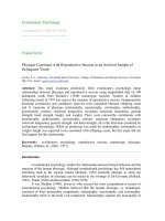
from the study cohort. The results were identical (Figure 1a
and e). As Thermo antibody was not suitable for western
blotting (producer recommendation), only the Invitrogen
antibody was subjected to analysis by western blotting.
The results showed a highly specific bond for COX-2
(Figure 1f).
Almost half of study specimens (44%) were evaluated
independently by two examiners (EP and AS) and kappa
interobserver was 0.73, indicating substantial agreement
(95% CI 0.6-0.9).
Statistical analysis
Associations between variables were examined using
Chi-square test, Fisher’s exact test and Mann-Whitney
test. Continuous variables were reported as median with
corresponding range or interquartile range (IQR). Unadjusted survival analysis was performed using the
Kaplan-Meier method, comparing curves using log-rank
test. Multivariable Cox regression analysis was used for
adjusted survival analysis. Possible interactions were evaluated by inclusion of an interaction term in the models.
For all tests, a two-sided p < 0.05 was considered statistically significant. Statistical analyses were performed in
SPSS 19 for Windows (SPSS Inc., Chicago, IL).
Results
The study cohort consisted of 230 patients consecutively
resected for adenocarcinomas originating from the ampulla (AC) (n = 62, 27%), distal bile duct (DBC) (n = 76,
33%), or pancreas (PC) (n = 92, 40%). Median age at time
of resection was similar for the three groups (67 years,
range 37-83; p = 0.463 Kruskal-Wallis). Overall 5-year
(actual) survival was 5% for PC, 16% for DBC, and 44%
for AC (p < 0.001).
COX-2 expression and prognosis in ampullary, distal bile
duct and pancreatic cancer
COX-2 staining was very similar in all three tumour
types, with a positivity rate of 71% in PC, 72% in DBC,
and 77% in AC. The COX-2 expression was detected in
the cytoplasm of cancer cells in all three types of adenocarcinoma. No COX-2 immunostaining was detected in
the stroma cells (Figure 1a,b, and e). The expression
pattern showed heterogeneity both among different tumours and within the individual tumour, as areas with
moderate to strong staining coexisted with negative
Page 3 of 10
areas within the same tumour (Figure 1c). Islet cells
expressed moderately to strong COX-2 staining in all cases
including those with no COX-2 expression in the tumour
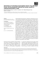
(Figure 1d). Irrespective of tumour origin, overall patient
survival was more favourable in COX-2 positive than
COX-2 negative tumours (Figure 2a-c). This was particularly prominent in AC (p = 0.011) and PC (p = 0.043)
whereas the same tendency was seen in DBC although not
reaching significance (p = 0.06). COX-2 expression varied
according to the type of histological differentiation. In
tumours with pancreatobiliary type of differentiation,
two thirds of the tumours were COX-2 positive irrespective of anatomical origin (67%, 69%, and 68% in AC,
DBC and PC, respectively). However there was no significant difference in overall survival when comparing
COX-2 positive and negative tumours in this group
(Figure 2d-f ). All PC and DBC tumours with intestinal
type of differentiation were COX-2 positive whereas
84% of the intestinal AC tumours expressed COX-2.
The survival data of the intestinal AC tumours showed
a favourable prognosis for patients with tumours expressing COX-2 (p = 0.003) (Figure 2g-i).
Factors associated with prognosis in pancreatic
adenocarcinoma
COX-2 expression status was compared across clinical
parameters associated with survival in the subgroup
consisting of the 92 patients resected for pancreatic
adenocarcinoma. The median survival for patients with
COX-2 positive tumours was 18 months (95% CI 14-22)
as compared to 11 months (95% CI 9.6-12) for patients
with COX-2 negative tumours (p = 0.043). COX-2 positive
tumours were more likely associated with high degree of
differentiation (p = 0.002) and with intestinal type of differentiation, although, the latter did not reach significance
(p = 0.099) (Table 1) probably due to the low number of
tumours of the intestinal differentiation type.
There was no association with COX-2 positivity and
R-status, lymph node ratio (LNR), lymph node status,
tumour diameter, T classification, and vascular or perineural infiltration (Table 1). Since tumours expressing
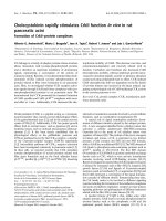
COX-2 were significantly more likely to be highly differentiated than COX-2 negative tumours, the joint effects
of COX-2 status and differentiation grade on survival
were assessed by Kaplan-Meier analysis, stratifying for
COX-2 status (positive vs. negative) and differentiation
grade (grade 1 and 2 vs. grade 3 and 4) (Figure 3a).
Patients whose tumours did not express COX-2 and
had a low differentiation grade (grade 3 and 4) had significantly poorer survival than the other three groups
(p = 0.006).
In a previous report we found that LNR independently
predicted prognosis in a multivariate model for survival
Pomianowska et al. BMC Cancer 2014, 14:458
/>
Page 4 of 10
Figure 1 COX-2 expression in tumour tissue from pancreatic cancer. a-d Double immunostaing with monoclonal anti-COX-2 antibody
(Thermo Fischer Scientific rabbit) and monoclonal anti-αSMA (Dako). COX-2 tumour positive cells (red colour), αSMA positive stromal cells (brown
colour). a magnification × 100, b magnification × 200, c Heterogeneity in COX-2 expression within pancreatic cancer tissue. Areas with moderate
to strong staining (thick arrow) coexist with COX-2 negative areas (thin arrow), (magnification x 100) d Moderately to strong COX-2 staining in islet cells
(thin arrow), pancreatic cancer negative for COX-2 staining, (magnification x 100). e Immunohistochemistry of COX-2 expression in tumour tissue from
pancreatic cancer. Immunostaining with monoclonal COX-2 mouse antibody Invitrogen (the same tumour as in a), magnification x 100. f Western blot
of COX-2 expression in the moderately differentiated pancreatic cancer cell lines BxPC3 and HPAFII known to overexpress COX-2, with and without
induction by interleukin 1 (Il-1), showed a specific bond for COX-2 (70 kDA) (monoclonal COX-2 mouse antibody Invitrogen).
in resected pancreatic cancer [38]. We thus also examined
the joint effects of COX-2 status and LNR, and found that
patients with COX-2 negative tumours and LNR >0.2 had
significantly worst prognosis (p < 0.001) (Figure 3b).
In a multivariate analysis model including COX-2
expression, LNR, tumour size, margin status, vascular
and perineural infiltration, COX-2 negative tumours
and LNR > 0.2 independently predicted poor prognosis
Pomianowska et al. BMC Cancer 2014, 14:458
/>
Page 5 of 10
Figure 2 Overall survival analysis stratified by COX-2 expression. a Ampullary cancer (AC), b Distal bile duct cancer (DBC), c Pancreatic
cancer (PC). d-f Overall survival analysis for AC, DBC and PC with pancreatobiliary differentiation stratified by COX-2 expression. g-i Overall survival
analysis for AC, DBC and PC with intestinal differentiation stratified by COX-2 expression.
Pomianowska et al. BMC Cancer 2014, 14:458
/>
Page 6 of 10
Table 1 Clinicopathological variables in 92 consecutive pancreatoduodenectomies for pancreatic cancer stratified by
COX-2 status
Characteristic
n(%)
COX2-neg. n(%)
COX2-pos. n(%)
pa
COX-2
Positive
65 (71%)
Negative
27 (29%)
Tumour size
≤ 20 mm
15 (16%)
3 (20%)
12 (80%)
> 20 mm
77 (84%)
24 (31%)
53 (69%)
N0, n (%)
25 (27%)
5 (20%)
20 (80%)
N1, n (%)
67 (73%)
22 (33%)
45 (67%)
0.54b
Lymph node metastasis
0.229
Lymph node ratio (LNR)c
≤ 0.2
54 (59%)
13 (24%)
41 (76%)
> 0.2
37 (41%)
13 (36%)
24 (65%)
No, n (%)
30 (33%)
12 (40%)
18(60%)
Yes, n (%)
62 (67%)
15 (24%)
47 (76%)
0.251
Vascular invasion
0.119
Perineural infiltration
No, n (%)
15 (16%)
3 (20%)
12 (80%)
Yes, n (%)
77 (84%)
24 (31%)
53 (69%)
3 (3%)
1 (33%)
2 (67%)
0.54b
T classification
T1
T2
6 (7%)
1 (17%)
5 (83%)
T3
83 (90%)
25 (30%)
58 (70%)
R0, n (%)
40 (44%)
10 (25%)
30 (75%)
R1, n (%)
52 (56%)
17 (33%)
35 (67%)
0.851b
R1 resection status, n (%)
0.422
Degree of differentiation
Grade I, II
53 (58%)
9 (17%)
44 (83%)
Grade III, IV
39 (42%)
18 (46%)
21 (54%)
Pancreaticobiliary, n (%)
84 (91%)
27 (32%)
57 (68%)
Intestinal, n (%)
8 (9%)
0 (0%)
8 (100%)
0.002
Type of differentiation
0.099b
PC, pancreatic adenocarcinoma.
a
Chi-square test, when not otherwise specified.
b
Fisher’s Exact Test.
c
LNR assessment of 91 patients since in one specimen no lymph nodes were retrieved.
(Table 2). Since there was a strong correlation between
COX-2 expression and differentiation grade (p = 0.002)
it was not possible to include differentiation grade in
the same model.
Only a minority of the patients received adjuvant
chemotherapy. Although the numbers are small, there
was no difference in survival between patients with
COX-2 positive and COX-2 negative tumours who received adjuvant treatment.
Discussion
There is a large body of epidemiological, clinical and
molecular evidence suggesting that COX-2 is implicated
in the oncogenesis and progression of gastrointestinal
malignancies, including adenocarcinomas derived from
pancreatic head structures. It has previously been shown
that COX-2 is upregulated in subsets of pancreatic, ampullary and distal bile duct adenocarcinomas although
the proportion of upregulated tumours varies in the
Pomianowska et al. BMC Cancer 2014, 14:458
/>
Page 7 of 10
a
b
Figure 3 Overall survival analysis for patients with pancreatic cancer stratified by COX-2 expression and a degree of differentiation,
b Lymph node ratio (LNR).
different reports. Furthermore, data on the prognostic
importance of COX-2 expression in these tumours is
conflicting. In pancreatic adenocarcinoma, two studies
reported that COX-2 expressing tumours were associated with worse overall prognosis [19,21] whereas other
studies have suggested a trend towards better prognosis
for tumours with high COX-2 expression [22] or no association at all [39-41]. The present data on pancreatic,
distal bile duct and ampullary adenocarcinomas indicates a more favourable overall survival for patients with
COX-2 expressing tumours.
In periampullary and pancreatic head tumours, we
have previously shown that histologic subtyping of these
tumours into intestinal and pancreatobiliary types correlates with cell-type specific markers [36] and prognosis
[3,37]. As COX-2 is thought to be expressed in epithelial
cells throughout the gastrointestinal tract [5,12,42] it
Table 2 Multivariate Cox regression analysis of
histopathologic factors in 92 patients with pancreatic
cancer
p-value
HR
95% CI
R-status (R1vs R0)
0.87
1.038
0.65 - 1.65
Vascular invasion
(Involved vs non- involved)
0.455
1.208
0.74 - 1.98
Perineural infiltration
(Involved vs non- involved)
0.359
1.369
0.70 - 2.68
Tumour size
(> 20 mm vs ≤ 20 mm)
0.315
1.434
0.71 - 2.90
COX-2 expression
(Negative vs Positive)
0.047
1.642
1.01 - 2.68
Lymph node ratio
(LNR) (> 0.2 vs ≤ 0. 2)
0.032
1.757
1.05 - 2.94
was of particular interest to examine whether there are
differences in COX-2 expression in the intestinal and
pancreatobiliary subtypes. Of note, most intestinal ampullary tumours (84%) were COX-2 positive, and in particular, all intestinal pancreatic and distal bile duct tumours
were COX-2 positive. Patients with ampullary cancers of
the intestinal subtype, which expressed COX-2, had a
favourable prognosis with a 5-year actual survival of 60%.
Histopathologic type of differentiation combined with biomarkers or gene expression profiles has recently attracted
interest as important factors for outcome as well as stratification for adjuvant chemotherapy in ampullary adenocarcinoma [43,44].
The finding in the present study that COX-2 expression correlates with a favourable prognosis in pancreatic
cancer can be explained by the fact that there is a statistically significant association between COX-2 positivity
and high degree of differentiation. More than 80% of tumours with high differentiation grade showed overexpression of COX-2. This result is consistent with previous
observations from studies of cultured pancreatic cancer
cells and pancreatic cancer tissue. In cultured tumour cells
COX-2 expression was found to be restricted to moderately and highly differentiated pancreatic cancer cell lines
[23,26,45]. In human pancreatic adenocarcinoma tissue,
well differentiated lesions expressed COX-2 to the highest
degree, whereas there was less expression of COX-2 in
moderately and poorly differentiated lesions [30]. In our
study, the subgroup of patients with COX-2 positive/well
differentiated tumours had a significantly better survival
compared to patients with COX-2 negative/poorly differentiated tumours, whereas COX-2 positive/poor differentiation and COX-2 negative/high differentiation formed
Pomianowska et al. BMC Cancer 2014, 14:458
/>
an intermediate group with respect to survival. Thus the
presence of COX-2 expression in these tumours appears
to be a marker of favourable prognosis closely linked to
the degree of tumour differentiation. Consistent with the
latter the strong statistical association between COX-2
expression and differentiation grade precluded inclusion
of both variables in the same multivariable model for
survival.
The precise function of COX-2 in pancreatic cancer
development is not known. In the normal pancreas, only
islet cells always express COX-2 [24]. In transgenic mice
models, overexpression of COX-2 in normal pancreatic
ductal cells results in development of dysplastic changes
resembling IPMNs and PanINs [46,47] suggesting a primary role of pancreatic cell COX-2 overexpression in
the initiation of ductal adenocarcinoma. Recent evidence
suggests that this is an intrinsic role of pancreatic cells
independent of prostaglandins from the tumour microenvironment [48]. These observations support the concept that COX-2 overexpression might be a causal factor
in pancreatic cancer development. It has also been suggested that pancreatic cancers that lack COX-2 (and
COX-1) depends on exogenic prostaglandins from stromal
fibroblasts for proliferation and other cancer-promoting
effects [49]. Since COX-2 overexpression is implicated in
tumour development, its expression in pancreatic cancer
was hypothesized to result in a poor patient prognosis
[19]. This hypothesis is difficult to reconcile with the observation that in fully developed tumours, COX-2 expression has been shown to be a function of differentiation
status, with highest expression in well differentiated
tumours [30]. In addition, it has been demonstrated
that COX-2 expression varies markedly throughout the
pathological process of pancreatic neoplasia. COX-2
expression increases in a stepwise manner with each
initial stage of neoplastic progression up to the PanIN
2 stage, whereas COX-2 expression was relatively lower in
invasive cancers [30].
Some of the discrepancies in results between our study
and the studies by Juuti et al [19] and Matsubayashi et al
[21] might be explained by methodological differences in
patient sampling and/or tumour immunohistochemistry
techniques. Since it is well known that it can be difficult
to determine the precise anatomical origin of tumours of
the pancreatic head, all cancers in the present series
were re-evaluated for correct sub-classification into ampullary, distal bile duct or pancreatic tumours. There are
also certain differences pertaining to the immunohistochemistry protocols that differ in our study compared to
the studies by Juuti et al [19] and Matsubayashi et al
[21]. In the work of Juuti, more than 30 years old specimens were included in the study cohort. It is known that
for immunohistochemical staining protocols aging of
fixed tumour tissue might interfere with staining [50].
Page 8 of 10
Not only aging of the waxed specimen itself, but also
variations in fixation protocols over time may result in
inadequate staining. This may partly explain the low frequency of COX-2 staining (36%) in their data, compared
to 55-80% in most other reports [20,22,23,25,26,41,51].
Since COX-2 expression in pancreatic tumours often is
heterogeneous [24,29,30], the actual number of COX-2
positive tumours might be underestimated unless immunohistochemistry is performed on whole slide sections
and assessed on multiple different high-power fields within
each tumour. In the study of Matsubayashi [21], assessment
of COX-2 staining was performed on tissue microarrays.
Although this method has many advantages, tissue microarrays might not be the optimal method for assessment
of COX-2 staining even if two cores of tumour tissues
were studied from each tumour. This may partly explain the lower proportion of tumours expressing
COX-2 in some studies [19,21] and hence the differences in patient survival.
Conclusion
COX-2 is overexpressed in pancreatic cancer, ampullary
cancer and distal bile duct cancer and confers a survival
benefit in all three cancer types. The overexpression is
consistently linked to the histopathological type of differentiation and to the degree of differentiation. In pancreatic
adenocarcinoma, COX-2 overexpression independently
predicts a favourable prognosis.
Competing interests
The authors declare that they have no competing interests.
Authors’ contributions
EP, OPC, IPG conceived and planned the study. EP and IPG conducted
acquisition of data. EP, ARS, and OPC performed immunohistochemistry. EP,
ARS, OPC and IPG analysed and discussed the results. EP and IPG drafted the
manuscript. All authors critically revised and approved of the final
manuscript.
Acknowledgements
The authors gratefully acknowledge Maria Einarsen Pretorius for technical
assistance with scanning of slides, and Knut Jørgan Labori for supplying
unpublished data on adjuvant chemotherapy. We thank Dagny Sandnes,
Vegard Tjomsland and Arne Westgaard for helpful discussions.
Author details
1
Institute of Clinical Medicine, Faculty of Medicine, University of Oslo, Oslo,
Norway. 2Department of Hepato-pancreato-biliary Surgery, Oslo University
Hospital, Rikshospitalet, PO Box 4950, Nydalen, 0424 Oslo, Norway.
3
Department of Pathology, Oslo University Hospital, Rikshospitalet, Oslo,
Norway.
Received: 3 October 2013 Accepted: 11 June 2014
Published: 20 June 2014
References
1. Pomianowska E, Grzyb K, Westgaard A, Clausen OP, Gladhaug IP:
Reclassification of tumour origin in resected periampullary
adenocarcinomas reveals underestimation of distal bile duct cancer.
Eur J Surg Oncol 2012, 38:1043–1050.
2. Verbeke CS, Gladhaug IP: Resection margin involvement and tumour
origin in pancreatic head cancer. Br J Surg 2012, 99:1036–1049.
Pomianowska et al. BMC Cancer 2014, 14:458
/>
3.
4.
5.
6.
7.
8.
9.
10.
11.
12.
13.
14.
15.
16.
17.
18.
19.
20.
21.
22.
23.
24.
25.
Westgaard A, Pomianowska E, Clausen OP, Gladhaug IP: Intestinal-type and
pancreatobiliary-type adenocarcinomas: how does ampullary carcinoma
differ from other periampullary malignancies? Ann Surg Oncol 2013,
20:430–439.
Kawai N, Tsujii M, Tsuji S: Cyclooxygenases and colon cancer. Prostaglandins
Other Lipid Mediat 2002, 68–69:187–196.
Sano H, Kawahito Y, Wilder RL, Hashiramoto A, Mukai S, Asai K, Kimura S,
Kato H, Kondo M, Hla T: Expression of cyclooxygenase-1 and −2 in human
colorectal cancer. Cancer Res 1995, 55:3785–3789.
Saukkonen K, Nieminen O, Van RB, Vilkki S, Harkonen M, Juhola M, Mecklin JP,
Sipponen P, Ristimaki A: Expression of cyclooxygenase-2 in dysplasia of the
stomach and in intestinal-type gastric adenocarcinoma. Clin Cancer Res
2001, 7:1923–1931.
Ristimaki A, Sivula A, Lundin J, Lundin M, Salminen T, Haglund C, Joensuu H,
Isola J: Prognostic significance of elevated cyclooxygenase-2 expression
in breast cancer. Cancer Res 2002, 62:632–635.
Hu M, Peluffo G, Chen H, Gelman R, Schnitt S, Polyak K: Role of COX-2 in
epithelial-stromal cell interactions and progression of ductal carcinoma
in situ of the breast. Proc Natl Acad Sci U S A 2009, 106:3372–3377.
Laga AC, Zander DS, Cagle PT: Prognostic significance of cyclooxygenase
2 expression in 259 cases of non-small cell lung cancer. Arch Pathol Lab
Med 2005, 129:1113–1117.
Sweeney CJ, Marshall MS, Barnard DS, Heilman DK, Billings SD, Cheng L,
Marshall SJ, Yip-Schneider MT: Cyclo-oxygenase-2 expression in primary
cancers of the lung and bladder compared to normal adjacent tissue.
Cancer Detect Prev 2002, 26:238–244.
Ristimaki A, Nieminen O, Saukkonen K, Hotakainen K, Nordling S, Haglund C:
Expression of cyclooxygenase-2 in human transitional cell carcinoma of
the urinary bladder. Am J Pathol 2001, 158:849–853.
Wang D, Mann JR, Dubois RN: The role of prostaglandins and other
eicosanoids in the gastrointestinal tract. Gastroenterology 2005, 128:1445–1461.
Wang D, Dubois RN: Eicosanoids and cancer. Nat Rev Cancer 2010, 10:181–193.
Wang D, Dubois RN: The role of COX-2 in intestinal inflammation and
colorectal cancer. Oncogene 2010, 29:781–788.
Sahin IH, Hassan MM, Garrett CR: Impact of non-steroidal anti-inflammatory
drugs on gastrointestinal cancers: current state-of-the science. Cancer Lett
2013, 345:249–257.
Cheng J, Fan XM: Role of cyclooxygenase-2 in gastric cancer development
and progression. World J Gastroenterol 2013, 19:7361–7368.
Dubois RN, Abramson SB, Crofford L, Gupta RA, Simon LS, Van De Putte LB,
Lipsky PE: Cyclooxygenase in biology and disease. FASEB J 1998, 12:1063–1073.
Allaj V, Guo C, Nie D: Non-steroid anti-inflammatory drugs, prostaglandins,
and cancer. Cell Biosci 2013, 3:8.
Juuti A, Louhimo J, Nordling S, Ristimaki A, Haglund C: Cyclooxygenase-2
expression correlates with poor prognosis in pancreatic cancer. J Clin
Pathol 2006, 59:382–386.
Kokawa A, Kondo H, Gotoda T, Ono H, Saito D, Nakadaira S, Kosuge T,
Yoshida S: Increased expression of cyclooxygenase-2 in human
pancreatic neoplasms and potential for chemoprevention by cyclooxygenase
inhibitors. Cancer 2001, 91:333–338.
Matsubayashi H, Infante JR, Winter J, Klein AP, Schulick R, Hruban R,
Visvanathan K, Goggins M: Tumor COX-2 expression and prognosis of
patients with resectable pancreatic cancer. Cancer Biol Ther 2007,
6:1569–1575.
Merati K, Said SM, Andea A, Sarkar F, Ben-Josef E, Mohammad R,
Philip P, Shields AF, Vaitkevicius V, Grignon DJ, Adsay NV: Expression
of inflammatory modulator COX-2 in pancreatic ductal adenocarcinoma
and its relationship to pathologic and clinical parameters. Am J Clin Oncol
2001, 24:447–452.
Molina MA, Sitja-Arnau M, Lemoine MG, Frazier ML, Sinicrope FA: Increased
cyclooxygenase-2 expression in human pancreatic carcinomas and cell
lines: growth inhibition by nonsteroidal anti-inflammatory drugs. Cancer
Res 1999, 59:4356–4362.
Okami J, Yamamoto H, Fujiwara Y, Tsujie M, Kondo M, Noura S, Oshima S,
Nagano H, Dono K, Umeshita K, Ishikawa O, Sakon M, Matsuura N, Nakamori
S, Monden M: Overexpression of cyclooxygenase-2 in carcinoma of the
pancreas. Clin Cancer Res 1999, 5:2018–2024.
Tucker ON, Dannenberg AJ, Yang EK, Zhang F, Teng L, Daly JM, Soslow RA,
Masferrer JL, Woerner BM, Koki AT, Fahey TJ III: Cyclooxygenase-2
expression is up-regulated in human pancreatic cancer. Cancer Res 1999,
59:987–990.
Page 9 of 10
26. Yip-Schneider MT, Barnard DS, Billings SD, Cheng L, Heilman DK, Lin A,
Marshall SJ, Crowell PL, Marshall MS, Sweeney CJ: Cyclooxygenase-2
expression in human pancreatic adenocarcinomas. Carcinogenesis 2000,
21:139–146.
27. Albazaz R, Verbeke CS, Rahman SH, McMahon MJ: Cyclooxygenase-2
expression associated with severity of PanIN lesions: a possible link
between chronic pancreatitis and pancreatic cancer. Pancreatology 2005,
547:361–369.
28. Hermanova M, Trna J, Nenutil R, Dite P, Kala Z: Expression of COX-2 is
associated with accumulation of p53 in pancreatic cancer: analysis of
COX-2 and p53 expression in premalignant and malignant ductal pancreatic
lesions. Eur J Gastroenterol Hepatol 2008, 20:732–739.
29. Maitra A, Ashfaq R, Gunn CR, Rahman A, Yeo CJ, Sohn TA, Cameron JL,
Hruban RH, Wilentz RE: Cyclooxygenase 2 expression in pancreatic
adenocarcinoma and pancreatic intraepithelial neoplasia: an
immunohistochemical analysis with automated cellular imaging. Am J
Clin Pathol 2002, 118:194–201.
30. Crowell PL, Schmidt CM, Yip-Schneider MT, Savage JJ, Hertzler DA,
Cummings WO: Cyclooxygenase-2 expression in hamster and human
pancreatic neoplasia. Neoplasia 2006, 8:437–445.
31. Perrone G, Santini D, Zagami M, Vincenzi B, Verzi A, Morini S, Borzomati D,
Coppola R, Antinori A, Magistrelli P, Tonini G, Rabitti C: COX-2 expression of
ampullary carcinoma: correlation with different histotypes and
clinicopathological parameters. Virchows Arch 2006, 449:334–340.
32. Santini D, Vincenzi B, Tonini G, Scarpa S, Vasaturo F, Malacrino C,
Vecchio F, Borzomati D, Valeri S, Coppola R, Magistrelli P, Nuzzo G,
Picciocchi A: Cyclooxygenase-2 overexpression is associated with a
poor outcome in resected ampullary cancer patients. Clin Cancer Res
2005, 11:3784–3789.
33. Kim HJ, Sohn TS, Lee KT, Lee JK, Paik SW, Rhee JC: Expression of
cyclooxygenase-2 and its correlation with clinicopathologic factors of
ampulla of vater cancer. J Korean Med Sci 2003, 18:218–224.
34. Kim HJ, Lee KT, Kim EK, Sohn TS, Heo JS, Choi SH, Choi DI, Lee JK, Paik SW,
Rhee JC: Expression of cyclooxygenase-2 in cholangiocarcinoma: correlation
with clinicopathological features and prognosis. J Gastroenterol Hepatol
2004, 19:582–588.
35. Westgaard A, Tafjord S, Farstad IN, Cvancarova M, Eide TJ, Mathisen O,
Clausen OP, Gladhaug IP: Resectable adenocarcinomas in the pancreatic
head: the retroperitoneal resection margin is an independent prognostic
factor. BMC Cancer 2008, 8:5.
36. Westgaard A, Schjolberg AR, Cvancarova M, Eide TJ, Clausen OP, Gladhaug
IP: Differentiation markers in pancreatic head adenocarcinomas: MUC1
and MUC4 expression indicates poor prognosis in pancreatobiliary
differentiated tumours. Histopathology 2009, 54:337–347.
37. Westgaard A, Tafjord S, Farstad IN, Cvancarova M, Eide TJ, Mathisen O,
Clausen OP, Gladhaug IP: Pancreatobiliary versus intestinal histologic type
of differentiation is an independent prognostic factor in resected
periampullary adenocarcinoma. BMC Cancer 2008, 8:170.
38. Pomianowska E, Westgaard A, Mathisen O, Clausen OP, Gladhaug IP:
Prognostic relevance of number and ratio of metastatic lymph nodes in
resected pancreatic, ampullary, and distal bile duct carcinomas. Ann Surg
Oncol 2013, 20:233–241.
39. Kong G, Kim EK, Kim WS, Lee KT, Lee YW, Lee JK, Paik SW, Rhee JC: Role of
cyclooxygenase-2 and inducible nitric oxide synthase in pancreatic
cancer. J Gastroenterol Hepatol 2002, 17:914–921.
40. Aprile G, Avellini C, Reni M, Mazzer M, Foltran L, Rossi D, Cereda S, Iaiza E,
Fasola G, Piga A: Biglycan expression and clinical outcome in patients
with pancreatic adenocarcinoma. Tumour Biol 2013, 34:131–137.
41. Hermanova M, Karasek P, Tomasek J, Lenz J, Jarkovsky J, Dite P: Comparative
analysis of clinicopathological correlations of cyclooxygenase-2
expression in resectable pancreatic cancer. World J Gastroenterol 2010,
16:1879–1884.
42. Warner TD, Mitchell JA: Cyclooxygenases: new forms, new inhibitors, and
lessons from the clinic. FASEB J 2004, 18:790–804.
43. Chang DK, Jamieson NB, Johns AL, Scarlett CJ, Pajic M, Chou A, Pinese M,
Humphris JL, Jones MD, Toon C, Nagrial AM, Chantrill LA, Chin VT, Pinho AV,
Rooman I, Cowley MJ, Wu J, Mead RS, Colvin EK, Samra JS, Corbo V, Bassi C,
Falconi M, Lawlor RT, Crippa S, Sperandio N, Bersani S, Dickson EJ,
Mohamed MA, Oien KA: Histomolecular phenotypes and outcome in
adenocarcinoma of the ampulla of vater.
J Clin Oncol 2013, 31:1348–1356.
Pomianowska et al. BMC Cancer 2014, 14:458
/>
Page 10 of 10
44. Overman MJ, Zhang J, Kopetz S, Davies M, Zhi-Qin J, Stemke-Hale K,
Rummele P, Pilarsky C, Grutzmann R, Hamilton S, Hwang R, Abbruzzese JL,
Varadhachary G, Broom B, Wang H: Gene expression profiling of ampullary
carcinomas classifies ampullary carcinomas into biliary-like and
intestinal-like subtypes that are prognostic of outcome. PLoS ONE 2013,
8:e65144.
45. Eibl G, Bruemmer D, Okada Y, Duffy JP, Law RE, Reber HA, Hines OJ: PGE(2)
is generated by specific COX-2 activity and increases VEGF production in
COX-2-expressing human pancreatic cancer cells. Biochem Biophys Res
Commun 2003, 306:887–897.
46. Colby JK, Klein RD, McArthur MJ, Conti CJ, Kiguchi K, Kawamoto T, Riggs PK,
Pavone AI, Sawicki J, Fischer SM: Progressive metaplastic and dysplastic
changes in mouse pancreas induced by cyclooxygenase-2 overexpression.
Neoplasia 2008, 10:782–796.
47. Muller-Decker K, Furstenberger G, Annan N, Kucher D, Pohl-Arnold A,
Steinbauer B, Esposito I, Chiblak S, Friess H, Schirmacher P, Berger I:
Preinvasive duct-derived neoplasms in pancreas of keratin 5-promoter
cyclooxygenase-2 transgenic mice. Gastroenterology 2006, 130:2165–2178.
48. Hill R, Li Y, Tran LM, Dry S, Calvopina JH, Garcia A, Kim C, Wang Y, Donahue TR,
Herschman HR, Wu H: Cell intrinsic role of COX-2 in pancreatic cancer
development. Mol Cancer Ther 2012, 11:2127–2137.
49. Omura N, Griffith M, Vincent A, Li A, Hong SM, Walter K, Borges M, Goggins M:
Cyclooxygenase-deficient pancreatic cancer cells use exogenous sources of
prostaglandins. Mol Cancer Res 2010, 8:821–832.
50. Walker RA: Quantification of immunohistochemistry–issues concerning
methods, utility and semiquantitative assessment I. Histopathology 2006,
49:406–410.
51. Koshiba T, Hosotani R, Miyamoto Y, Wada M, Lee JU, Fujimoto K, Tsuji S,
Nakajima S, Doi R, Imamura M: Immunohistochemical analysis of
cyclooxygenase-2 expression in pancreatic tumors. Int J Pancreatol 1999,
26:69–76.
doi:10.1186/1471-2407-14-458
Cite this article as: Pomianowska et al.: COX-2 overexpression in resected
pancreatic head adenocarcinomas correlates with favourable prognosis.
BMC Cancer 2014 14:458.
Submit your next manuscript to BioMed Central
and take full advantage of:
• Convenient online submission
• Thorough peer review
• No space constraints or color figure charges
• Immediate publication on acceptance
• Inclusion in PubMed, CAS, Scopus and Google Scholar
• Research which is freely available for redistribution
Submit your manuscript at
www.biomedcentral.com/submit