Stability of the CpG island methylator phenotype during glioma progression and identification of methylated loci in secondary glioblastomas
Bạn đang xem bản rút gọn của tài liệu. Xem và tải ngay bản đầy đủ của tài liệu tại đây (1.24 MB, 12 trang )
Hill et al. BMC Cancer 2014, 14:506
/>
RESEARCH ARTICLE
Open Access
Stability of the CpG island methylator phenotype
during glioma progression and identification of
methylated loci in secondary glioblastomas
Victoria K Hill1, Thoraia Shinawi1, Christopher J Ricketts1, Dietmar Krex2, Gabriele Schackert2, Julien Bauer3,
Wenbin Wei4, Garth Cruickshank5, Eamonn R Maher1 and Farida Latif1*
Abstract
Background: Grade IV glioblastomas exist in two forms, primary (de novo) glioblastomas (pGBM) that arise without
precursor lesions, and the less common secondary glioblastomas (sGBM) which develop from earlier lower grade
lesions. Genetic heterogeneity between pGBM and sGBM has been documented as have differences in the
methylation of individual genes. A hypermethylator phenotype in grade IV GBMs is now well documented however
there has been little comparison between global methylation profiles of pGBM and sGBM samples or of
methylation profiles between paired early and late sGBM samples.
Methods: We performed genome-wide methylation profiling of 20 matched pairs of early and late gliomas using
the Infinium HumanMethylation450 BeadChips to assess methylation at >485,000 cytosine positions within the
human genome.
Results: Clustering of our data demonstrated a frequent hypermethylator phenotype that associated with IDH1
mutation in sGBM tumors. In 80% of cases, the hypermethylator status was retained in both the early and late tumor of
the same patient, indicating limited alterations to genome-wide methylation during progression and that the CIMP
phenotype is an early event. Analysis of hypermethylated loci identified 218 genes frequently methylated across grade
II, III and IV tumors indicating a possible role in sGBM tumorigenesis. Comparison of our sGBM data with TCGA pGBM
data indicate that IDH1 mutated GBM samples have very similar hypermethylator phenotypes, however the methylation
profiles of the majority of samples with WT IDH1 that do not demonstrate a hypermethylator phenotype cluster
separately from sGBM samples, indicating underlying differences in methylation profiles. We also identified 180 genes
that were methylated only in sGBM. Further analysis of these genes may lead to a better understanding of the
pathology of sGBM vs pGBM.
Conclusion: This is the first study to have documented genome-wide methylation changes within paired early/late
astrocytic gliomas on such a large CpG probe set, revealing a number of genes that maybe relevant to secondary
gliomagenesis.
Keywords: Primary and secondary glioblastoma (pGBM, sGBM), HumanMethylation450, Methylation, IDH1, CIMP
* Correspondence:
1
Centre for Rare Diseases and Personalised Medicine and Department of
Medical & Molecular Genetics, School of Clinical and Experimental Medicine,
University of Birmingham College of Medical and Dental Sciences,
Edgbaston, Birmingham, UK
Full list of author information is available at the end of the article
© 2014 Hill et al.; licensee BioMed Central Ltd. This is an Open Access article distributed under the terms of the Creative
Commons Attribution License ( which permits unrestricted use, distribution, and
reproduction in any medium, provided the original work is properly credited. The Creative Commons Public Domain
Dedication waiver ( applies to the data made available in this article,
unless otherwise stated.
Hill et al. BMC Cancer 2014, 14:506
/>
Background
Gliomas are classified into 4 grades according to the
WHO classification system. These range from curable
World Health Organization (WHO) grade I tumors
(pilocytic astrocytomas) to the highly malignant WHO
grade IV glioblastoma (GBM) with mean survival <
1year. In between these two grades are WHO grade III
malignant tumors (anaplastic astrocytomas) with median
survival rates of 2–3 years after diagnosis and WHO
grade II (diffuse astrocytomas) considered as low grade
gliomas with median survival rates of 6–8 years after
diagnosis [1,2]. Glioblastomas are subdivided into 2 distinct types, primary grade IV glioblastoma (pGBM or de
novo glioblastomas) that account for >90% of the cases,
usually affecting older patients and develop rapidly after
a short clinical history and without evidence of a less
malignant precursor lesion. While secondary glioblastomas (sGBM) develop slowly through progression from
lower grade diffuse or anaplastic astrocytomas and more
commonly occur in younger patients. pGBM and sGBM
represent not only clinically distinct entities but also
demonstrate distinct genetic heterogeneity. For example,
pGBM demonstrate mutation of the PTEN gene and frequent loss of heterozygosity on chromosome 10q (inclusive of the PTEN gene locus), amplification of EGFR,
deletions of CDKN2A (p16), while sGBM and their lower
grade precursor lesions have frequent mutations of the
TP53 gene and the IDH1 gene [3-7]. Recent studies have
also looked at genetic alterations in early and late paired
secondary samples [8].
In recent years large scale genome-wide epigenetic
studies have been performed with the aim of developing
clinically relevant biomarkers for glioblastoma [9-11]. A
good example is the epigenetic silencing of the MGMT
promoter that has provided an exciting and clinically
relevant epigenetic marker in gliomas. The MGMT gene
encodes for an O-6-methylguanine methyltransferase
that removes alkyl groups from the O-6 position of
guanine. Thus loss of its activity greatly impairs a cells
ability to tolerate alkylating agents and studies have
shown that MGMT-promoter methylation is associated
with longer survival of patients treated with alkylating
agents such as temozolomide [12,13]. Recently, the Cancer Genome Atlas (TCGA) research network identified
a CpG island methylator phenotype (CIMP) in a subset
of human gliomas with distinct clinical and molecular
features, including improved survival outcomes for
those gliomas demonstrating CIMP [10]. The gain of
function mutations within the isocitrate dehydrogenase
1 gene (IDH1) are thought to be largely responsible for
the glioma hypermethylator phenotype due to the massively increased production of the 2-hydroxyglutarate
oncometabolite and have recently been shown to be sufficient to result in a hypermethylator phenotype in
Page 2 of 12
glioma cell lines [14,15]. At least some individual genes
have demonstrated differential methylation frequencies
in grade IV pGBM and sGBM samples [16] and although much progress has been made in assessing
genome-wide methylation of pGBM tumors, much less
is known about genome-wide methylation in early grade
tumors and their subsequent higher grade sGBM
manifestations.
Recent technological advances have made it possible
to quantitatively assess genome-wide methylation at the
individual CpG loci level using the Illumina Infinium
BeadChips. The most recent version of this BeadChip
(Infinium HumanMethylation450 BeadChip) is able to
quantitatively assess the levels of methylation at specific
CpG loci throughout the genome, including CpG
islands and regions of much lower CpG dinucleotide
density. In this report we utilized these comprehensive
Infinium HumanMethylation450 BeadChip arrays to define genome-wide methylation in paired samples of
early/late astrocytic gliomas and to demonstrate any alterations induced by progression.
Methods
DNA samples
Forty DNA samples from 20 astrocytoma/glioma patients
were used in this study. These patient samples consisted
of; 10 WHO grade II astrocytomas, 15 WHO grade III astrocytomas and 15 WHO grade IV glioblastomas. The 40
DNA samples represent 20 cases of paired early and late
lesions from the same patient. The DNA was extracted
from tissue samples consisting of a minimum of 80%
tumor. The DNA from four non-disease brain samples
was used to provide the normal, expected levels of methylation. Ethical guidelines were followed for patient sample
collection and all samples have been anonymised. Research was conducted according to the principles
expressed in the Declaration of Helsinki. Patients gave
written informed consent for analysis of tumor samples.
The study was approved by the Institutional Ethics Committees of University of Technology Dresden and University of Birmingham.
Illumina array
The Illumina Infinium HumanMethylation450 array
(Illumina, San Diego, CA, USA) was performed on 0.5
μg bisulfite modified patient DNA according to manufacturers’ instructions. Bisulfite modification of DNA
and array hybridization was carried out by Cambridge
Genomics Services. Raw data was obtained using Genome Studio software from Illumina. The raw data were
processed using the lumi R [17] package to correct for
the color bias present due to the use of different dye on
the array. To correct this bias, Infinium type I and type
II are separated, then both channel are also separated
Hill et al. BMC Cancer 2014, 14:506
/>
and the color bias is corrected using a within array
smooth quantile normalization. After correction the
two channels and probe types are combined and a between array quantile normalization is performed. The
beta score are then calculated. The raw files have been
deposited in NCBI’s Gene Expression Omnibus [18] and
are accessible through GEO Series accession number
GSE58298.
Probes demonstrating detection p-values greater than
0.01 in any sample were removed along with probes located on the X and Y chromosomes. To ensure tumor
specific hypermethylation, probes showing a beta value
≥0.25 in any of the four normal samples were also removed. Hypermethylation was subsequently determined
as a beta value ≥0.5. This was considered relevant if
present in >30% tumor samples. Additional filtering was
achieved limiting selection to genes for which the
hypermethylation criteria were met in ≥3 probes associated to that gene.
Page 3 of 12
as a percentage of the number of methylated CpGs out of
the total number of CpGs sequenced.
IDH1 and IDH2 mutation status
Previously described primers were used to amplify 129 bp
and 150 bp fragments of the IDH1 and IDH2 genes [19].
The IDH1 forward primer 5′-CTCCTGATGAGAA
GAGGGTTG-3′ and IDH1 reverse primer 5′-TGGAAA
TTTCTGGGCCATG-3′ were used to sequence codon
132 and the IDH2 forward primer 5′-TGGAACTATCCG
GAACATCC-3′ and IDH2 reverse primer 5′-AGTCTG
TGGCCTTGTACTGC-3 were used to sequence codon
172 of IDH2. Twenty nanograms of genomic DNA were
used as starting material for a 25 μl total volume PCR
reaction using Go Taq polymerase. An annealing
temperature of 58°C was used for 35 cycles. PCR products
were bi-directionally sequenced using cycle sequencing on
an ABI 3730x (Applied Biosystems, Carlsbad, CA, USA).
TCGA samples
Clone sequencing
Illumina Infinium HumanMethylation450 BeadChip
array data was used for the following 19 TCGA primary
glioblastomas: TCGA-06-5416, TCGA-06-0171, TCGA-265136, TCGA-06-0190, TCGA-06-5418, TCGA-06-0210,
TCGA-26-5135, TCGA-26-5134, TCGA-26-5132, TCG
A-12-5295, TCGA-06-5414, TCGA-06-0211, TCGA-265133, TCGA-06-5417, TCGA-06-0221, TCGA-26-1442,
TCGA-06-6389, TCGA-06-6701, TCGA-15-1444. All
array data was downloaded from the TCGA Data Portal
( />IDH1 and IDH2 mutation status for these tumors was
identified using the cBioPortal for Cancer Genomics
( />
Clone sequencing was used for array validation. 0.5 μg of
DNA for each sample was bisulfite modified using the
Qiagen EpiTect kit (Qiagen, Heidelberg, Germany) according to manufacturers’ instructions. PCR reactions
were performed using FastStart Taq DNA polymerase
(Roche, West Sussex, UK) on a semi-nested basis for all
genes using the primers listed in Additional file 1. A
touchdown PCR program for primary and secondary reactions using gene specific annealing temperatures was performed. Selected PCR products were cloned into the
pGEM-T easy vector (Promega, Madison, WI, USA) according to manufacturers’ instructions and cultured overnight at 37°C. Up to 12 colonies were selected for single
colony PCR using primer sequences F: 5′- TAATAC
GACTCACTATAGGG -3′ and R: 5′- ACACTATAGA
ATACTCAAGC -3′. PCR products were cleaned for
sequencing using thermosensitive alkaline phosphatase
(Fermentas UK, York, UK) and Exonuclease I (NEB,
Ipswich, MA, USA) and then sequenced using cycle
sequencing on an ABI 3730 (Applied Biosystems,
Carlsbad, CA, USA). Methylation indexes were calculated
Results
To determine whether aberrant DNA methylation differs
between early and late secondary glioma lesions we have
used the new Illumina Infinium HumanMethylation450
BeadChip array on 40 astrocytic secondary glioma tumors, consisting of 20 pairs of early and late lesions for
individual patients and four normal brain samples. Of
the 20 patient paired samples; 5 pairs are WHO grade II
astrocytomas progressing to grade III astrocytomas, 5
pairs are WHO grade II astrocytomas progressing to
WHO grade IV glioblastomas, and 10 pairs are grade III
astrocytomas progressing to grade IV glioblastomas. In
order to adjust for potential bias based on the differences in probe design between Illumina Type I/II probes
we ran all raw data through a correction pipeline prior
to analysis. In addition, these samples had been assessed
for IDH1 and IDH2 mutation status, 14 out of 20 (70%)
samples demonstrated mutation in the IDH1 R132
codon. No IDH2 mutations were detected (Additional
file 2: Table S1).
Clustering
The top 2000 most variable loci for each clustering
event were determined by selecting the 2000 probes
with the greatest standard deviation across all the given
samples. Clustering was performed using the Cluster3
program ( />software.htm#ctv) and visualized using the Java TreeView
program ( Unsupervised
hierarchical clustering was performed using the Euclidean
based algorithm.
Hill et al. BMC Cancer 2014, 14:506
/>
CIMP is an early event in secondary gliomagenesis that
can be retained throughout progression
Unsupervised clustering of the 2000 most variable loci
in all 40 samples plus normal controls produces two
major clusters: major cluster 1 (n = 20 samples; mean
beta value = 0.21) and major cluster 2 (n = 24 samples;
mean beta value = 0.60) (p < 0.001; ANOVA) (Figure 1a,
b). Each major cluster can be further sub-divided into 2
sub-clusters: sub-clusters 1a and 1b (n = 13 and n = 7
samples respectively; mean beta values 0.14 and 0.34 respectively) and sub-clusters 2a and 2b (n = 12 samples in
each cluster; mean beta values 0.50 and 0.69 respectively) (p < 0.001; ANOVA). Mean beta values for samples
within each sub-cluster differ significantly in all comparisons (p < 0.05; ANOVA) (Figure 1b). Samples within
major cluster 2 demonstrate a high level of methylation
throughout the most variable 2000 loci indicating the
CpG island methylator phenotype (CIMP) and these
samples were designated CIMP+ve with all but one sample (P19E) demonstrating an IDH1 mutation (Figure 1).
Within our most variable 2000 loci were probes for
genes previously associated with a CIMP phenotype in
GBM [10]. Samples in major cluster 1 appear to be
negative for the CIMP phenotype and were designated
CIMP–ve with sub-cluster 1a appearing to be notably
normal-like, including all the control normal samples,
whilst sub-cluster 1b has low level methylation. Interestingly, major cluster 1 included several IDH1 mutation
positive samples as well as all the IDH1 mutation negative samples except for P19E (Figure 1a). In general,
IDH1 mutation negative samples (P2, P3, P4 and P9)
demonstrated very similar methylation patterns between
early and late grades (Figure 1a). For all but one (P16) of
the IDH1 positive samples, the lower grade sample demonstrated distinct CIMP and this is suggestive that it is a
very early event in secondary gliomagenesis. In addition,
no sample gained CIMP during progression suggesting
that it occurs early on or not at all. In progression to the
later grades the IDH1 positive sample split into two categories; those samples that retain a very similar methylation profile after progression (P5, P8, P10, P11, P15,
P17) and those that demonstrate a partially remaining
CIMP+ve between early and late lesions or greatly reduced (becoming CIMP-ve) degree of methylation after
progression (P1, P7, P12, P13, P14, P18, P20) (Figure 1a).
Thus, in total, progression through to higher grades had
little effect on the genome-wide methylation for 10 of
the 20 pairs (50%) and no effect on CIMP status for 16
of the 20 pairs (80%). The P19 sample acted as though
an IDH mutation was present and that it fell into the
second category of IDH1 mutation positive samples. Although the sample was negative for IDH1 or IDH2 mutation it could possibly have another mutation capable
of causing a similar effect, such as a TET2 mutation, that
Page 4 of 12
was not assessed for. The IDH1 positive P16 sample
acted more like an IDH1 negative sample for unknown
reasons and was retained for further analysis.
Identification of hypermethylated loci dependent upon
glioma grade
To initially discern a list of differentially methylated loci
between normal and tumor samples we first split the
samples into grade II, III and grade IV groups and identified hypermethylated loci within each group. Following
removal of all probes showing a β-value ≥0.25 in any of
the four normal samples, the remaining probes were
considered hypermethylated if >30% of tumor samples
showed a β-value of ≥0.5. When using these criteria:
6024 CpG loci were identified as being hypermethylated
in grade II astrocytomas of which 4374 were associated
with a gene; 5295 CpG loci were identified as being
hypermethylated in grade III astrocytomas of which
3772 were associated with a gene; 3329 CpG loci were
identified as being hypermethylated in grade IV glioblastomas of which 2397 were associated with a gene.
This trend of decreasing methylation levels is in agreement with our clustering data above and with previous
studies [11,20]. Further analysis was carried out only
with probes that were associated with genes. The location of differentially methylated loci with respect to gene
features was very similar for each grade, the majority being within the gene body (34.9%, 34.9% and 36.1% for
grades II, III and IV respectively) and within 1500 bp of
the transcription start site (22.6%, 21.7% and 20.2% for
grades II, III and IV respectively), this largely followed
the distribution of analyzed CpG probes as determined
by array design (Additional file 3: Figure S1). However,
we saw a very different distribution of hypermethylated
CpG loci compared to design array when assessing the
genomic location. In this case, the majority of hypermethylated probes fell within CpG islands (67.9%, 72.7%
and 73.8% for grade II, III and IV respectively) while
only 35.6% of total analyzed probes fell within these regions. In contrast, we saw very few hypermethylation
events within open sea locations (7.1%, 7.1% and 5.5%
for grades II, III and IV, respectively) compared to the
total number of CpG loci analyzed within these locations
(31.4%) (Additional file 3: Figure S1). Due to the large
number of probes per gene in the Infinium HumanMethylation450 BeadChip array we were able to further
refine our gene lists by removing genes that had limited
CpG hypermethyaltion events. Removal of genes that
were not represented by ≥3 probes resulted in 2189 relevant
hypermethylated probes representing 496 genes in astrocytoma grade II samples, 1837 relevant hypermethylated
probes representing 427 gene in astrocytoma grade III samples and 1208 relevant hypermethylated probes representing
279 genes in grade IV glioblastomas. Of the 2189 loci that
Hill et al. BMC Cancer 2014, 14:506
/>
Page 5 of 12
Figure 1 Clustering analysis. 1a. Hierarchical euclidean based clustering of the 2000 most variable loci. Samples split into 2 major cluster
groups designated as being either CIMP+ve or CIMP-ve with each major cluster splitting into 2 sub-groups. Normal samples clustered together and
are labeled N1-N4, tumor samples are labeled with their pair number (P#) followed by either E or L to denote early or late lesion respectively. 1b.
Box and whisker plots of cluster group ANOVAs. 1c. CIMP status as determined by clustering is shown with a black or white circle representing
CIMP+ve and CIMP-ve respectively. IDH1 mutation status is shown as either mutant (mut) or wild-type (wt) for the p.R132H change.
Hill et al. BMC Cancer 2014, 14:506
/>
Page 6 of 12
are hypermethylated in grade II astrocytomas, approximately 24.9% (n = 544) are specifically hypermethylated
within this group, in contrast, grade III and IV samples
showed a lower level of specific grade methylation
(10.8%, n = 198 and 8.4%, n = 102) respectively (Figure 2).
Identification of the hypermethylated loci conserved
during tumor grade progression
To try and identify genes important throughout secondary gliomagenesis it was assumed that genes hypermethylated in all glioma grades would be the most
relevant. This analysis identified 939 hypermethylated
CpG loci across all grades for further analysis. This list
represents 232 genes and was, as before, reduced to 218
genes (represented by 914 CpG loci) by selecting genes
that were represented in the list by ≥3 CpG loci probes.
The gene list and beta values for these probes are provided in Additional file 4: Tables S2 and S3 respectively.
Three genes (ALS2CL, GNMT and WNK2) were chosen
from the list of 218 genes to confirm array values with
regard to methylation. We chose two genes that had not
previously been shown to be methylated in GBM
(ALS2CL and GNMT) and one gene that has (WNK2)
[21] for this technical validation of array results. Results
from clone sequencing confirmed β-values >0.5 are representative of methylation and that very low β-values
correspond to no methylation (Additional file 5: Figure S2;
Additional file 1). Use of the Ingenuity Pathway Analysis
software identified 47.7% (104/218) of genes as falling
within five molecular and cellular function groups; cell
morphology, cellular movement, cellular development,
cellular growth and proliferation, and cellular assembly and organization. Of these 104 genes, 39 have
been previously associated with cancer (Additional file 6:
Table S4-S5).
Identification of sGBM preferentially methylated targets
Since there is evidence for primary and secondary gliomas having different genetic attributes and this is one
of the first examples of the Illumina Infinium HumanMethylation450 BeadChip arrays on secondary gliomas
we used a subset of the publically available Infinium
HumanMethylation450 BeadChip primary GBM TCGA
datasets to compare methylation in primary and secondary grade IV glioblastomas to determine any global
methylation differences. To avoid any bias due to our
sGBM data being adjusted for Illumina Type I/II probes
(see Methods), we chose to use our sGBM data prior to
adjustment for this particular analysis. Using 15 of
TCGA grade IV pGBM and our 15 grade IV sGBM data,
we clustered the most variable 2000 loci and observed
that the pGBM samples largely clustered together, while
the sGBM samples clustered into two separate groups
dependent upon their CIMP phenotype (data not
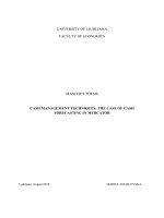
Grade II and III
1558 CpG loci probes
Grade II
2189 probes
Grade III
1837 probes
Grade II, III & IV
938 CpG loci probes
Grade III and IV
1019 CpG loci probes
Grade II and IV
1025 CpG loci probes
Grade IV
1208 probes
Figure 2 The cross-over between hypermethylated CpG loci probes in grade II, III and IV samples is illustrated with a venn diagram.
Numbers refer to the number of hypermethylated probes that belong to genes present in the list by ≥3 probes.
Hill et al. BMC Cancer 2014, 14:506
/>
shown). This suggests there is also epigenetic heterogeneity between pGBM and sGBM, at least in terms of
DNA methylation, but interestingly there were three of
the pGBM samples that clustered within the sGBM
CIMP+ve group, one of which had the IDH1 p.R132H
change. To further expand this analysis we then downloaded all pGBM IDH1 p.R132H mutated samples that
had available Infinium HumanMethylation450 BeadChip
methylation data (4 additional samples, there were no
IDH2 mutated samples). Clustering of the total 19
pGBM samples with our 15 grade IV sGBM samples
showed the CIMP phenotype within all five pGBM IDH1
mutated samples, which clustered together with our
sGBM CIMP+ve IDH1 mutation positive samples, indicating the CIMP+ve phenotype induced by IDH1 mutation in pGBM is similar in sGBM. Two additional
pGBM samples also clustered within this group, completing the smaller of the two major cluster groups
(Figure 3). Of the larger, CIMP-ve cluster, samples split
into four sub-clusters, dependent predominantly on
pGBM/sGBM status. Sub-clusters 1c and 1d contained
all but two pGBM samples and only one sGBM sample
whilst the remaining two sub-clusters contain all but
one sGBM CIMP-ve samples (Figure 3), indicating methylated targets of CIMP-ve primary and secondary grade
IV glioblastomas differ significantly. Three of the four
sGBM CIMP-ve IDH1 mutation positive samples are the
samples that exhibited CIMP in the earlier lesion but
not the later lesion, whilst the fourth sGBM and its
paired earlier lesion were CIMP-ve. Comparison of methylated gene lists for sGBM and pGBM samples (irrelevant of CIMP status) identified 180 genes that were only
methylated in sGBM samples according to our criteria
(Additional file 7: Table S6). We also identified 338
genes that were only methylated in pGBM samples
(Additional file 7: Table S6) and 123 genes methylated in
both. Reassuringly, only two genes were present in the
pGBM specific list and our earlier list of 218 genes that
were methylated across grade II, III and IV secondary
gliomas. Of the 180 genes that were sGBM specific from
this analysis, 115 were present in our list of 218 genes
across grade II, III and IV secondary gliomas (Additional
file 8: Figure S3). This discrepancy is most probably due
to a combination of looking only at grade IV samples
and using data unadjusted for Type I/II Illumina probes.
Ingenuity analysis identified a substantial number of
genes associated with cancer in both lists, with substantially more in the pGBM only list; 20% and 54% of genes
within the sGBM and pGBM lists respectively. Considerable differences were observed between the molecular
and cellular functions of genes within each list (Table 1).
The pGBM gene list is enriched for genes that alter or
control gene expression which in turn may affect cellular
development and growth and proliferation. In contrast,
Page 7 of 12
the sGBM only list is enriched for genes that affect cell
death, survival and maintenance pathways that would
need to be altered or abrogated for tumorigenesis. Thus,
the differing patterns of methylation between these two
subtypes of glioma may provide differing advantages to
these tumor cells.
Discussion
Secondary GBM represents a smaller subset (5%) of GBM
tumors which develop from preexisting lower grade tumors (grade II/III), are more often seen in younger patients and patients with sGBM have longer survival times
[3]. These tumors demonstrate distinct genetic heterogeneity compared to primary GBM, including a considerably
greater mutation rate of the IDH1 gene that has been
shown to result in a CpG island methylator phenotype
(CIMP). In this report we have used the latest Illumina
Infinium HumanMethylation450 BeadChips to assess the
genome-wide methylation of 20 secondary glioblastomas
and their matching lower grade precursors. Sandoval et al.
[22] recently validated the Illumina Infinium HumanMethylation450 BeadChip array and demonstrated that
this latest array consistently and significantly detects CpG
methylation changes in the HCT-116 colorectal tumor cell
line in comparison with normal colon mucosa or HCT116 cells with defective DNA methytransferases [22].
While whole-genome bisulfite sequencing is the gold
standard for comprehensive mapping of methylation
events, it is still expensive and requires a high level of
specialization. However, the Illumina Infinium HumanMethylation450 BeadChip offers a powerful technique for
better understanding of the DNA methylation changes occurring in human diseases at a reasonable cost. Our study
represents the first to utilize the Illumina Infinium
HumanMethylation450 BeadChips to evaluate epigenetic
changes occurring during glioma progression.
We demonstrated that these samples had the expected
high levels of IDH1 mutation and that in the lower grade
precursors this nearly uniformly resulted in a CIMP
phenotype. We saw one case (P16, early and late lesions)
where there was evidence of an IDH1 mutation but no
CIMP phenotype. We also saw one case (P19.E) where
there was no evidence of IDH1 or IDH2 mutation but
was CIMP positive. However, it has previously been suggested that even when negative for the known IDH1 p.
R132H mutation, it is possible that other IDH1 mutations could be present in some cases that might therefore potentially affect CIMP status [23]. The early
presentation of IDH1 mutation and CIMP that we have
seen in our study suggests this is an early and important
event in gliomagenesis and that if not acquired at an
early stage is not gained during progression as no later
stage glioblastoma presented with CIMP where the precursor did not. Although the total number of samples is
sGBM (P20.L)
sGBM (P4.L)
sGBM (P3.L)
sGBM (P1.L)
sGBM (P9.L)
sGBM (P19.L)
sGBM (P18.L)
sGBM (P16.L)
sGBM (P6.L)
pGBM (TCGA-06-5416)
pGBM (TCGA-06-0171)
pGBM (TCGA-26-5136)
sGBM (P2.L)
pGBM (TCGA-06-0190)
pGBM (TCGA-06-5418)
pGBM (TCGA-06-0210)
pGBM (TCGA-26-5135)
pGBM (TCGA-26-5134)
pGBM (TCGA-26-5132)
pGBM (TCGA-12-5295)
pGBM (TCGA-06-5414)
pGBM (TCGA-06-0211)
pGBM (TCGA-26-5133)
pGBM (TCGA-06-5417)
pGBM (TCGA-06-0221)
pGBM (TCGA-26-1442)
pGBM (TCGA-06-6389)
pGBM (TCGA-06-6701)
sGBM (P7.L)
sGBM (P10.L)
sGBM (P13.L)
sGBM (P11.L)
pGBM (TCGA-15-1444)
sGBM (P14.L)
Hill et al. BMC Cancer 2014, 14:506
/>
Sub-cluster
1a
Figure 3 (See legend on next page.)
Sub-cluster
1b
Page 8 of 12
IDH1 Mutation
Positive
Sub-cluster
1c
Major cluster 1 CIMP -ve
Sub-cluster
1d
Major cluster 2
CIMP +ve
Hill et al. BMC Cancer 2014, 14:506
/>
Page 9 of 12
(See figure on previous page.)
Figure 3 Hierarchical Euclidean based clustering of the 2000 most variable loci. Samples split into 2 major cluster groups containing either
CIMP+ve or CIMP-ve samples (major cluster 2 and 1 respectively). Major cluster 1 split into four sub-clusters. IDH1 mutated samples are highlighted
with a black box. sGBM samples are labeled with their pair number (P#) followed by L to denote late lesion. pGBM samples are labeled with
respective TCGA sample names.
small the large degree of IDH1 mutation and CIMP argues strongly that this is true. In addition to increased
overall survival, IDH1 mutation status has been shown
to correlate with genetic features including the presence
of MGMT methylation and codeletion of 1p and 19q, as
well as inversely correlating with EGFR amplification,
chromosome 10 loss and chromosome 7 polysomy
[5,24] and therefore if we had been able to analyze a larger sample set, it would have been interesting to look at
the relationship between these factors.
The effects of tumor grade progression on the genomewide methylation of these paired samples of sGBM tumors and their earlier lower grade lesions could be
assessed in the most comprehensive manner to date due
to the large amount of data provided by the Illumina Infinium HumanMethylation450 BeadChips. Firstly, as mentioned above samples lacking CIMP in their precursor
lesions never gained it via progression, presumably due to
the early gain of some other genetic or environmental factor capable of driving gliomagenesis without the subsequent need for hypermethylation. While those samples
presenting with CIMP in their precursor lesion, largely in
association with IDH1 mutation, split approximately in
half to follow two paths after progression. Some samples
appeared to fully retain and maintain CIMP in their higher
grade lesions whatever level CIMP hypermethylation was
observed within the lower grade precursor lesions, presumably due to the importance of this high level of
general hypermethylation to the tumors survival. Interestingly, some samples notably reduced their levels of
general hypermethylation, some retaining what we defined as CIMP and some losing it. This could potentially
be due the initial lower grade lesion demonstrating epigenetic heterogeneity with different cells having differing hypermethylation patterns that together present as
CIMP positive. If a subset of these cells contained
hypermethylation of a particular tumor suppressor that
resulted in a considerable growth advantage then these
cells could grow out and progress to be the higher grade
lesion. This lesion would still have the evolutionary
pressure to maintain the hypermethylation of this specific tumor suppressor but not necessarily the need to
maintain a global methylation phenotype, although in
general you would expect some degree of maintenance
by the IDH1 mutation, it is plausible that due to changing tumor heterogeneity this would be visualized at a
lesser extent. Unfortunately we were unable to assess
different regions from within the same tumor to investigate this hypothesis. Furthermore, we observed that
these differences were not simply due to pairs progressing from grade II to grade III compared to grade III to
grade IV or grade II to grade IV. Due to the relatively
Table 1 Ingenuity Pathway Analysis software assessment of molecular and cellular functions of exclusively methylated
genes in either pGBM grade IV glioblastomas or sGBM grade IV glioblastomas
sGBM Ingenuity analysis(w)
Category(x)
No. of Genes(y)
p-value range(z)
Diseases and disorder
Cancer
36/180 (20.0%)
2.52E-04 – 1.05E-02
Molecular and cellular functions
Cellular compromise
12/180 (6.7%)
3.53E-06 – 8.01E-03
Molecular and cellular functions
Cellular assembly and organization
44/180 (24.4%)
1.56E-05 – 7.76E-03
Molecular and cellular functions
Cell morphology
45/180 (25.0%)
1.29E-04 – 1.29E-04
Molecular and cellular functions
cell death and survival
47/180 (26.1%)
2.52E-04 – 1.02E-02
Molecular and cellular functions
Cellular function and maintenance
38/180 (21.1%)
2.52E-04 – 1.05E-02
pGBM Ingenuity analysis(w)
Category(x)
No. of Genes(y)
p-value range(z)
Diseases and disorder
Cancer
183/338 (54.1%)
7.67E-09 – 2.68E-04
Molecular and cellular functions
Gene expression
104/338 (30.8%)
7.23E-23 – 1.28E-04
Molecular and cellular functions
Cellular development
124/338 (36.7%)
4.44E-20 – 7.94E-04
Molecular and cellular functions
Cell morphology
73/338 (21.6%)
1.16E-09 – 8.32E-04
Molecular and cellular functions
Cellular movement
90/338 (26.6%)
4.41E-09 – 6.52E-04
Molecular and cellular functions
Cellular growth and proliferation
116/338 (34.3%)
2.45E-08 – 7.69E-04
Table 1 shows the top diseases and disorders and 5 molecular and cellular functions of genes exclusively methylated in sGBM samples (top) and pGBM (bottom)
samples. For each table: (w) type of ingenuity analysis used (diseases and disorders or molecular and cellular functions); (x) category of either the disease/disorder
or molecular/cellular function; (y) the total number of genes within the list per category out of the total number of genes expressed, also shown as a percentage;
(z) p-value range of this number of genes within the list falling into each category.
Hill et al. BMC Cancer 2014, 14:506
/>
small size of our cohort we were unable to identify the
specific genetic differences that may support this hypothesis as we would assume them to be tumor specific.
Nonetheless this is an interesting observation that could
possibly affect the effectiveness of therapies based on
demethylating agents on these tumors. Naturally, we
would assume they would be more effective in samples
that at some stage demonstrated CIMP but they may
still be effective in samples that do not demonstrate
CIMP in the later grades if CIMP was present in the
precursor lesion. It is hard to estimate whether a
demethylating agent would be more effective on tumors
dependent on global hypermethylation or are reliant on
the hypermethylation of only a small number of targets.
Promisingly, 5-azacitidine has recently been shown to
be effective in reducing selected promoter methylation,
tumor growth, cell proliferation and inducing differentiation in an in vivo primary xenograft IDH1 mutant glioma [25].
Further evidence for the loss of some hypermethylation due to tumor grade progression was observed when
the levels of hypermethylated loci and genes were
assessed simply by the grade of each tumor rather than
looking for differences between paired samples. We noticed a trend towards decreasing levels of methylated
targets with increasing tumor grade which has previously been documented [11,20]. This loss of methylation
as tumors progress to later grades may indicate changes
in tumor heterogeneity resulting in refinement of the
most beneficial effects of hypermethylation as proposed
above, but could also represent a potential increase in
normal contamination as the tumor becomes more invasive and thus the tumor sample more intermingled with
normal.
By analyzing grade II, III and IV tumors separately, we
were able to identify a list of genes where hypermethylation was retained in all 3 grades, likely representing the
most generally important methylated genes within this
cohort of sGBM tumors. This identified preferential
hypermethylation of several genes associated with cell
morphology, cellular movement, cellular development,
cellular growth and proliferation, and cellular assembly
and organization, with many of these select genes having been previously associated with cancer. Due to the
relatively small number of tumors assessed, this analysis
would greatly benefit from expansion into a larger
cohort that could highlight which genes and pathways
are most important to sGBM gliomagenesis and
progression.
By comparison of methylation profile of our grade IV lesions with a subset of the publically available methylation
profiles of grade IV pGBM provided by the Cancer Genome Atlas (TCGA) network we demonstrated that in general the methylation profiles between these two tumor
Page 10 of 12
types differ in a similar manner to their respective genetic
alterations. This was further observed when comparing
the functions of genes commonly hypermethylated in
grade IV sGBMs compared to grade IV pGBMs with
sGBMs preferentially hypermethylating genes involved in
cell death, survival and maintenance pathways and
pGBMs preferentially hypermethylating genes that alter or
control gene expression. Interestingly, a small number of
the pGBM tumors demonstrated CIMP that was also
largely associated with IDH1 mutation, demonstrating a
very similar hypermethylation profile to CIMP positive
grade IV sGBM. This represented a specific epigenetic
overlap between a subset of the pGBM and sGBM tumors.
Included in this were two pGBM tumors exhibiting CIMP
that lacked mutation in IDH1 or IDH2 that could possibly
retain other mutations capable of resulting in CIMP such
as could be present in our 19th pair. Overall, this small
sGBM/pGBM analysis offers an insight into different
tumorigenic processes giving rise to these different types
of GBM tumors.
Conclusions
In summary, this data offers an insight into different epigenetic, methylation-related processes that give rise to
these different types of GBM tumors and provides interesting rationales for further study of this kind on much
larger cohorts. The increased use of genome-wide analysis
of methylation using technologies such as the Illumina
Infinium HumanMethylation450 BeadChips, that are relatively cheap and can be performed using both archival tissue DNA from FFPE blocks and small amounts of DNA
acquired from biopsies, may well increase their usefulness
as diagnostic or therapeutic markers. Thus, providing a
greater understanding on these tumor specific methylation
patterns may prove useful in a number of ways.
Additional files
Additional file 1: Primer sequences are provided for ALS2CL, GNMT
and WNK2 beta value validation analysis.
Additional file 2: Table S1. Sample information is provided for the
patient samples used in this study. (a) Pair numbers used in this study.
(b) sample numbers used in this study. (c) WHO grade of each tumour
given as either astrocytoma (astro) grade II or III; or GBM (glioblatoma
multiforme); astrocytoma WHO grade IV. (d) KPS; Karnofsky Performance
Score at the time of initial admission. (e) tumor location (hemisphere)
(f) RTx; whether radiotherapy was received (g) CTx; whether
chemotherapy was received (h) IDH1 mutation status; mutated (mut) or
wild type (wt) for the recurrent p.R132H mutation.
Additional file 3: Figure S1. Pie charts for each grade illustrate the
distribution of hypermethyaled CpG loci with respect to gene features or
genomic location. Gene features include CpG loci within the following
regions: 1st exon, 3′UTR, 5′UTR, gene body, within 1500 base pairs of the
transcription start site (TSS1500) or within 200 bp of the transcription
start site (TSS200). Genomic locations include: CpG islands (island), north
CpG island shelves (N shelf), south CpG island shelves (S shelf), north CpG
island shores (N shore) south CpG island shores (S shore) or unclassified
Hill et al. BMC Cancer 2014, 14:506
/>
regions (open sea). Hypermethylated CpG loci distributions are almost
identical in each grade. We also show the distribution of CpG islands that
were analyzed for hypermethylation events with respect to gene feature
or genomic location, as determined by the array design. These include all
probes on the array that associated with a gene, were not on either the
X or Y chromosome and were not associated with a SNP.
Additional file 4: Table S2. This table contains the gene symbols for
probes methylated in grade II, III and IV samples in 3 or more
probes. Table S3. This table contains probe beta values for the
genes listed in Table S2.
Additional file 5: Figure S2. Clone sequencing results are shown for
CpG island regions of three genes; ALS2CL, WNK2 and GNMT. Black and
white circles represent methylated and unmethylated CpG dinucleotides
respectively and each line represents a single clone. Methylation indexes
are given for each sample as a percentage of methylated CpGs out of
the total number of CpGs analysed. CpG dinucleotides analysed by the
infinium assay are indicated by an arrow and beta values for these loci
are shown next to each sample.
Additional file 6: Ingenuity pathway analysis results for the 218
genes hypermethylated across grade II, III and IV tumor samples.
Table S4. shows the top 5 molecular and cellular functions alongside the
number of genes falling within each category and the p-value range.
Gene symbols are given for genes that fall within any of these 5
categories. Gene symbols in bold indicate genes that have previously
been associated with cancer. Table S5. illustrates the top 3 gene
networks of the 218 hypermethylated genes and the genes present
within each of these networks.
Additional file 7: Table S6. Gene lists for hypermethylated genes
exclusively in sGBM or pGBM samples, following comparison between
the lists for the two tumor types.
Additional file 8: Figure S3. Venn diagram to illustrate the cross-over
between hypermethylated genes within the grade IV pGBM and grade IV
sGBM specific lists in addition to the original list of universally methylated
genes across grade II, III and IV secondary glioma samples.
Competing interests
The authors declare that they have no competing interests.
Authors’ contributions
FL and VKH designed the study and drafted the manuscript. ERM and GC
helped design the study. VKH carried out the clone sequencing and in
silico/bioinformatics analysis. TS carried out IDH1 mutation analysis. CJR
carried out in silico/bioinformatics analysis. DK and GS provided the DNA
samples and clinical information. JB developed the correction pipeline.
WW helped with bioinformatic analysis. All authors’ read and approved the
final manuscript.
Acknowledgments
VH was sponsored in part by the Department of Neurosurgery, University
Hospital Dresden, Germany. TS were sponsored by King Abdulaziz University,
Jeddah, Saudi Arabia.
Author details
1
Centre for Rare Diseases and Personalised Medicine and Department of
Medical & Molecular Genetics, School of Clinical and Experimental Medicine,
University of Birmingham College of Medical and Dental Sciences,
Edgbaston, Birmingham, UK. 2Department of Neurosurgery, University
Hospital Carl Gustav Carus Dresden, Technical University of Dresden,
Dresden, Germany. 3Department of Pathology, University of Cambridge,
Tennis Court Road, Cambridge CB2 1QP, UK. 4School of Cancer Sciences,
University of Birmingham, Birmingham, UK. 5Department of Neurosurgery,
University of Birmingham and Queen Elizabeth Hospital Birmingham,
Birmingham, UK.
Received: 13 December 2013 Accepted: 2 July 2014
Published: 10 July 2014
Page 11 of 12
References
1. Tortosa A, Vinolas N, Villa S, Verger E, Gil JM, Brell M, Caral L, Pujol T, Acebes
JJ, Ribalta T, Ferrer I, Graus F: Prognostic implication of clinical, radiologic,
and patholofic features in patients with anaplastic gliomas. Cancer 2003,
15:1063–1071.
2. Kaye AH, Walker DG: Low grade astrocytomas: controversies in
management. J Clin Neurosci 2000, 7:475–483.
3. Ohgaki H, Kleihues P: The definition of primary and secondary
glioblastoma. Clin Cancer Res 2013, 19:764–772.
4. Guo C, Pirozzi CJ, Lopez GY, Yan H: Isocitrate dehydrogenase mutations in
gliomas: mechanisms, biomarkers and therapeutic target. Curr Opin
Neurol 2011, 24:648–652.
5. Parsons DW, Jones S, Zhang X, Lin JC, Leary RJ, Angenendt P, Mankoo P,
Carter H, Siu IM, Gallia GL, Olivi A, McLendon R, Rasheed BA, Keir S,
Nikolskaya T, Nikolsky Y, Busam DA, Tekleab H, Diaz LA Jr, Hartigan J, Smith
DR, Strausberg RL, Marie SK, Shinjo SM, Yan H, Riggins GJ, Bigner DD,
Karchin R, Papadopoulos N, Parmigiani G: An integrated genomic analysis
of human glioblastoma multiforme. Science 2008, 321:1807–1812.
6. Yan H, Parsons DW, Jin G, McLendon R, Rasheed BA, Yuan W, Kos I,
Batinic-Haberle I, Jones S, Riggins GJ, Friedman H, Friedman A, Reardon D,
Herndon J, Kinzler KW, Velculescu VE, Vogelstein B, Bigner DD: IDH1 and
IDH2 mutations in gliomas. N Engl J Med 2009, 360:765–773.
7. Jiao Y, Killela PJ, Reitman ZJ, Rasheed AB, Heaphy CM, de Wilde RF,
Rodriguez FJ, Rosemberg S, Oba-Shinjo SM, Nagahashi Marie SK, Bettegowda
C, Agrawal N, Lipp E, Pirozzi C, Lopez G, He Y, Friedman H, Friedman AH,
Riggins GJ, Holdhoff M, Burger P, McLendon R, Bigner DD, Vogelstein B, Meeker
AK, Kinzler KW, Papadopoulos N, Diaz LA, Yan H: Frequent ATRX, CIC, FUBP1
and IDH1 mutations refine the classification of malignant gliomas.
Oncotarget 2012, 3:709–722.
8. Juratli TA, Kirsch M, Robel K, Soucek S, Geiger K, von Kummer R, Schackert
G, Krex D: IDH mutations as an early and consistent marker in low-grade
astrocytomas WHO grade II and their consecutive secondary high-grade
gliomas. J Neurooncol 2012, 108:403–410.
9. Martinez R, Esteller M: The DNA methylome of glioblastoma multiforme.
Neurobiol Dis 2010, 39:40–46.
10. Noushmehr H, Weisenberger DJ, Diefes K, Phillips HS, Pujara K, Berman BP,
Pan F, Pelloski CE, Sulman EP, Bhat KP, Verhaak RG, Hoadley KA, Hayes DN,
Perou CM, Schmidt HK, Ding L, Wilson RK, Van Den Berg D, Shen H,
Bengtsson H, Neuvial P, Cope LM, Buckley J, Herman JG, Baylin SB, Laird PW,
Aldape K, Cancer Genome Atlas Research Network: Identification of a CpG
island methylator phenotype that defines a distinct subgroup of glioma.
Cancer Cell 2010, 17:510–522.
11. Wu X, Rauch TA, Zhong X, Bennett WP, Latif F, Krex D, Pfeifer GP: CpG
island hypermethylation in human astrocytomas. Cancer Res 2010,
70:2718–2727.
12. Esteller M, Garcia-Foncillas J, Andion E, Goodman SN, Hidalgo OF,
Vanaclocha V, Baylin SB, Herman JG: Inactivation of the DNA-repair gene
MGMT and the clinical response of gliomas to alkylating agents. N Engl J
Med 2000, 343:1350–1354.
13. Weller M, Stupp R, Reifenberger G, Brandes AA, van den Bent MJ, Wick W,
Hegi ME: MGMT promoter methylation in malignant gliomas: ready for
personalized medicine? Nat Rev Neurol 2010, 6:39–51.
14. Dang L, White DW, Gross S, Bennett BD, Bittinger MA, Driggers EM, Fantin
VR, Jang HG, Jin S, Keenan MC, Marks KM, Prins RM, Ward PS, Yen KE, Liau
LM, Rabinowitz JD, Cantley LC, Thompson CB, Vander Heiden MG, Su SM:
Cancer-associated IDH1 mutations produce 2-hydroxyglutarate.
Nature 2009, 462:739–744.
15. Turcan S, Rohle D, Goenka A, Walsh LA, Fang F, Yilmaz E, Campos C, Fabius
AW, Lu C, Ward PS, Thompson CB, Kaufman A, Guryanova O, Levine R,
Heguy A, Viale A, Morris LG, Huse JT, Mellinghoff IK, Chan TA: IDH1
mutation is sufficient to establish the glioma hypermethylator
phenotype. Nature 2012, 483:479–483.
16. Hill VK, Underhill-Day N, Krex D, Robel K, Sangan CB, Summersgill HR, Morris
M, Gentle D, Chalmers AD, Maher ER, Latif F: Epigenetic inactivation of the
RASSF10 candidate tumor suppressor gene is a frequent and an early
event in gliomagenesis. Oncogene 2011, 30:978–989.
17. Du P, Kibbe WA, Lin SM: lumi: a pipeline for processing Illumina
microarray. Bioinformatics 2008, 24:1547–1548.
18. Edgar R, Domrachev M, Lash AE: Gene Expression Omnibus: NCBI gene
expression and hybridization array data repository. Nucleic Acids Res 2002,
30:207–210.
Hill et al. BMC Cancer 2014, 14:506
/>
Page 12 of 12
19. Hartmann C, Meyer J, Balss J, Capper D, Mueller W, Christians A, Felsberg J,
Wolter M, Mawrin C, Wick W, Weller M, Herold-Mende C, Unterberg A,
Jeuken JW, Wesseling P, Reifenberger G, von Deimling A: Type and
frequency of IDH1 and IDH2 mutations are related to astrocytic and
oligodendroglial differ- entiation and age: a study of 1,010 diffuse
gliomas. Acta Neuropathol 2009, 118:469–474.
20. Christensen BC, Smith AA, Zheng S, Koestler DC, Houseman EA, Marsit CJ,
Wiemels JL, Nelson HH, Karagas MR, Wrensch MR, Kelsey KT, Wiencke JK:
DNA methylation, isocitrate dehydrogenase mutation, and survival in
glioma. J Natl Cancer Inst 2011, 103:143–153.
21. Moniz S, Martinho O, Pinto F, Sousa B, Loureiro C, Oliveira MJ, Moita LF,
Honavar M, Pinheiro C, Pires M, Lopes JM, Jones C, Costello JF, Paredes J,
Reis RM, Jordan P: Loss of WNK2 expression by promoter gene
methylation occurs in adult gliomas and triggers Rac1-mediated tumour
cell invasiveness. Hum Mol Genet 2013, 22:84–95.
22. Sandoval J, Heyn H, Moran S, Serra-Musach J, Pujana MA, Bibikova M,
Esteller M: Validation of a DNA methylation microarray for 450,000 CpG
sites in the human genome. Epigenetics 2011, 6:692–702.
23. Gravendeel LA, Kloosterhof NK, Bralten LBC, van Marion R, Dubbink HJ,
Dinjens W, Bleeker FE, Hoogenraad CC, Michiels E, Kros JM, van den Bent M,
Smitt PA, French PJ: Segregation of Non-p.R132H Mutations in IDH1 in
Distinct Molecular Subtypes of Glioma. Hum Mutat 2010, 31:E1186–E1199.
24. van den Bent MJ, Dubbink HJ, Marie Y, Brandes AA, Taphoorn MJ, Wesseling
P, Frenay M, Tijssen CC, Lacombe D, Idbaih A, van Marion R, Kros JM,
Dinjens WN, Gorlia T, Sanson M: IDH1 and IDH2 mutations Are prognostic
but not predictive for outcome in anaplastic oligodendroglial tumors: a
report of the European organization for research and treatment of
cancer brain tumor group. Clin Cancer Res 2010, 16:1597–1604.
25. Borodovsky A, Salmasi V, Turcan S, Fabius AW, Baia GS, Eberhart CG,
Weingart JD, Gallia GL, Baylin SB, Chan TA, Riggins GJ: 5-azacytidine
reduces methylation, promotes differentiation and induces tumor
regression in a patient-derived IDH1 mutant glioma xenograft.
Oncotarget 2013, 4:1737–1747.
doi:10.1186/1471-2407-14-506
Cite this article as: Hill et al.: Stability of the CpG island methylator
phenotype during glioma progression and identification of methylated
loci in secondary glioblastomas. BMC Cancer 2014 14:506.
Submit your next manuscript to BioMed Central
and take full advantage of:
• Convenient online submission
• Thorough peer review
• No space constraints or color figure charges
• Immediate publication on acceptance
• Inclusion in PubMed, CAS, Scopus and Google Scholar
• Research which is freely available for redistribution
Submit your manuscript at
www.biomedcentral.com/submit