Antimicrobial activity of medicinally important essential oils against selected dental microorganisms
Bạn đang xem bản rút gọn của tài liệu. Xem và tải ngay bản đầy đủ của tài liệu tại đây (295.84 KB, 14 trang )
Int.J.Curr.Microbiol.App.Sci (2017) 6(6): 1562-1575
International Journal of Current Microbiology and Applied Sciences
ISSN: 2319-7706 Volume 6 Number 6 (2017) pp. 1562-1575
Journal homepage:
Original Research Article
/>
Antimicrobial Activity of Medicinally Important Essential Oils
against Selected Dental Microorganisms
Nisheet Bhoot and Kalpesh B. Ishnava*
Ashok and Rita Patel Institute of Integrated Study and Research in Biotechnology and Allied
Sciences (ARIBAS), New Vallabh Vidyanager, Anand, Gujarat-388120, India
*Corresponding author
ABSTRACT
Keywords
Essential oils,
Oral diseases,
Anticariogenic
activity, TLC,
Bioautography
Article Info
Accepted:
21 May 2017
Available Online:
10 June 2017
Oral diseases are among the major public health problems and the most common of
chronic diseases that affect mankind. Essential oils could serve as an important natural
alternative to prevent microbial growth in oral infection diseases. This study was
undertaken to determine the in vitro anticariogenic activities of 11 essential oils against
dental pathogenic bacteria (Staphylococcus aureus, Streptococcus mutans and
Streptococcus pyogenes) and fungi (Candida albicans and Candida parapsilosis) using
agar well diffusion method, followed by determination of MIC. Most of the tested essential
oils exhibited anticariogenic activity against all tested microbes. 16 formulations were
made using them Formulations 10 and 13 showing good activity against C. albicans and
C. parapsilosis. The formulations No. 10 and 13 showed strong antimicrobial activities
with MIC ≥ 0.2mg/ml against C. albicans. Active components of oil were separated by
TLC. Separation of the compounds of formulation 10 using TLC shows 5 different bands
present. Among 5 bands, only 1 band was active against C. albicans. These materials can
be served as an important natural alternative to prevent microbial growth in dental
diseases. The prepared formulation also uses as natural alternative and also less expensive
compared to the commercial product.
Introduction
Oral diseases are among the major public
health problems and the most common of
chronic diseases that affect mankind. Bacteria
are the dominant inhabitants of the oral cavity
but other microorganisms are also seen which
includes species of fungi, viruses and
protozoa. The oral cavity is inhabited by more
than 700 microbial species and many intrinsic
and extrinsic factors affect the composition,
metabolic activity and pathogenicity of the
highly diversified oral micro flora (Aniebo et
al., 2012; Samaranayake et al., 1986; Aas et
al., 2005; Nejad et al., 2011). The most
prevalent oral infectious diseases, caries and
periodontal disease, are historically the
province of dentists for diagnosis and
treatment. However, the effect of these oral
diseases
often
extends
systemically,
particularly in older adults. Hematogenous
seeding from an oral source is a dominant
cause of bacterial endocarditis and is
implicated in late prosthetic joint infection
(LPJI). Periodontal disease impairs glycemic
control in people with diabetes, and poorly
controlled
diabetes
may
exacerbate
periodontal disease (Collin et al., 1998;
1562
Int.J.Curr.Microbiol.App.Sci (2017) 6(6): 1562-1575
Taylor et al., 1998). Aspiration of
oropharyngeal secretions is the predominant
cause of nosocomial pneumonia in elderly
persons (Scannapieco et al., 1997).
Periodontopathic bacteria in the bloodstream
have been linked to atherosclerosis, coronary
artery disease, and stroke (Beck et al., 1996).
Dental plaque is formed by the colonization
and accumulation of oral microorganisms in
the insoluble glucan layer that are synthesized
by
glucosyltransferase
(GTase)
from
Streptococcus mutans (Loesche, 1986).
Actinomyces naeslundii and Actinomyces
visosus are usually associated with dental
caries particularly human root surface caries.
To avoid dental caries due to cariogenic
bacteria, inhibition of glucosyltransferase
activity by specific enzyme inhibitor
(Yanagida et al., 2000), inhibition of initial
cell adhesion of S. mutans by polyclonal and
monoclonal antibodies and inhibition of cell
growth of S. mutans by antibacterial agents
have been investigated (Raamsdonk et al.,
1995). Antibiotics such as penicillin and
erythromycin have been reported to
effectively prevent dental caries in animal and
humans but they are never used clinically
because of many adverse effects such as
hypersensitivity reaction, supra infections and
teeth staining (Kubo et al., 1992).
Furthermore, viridians group Streptococci
including S. mitis, S. sanguis and S. mutans,
the most representative human cariogenic
bacteria are moderately resistant to antibiotics
(Venditti et al., 1989). These drawbacks
justify further research and development of
natural antibacterials that are safe for the host
or specific for oral pathogens. The natural
phytochemicals could offer an effective
alternative to antibiotics and represent a
promising approach in prevention and
therapeutic strategies for dental caries and
other oral infections. Although, plant products
are greatly exploit for therapeutic potential to
cure various oral ailments.
Medicinal plants have been recognized as
valuable source of therapeutic components for
centuries, and about 60% of world’s
population is known to use traditional
medicines derived from medicinal plants.
Natural products have been recently
investigated more thoroughly as promising
agents for the prevention of oral diseases,
especially plaque-related diseases such as
dental caries (Pai et al., 2004; FernandesFilho et al., 1998). The increasing resistance
to available antimicrobials has attracted the
attention of the scientific community
regarding a search for new cost-effective
drugs of natural or synthetic origin (Fine et
al., 2000). Essential oils in general
demonstrate antimicrobial activity against
cariogenic microbes (Takarada et al., 2004)
and fungal filaments as well (Prashar et al.,
2003). Some studies have pointed out that
plant-derived essential oils may be an
effective alternative to overcome microbial
resistance (Didry et al., 1994). This study was
undertaken to determine the in vitro
antimicrobial activities of 11 essential oils
against
dental
pathogenic
bacteria
(Staphylococcus
aureus,
Streptococcus
mutans and Streptococcus pyogenes) and
fungi (Candida albicans and Candida
parapsilosis) using.
Materials and Methods
Plant materials
The different plant species were selected and
collected between May to June (2015), from
different areas of Gujarat and surroundings of
Ashok & Rita Patel Institute of Integrated
Study and Research in Biotechnology and
Allied Sciences (ARIBAS), medicinal plant
garden of New Vallabh Vidyanagar (Table 1).
The plant was identified by Dr. Kalpesh
Ishnava (Plant taxonomist) at Ashok and Rita
Patel Institute of Integrated Study and
Research in Biotechnology and Allied
1563
Int.J.Curr.Microbiol.App.Sci (2017) 6(6): 1562-1575
Sciences
(ARIBAS),
New
Vallabh
Vidyanagar, Gujarat, India. The leaves and
seeds of all the healthy and disease free plants
were used for oil extraction for the test of
antimicrobial activity.
Bioassay for antimicrobial activity
Extraction of essential oils
In the present study, to test antimicrobial
activity, eleven different plant essential oils
were used. The antimicrobial activity was
studied by agar well diffusion method (Perez
et. al., 1990). From the stock, 10 mg, 30 mg,
50mg concentrations of essential oils were
suspended in one millilitre of Dimethyl
sulfoxide (DMSO). In order to make agar
plates, the Petri plates were thoroughly
washed using detergent, dried and sterilized in
autoclave at 15 lbs pressure (121˚C) for 15
minutes. Approximately 25ml of sterilized
medium was poured into Petri plates and
solidified at room temperature. The plates
were incubated at 37˚ C for overnight for
sterility testing. A fresh microbial culture of
300 µl was spread on agar plates with glass
spreader. A well of 9 mm diameter punched
off in Petri plates with sterile cup borer and
then 100µl particular plant essential oil was
loaded. Plates were placed for 30 minutes in
refrigerator for diffusion of oil and then
incubated at 37˚C for 24 hours or more
depending upon the organisms, until
appearance of zone of inhibition. The zone of
inhibition was measured as a property of
antimicrobial activity. In the present study,
ampicilin and amoxicilin antibiotics were
used as positive control to compare the zone
of inhibition with the antibacterial assay.
Hydro distillation method
Hydro distillation method was used for the
extraction of essential oils form the selected
plants. Selected plants were collected and
washed with tap water. After that leaves were
cut into small pieces and weighed 70g. It was
placed in a 2-liter round bottomed flask with
distilled water (300 ml for 70g fresh material)
and the assembly was placed at rotating
mantle at 80˚ C for 3 hours.
The essential oil was extracted and then
collected in Eppendorf tubes and stored at
room temperature.
The essential oil content was determined on
an oil volume to tissue weight. Oil stocks
were
prepared
by
using
different
concentrations 10mg, 30mg, 50mg of oil in
50% DMSO and used for further experiment
use (Charles et al., 1990).
Cariogenic microbial strains
A group of microorganisms known to cause
tooth decay were selected (Candida albicansMTCC-3017; Candida parapsilosis-MTCC6510; Lactobacillus casei- MTCC-1423;
Staphylococcus
aureus-MTCC-96;
Streptococcus
mutans-MTCC-890;
Streptococcus pyogenes-MTCC-442) and
purchased from Microbial Type Culture
Collection (MTCC) bank, Chandigarh as a
freeze dried pure culture. The microbial
cultures were revived by using MTCC
specified selective growth medium and
preserved as glycerol stocks.
Antibacterial activity
Agar well diffusion method
Minimum Inhibitory Concentration (MIC)
determination (for bacteria)
Minimum inhibitory concentration was
evaluated by serial broth dilution method
(Chattopadhyay et. al., 1998). Essential oils
showing more than 08 mm inhibition zone
were selected for MIC. Selective broth
medium was used for dilutions as well as
1564
Int.J.Curr.Microbiol.App.Sci (2017) 6(6): 1562-1575
preparing inoculums. The bacterial cell
density was maintained uniformly throughout
the experimentation at 1×10⁸ CFU/ml by
comparing with 0.5 McFarland turbidity
standards. Plants essential oil of 400 µl from
stock solution was taken into first dilution
tube containing 1600 µl of selective medium
broth and mixed it well. From these, 1000 µl
were transferred to second tube containing
1000 µl broth. This step is repeated nine times
and from the last tube 1000 µl was discarded.
100 µl of test organisms was added in each
tube. The final volume of solution in each
tube was made up to 1 ml. The MIC was
tested in the concentration range between
20mg/ml to 0.2mg/ml. Tubes were incubated
at optimal temperature and time in an
incubator.
Growth indicator 2, 3, 5-triphenyl tetrazolium
chloride solution (100 µl of 0.1%) was
incorporated in each tube to find out the
bacterial growth inhibition. Tubes were
further incubated for 30 minutes under dark
conditions. Bacterial growth was visualized
when colourless 2, 3, 5-triphenyl tetrazolium
chloride was converted red colour formazone
in the presence of bacteria. Each assay was
done by using DMSO and selective medium
as control.
Antifungal activity
Agar
well
diffusion
method
A drop of fungal spore suspension was placed
in the centre of PDA plates and spreader all
over with sterile glass spreader. Cups were
pored with sterile cup borer and filled with
100 µl of extract. Plates were place in
refrigerator for 10 min and then transferred to
incubator held at 28 ˚ C and incubated for 72
hours then after plates were observed for zone
of inhibition. Antifungal activity was
measuring by diameter of zone. The
experiment was carried out in duplicate and
mean of diameter of inhibition zone was
calculated. 100% DMSO used as a control.
Minimum Inhibitory Concentration (MIC)
determination (for fungus)
Minimum inhibitory concentration was
evaluated by Agar well diffusion method.
Essential oils showing more than 08 mm
inhibition zone were selected for MIC. From
the stock, 10mg, 30mg, 50mg concentrations
of essential oils were suspended in one
millilitre of Dimethyl sulfoxide (DMSO). In
order to make agar plates, the Petri plates
were thoroughly washed using detergent,
dried and sterilized in autoclave at 15 lbs
pressure
(121˚C)
for
15
minutes.
Approximately 25ml of sterilized medium
was poured into Petri plate and solidified at
room temperature. The plates were incubated
at 37˚ C for overnight for sterility testing. A
fresh microbial culture of 100 µl was spread
on agar plates with glass spreader. A well of 9
mm diameter punched off in Petri plates with
sterile cup borer and then 2µl, 4µl, 6µl, 8µl,
10µl, 12µl, 14µl, 16µl,18µl, 20µl, 22µl, 24µl,
26µl, 28µl, 30µl and 100µl particular plant
essential oil formulation was loaded. Plates
were placed for 30 minutes in refrigerator for
diffusion of oil and then incubated at 37˚ C
for 48 hours or more depending upon the
organisms, until appearance of zone of
inhibition. The zone was measured and
minimum activity zone is considered as the
MIC of that essential effect on oral fungal
pathogen. Fluconazole was used as a positive
control to compare the zone of inhibition with
the antifungal assay. DMSO was used as a
negative control in both assays respectively.
A preparation of essential oils formulation
Antibacterial and antifungal activity evaluate
of the 11 essential oils (Table 2). 11 out of
selected essential oils based on the criteria of
1565
Int.J.Curr.Microbiol.App.Sci (2017) 6(6): 1562-1575
minimum inhibitory concentration (MIC) of
bacteria and fungus selected. 7 out of 11
essential oils selected for the preparation of
the formulation. Essential oils showing more
than 08 mm inhibition zone were selected for
MIC. Formulations were made by using seven
different essential oils for antimicrobial assay.
Analytical thin layer chromatography
Analytical TLC was performed to find out
suitable solvent system for the development
of chromatogram. The following solvent
mixtures were tried on percolated TLC plates
(Merck, silica gel 60 F254 plate, 0.25mm).
Take the 0.1ml essential oil and 0.9ml
formulation is diluted with 0.9 ml toluene
prepared sample. This sample further used of
the separation of the compound in thin layer
chromatography. The 5µl sample is used for
TLC for separation of the compound. The
Adsorbent - Silica gel 60F254- Percolated TLC
plates used. The system is Toluene: ethyl
acetate: (93:7) used for the separation of
compound from the selected formulation.
After the run the plate observed under the UV
trans-illuminator at 265 nm and 365 nm of
TLC plate. Spray reagent Vanillin-Sulphuric
acid is used for the detection of the compound
present in the formulation. Some other spray
reagents apply for the detection of the
compound on the TLC plate. After that the
plate is evaluated and not down the Rf value.
Iodine vapours use for the developed the TLC
bands in iodine chamber.
Bio autography
Out of 11 essential oils tested for
antimicrobial activity, only one showing
maximum growth inhibition against Candida
albicans was selected and used for
bioautography. By using capillaries 5 µL of
essential oil of formulation no. 10 (100mg/mL
stock solution) was spotted on to 0.25mm
thick precoated silica gel 60 F254 plate
(Merck, Germany). The band length was
2mm thick. After air drying the TLC plate
was run using pre-standardized solvent
system, toluene: ethyl acetate: (93:7). The
chromatogram was observed under UV
illumination and used for bioautography.
Organism specific agar medium, seeded with
specific organism Candida albicans was
overlaid on to the silica gel plate loaded with
sample and incubated at 37°C for 24 hrs. On
the next day, the plate was flooded with 2, 3,
5-Tri phenyl tetrazolium chloride (0.1%) to
visualize growth inhibition. The area of
inhibition zone was appeared as transparent
against reddish background (lawn of living
fungus).
Results and Discussion
Essential oils are rich sources of biologically
active
compounds
which
possess
antibacterial, antifungal, antiviral, insecticidal
and
antioxidant
properties
against
microorganisms. These essential oils are
considered as non-phytotoxic compounds and
potentially
effective
against
several
microorganisms including many fungal
pathogens (Pandey et al., 1982). Conner
(1993) found that cinnamon, clove, pimento,
thyme, oregano, and rosemary plants had
strong inhibitory effect against several
bacterial pathogens. It has been also reported
that essential oils extracted from some
medicinal plants had the antibacterial effects
against all the oral pathogens due to presence
of phenolic compounds such as carvacrol,
eugenol and thymol (Kim et al., 1995). The
essential oils and their components have been
used broadly against moulds. The essentials
oils extracts from many plants such as basil,
citrus, fennel, lemon grass, oregano, rosemary
and thyme have shown their considerable
antifungal activity against the wide range of
fungal pathogens (Kivanc, 1991). Therefore,
use of essential oils is increased for treatment
of oral infection.
1566
Int.J.Curr.Microbiol.App.Sci (2017) 6(6): 1562-1575
In the present study the antimicrobial assay of
plant essential oils and different formulation
made from the effective oils is carried out for
the purpose of checking the sensitivity of oral
pathogens. The different concentration of 11
essential oils was screened against selected
oral pathogens and formulation was prepared
from them.
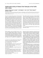
Antimicrobial activity of essential oils
10 out of 11 essential oils against C. albicans
give good antifungal activity. The diameters
of the inhibition zones are presented in figure
1. The results showed that the isolates
sensitivity was increased with the increase of
antifungal concentration (p<0.05).The range
of the 10 to 31mm zone of inhibition
observed. A. indica not give any antifungal
activity. Maximum activity showed in the S.
aromaticum against all the selected
concentration and also pure sample of the oil.
Maximum activity showed in the S.
aromaticum in pure oil sample.
05 out of 11 essential oils against C.
parapsilosis give good antifungal activity.
The diameters of the inhibition zones are
presented in figure 1. The results showed that
the isolates sensitivity was increased with the
increase of antifungal concentration (p<0.05).
The range of the 14 to 32mm zone of
inhibition observed. A. indica, E. globuls, C.
citrates, O. sanctum and M. elengi not give
any antifungal activity. Maximum activity
showed in the C. martini against all the
selected concentration and also pure sample
of the oil. Maximum activity showed in the S.
aromaticum in pure oil sample.
The activity is compared with negative
control DMSO. Which show no zone of
inhibition
against
microorganisms
as
compared to antifungal and antibacterial
positive controls used. Amoxiciilin and
ampicillin are used as a positive control.
The action of mechanism of phenolic
compounds was related to the ability of
phenolic compounds to alter microbial cell
permeability, thereby permitting the loss of
macromolecules from the cell interior, could
help explain some of the antimicrobial
activity. Another explanation might be that
phenolic compounds interfere with membrane
function and interact with membrane proteins,
causing deformation in structure and
functionality (Bajpai et al., 2008).
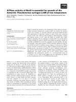
02 out of 11 essential oils against S. aureus
give good antibacterial activity. The
diameters of the inhibition zones are
presented in figure 2. The results showed that
the isolates sensitivity was increased with the
increase of antibacterial concentration
(p<0.05).The range of the 10 to 37mm zone
of inhibition observed. V. negundo and S.
aromaticum give antibacterial activity and
rest of the oils not give any activity.
Maximum activity showed in the V. negundo
and S. aromaticum against all the selected
concentration and also pure sample of the oil.
Maximum activity showed in the S.
aromaticum in pure oil sample.
05 out of 11 essential oils against S. mutans
give good antibacterial activity. The
diameters of the inhibition zones are
presented in figure 2. The results showed that
the isolates sensitivity was increased with the
increase of antibacterial concentration
(p<0.05).
The range of the 10 to 27mm zone of
inhibition observed. V. negundo, S.
aromaticum, O. sanctum, M. elengi and P.
pinnata give antibacterial activity and rest of
the oils not give any activity. Maximum
activity showed in the V. negundo and S.
aromaticum against all the selected
concentration and also pure sample of the oil.
Maximum activity showed in the S.
aromaticum in pure oil sample.
1567
Int.J.Curr.Microbiol.App.Sci (2017) 6(6): 1562-1575
06 out of 11 essential oils against L. casei
give good antibacterial activity. The
diameters of the inhibition zones are
presented in figure 2. The results showed that
the isolates sensitivity was increased with the
increase of antibacterial concentration
(p<0.05).The range of the 10 to 27mm zone
of inhibition observed. P. granatum, V.
negundo, S. aromaticum, O. sanctum, M.
elengi and P. pinnata give antibacterial
activity and rest of the oils not give any
activity. Maximum activity showed in the V.
negundo and S. aromaticum against all the
selected concentration and also pure sample
of the oil. Maximum activity showed in the S.
aromaticum in pure oil sample.
03 out of 11 essential oils against S. pyogenes
give good antibacterial activity. The
diameters of the inhibition zones are
presented in figure 2. The results showed that
the isolates sensitivity was increased with the
increase of antibacterial concentration
(p<0.05). The range of the 14 to 27mm zone
of inhibition observed. V. negundo, S.
aromaticum and C. martini antibacterial
activity and rest of the oils not give any
activity. Maximum activity showed in the S.
aromaticum against all the selected
concentration and also pure sample of the oil.
Maximum activity showed in the S.
aromaticum in pure oil sample.
Essential oils have been tested for in vivo
and in vitro antimicrobial activity and some
have demonstrated to be possessing
potential antimicrobial potential. Their
mechanism of action appears to be
predominantly on the cell membrane by
disrupting its structure thereby causing cell
leakage and cell death, secondary actions
maybe by blocking the membrane synthesis;
and inhibition of cellular respiration (Cristiane
et al., 2008). They readily penetrate into the
cell membrane and exert their biological
effect because of high volatility and
lipophilicity of the essential oils (Inouye,
2003).
The elimination of cariogenic bacteria from
the oral cavity using antibacterial agents is
one of primary strategies for prevention of
dental caries. Herbs are being widely explored
to discover alternatives to synthetic
antibacterial agents. Essential oils have been
shown to possess antibacterial, antiviral,
insecticidal and antioxidant properties.
Similar to antifungal activity of essential oils
oral bacteria are also screened for sensitivity
assay. The results obtained from our study
shows that the five essential oils have got a
very good antibacterial activity against
Streptococcus mutans. Regardless of which
agent is the drug of choice for the treatment of
oral diseases, dental scientists are still
searching for new therapeutic applications to
prevent and treat them. Toxicity, mucosal
ulceration, and development of resistant
bacterial strains are the adverse effects found
with several other antibacterial agents.
Collectively, these adverse effects of dental
medications motivate dentists to use
conventional natural therapeutics for the oral
cavity ailments (Takahashi et al., 2003).
In this study, the essential oil of Syzygium
aromaticum was obtained, eugenol was
identified as a compound and its antimicrobial
activity was assessed, agreeing with what has
been reported in several studies (Chaieb et al.,
2007). Its activity against Streptococcus
mutans was observed, agreeing with several
studies which reported its growth inhibitory
activity in oral pathogens (Ayoola et al.,
2008). Many essential oils have been
advocated for use in complementary medicine
for bacterial infections. However, few of the
many claims of therapeutic efficacy have
been validated adequately by either in vitro
testing or in vivo clinical trials. From the
above results the most effective seven
essential oils are used for preparing different
1568
Int.J.Curr.Microbiol.App.Sci (2017) 6(6): 1562-1575
formulations which are further used to check
anticariogenic activity of the formulations.
Antimicrobial activities of formulation of
essential oils
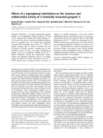
C. albicans
15 out of 15 essential oils formulation against
C. albicans give good antifungal activity. The
diameters of the inhibition zones are
presented in figure 3. The range of the 18 to
30mm zone of inhibition observed. Maximum
activity showed in the Formulation No. 14
and Formulation no. 15 (30 mm). Maximum
activity showed in the selected oral
microorganism out of C. albicans against the
Formulation No. 14 and Formulation no. 15.
Table.1 Plant selected for oils extraction and antimicrobial activity
Sr. No
1
2
3
4
5
6
7
8
9
10
11
Botanical Names
Azadirachta indica
Pongamia pinnata
Eucalyptus globus
Cymbopogon citratus
Punica granatum
Vitex negundo
Syzygium aromaticum
Oscimum sanctum
Cymbopogon martini
Mimusops elengi
Jatropa curcas
Local Names
Neem
Karanj
Nilgri
Lemon grass
Dadam
Nagod
Lavige
Tulsi
Palm roza
Borsalli
Ratanjot
Part Use
Leaf
Seed
Leaf
Leaf
Seed
Leaf
Fruit
Leaf
Leaf
Seed
Seed
Table.2 Different formulation of essential oils
Formulations
Neem
(µl)
Eucalyptus
(µl)
Tulsi
(µl)
Lemon
Grass (µl)
Palm
Roza(µl)
Clove
(µl)
Dadam
(µl)
F1(10mg/ml)
F2(10mg/ml)
F3(10mg/ml)
F410(mg/ml)
F5(30mg/ml)
F6(30mg/ml)
F7(30mg/ml)
F8(30mg/ml)
F9(50mg/ml)
F10(50mg/ml)
F11(50mg/ml)
F12(50mg/ml)
F13(pure)
F14(pure)
F15(pure)
F16(pure)
200
300
100
100
100
100
100
200
100
100
100
100
100
100
100
100
200
100
200
100
200
100
100
200
200
100
200
100
100
200
300
50
100
200
300
10
100
100
300
100
100
200
200
300
100
200
100
300
100
100
100
200
200
100
100
100
100
200
100
100
100
100
200
50
200
100
100
200
200
300
100
100
200
100
100
100
300
100
100
400
100
100
100
20
100
100
200
100
200
100
200
100
100
200
100
50
100
100
100
100
100
200
100
200
100
200
100
200
200
100
100
50
1569
Int.J.Curr.Microbiol.App.Sci (2017) 6(6): 1562-1575
Fig.1 Antifungal activities of essential oils against C. albicans and
C. parapsilosis and their zone of inhibition (in mm)
Fig.2 Antibacterial activities of essential oils against S. aureus, S. mutans, L. casei and
S. pyogenes and their zone of inhibition (in mm)
1570
Int.J.Curr.Microbiol.App.Sci (2017) 6(6): 1562-1575
Table.3 The MIC (mg / mL) of selected essential oils formulations against microorganisms
TEST ORGANISMS
FORMULATIONS
1
2
3
4
5
6
0
0
0
0
3
0
F6
0
0
0
0
0
3
F8
0
0
0
5
2.5
20
F9
0.2
0.4
2.5
0
0
0
F10
0.2
0
0
0
0
0
F13
0
0
0
0
0
6.5
F14
1- C. albicans; 2- C. parapsilosis; 3-S. aureus;4- S. aureus; 5-L. Casei; 6- S.Pyogenes
Fig.3 Antimicrobial activities of formulation of essential oils against C. albicans C. parapsilosis,
S. aureus, S. mutans, L. casei and S. pyogenes and their zone of inhibition (in mm)
C. parapsilosis
15 out of 15 essential oils formulation against
C. parapsilosis give good antifungal activity.
The diameters of the inhibition zones are
presented in figure 3. The range of the 10 to
25mm zone of inhibition observed. Maximum
activity showed in the Formulation No. 10 (25
mm). Formulation No. 10 compare to C.
albicans is less active against this organism.
S. aureus
15 out of 15 essential oils formulation against S.
aureus give moderate antibacterial activity. The
diameters of the inhibition zones are presented
in figure 3. The range of the 06 to 08 mm zone
of inhibition observed. In this organism showed
the moderate activity against all formulation.
S. mutans
15 out of 15 essential oils formulation against S.
mutans give very poor antibacterial activity
among all the selected microorganisms. The
diameters of the inhibition zones are presented
in figure 3. The range of the 03 to 08 mm zone
of inhibition observed. In this organism showed
the moderate activity against all formulation.
1571
Int.J.Curr.Microbiol.App.Sci (2017) 6(6): 1562-1575
L. casei
15 out of 15 essential oils formulation against S.
mutans give moderate antibacterial activity. The
diameters of the inhibition zones are presented
in figure 3. The range of the 05 to 09 mm zone
of inhibition observed. In this organism showed
the very less activity against all formulation.
S. pyogenes
15 out of 15 essential oils formulation against S.
mutans give good antibacterial activity. The
diameters of the inhibition zones are presented
in figure 3. The range of the 03 to 15 mm zone
of inhibition observed. In this organism showed
the good activity against all formulation and
also among all oral bacteria. Maximum activity
showed in the Formulation No. 08, 09 and 14
(15 mm).
Selected microorganisms among antifungal
activity give more responded against bacteria.15
out of the formulation best formulation No. 14
and formulation No.15 among both antifungal
and anticariogenic activity. Formulation No. 13,
14 and 15 highly active against C. albicans < C.
parapsilosis < S. pyogenes< S. aureus < S.
mutans. Formulation No. 13 include the
formulation pure oils more response to other
combination of the different concentration of
the oils. In this formulation included the Neem
(100), Eucalyptus (100), Tulsi (100), Lemon
(100), Grass (100), Palm roza (300), Clove,
(100) and Punica (200). In this formulation
maximum quantity takes for the formulation
preparation from the clove oils. Therefore, it is
responsible compound available in the clove
oils. Components of clove oil are eugenol,
eugenol acetate, isoeugenol and caryophyllene.
Clove oil is useful for its disinfecting properties,
relieving of pain, especially toothache, arthritis
and rheumatism. Studies conducted by Dorman
et al., (2000) in UK in 2000 and Betoni et al.,
(2006) in Brazil in 2006 have proved the
antimicrobial
potential
of
clove
oil.
Components of eucalyptus oil that are thought
to be responsible for its antibacterial property
are pinene, limonene, terpinenol, piperitone and
globulol. Antimicrobial potential of eucalyptus
oil has been proved in studies conducted by
Sattari et al., (2010) in Iran in 2009 and Filoche
et al., (2005) in New Zealand in 2005.
MIC (mg / mL) of selected essential oils
formulations against microorganisms
The Minimum Inhibitory Concentration (MIC)
values of different formulations of essential oils
of all the selected plants showing highest
activity against selected organisms was assessed
and determined. Examining the MIC values of
six formulations of different essential oils
generated the data where the maximum MIC
value was found to be 20 mg/ml and the
minimum value as 0.2 mg/ml.
The Minimum Inhibitory Concentration (MIC)
values of essential oil formulation of all
selected formulations showing highest activity
against selected organisms was assessed and
summarized in table 3.
Examining the MIC values of six samples of
various essential oils formulation showed the
maximum MIC value was found to be 20
mg/mL and minimum value as 0.2 mg/mL.
The MIC value of essential oils formulation of
formulation No.06 against LC is 3 mg/mL.
The MIC value of essential oils formulation of
formulation No.08 against SP is 3 mg/mL.
The MIC value of essential oils formulation of
formulation No.09 against SM, LC and SP is 5
mg/mL, 2.5 mg/mL and 20 mg/mL respectively.
The MIC value of essential oils formulation of
formulation No.10 against CA, CP and SA is
0.2 mg/mL, 0.4 mg/mL and 2.5 mg/mL
respectively
The MIC value of essential oils formulation of
formulation No.13 against CA was 0.2 mg/mL.
The MIC value of essential oils formulation of
formulation No.14 against SP was 6.5 g/mL.
1572
Int.J.Curr.Microbiol.App.Sci (2017) 6(6): 1562-1575
This formulation exhibited moderate MIC
values ranging from 3 mg /mL to 20 mg/mL
against cariogenic bacteria of SP.
According to these results, Formulation No.13
exhibits good MIC value ranging from 0.2
mg/ml to 20 mg/ml against selected oral
pathogenic organisms.
TLC analysis
Based on the MIC value and thin layer
chromatography results, formulations are
selected for the further study of the
characterization of phytochemical constituents
using TLC. In this study, formulation No.10 of
essential oil used for the characterization. In
TLC analysis of formulation no. 10, band no. 4
having active compound against cariogenic
fungus C. albicans. This band appear as black
colored under 254 lower intensity and 365
colored under higher intensity not observed and
band. The active compound Rf value is 0.78. In
TLC analysis of formulation No. 10 having
active compound against cariogenic bacteria C.
albicans. This band appear as black colored
under 254 lower intensity and no color 365
colored under higher intensity. The active
compound Rf value is 0.78.
potential of essential oils of Neem, Eucalyptus,
Tulsi, Lemongrass, Palmrosa, Clove and
Punica. The formulations No. 10 and 13 showed
strong antimicrobial activities with MIC ≥
0.2mg/ml against C. albicans. Active
components of oil were separated by TLC.
Separation of the compounds of formulation 10
using TLC shows 5 different bands present.
Among 4 bands, only 1 band was active against
C. albicans. The result shows that oils at
different
concentrations
exhibited
antimicrobial activity against dental pathogens.
These materials could be served as an important
natural alternative to prevent bacterial growth in
dental diseases. Essential oils have great
potential as antimicrobial compound against
pathogenic microorganisms, which can be used
to treat oral infectious diseases.
Acknowledgements
Authors are thankful to Charutar Vidya Mandal
(CVM), Vallabh Vidyanagar and Director of
Ashok and Rita Patel Institute of Integrated
Studies and Research in Biotechnology and
Allied Sciences (ARIBAS), New Vallabh
Vidyanagar, Gujarat, India for providing
necessary support for research and laboratory
facility.
References
Bio-autographical study
In order to find out active principles present in
formulation no.10 of oil, TLC solvent system
was standardized (Toluene: chloroform: 0.9:
0.3) and used for subsequent analysis. The
bioactive compounds were separated from crude
extracts by using TLC technique.
To locate the major active compounds
responsible for the anticariogenic activity in
formulation no.3, chromatogram was used for
TLC – Bioautography against CA. Further
chromatographic and spectroscopic analysis of
plant formulation extracts is necessary for
determination of structures of bioactive
compounds.
The present study, the very good inhibitory
Aas, J.A., Paster, B.J., Stokes, L.N., Olsen. I.
and Dewhirst, F.E. 2005. Defining the
normal bacterial flora of the oral cavity.
J. Clin. Microbiol., 43: 5721-5732.
Ayoola, G.A., Lawore, F.M., Adelowotan, T.,
Aibinu, I.E., Adenipekun, E., Coker,
H.A.B. and Odugbemi, T.O. 2008.
Chemical analysis and antimicrobial
activity of the essential oil of Syzigium
aromaticum (clove). Afr. J. Microbiol.
Res., (2):162-166.
Bajpai, V.K., Rahman, A., Nguyen, T.D., Huh,
M.K. and Kang, S.C. 2008. In vitro
inhibition of food spoilage and foodborne pathogenic bacteria by essential
oil and leaf extracts of Magnolia
liliflora. Desr. J. Food Sci., 73: 314320.
1573
Int.J.Curr.Microbiol.App.Sci (2017) 6(6): 1562-1575
Beck, J.D., Garcia, R., Heiss, G. and Vokonas,
P.S.1996. Offen bacher S. Periodontal
disease and cardiovascular disease. J
Periodontol., 67: 1123-37.
Betoni, J.E., Mantovani, R.P., Barbosa, L.N., Di
Stasi, L.C. and Fernandes Junior, A.
2006. Synergism between plant extract
and antimicrobial drugs used on
Staphylococcus aureus diseases. Mem.
Inst. Oswaldo Cruz., 101(4):387-90.
Chaieb, K., Hajlaoui, H., Zmantar, T., KahlaNakbi,
A.B.,
Rouabhia,
M.,
Mahdoua¬ni, K. and Bakhrouf, A.
2007. The chemical composi¬tion and
biological activity of clove essential oil,
Eugenia
caryophyllata
(Syzigium
aromaticum L. - Myrtaceae): a short
review. Phytother. Res., 21(6):501-6.
Charles, D.J., Joly, R.J. and Simon, J.E. 1990.
Effect of osmotic stress on the essential
oil content and commercial plant
extracts. Turkish J. Biol., 27: 157–162.
Chattopadhyay, D., Maiti, K., Kundu, A.P.,
Chakraborty, M.S., Bhadra, R., Mandal
S.C. and Manda, A.B. 2001.
Antimicrobial activity of Alstonia
macrophylla: a folklore of Bay Island. J.
Ethnopharmacol., 77:49–55.
Collin, H.L., Uusitupa, M. and Niskanen, L., et
al., 1998. Periodontal findings in elderly
patients with non-insulin dependent
diabetes mellitus. J Periodontol.,
69:962-6.
Conner, D.E. 1993. Naturally occurring
compounds. In: Davidison PM, Branen
AL (Eds). Antimicrobials in foods.
Marcel Dekker, New York. pp. 441-468
Cristiane, da silva, Gutteres, S.S., Weisheimer,
V. and Schapoval, E.E.S. 2008.
Antibacterial activity of lemongrass oil
and citral against candida spp. Braz. J.
Infect. Dis., 12(1): 63-66.
Didry, N., Dubreuil, L. and Pinkas, M.1994.
Activity
of
thymol,
carvacrol,
cinnamaldehyde and eugenol on oral
bacteria. Pharm. Acta Helv., 69: 25-28.
Dorman,
H.J.
and Deans,
S.G.2000.
Antimicrobial agents from plants:
Antibacterial activity of plant volatile
oils. J. Appl. Microbiol., 88(2):308-16.
Fernandes-Filho, E.S., Morais, S.M., Fonseca,
S.G. and Mota, O.M.1998. Preparação e
avaliação clínica do óleo essencial da
planta medicinal Lippia sidoides Cham
(Alecrim-pimenta). Rev. Ass. Bras.
Odonto., 6: 323-325.
Filoche, S.K., Soma, K. and Sissons, C.H. 2005.
Antimicrobial effects of essential oils in
combination
with
chlorhexidine
digluconate. Oral Microbiol. Immunol.,
20(4):221-25.
Fine, D.H., Furang, D., Barnett, M.L., Drew, C.,
Steinberg, L. and Charles, C.H. 2000.
Effect of essential oil containing
antiseptic mouth rinse on plaque and
salivary Streptococcus mutans levels. J.
Clin. Periodontal., 27: 157-161.
Inouye, S.2003. Laboratory evaluation of
gaseous essential oils (Part 1). Int. J.
Aromather., 13:95-107.
Kim, J., Marshall, M.R. and Wei, C. 1995.
Antibacterial activity of some essential
oils components against five foodborne
pathogens. J. Agric. Food Chem.,
43(11): 2839-2845.
Kivanc, M., Akgul, A. and Dogan, A.1991.
Inhibitory and stimulatory effects of
cumin, oregano and their essential oils
on growth and acid production of
Lactobacillus
plantarum
and
Leuconostoc mesenteroides. Int. J. Food
Microbiol., 13(1): 81-85.
Kubo, I., Muroi, H. and Himejima, M.1992.
Antimicrobial
activity
against
Streptococcus mutans of mate tea flavor
components. J. Agric. Food Chem., 40:
245-8.
Loesche, W.J. 1986. Role of Streptococcus
mutans in human dental decay.
Microbiol Rev., 50: 353-80.
Mbakwem Aniebo, C., Odoemelam, H.A. and
Okonko, I.O.2012. Isolation and
identification of Candida albicans and
Staphylococcus aureus from oral swabs
among primary school pupils in
Uzuakoli, Abia State, Nigeria. Nat. Sci.,
10(8):9-16.
1574
Int.J.Curr.Microbiol.App.Sci (2017) 6(6): 1562-1575
Nejad, B.S., Rafiei, F. and Moosanejad, A.
2011. Prevalence of Candida species in
the oral cavity of patients with
periodentitis. Afr. J. Biotechnol.,
10(15):2987-2990.
Pai, M.R., Acharya, L.D. and Udupa, N.2004.
Evaluation of antiplaque activity of
Azadirachta indica leaf extract gel - a 6week clinical study. J Ethnopharmacol.,
90: 99-103.
Pandey, D.K., Tripathi, N.N., Tripathi, R.D. and
Dixit, S.N. 1982. Fungitoxic and
phytotoxic properties of the essential oil
Caesulia axillaris Roxb. Angew Bot.,
56:259–267.
Perez, C., Pauli, M. and Bazerque, P. 1990. An
antibiotic assay by agar-well diffusion
method. Acta Biologiae ET Medecine
Experimentaalis, 15:113-115.
Prashar, A., Hili, P., Veness, R.G. and Evans,
C.S.2003. Antimicrobial action of
palmarosa oil (Cymbopogon martinii)
on
Saccharomyces
cerevisiae.
Phytochemistry, 63: 569-575.
Raamsdonk, M., Mei, H.C., Soet, J.J., Busscher,
H.J. and Graff, J.1995. Effect of
polyclonal and monoclonal antibodies
on surface properties of Streptococcus
sorbrinus. Infec. Immun., 63: 16981702.
Samaranayake, L.P., MacFarlane, T.W., Lamey,
P.J. and Ferguson, M.M.1986. A
comparison of oral rinse and imprint
sampling techniques for the detection of
yeast, coliform and Staphylococcus
aureus carriage in the oral cavity. J Oral
Pathol., 15: 386–388.
Sattari, M. et al., 2010. An inspection of
antimicrobial activities of essential oils
Eucalyptus
polycarpa,
Eucalyptus
largiflorence, Eucalyptus mallidora and
Eucalyptus
camaldulensis
on
Staphylococcus
aureus
in
the
laboratory. Iran. J. Pharm. Res., 9(1).
Scannapieco, F.A. and Mylotte, J.M.1996.
Relationships between periodontal
disease and bacterial pneumonia. J.
Periodontol., 67:1114-22.
Takahashi, K., Fukazawa, M., Motohia, H.,
Ochiai, K., Nishikawa, H. and Miyata,
T. A. 2003. Pilot study on antiplaque
effects of mastic chewing gum in the
oral cavity. J. Periodontol., 74: 501505.
Takarada, K., Kimizuka, R., Takahashi, N.,
Honma, K., Okuda, K. and Kato, T.
2004. A comparison of the antibacterial
efficacies of essential oils against oral
pathogens. Oral Microbiol. Immunol.,
19: 61-64.
Taylor, G.W., Burt, B.A. and Becker MP, et
al.,1998.
Non-insulin
dependent
diabetes mellitus and alveolar bone loss
progression
over
2
years.
J.
Periodontol., 69:76-83.
Venditti, M., Baiocchi, P., Santini, C.,
Brandimarte, C., Serra, P. and Gentile,
G. et al., 1989. Antimicrobial
susceptibilities of Streptococcus species
that cause septicemia in neutropenic
patients.
Antimicrob.
Agents
Chemother., 33: 580-582.
Yanagida, A., Kanda, T., Tanabe, M.,
Matsndaira,
F.
AND
Cordeiro,
J.G.O.2000. Inhibitory effects of apple
polyphenols and related compounds on
cariogenic
factors
of
mutans
Streptococci. J. Agric. Food Chem., 48:
5666-71.
How to cite this article:
Nisheet Bhoot and Kalpesh B. Ishnava. 2017. Antimicrobial Activity of Medicinally Important
Essential Oils against Selected Dental Microorganisms. Int.J.Curr.Microbiol.App.Sci. 6(6): 15621575. doi: />
1575