Isolation, molecular identification and antibiotic resistance of enterococcus faecalis from diseased tilapia
Bạn đang xem bản rút gọn của tài liệu. Xem và tải ngay bản đầy đủ của tài liệu tại đây (236.16 KB, 11 trang )
Int.J.Curr.Microbiol.App.Sci (2017) 6(6): 136-146
International Journal of Current Microbiology and Applied Sciences
ISSN: 2319-7706 Volume 6 Number 6 (2017) pp. 136-146
Journal homepage:
Original Research Article
/>
Isolation, Molecular Identification and Antibiotic Resistance of
Enterococcus faecalis from Diseased Tilapia
Uma Arumugam*, Nattan Stalin and Gnanadesika Pandian Rebecca
Tamil Nadu Fisheries University, State Referral Laboratory for Fish Disease Diagnosis and
Aquatic Animal Health, Fisheries Research and Extension Centre, Madhavaram Milk Colony,
Chennai-600051, Tamil Nadu, India
*Corresponding author
ABSTRACT
Keywords
Tilapia,
Enterococcus
faecalis,
Multiple-antibiotic
resistance,
16s rRNA
amplification and
sequencing
Article Info
Accepted:
04 May 2017
Available Online:
10 June 2017
Aquaculture is one of the fastest growing food production sectors globally. Tilapia is the
second widely farmed fish species in the global fish production. Enterococcus sp. is one of
the leading causes of nosocomial infections in urinary tract, surgical wound and
endocarditis in humans. These infections can be hard to treat because of the rising
incidence of multiple antibiotic-resistances. The spread of antibiotic resistance has become
a major concern in both human and veterinary medicine. In this study, we isolated and
characterized an Enterococcus faecalis isolate from a diseased cultured Tilapia. Initial
isolation of putative E. faecalis was carried out on streptococcus enrichment broth for 36
h. Characteristic, gram-positive, black color colony was selectively sub-cultured and
subsequently identified by 16 rRNA sequencing analysis as Enterococcus faecalis
(Genbank Acc no. KT877352). Furthermore, when the isolate was subjected to profiling
against 16 antibiotics, it was found to be highly resistant to amoxyclave, ampicillin,
erythromycin, gentamicin, kanamycin, nitrofurantoin, penicillin G, streptomycin,
sulphafurazole, and vancomycin. The findings of present study showed that E. faecalis
infects the cultured Tilapia species and the isolate (SRLFDA/TIL-1/15) possess multiple
antibiotic resistance, which emphasizes the urgent need for targeted alternate bio-control
strategies for control of emerging diseases like Enterococcus sp. infection in Tilapia
culture.
Introduction
Aquaculture has emerged as one of the
important food production sector over recent
decades (FAO, 2000). Food fish supply has
been reported to increase at the rate of 8.3 %
annually (FAO, 2014). Tilapia is the second
most farmed fish species in the world with the
estimated global production of around 5.5
million metric tons (mmt) in 2016, but is
predicted to increase by 4.5 % in 2017
reaching 5.8 mmt (RGCA, 2016). Tilapia may
play an important role in the growth of
aquaculture and continue to contribute in the
future food demand in developing and
developed countries.
As in any other fish culture operations,
disease is the major factor that adversely
affects the production of farmed Tilapia.
Although, Tilapia is considered hardy with
high disease resistance, bacterial diseases
caused due to Streptococcus sp (Atyah et al.,
2010; Chen et al., 2015; Li et al., 2015; Shen
et al., 2016) and Enterococcus sp (Martins et
al., 2009) have been reported. Enterococcus
136
Int.J.Curr.Microbiol.App.Sci (2017) 6(6): 136-146
sp. is a commensal gram-positive, diplococcal
bacterium which are dominant in fish,
shellfish, and other aquatic animals (Wilson
and McAfee, 2002; Sonsa Ard et al., 2015;
Chajecka-Wierzchowsk et al., 2016; Paganelli
et al., 2017). Nonetheless, Enterococcus sp. in
contaminated food fishes can lead to lifethreatening illness in human such as
endocarditis (Dahl and Bruun, 2013),
bacteremia (Stuart et al., 2006), urinary tract
infection and meningitis (Tebruegge et al.,
2011) and its resistance to antibiotics is
emerging as a major problem in treating these
infections (Koch et al., 2004). In addition, the
ubiquity of Enterococcus sp in food has been
reported to be mainly a result of their
resistance to unfavorable environmental
conditions during production technology with
food fish storage conditions and their
adaptability (Sarra et al., 2013). Because of
their relative abundance and their resistance
to environmental factors, Enterococcus sp
have been proposed as an indicator bacteria
for hygiene quality, as well as for
antimicrobial resistance in food and water
(Boehm and Sassoubre, 2014). Enterococcus
sp. has emerged as important healthcare
associated pathogen (Arias and Murray, 2012;
Khan et al., 2015), as they are intrinsically
resistant and tolerant to numerous commercial
antibiotics and are able to acquire drug
resistance either by chromosome, transfer of
plasmid or transposing acquisition containing
genetic sequences that confer resistance in
other bacteria (Eaton and Gasson, 2001; Ben
Belgacem et al., 2010; Hammerum Lester and
Heuer, 2010). In the last decade, several
virulence factors have been described in
Enterococci
including
cytolysins
(Vankerckhoven et al., 2004), gelatinase
(Mannu et al., 2003), serine protease
(Mohamed et al. 2004), hyaluronidase,
aggregation substance (Muscholl-silberhorn et
al., 2000) and extracellular surface protein
(Shankar et al., 1999). The cell wall adhesion
and biofilm formation properties of
Enterococcus sp. have also been described
(Barbosa, Gibbs and Teixeira, 2010).
The presence of commensal microbiota in
environmental ecosystems (Salyers and
Shoemakers, 2006), human ecosystems and in
food suggest that microorganisms can play a
essential role in transfer of antibiotic resistant
genes and the food chain may play a key role
in the transmission of resistance between the
environment and human (Marshall and Levy,
2009). Although, the detection of virulence
factors may indicate a virulence potential in
food isolates, food-borne Enterococcal
infection have never been reported (Giraffa,
2002; Foulquie-Moreno et al., 2006;
Valenzuela et al., 2010). However, the
consumption of food carrying antibioticresistant bacterial populations is considered a
possible transfer route (Kruse and Sorum,
1994). In recent years, growing interest in the
consumption of fish foods, which are
considered balanced healthy foods have been
observed. In the present study E. faecalis was
isolated from a diseased Tilapia, identified by
16S rRNA amplification and sequencing and
its antibiotics resistance was studied.
Materials and Methods
Samples
Diseased Tilapia sample (Average weight 57
g, average 18 cm) was collected from a fresh
water aquaculture farm in Chennai,
Tamilnadu, India. At site, behavioral
abnormalities, gross and clinical sign of the
diseased Tilapia were recorded (Heil, 2009).
Morbid tilapia fish with typical disease
symptom was first rinsed in sterile saline and
dissected aseptically. Inoculum from the
kidney of tilapia was collected aseptically and
was spread plated onto brain heart infusion
agar (BHIA) and Streptococcus selective
isolation broth (SIB) supplemented with 6.5%
NaCl. Presumptive single (black; 1mm dia)
137
Int.J.Curr.Microbiol.App.Sci (2017) 6(6): 136-146
colony on bile-esculin agar (Himedia, India)
were streaked on to Streptococcus selective
isolation agar (SIA) (Himedia, India) plates
for further purification at 30±2°C for 24-36 h
and maintained on BHIA slants at room
temperature (28±2°C).
CLSI guidelines, 2012).
Molecular identification
DNA extraction
The genomic DNA was extracted from the
Enterococcus sp isolate (SRLFDA/TIL-1/15)
using QIAamp genomic DNA kit (Qiagen,
Germany) as per manufacturerʹs protocol.
Phenotypic characterization
A series of biochemical tests were performed
to identify the isolate up to genus level (Svec
and
Devriese,
2015).
Biochemical
characterization like, gram strain, catalase
test, and growth at 6.5% NaCl/ 45°C, was
done using Rapid HiStrepTM biochemical
test kit specific for Streptococcus sp.
(HiMedia,
India).
The
phenotypic
characteristics documented in earlier reports
(Murray, 1990; Teixeira et al., 2011) were
compared for presumptive identification of
the isolate.
16S rRNA gene amplification and sequence
analysis
The 16S ribosomal RNA gene (16S rRNA) of
the isolate was amplified using a set of
universal prokaryotic primers 8F, 5’AGAGTTTGATCCTGGCTCAG-3’
and
1492R, 5’-GGTTACCTTGTTACGACTT-3’
(Eden et al., 1991). The PCR amplification
was performed in a 50 µl reaction volume
with 25 µl of PCR master mix (Ampliqon,
Denmark), 2.0 µl each of forward and reverse
primers, and 2.0 µl (100 ng) of genomic DNA
template and (19 µl) nuclease-free water. The
PCR reaction was carried out in T-100 TM
thermal
cycler
(Bio-Rad,
USA).
Amplification
was
done
by
initial
denaturation at 95 °C for 5 min, followed by
30 cycles of denaturation at 94 °C for 30 s,
annealing at 55 °C for 30 s and extension at
72 °C for 60 s with a final extension at 72 °C
for 5 min. The PCR product was resolved on
a 1.5% agarose gel containing 0.5µg/mL
ethidium bromide in 1X Tris-Borate-EDTA
(TBE) buffer and electrophoresed at 100 V.
Antibiotics susceptibility test
The antimicrobial susceptibility of the isolate
was determined by disc diffusion technique
using Muller-Hinton’s agar (Bauer et al.,
1966). The isolate was tested against 16
antibiotics (Himedia, India) viz., amoxyclave
(AMC), ampicillin (AMP), chloramphenicol
(C), ciprofloxacin (CIP), clindamycin (CO),
co-trimoxazole (COT), erythromycin (E),
gentamicin
(GEN),
kanamycin
(K),
nitrofurantoid (NIT), norfloxacin (NX),
oxytetracycline
(O),
penicillin-G (P),
streptomycin (S), sulphafurazole (SF), and
vancomycin (VA). The isolate (Himedia,
India) was grown overnight (OD 2 at 600nm)
in tryptic soy agar and spreaded on MullerHinton’s agar. After 30 min, four dissimilar
antibiotics discs were positioned on the plates
and incubated for 10-24 h at 37°C. After
incubation, the zone of inhibition (by mean of
diameter in mm) was measured around the
discs and compared with the interpretive chart
(Clinical and Laboratory Standards Institute,
Sequencing analysis
The amplified 16S rRNA gene PCR product
was purified using HiyieldTM Gel/PCR DNA
mini kit (real genomic, Taiwan) as per the
manufacturer’s
instructions.
Nucleotide
sequencing (forward and reverse) was done
with a commercial sequencing service
(Eurofins, India). The forward and reverse
138
Int.J.Curr.Microbiol.App.Sci (2017) 6(6): 136-146
sequences were assembled by DNA baser
sequence assembler v3.5.3 (2012) to form
consensus sequence and identified by NCBI
BLAST ( />cgi) search algorithm.
bacterial contamination in seafood products
have been documented (Wilson and McAfee,
2002; Sonsa-Ard et al., 2015).
PCR detection of tetracycline resistance
genes
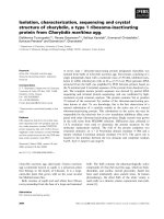
PCR amplification of the 16S rRNA of the
isolate (SRLFDA/TIL-1/15) resulted in an
amplified product of 1450 bp size (Fig. 1). On
the basis of gene sequence similarity carried
out by BLAST NCBI, the isolate was
identified as E. faecalis (Genbank Acc. No.
KT877352) with 99-100 % homology with
other E. faecalis strains in the GenBank
database (NCBI).
Molecular confirmation by 16S rRNA
The isolate SRLFDA/TIL-1/15 was examined
for the presence of the tetracycline resistance
encoding genes viz., tet (K) tet (L), following
the primers and protocols of the previous
researchers (Aarestrup et al., 2000; Garofalo
et al., 2007; Ullah et al., 2012).
16S ribosomal RNA present in bacteria plays
a major role in gene coding due to the highly
conserved region. It is considered as a
standard marker for bacterial phylogenetic
analysis to differentiate the species (Nagpal et
al., 1998). Recent studies demonstrated that
the different Enterococcus strains isolated
from diverse sea water environment
elucidated unique nucleotide position and
evolution of Enterococcus and its related
species Chajęcka -Wierzchowska et al., 2016;
Prichula et al. 2016). Moreover, many recent
reports have been published on the 16S rRNA
sequences of Enterococcus sp and the
phylogenetic relationship deduced from
analysis of these sequences (Deasy et al.,
2000; Mannu et al., 2003; Ben Belgacem et
al., 2010; Galimand et al., 2011; GallowayPena et al., 2012).
Results and Discussion
Isolation and identification of E. faecalis,
SRLFDA/TIL-1/15
The clinical symptoms recorded in the Tilapia
sample were lethargy, abdominal ascites,
organ discoloration, necrosis of the spleen and
haemorrhages in kidney. The isolate
recovered from kidney samples yielded a
predominant black colony on bile-esculin agar
(BEA) and Streptococcus selective isolation
(SIA) agar. Microscopic observation of the
stained smear revealed Gram-positive cocci
arranged in diplococci or short chain and
displayed oxidase and catalase negative
activity. The isolate could be grown at above
45 °C on BHI medium containing 6.5 %
NaCl, at pH 7.5. Biochemical characterization
of the isolate (MLTEC) as assessed by Rapid
HiStrepTM biochemical test is presented in
table 1.
Antibiotic susceptibility profile
Antibiotic sensitivity test showed that the E.
faecalis (SRLFDA/TIL-1/15) was either
resistant and/or intermediately resistant to
more than nine classes of antibiotic groups
(Table 2). The isolate showed resistance to
amoxyclave (AMC), ampicillin (AMP),
erythromycin (E), gentamicin (GEN),
kanamycin (K), nitrofurantoin (NIT),
Enterococci are one of the most common
group of bacteria present in foods (Paganelli
et al., 2017), mainly due to their resistance to
adverse environmental conditions during
production technology, as well as food
storage conditions and their high adaptability
(Boehm and Sassoubre, 2014). Enterococci
139
Int.J.Curr.Microbiol.App.Sci (2017) 6(6): 136-146
oxytetracycline
(O),
penicillin-G (P),
streptomycin (S), and sulphafurazole (SF) and
was
intermediately
resistant
to
chloramphenicol (C), ciprofloxacin (CIP),
clindamycin (CO), norfloxacin (NX), and
vancomycin (VA). The isolate was observed
to be susceptible only to co-trimoxazole
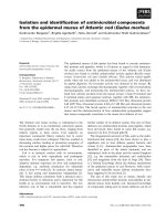
(COT), (Table 2). PCR amplification of the
tet (tet K and tet L) genes showed that the E.
faecalis isolate (SRLFDA/TIL-1/15) from
Tilapia carry tet K (360 bp) and tet L (1077
bp) genes (Fig. 2).
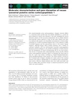
Table.1 Biochemical characterization of Enterococcus faecalis strain
(SRLFDA/TIL-1/15) isolated from diseased Tilapia
Biochemical tests
Enterococcus faecalis
SRLFDA/TIL-1/15
+
+
+
+
+
–
nd
+
+
–
+
+
+
–
+
Gram reaction
Voges–Proskauer’s
Bile-Esculin agar (black)
Esculin hydrolysis
PYR
ONPG
Arginine utilization
Glucose
Lactose
Arabinose
Sorbitol
Sucrose
Mannitol
Raffinose
Salt tolerance (6.5 %
NaCl)
Fig.1 PCR amplification of 16S rRNA of E. faecalis isolate from Tilapia
140
Int.J.Curr.Microbiol.App.Sci (2017) 6(6): 136-146
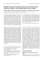
Table.2 Antibiotic resistance profile of Enterococcus faecalis strain
(SRLFDA/TIL-1/15) isolated from diseased Tilapia
S. No
1
2
3
4
5
6
7
8
9
13
10
11
12
14
15
16
Antibiotics
Amoxyclave (AMC)
Ampicillin (AMP)
Chloramphenicol (C)
Ciprofloxacin (CIP)
Co-Clindamycin (CO)
Co-Trimoxazole (COT)
Erythromycin (E)
Gentamicin (GEN)
Kanamycin (K)
Nitrofurantoin (NIT)
Norfloxacin (NX)
Oxytetracycline (O)
Penicillin-G (P)
Streptomycin (S)
Sulphafurazole (SF)
Vancomycin (VA)
Ratio S: I: R
TE
0(R)
7(R)
17(1)
18(1)
12(1)
20(S)
9(R)
7(R)
0(R)
12(R)
16(1)
0(R)
17(R)
0(R)
11(R)
16(1)
1: 5: 10
Zone of inhibition measured (mm) S=sensitive, I=intermediate, R=resistant, amoxyclav (30 µg), Ampicillin (10
µg), Penicillin-G (10 units), streptomycin (10 µg), kanamycin (30 µg), vancomycin (30 µg), erythromycin (15 µg),
clindamycin (2 µg), norfloxacin (10 µg), ciprofloxacin (5 µg), chloramphenicol (30 µg), co-trimoxazole (25 µg),
gentamicin (10 µg), nitrofurantoin (300 µg), oxytetracycline (30 µg) and sulphafurazole (300 µg).
Fig.2 PCR amplified fragments of the tetracycline resistance genes in
E. faecalis isolated from diseased Tilapia. Lane M- 100 bp DNA ladder;
Lane 1, tet K gene (360 bp); lane 2, tet L gene (1077 bp)
E. faecalis isolate (SRLFDA/TIL-1/15) was
resistant to drugs frequently used to treat
bacterial infections in humans and veterinary
medicine,
including
erythromycin,
ciprofloxacin, norfloxacin and vancomycin
(Bates et al., 1994; Prichula et al., 2016).
141
Int.J.Curr.Microbiol.App.Sci (2017) 6(6): 136-146
Antibiotics represent one of the most
prominent aquatic pollutants (Tello et al.,
2012). The presence of antibiotics in water
can cause serious environmental issues, such
as the emergence of resistance due to
selective pressure (Baquero et al., 2008).
Recently, several studies have reported the
development of multiple antibiotic resistance
in the microbes of the aquaculture systems
(Stalin and Srinivasan, 2016; Prichula et al.,
2016; Uma and Ronald, 2016). Although,
most of the studies on antibiotic resistance
and virulence of Enterococci sp have been
carried out in strain isolated from clinical
samples, recent reports have suggested that
environment and food could play a significant
role in the transmission of resistance to
humans (Gomes et al., 2008; Koluman, 2009;
Barbosa et al., 2010; Chajęcka Wierzchowska et al., 2016). The E. faecalis
isolate (SRLFDA/TIL-1/15) was found to be
resistant to 62% of the antibiotics tested in
this study with multiple resistances to ten
different antibiotics. Resistances to upto eight
antibiotics have been reported in isolates from
other aquaculture sources (Akinbowale et al.,
2006). The amplification of (tet K and tet L)
resistant genes and the tetracycline resistance
of the E. faecalis isolate (SRLFDA/TIL-1/15)
in the antibiotics sensitivity test showed that
the resistance shown against tetracycline
could be due to the expression of these genes.
tet genes are reported to be widely
disseminated in the environment (Pallecchi et
al., 2008; Di Cesare et al., 2012). The
identification of tetracycline resistance
determinants may be used as additional
genotypic markers for the purpose of outbreak
investigation and evolution of gene exchange
(Koike et al., 2007; Ng et al., 2001; Ullah et
al., 2012; Rico et al., 2014; Prichula et al.,
2016).
of antibiotic resistant genes, tet K and tet L
shows that there is a need for judicious use of
antibiotics in aquaculture and to adopt
alternate and safe measures for the
management of disease in aquaculture.
Acknowledgments
We are thankful to the National Agriculture
Development Programme (NADP), (Grant
No.30/FREC-NADP 2016-17) for financial
support.
References
Aarestrup, F.M., Agerso, Y. Gerner-Smidt, P.
Madsen, M. Jensen, L.B. 2000.
Comparison of antimicrobial resistance
phenotypes and resistance genes in
Enterococcus faecalis and Enterococcus
faecium
from humans
in
the
community, broilers, and pigs in
Denmark. Diagn. Microbiol. Infect.
Dis., 37, 127–137.
Akinbowale, O.L., Peng, H. Barton, M.D.
2006. Antimicrobial resistance in
bacteria isolated from aquaculture
sources in Australia. J Appl Microbiol.,
100, 1103–1113.
Arias, C.A., and Murray, B.E. 2012. The rise
of
the
Enterococcus:
beyond
vancomycin resistance. Nat. Rev.
Microbiol., 10, 266–78.
Atyah, M.A.S., Zamri-Saad, M. Siti-Zahrah,
A. 2010. First report of methicillinresistant Staphylococcus aureus from
cage-cultured tilapia (Oreochromis
niloticus). Vet. Microbiol., 144, 502–
504.
Barbosa, J., Gibbs, P.A. Teixeira, P. 2010.
Virulence factors among Enterococci
isolated from traditional fermented meat
products produced in the North of
Portugal. Food Control, 21, 651–656.
Bates, J., Jordens, J. Griffith, D.T. 1994. Farm
animals as putative reservoir for
In conclusion, this is the first report from
India on the isolation and confirmation of E.
faecalis from diseased Tilapia. The presence
142
Int.J.Curr.Microbiol.App.Sci (2017) 6(6): 136-146
vancomycin resistant enterococcal
infections in man. J. Antimicrob.
Chemother, 34, 507–516.
Bauer, A.W., Kirby, W.M.M, Sherris, J.C,
Turck,
M.
1966.
Antibiotic
susceptibility testing by a standardized
single disk method. Am J Clin Pathol.,
45(4):493-6?
Ben Belgacem, Z., Ben, Abriouel, H. Omar,
N. Ben, Lucas, R. Martínez-Canamero,
M. Gálvez, A. Manai, M. 2010.
Antimicrobial activity, safety aspects,
and some technological properties of
bacteriocinogenic Enterococcus faecium
from artisanal Tunisian fermented meat.
Food Control, 21, 462–470.
Boehm, A.B., Sassoubre, L.M. 2014.
Enterococci
as
Indicators
of
Environmental Fecal Contamination.
2014 Feb 5. In: Gilmore, M.S., Clewell,
D.B, Ike Y, et al., editors. Enterococci:
From Commensals to Leading Causes
of Drug Resistant Infection. Boston:
Massachusetts Eye and Ear Infirmary;
2014-.
Available
from:
/>BK190421/
Chajęcka-Wierzchowska, W., Zadernowska,
A. Łaniewska-Trokenheim, Ł. 2016.
Virulence
factors,
antimicrobial
resistance and biofilm formation in
Enterococcus spp. isolated from retail
shrimps. LWT - Food Sci. Technol., 69,
117–122.
Chen, M., Wang, R. Luo, F.G. Huang, Y.
Liang, W.W. Huang, T. Lei, A.Y. Gan,
X. Li, L.P. 2015. Streptococcus
agalactiae isolates of serotypes Ia, III
and V from human and cow are able to
infect tilapia. Vet. Microbiol, 180, 129–
135.
CLSI, 2012. Performance standards for
antimicrobial susceptibility testing;
Twenty-Second
Informational
Supplement. Clinical and Laboratory
Standards Institute, Wayne, PA 19087,
USA, CLSI document M100-S22, Vol.
32(3).
Dahl, A., and Bruun, N.E. 2013.
Enterococcus
faecalis
infective
endocarditis: focus on clinical aspects.
Expert Rev. Cardiovasc. Ther, 11,
1247–57.
Deasy, B.M., Rea, M.C. Fitzgerald, G.F.
Cogan, T.M. Beresford, T.P. 2000. A
Rapid PCR Based Method to
Distinguish between Lactococcus and
Enterococcus. Syst. Appl. Microbiol.,
23, 510–522.
Di Cesare, A., Vignaroli, C. Luna, G.M.
Pasquaroli, S. Biavasco, F. 2012.
Antibiotic-Resistant Enterococci in
Seawater and Sediments from a Coastal
Fish Farm. Microb. Drug Resist, 18,
502–509.
Eaton, T.J., and Gasson, M.J. 2001.
Molecular screening of Enterococcus
virulence determinants and potential for
genetic exchange between food and
medical isolates. Appl. Environ.
Microbiol, 67, 1628–1635.
Eden, P.A., Schmidt, T.M. Blakemore, R.P.
Pace, N.R. 1991. Phylogenetic analysis
of Aquaspirillum magnetotacticum
using polymerase chain reactionamplified 16S rRNA-specific DNA. Int.
J. Syst. Bacteriol., 41, p. 324–325.
FAO, 2014. The state of world fisheries and
aquaculture. FAO Rome, Italy.
Food Agriculture Organization of the United
Nations, 2000.The State of World
Fisheries and Aquaculture – 2000.
Available at: />Foulquié Moreno, M.R., Sarantinopoulos, P.
Tsakalidou, E. De Vuyst, L. 2006. The
role and application of Enterococci in
food and health. Int. J. Food Microbiol,
106, 1–24.
Galimand, M., Schmitt, E. Panvert, M.
Desmolaize,
B.
Douthwaite,
S.
Mechulam, Y. Courvalin, P. 2011.
Intrinsic resistance to aminoglycosides
143
Int.J.Curr.Microbiol.App.Sci (2017) 6(6): 136-146
in Enterococcus faecium is conferred by
the 16S rRNA m5C1404-specific
methyltransferase EfmM. RNA., 17,
251–62.
Galloway-Peña, J.R., Rice, L.B. Murray, B.E.
2011. Analysis of PBP5 of early U.S.
isolates of Enterococcus faecium:
Sequence variation alone does not
explain increasing ampicillin resistance
over
time.
Antimicrob.
Agents
Chemother., 55, 3272–3277.
Garofalo, C., Vignaroli, C. Zandri, G.
Aquilanti, L. Bordoni, D. Osimani, A.
Clementi, F. Biavasco, F. 2007. Direct
detection of antibiotic resistance genes
in specimens of chicken and pork meat.
Int. J. Food Microbiol, 113, 75–83.
Giraffa, G., 2002. Enterococci from foods.
FEMS Microbiol. Rev., 26, 163–171.
Gomes, B.C., Esteves, C.T. Palazzo, I.C. V,
Darini, A.L.C. Felis, G.E. Sechi, L.A.
Franco, B.D.G.M. De Martinis, E.C.P.
2008. Prevalence and characterization
of Enterococcus spp. isolated from
Brazilian foods. Food Microbiol, 25,
668–675.
Hammerum, A.M., Lester, C.H. Heuer, O.E.
2010.
Antimicrobial-resistant
Enterococci in animals and meat: a
human health hazard. Foodborne
Pathog. Dis., 7, 1137–1146.
Heil, N., 2009. National wild fish health
survey. Laboratory procedures manual,
5th edn. U.S. Fish and Wildlife Service,
warm springs, GA. p. 409.
Khan, H.A., Ahmad, A. Mehboob, R. 2015.
Nosocomial infections and their control
strategies. Asian Pac. J. Trop. Biomed.,
5, 509–514.
Koch, S., Hufnagel, M. Theilacker, C.
Huebner, J. 2004. Enterococcal
infections: Host response, therapeutic,
and prophylactic possibilities. Vaccine,
22, 822–830.
Koike, S., Krapac, I.G. Oliver, H.D.
Yannarell, A.C. Chee-Sanford, J.C.
Aminov, R.I. Mackie, R.I. 2007.
Monitoring and source tracking of
tetracycline resistance genes in lagoons
and groundwater adjacent to swine
production facilities over a 3-year
period. Appl. Environ. Microbiol, 73,
4813–4823.
Koluman, A., Akan, L.S. Çakiro-lu, F.P.
2009. Occurrence and antimicrobial
resistance of Enterococci in retail foods.
Food Control, 20, 281–283.
Kruse, H., and Sørum, H., 1994. Transfer of
multiple drug resistance plasmids
between bacteria of diverse origins in
natural
microenvironments.
Appl.
Environ. Microbiol., 60, 4015–21.
Li, L.P., Wang, R. Liang, W.W. Huang, T.
Huang, Y. Luo, F.G. Lei, A.Y. Chen,
M. Gan, X. 2015. Development of live
attenuated Streptococcus agalactiae
vaccine for tilapia via continuous
passage in vitro. Fish Shellfish
Immunol, 45, 955–963.
Mannu, L., Paba, A. Daga, E. Comunian, R.
Zanetti, S. Duprè, I. Sechi, L.A. 2003.
Comparison of the incidence of
virulence determinants and antibiotic
resistance
between
Enterococcus
faecium strains of dairy, animal and
clinical origin. Int. J. Food Microbiol,
88, 291–304.
Marshall, B.M., Ochieng, D.J. Levy, S.B.
2009. Commensals: Underappreciated
Reservoir of Antibiotic Resistance.
Microbe, 4, 231–238.
Mohamed, J.A., Huang, W. Nallapareddy,
S.R. Teng, F. Murray, B.E. 2004.
Influence of Origin of Isolates,
Especially Endocarditis Isolates, and
Various Genes on Biofilm Formation by
Enterococcus faecalis. Infect. Immun.
72, 3658–3663.
Murray, B.E., 1990. The life and times of the
Enterococcus. Clin. Microbiol. Rev., 3,
46–65.
144
Int.J.Curr.Microbiol.App.Sci (2017) 6(6): 136-146
Muscholl-silberhorn, A., Wirth, R. Susa, M.
Marre, R. Rozdzinski, E.V.A. 2000.
Aggregation
substance
promotes
adherence,
phagocytosis,
and
intracellular survival of Enterococcus
faecalis within human macrophages and
suppresses respiratory burst. Infect
Immun., 68, 4900–4906.
Nagpal, M.L., Fox, K.F. Fox, A. 1998. Utility
of 16S–23S rRNA spacer region
methodology:
how
similar
are
interspace regions within a genome and
between strains for closely related
organisms? J. Microbiol. Methods., 33,
211–219.
Ng, L.K., Martin, I. Alfa, M. Mulvey, M.
2001. Multiplex PCR for the detection
of tetracycline resistant genes. Mol.
Cell. Probes., 15, 209–215.
Paganelli, F.L., van de Kamer, T., Brouwer,
E.C., Leavis, H.L., Woodford, N.,
Bonten, M.J.M., Willems, R.J.L.,
Hendrickx, A.P.A., 2017. Lipoteichoic
acid synthesis inhibition in combination
with antibiotics abrogates growth of
multidrug-resistant
Enterococcus
faecium. Int. J. Antimicrob. Agents, 49,
355–363.
Pallecchi, L., Bartoloni, A. Paradisi, F.
Rossolini, G.M. 2008. Antibiotic
resistance
in
the
absence
of
antimicrobial use: mechanisms and
implications. Expert Rev. Anti. Infect.
Ther., 6, 725–732.
Prichula, J., Pereira, R.I. Wachholz, G.R.
Cardoso,
L.A.
Tolfo,
N.C.C.
Santestevan, N.A. Medeiros, A.W.
Tavares, M. Frazzon, J. D’Azevedo,
P.A. Frazzon, A.P.G. 2016. Resistance
to
antimicrobial
agents
among
Enterococci isolated from fecal samples
of wild marine species in the southern
coast of Brazil. Mar. Pollut. Bull., 105,
51–57.
RGCA, 2016. Global Farmed Finfish
Production Outlook: Slower-paced
growth November 4, 2016 by Ragnar
Tveteras. />global-farmed-finfish-production-out
look-slower-pacedgrowth/#sthash.NvVHd6mE.dpuf
Rico, A., Oliveira, R. McDonough, S. Matser,
A. Khatikarn, J. Satapornvanit, K.
Nogueira, A.J.A. Soares, A.M.V.M.
Domingues, I. Van Den Brink, P.J.
2014. Use, fate and ecological risks of
antibiotics applied in tilapia cage
farming in Thailand. Environ. Pollut,
191, 8–16.
Salyers, A. Shoemaker, N.B. 2006. Reservoirs
of antibiotic resistance genes. Anim.
Biotechnol., 17, 137–146.
Sarra, M., Taoufik, G. Patrick, L.C.
Benjamin, B. Yannick, F. Khaled, H.
2013. Isolation and Characterization of
Enterococci Bacteriocinic Strains from
Tunisian Fish Viscera. Food Nutr. Sci.,
4, 701–708.
Shankar, V., Baghdayan, A.S. Huycke, M.M.
Lindahl, G. Gilmore, M.S. 1999.
Infection-derived Enterococcus faecalis
strains are enriched in esp, a gene
encoding a novel surface protein. Infect.
Immun., 67, 193–200.
Shen, Y., Ma, K. Liu, F. Yue, G.H. 2016.
Characterization of two novel gadd45a
genes in hybrid tilapia and their
responses to the infection of
Streptococcus agalactiae. Fish Shellfish
Immunol, 54, 276–281.
Sonsa-Ard, N., Rodtong, S. Chikindas, M.L.
Yongsawatdigul,
J.
2015.
Characterization
of
bacteriocin
produced by Enterococcus faecium CN25 isolated from traditionally Thai
fermented fish roe. Food Control, 54,
308–316.
Stalin, N. and Srinivasan, P. 2016. Molecular
characterization of antibiotic resistant
Vibrio harveyi isolated from shrimp
aquaculture environment in the south
145
Int.J.Curr.Microbiol.App.Sci (2017) 6(6): 136-146
east coast of India. Microb. Pathog., 97,
110–118.
Stuart, C.H., Schwartz, S.A. Beeson, T.J.
Owatz, C.B. 2006. Enterococcus
faecalis: Its role in root canal treatment
failure and current concepts in
retreatment. J. Endod., 32, 93–98.
Svec, P., and Devriese, L. A. 2015.
Enterococcus. Bergey's Manual of
Systematics of Archaea and Bacteria, 1–
25.
Tebruegge, M., Pantazidou, A. Clifford, V.
Gonis, G. Ritz, N. Connell, T. Curtis, N.
2011. The age-related risk of coexisting meningitis in children with
urinary tract infection. PLoS One., 6, 4–
9.
Teixeira, 2011. Manual of clinical
microbiology. 10th ed. American
Society for Microbiology; Washington,
DC: 2011. 2630
Tello, A., Austin, B. Telfer, T.C. 2012.
Selective pressure of antibiotic pollution
on bacteria of importance to public
health. Environ. Health Perspect, 120,
1100–1106.
Ullah, F.F., Malik, S.A. Ahmed, J. Ullah, F.F.
Ayaz, M. Hussain, S. Khatoon, L. Shah,
S.M. Ayaz, M. Hussain, S. Khatoon, L.
2012. Investigation of the Genetic Basis
of
Tetracycline
Resistance
in
Staphylococcus aureus from Pakistan.
Trop. J. Pharm. Res., 11, 925–931.
Uma, A., and Ronald, B.S. 2016. Drug
Resistance in Mycobacterium fortuitum
isolated from gold fish, Carassius
auratus. Int. J. Sci. Environ., 5, 4411–
4417.
Valenzuela, A.S., Benomar, N. Abriouel, H.
Cañamero, M.M. Gálvez, A. 2010.
Isolation
and
identification
of
Enterococcus faecium from seafoods:
Antimicrobial resistance and production
of bacteriocin-like substances. Food
Microbiol, 27, 955–961.
Vankerckhoven, V., Van Autgaerden, T.
Vael, C. Lammens, C. Chapelle, S.
Rossi, R. Jabes, D. Goossens, H. 2004.
Development of a multiplex PCR for
the detection of asaI, gelE, cylA, esp,
and hyl genes in Enterococci and survey
for virulence determinants among
european
hospital
isolates
of
Enterococcus
faecium.
J.
Clin.
Microbiol, 42, 4473–4479.
Wilson, I.G., and McAfee, G.G. 2002.
Vancomycin-resistant Enterococci in
shellfish, unchlorinated waters, and
chicken. Int. J. Food Microbiol, 79,
143–151.
How to cite this article:
Uma Arumugam, Nattan Stalin and Gnanadesika pandian Rebecca. 2017. Isolation, Molecular
Identification and Antibiotic Resistance of Enterococcus faecalis from Diseased Tilapia.
Int.J.Curr.Microbiol.App.Sci. 6(6): 136-146. doi: />
146