Epistemology of the origin of cancer: A new paradigm
Bạn đang xem bản rút gọn của tài liệu. Xem và tải ngay bản đầy đủ của tài liệu tại đây (778.4 KB, 15 trang )
Brücher and Jamall BMC Cancer 2014, 14:331
/>
HYPOTHESIS
Open Access
Epistemology of the origin of cancer: a new
paradigm
Björn LDM Brücher1,2,3,4,5,6,7* and Ijaz S Jamall1,2,3,4,5,6,8*
Abstract
Background: Carcinogenesis is widely thought to originate from somatic mutations and an inhibition of growth
suppressors, followed by cell proliferation, tissue invasion, and risk of metastasis. Fewer than 10% of all cancers are
hereditary; the ratio in gastric (1%), colorectal (3-5%) and breast (8%) cancers is even less. Cancers caused by infection are
thought to constitute some 15% of the non-hereditary cancers. Those remaining, 70 to 80%, are called “sporadic,” because
they are essentially of unknown etiology. We propose a new paradigm for the origin of the majority of cancers.
Presentation of hypothesis: Our paradigm postulates that cancer originates following a sequence of events that include
(1) a pathogenic stimulus (biological or chemical) followed by (2) chronic inflammation, from which develops (3) fibrosis
with associated changes in the cellular microenvironment. From these changes a (4) pre-cancerous niche develops,
which triggers the deployment of (5) a chronic stress escape strategy, and when this fails to resolve, (6) a transition
of a normal cell to a cancer cell occurs. If we are correct, this paradigm would suggest that the majority of the findings
in cancer genetics so far reported are either late events or are epiphenomena that occur after the appearance of the
pre-cancerous niche.
Testing the hypothesis: If, based on experimental and clinical findings presented here, this hypothesis is plausible, then
the majority of findings in the genetics of cancer so far reported in the literature are late events or epiphenomena that
could have occurred after the development of a PCN. Our model would make clear the need to establish preventive
measures long before a cancer becomes clinically apparent. Future research should focus on the intermediate steps of
our proposed sequence of events, which will enhance our understanding of the nature of carcinogenesis. Findings on
inflammation and fibrosis would be given their warranted importance, with research in anticancer therapies focusing on
suppressing the PCN state with very early intervention to detect and quantify any subclinical inflammatory change and
to treat all levels of chronic inflammation and prevent fibrotic changes, and so avoid the transition from a normal cell to
a cancer cell.
Implication of the hypothesis: The paradigm proposed here, if proven, spells out a sequence of steps, one or more of
which could be interdicted or modulated early in carcinogenesis to prevent or, at a minimum, slow down the
progression of many cancers.
Keywords: Cancer, Paradigm, Inflammation, Fibrosis, Carcinogenesis, Tumor, Neoplasm
Background
Cancer is a complex and heterogeneous set of diseases
with no simple definition [1]. A century ago, tumor growth
alone was considered the fundamental derangement, and
tumors were classified and described in terms of their
growth rates: (1) slow, (2) moderately rapid, and (3) rapid
[2]. Today, carcinogenesis is thought to be triggered by
* Correspondence: ;
1
Theodor-Billroth-Academy®, Munich, Germany
2
Theodor-Billroth-Academy®, Richmond, VA, USA
Full list of author information is available at the end of the article
mutations [3] and an inhibition of growth suppressors,
which, in turn, gives rise to the cell proliferation, tissue
invasion, and risk of metastasis [4].
Mutation and polymorphism
Over the past several decades, the theory that somatic
mutations are the primary trigger for carcinogenesis has
become the predominant paradigm to explain the origin
of most cancers. In fact, the German surgeon and cancer
researcher, Karl-Heinrich Bauer (1928), on observing
© 2014 Brücher and Jamall; licensee BioMed Central Ltd. This is an Open Access article distributed under the terms of the
Creative Commons Attribution License ( which permits unrestricted use,
distribution, and reproduction in any medium, provided the original work is properly credited. The Creative Commons Public
Domain Dedication waiver ( applies to the data made available in this
article, unless otherwise stated.
Brücher and Jamall BMC Cancer 2014, 14:331
/>
mutations in plants and animals, offered the then plausible
biological explanation that cancers were likely caused by
mutations [5]. Some rare cancers have indeed been shown
to involve mutations, most notably the deoxyribonucleic
acid (DNA) damage that ensues from exposure to nonlethal doses of ionizing radiation [6]. The Watson and
Crick discovery, aided by Rosalyn Franklin’s X-ray diffraction study of DNA [7], achieved in large measure by
“theoretical conversation…little experimental activity” [8],
served to elucidate the three-dimensional structure of
DNA [9] and gave credence to the concept that damage to
DNA molecules can lead to cancer. Although some
50 years ago, Ashley stated that cancer may be the result
of just 3 to 7 mutations [10], and since then, others have
proposed different possible numbers of critical mutations
[11,12], the number necessary to cause a normal cell to
change to a cancer cell is not yet known. The clinical
and laboratory evidence suggests that carcinogenesis
requires more than mutations since, in order for a cancer to develop, the DNA repair mechanism would have to
be absent, defective, or inefficient, as seen, for example,
in children with Xeroderma pigmentosum [13]. Somatic
mutations are increasingly questioned as drivers of carcinogenesis [14,15], and some cancers are not associated
with any mutation [16,17]. Furthermore, the inactivation
of tumor suppressor genes is also involved in the cell
transformation process [18]. In this context, one group of
researchers has suggested illuminating the process by
comparing genomes among different species for example,
those of a mouse or rat to those of the naked mole rat,
which is resistant to cancer [19]. In recent years, the
contribution of chronic inflammation to cell transformation
has been revisited, although the mechanism of inflammation and its importance have yet to be elucidated [20]. Long
thought to play a role in the development of cancer, inflammation is again under scrutiny, in light of recent data.
Until recently, the source of cancers was thought to be
(1) hereditary, (2) infectious or (3) sporadic. Hereditary
cancers occur in 5 to 10% of all cancers and in some 8%
of breast and ovarian cancers, which are associated with
genetic changes as BRCA1 or BRCA2 [21]; the equivalent
figure for colorectal cancer is between 3 and 5%. Some
15% are thought to be caused by infection [22,23], a ratio
perhaps misleading, as it is about 60% of gastric cancers
and as high as 80% of hepatic cancer [24]. The remaining
cancers (70-80%) are considered sporadic, a euphemism
for “unknown cause”. Only 15% of sporadic cancers are
traced to somatic mutations [25], but a carrier is not
automatically afflicted, although his risk for the associated
cancer may be greater than 50%. Intra-patient heterogeneity and variability have always hampered the search
for uniform and effective therapies, and heterogeneity
remains a huge impediment to assigning one origin to
many different types of cancer.
Page 2 of 15
Fully 99.9% of all mutations that occur within the coding
regions of the genome are not understood, nor have they
been investigated. Additionally, the number of mutated
genes and mutations per cancer are, a small percentage
of mutations in a coding region varies greatly [26]: 97%
of mutations are single-base substitutions and about 3%
are insertions or deletions. Furthermore, of the reported
single-base mutations, 90.7% are missense changes, 7.6%
are nonsense, and 1.7% involve splice sites located in
non-translated regions that immediately follow a start
or stop codon. The number of mutated genes varies, with
a smaller number of somatic mutations observed in the
population of younger patients with a cancer than that
of older patients with the same cancer. The number of
observed mutations varies among tissues of the source
cancer: tissue of cancers with high rates of cell division, such
as the colon, exhibit more mutations per cell than that of
cancers in slowly dividing tissues, such as the brain [26,27].
The enormous variability of mutations, combined with
the fact that more than half of these occur even before
the cancer phenotype is established, leads to an elevated
“noise to signal” ratio in the exon sequencing data [26,27].
Mutations are assumed to occur over long periods of
time - even as long as several decades. Because of the
long time frame, it is reasonable to assume that the
data from sequencing vary greatly according to the time
of sample collection. Investigation to understand mutations
is of significant importance to understanding even more
profound underlying biological processes.
Genetic polymorphism is also important for understanding the processes, as two or more different phenotypes may
exist in the same individual. Biologists usually investigate
certain point mutations in the genotype, such as singlenucleotide polymorphisms (SNPs) or variations in homologous DNA by restriction fragment length polymorphisms
(RFLPs), with chromatography, chromosome cytology, or
by exploiting genetic data. Neither the mechanisms nor the
distribution of different polymorphisms among individual
genes are well understood, although the latter is considered
a major reason for the evolutionary disparity that survives
natural selection [28]. Polymorphisms are necessary to
understanding biology - including tumor biology - but
are not be the key to solving cancer genomics.
The reasons why polymorphisms are not a viable route
for unraveling cancer genomics are multiple: (1) We do not
understand how polymorphisms reflect a disease or respond to a treatment, or even if they react in coordination
with other polymorphisms in other genes. (2) On 23 July
2013, the number of SNPs published in the Single Nucleotide Polymorphism Database (dbSNP) was 62,676,337
[29]. (3) Human beings have 23 paired chromosomes
(46 in each cell) and, according to data from the Human
Genome Project, humans probably have 21,000 haploid
coding genes with approximately 3.3 × 10⁹ base pairs [30].
Brücher and Jamall BMC Cancer 2014, 14:331
/>
(4) Chromosome 1 of the 46, with its 249,250,621 base
pairs, has 4,401,091 variations [31]. (5) The mutation
rate is estimated to be 10−6 to 10−10 in eukaryotes [32],
a piece of data that could permit a calculation of the
possible combinations. (6) However, the number of
pseudogenes - about 13,000 [30] - and (7) the wide variation of transposable (mobile) genetic DNA sequences
complicate such a calculation [33,34]. For example, Alu
has about 50,000 active copies/genome, while another,
LINE-1 (=long interspersed element 1), has 100. (8) To
the best of our knowledge, mobile genetic elements classified under CLASS I DNA transposons as LTRs
(long terminal transposanable retroposons) and non-LTRs,
such as long interspersed elements (=LINEs) and short
interspersed elements (=SINEs), and CLASS II DNA
transposons - account for more than 40% of the total
genetic elements [35].
In addition to these eight reasons, we note that neither
the genetic information nor the different cells alone
influence biological processes [36]; the extracellular matrix
(ECM) is essential for cellular differentiation and thus
influences that differentiation directly, as well as providing
stabilizing ligament fibroblasts [37]. Moreover, only 50%
of patients with disseminated tumor cells and circulating
tumor cells (CTCs) develop clinically evident metastatic
cancer, and only 0.01% of disseminated cells and CTCs
develop metastasis [38,39]. Even the fact that cancerous
cells have been observed in vitro without inflammation or
fibrosis does not account for the vast majority of cancers
for which mutations cannot explain their development.
Normal cellular processes that damage DNA include the
generation of reactive oxygen species (ROS), alkylation,
depurination, and cytidine deamination [40]. The magnitude of DNA damage affected by normal cellular processes
is enormous, estimated at approximately ten thousand
depurinated sites generated per cell per day; an even
greater number of alterations results from ROS [41,42].
This DNA damage is continuously monitored and
repaired; over 130 DNA repair products have been
identified [43]. In normal cells, DNA replication and
chromosomal segregation are exceptionally accurate
processes. Measurements of the mutagenesis of cells
grown in culture yield values of approximately 2×10⁻¹⁰
single base substitutions per nucleotide in DNA per cell
division, or 1×10⁻⁷ mutations/gene/cell division. An
even lower number has been demonstrated in cultured
stem cells [40,44]. Taking into account this very low
frequency of mutation, the spontaneous mutation rate
of normal cells seems insufficient to generate the large
number of genetic alterations observed in human cancer
cells. If a cancer arises in a single stem cell, then the spontaneous mutation rate would account for less than one
mutation per tumor. That discrepancy led to a hypothesis,
as yet unproven, of a “mutator phenotype,” which - by
Page 3 of 15
envoking genomic instability - might account for the
greater number of somatic mutations observed [45].
These sobering considerations reflect the complexity
of biological processes. We think it unlikely, logically and
computationally, to find the needle - the origin of cancers in this huge haystack. After depending on the somatic
mutation paradigm for some 85 years, these considerations justify contemplating a paradigm shift. Biological
processes as well as cell-cell communication and signaling are themselves a multidimensional musical opera in
different acts, which are played differently by different
symphony orchestras rather than by a soloist. Even the
composition of the music, which is needed before it can
be played, is not well understood.
We propose an alternate hypothesis for the origin of
the majority of cancers. Our paradigm postulates that
cancer originates following a sequence of events that
include (1) a pathogenic stimulus (biological or chemical),
followed by (2) subclinical chronic inflammation, from
which develops (3) fibrosis with associated changes in
the cellular microenvironment. From these changes, (4)
a pre-cancerous niche (PCN) develops, which triggers
(5) deployment of a chronic stress escape strategy (CSES)
with (6) a normal cell-cancer cell transition (NCCCT)
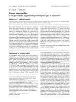
(Figure 1). In this paper, we justify our hypothesis by
showing why it deserves consideration as the explanation
for the genesis of most cancers.
Presentation of the hypothesis
(1) Pathogenic Stimulus
The earliest information a cell receives is a pathogenic
(biological or chemical) stimulus. The first receiver
seems to play a major role in processing the stimulus.
Chemical carcinogenesis is thought to be a two-step
process: in the first step, called “initiation,” the
carcinogen damages or binds to nuclear DNA; in the
second step, referred to as “promotion,” some other
chemical or physiologic event facilitates the aberrant
growth that ultimately results in cancer. The classic
example was reported by Yamigawa and Ichikawa in
1915, when they applied coal tar derivatives to rabbit
ears and observed skin cancer [46]. Subsequent work
showed that dermal application of several different
polyaromatic hydrocarbons (PAHs), such as benzo
[a]pyrene and benzo[a]anthracene, followed by a
phorbol ester (a promoter), generated skin cancers
in a dose-dependent manner. Over time, alkylating
agents, such as sulfur mustard, ethylene dibromide,
and many nitrosoamines, were included in the list
of chemicals that could give rise to cancer, both in
experimental animals and in humans. The list grew to
include arsenic, hexavalent chromium, mycotoxins notably aflatoxins - ionizing and ultraviolet radiation,
Brücher and Jamall BMC Cancer 2014, 14:331
/>
Page 4 of 15
Figure 1 Schematic drawing of “Epistemology of the Origin of Cancer”. Abbreviations: CSES, chronic stress escape strategy; NCCCT, normal
cell cancer cell transition; npGC, neutrophil Granulocyte; TGFβ, tumor growth factor beta; LOX, Lysyl oxidase; ECM, extracellular matrix.
cigarette smoke, and asbestos, to name the most
egregious compounds linked to cancer. Phenotypes
of cancer cells can be the result of mutations, i.e.,
changes in the nucleotide sequence of DNA, which
accumulate as tumors progress. Such mutations
can arise as a result of DNA damage or by the
incorporation of non-complementary nucleotides
during DNA replication. In the past decade or so,
it has been postulated that a cancer must exhibit a
“mutator phenotype” that leads to genomic instability,
but whether or not the acquisition of a mutator
phenotype is necessary for tumor progression
remains unproven [45].
We have long known that nearly all cells are coated
with a thin layer of glycoprotein and acidic material
outside the plasma membrane, called the glycocalyx
[47], which consists of polysaccharides covalently
bonded to membrane proteins (90% glycoproteins
and 10% glycolipids). The surface and size of the
glycocalyx that coats biological membranes differ in
their specific function. The glycocalyx in mammalian
cells contains 5 classses of phylogenetically conserved
molecules for adhesion: (1) immunoglobulins (2)
integrins (3) cadherins (4) selectins, and (5) cell
adhesion-molecules. Through these, the glycocalyx
contacts the microfilament (cytoskeletal) system of the
cells, couples with GTP-binding proteins of the cell
membrane, and communicates between cells and their
microenvironment. Other functions include protecting
the cell and underlying tissues from dehydration or
phagocytosis, providing adherence on the surfaces,
acting against a pathogenic factors, interacting in
cell-to-cell communication, and in vessels, housing
vascular protective enzymes [48].
Due to its oligopolysacharide polymers and sialic
acids, the glycocalyx surrounding mammalian cells is
negatively charged. The resulting electrostatic
repulsion is thought to be important in protecting
cells from non-specific adhesions [49] and, reportedly,
that “specific lock-and-key-type adhesion molecules
overcome this repellent force” [50]. Downregulation
of the repelling components of the glycocalyx in
oligodendrocytes brings extracellular surfaces
separated by long distances closer together, a
finding that could explain the way changes in
pH or ion concentrations seem to influence myelin
destabilization in multiple sclerosis [51]. Moreover,
ROS cause proteinuria by modulating the barrier
function of the glomeruli endothelial glycocalyx
[52]. Disruption of the glycocalyx in vascular tissue
results in inflammation and thrombosis, and is under
investigation in the search for new cardiovascular
drugs [53]. We think that because it receives
information first from a pathogenic stimulus the
glycocalyx deserves greater emphasis in the effort
to elucidate its significance in cancer.
Brücher and Jamall BMC Cancer 2014, 14:331
/>
(2)Chronic inflammation
Some 230 years ago, the British physician, Sir
Percival Pott, reported a high incidence of scrotal
cancers in chimney sweeps, suggesting that irritation
by soot led to a chronic inflammation of the
scrotum and that, in turn, resulted in the scrotal
cancers in this cohort [54]. Later, in 1863, Virchow
observed leukocytes in neoplastic tissue [55],
indicative of inflammation, but he could not
determine whether the inflammation was a cause or
an effect of the accompanying neoplasia. John
Chalmers da Costa reported two cases of squamous
cell carcinoma within chronic ulcers and noted, “[it
is] believed, that cancer may arise … in an area of
chronic inflammation” [56]. As mentioned above, in
the early 20th century, Yamagiwa and Ishikawa
repeatedly applied coal tar to rabbit ears and
observed the resultant tumor growth, which was
preceded by chronic inflammation [46]. William Gye
used acriflavine, other antiseptics, and heat
treatment to inactivate filtrates from the Rous
sarcoma, which were free of tumor cells, and
demonstrated that these filtrates gave rise to chronic
inflammation before the onset of the cancer [57].
All organisms attempt to resolve the disruption of
cells and tissues caused by inflammation, a complex
and multifactorial process that usually results in
wound healing. Persistent acute inflammation due to
non-degradable pathogenic stimuli such as a viral or
bacterial infection, a persistent foreign body, or an
autoimmune reaction results in unresolved wound
healing with consequent chronic inflammation.
Between acute and chronic inflammation lye a
wide range of overlapping processes; the kind of
inflammation found at the midway point of that
range is often referred to as sub-acute inflammation
[1]. In addition to the differences between acute and
chronic inflammation, a difference between local and
systemic wound responses, in terms of inter-tissue and
organ communications, also exists [58]. Modulation of
cell interacting junctions is maintained for epithelial
integrity and, in particular, desmosomes, connexins,
and adhesion complexes are downregulated at the
wound edge [59,60]. The major cells involved are
mononuclear: monocytes, lymphocytes, plasma
cells, fibroblasts, and, especially, mast cells (MCs).
Paul Ehrlich, in 1878, first described MCs in detail
[61]; more recently, they have been reported as a
component of the tumor microenvironment reviewed
in [62]. MCs are thus a significant communication link
between a pathogenic stimulus, the glycocalyx, and the
cell stroma directly and/or via fibroblasts. MCs can be
activated directly by a pathogen or indirectly by binding
to such receptors as the high-affinity immunoglobulin
Page 5 of 15
E (IgE) receptor FcεRI, as well as through pattern
recognition receptors (PRRs), e.g., toll-like receptors
(TLRs) [63,64] and G-protein-coupled receptors
(GPCRs) [63]. MCs present native protein antigens to
CD4+ T-cells and act as antigen-presenting cells
(APC); both cell types influence each other in an
antigen-dependent manner [65]. CD4+ T-cell
populations, with their regulatory interactions, play
a role in the host response to pathogenic stimuli [66].
Contact-mediated activation of endothelial cells by
T-cells involving a ligand such as CD40 may serve as
one mechanism for the continuous progression of
inflammatory diseases in atherosclerosis and
rheumatoid arthritis [67]. Immune cells and their
cytokines have been reported to be associated with
carcinogenesis and T-cell-infiltrating tumors such as
ovarian, breast, prostate, renal, esophageal, colorectal
carcinomas, and melanomas, all of which have been
correlated with patient outcome [68-74]. Stromal
cell-related cytokines of inflammation such as tumor
necrosis factor alpha (TNF-α) activate the nuclear factor
kappa-light-chain-enhancer of activated B cells (NF-κB),
which plays an important role - not completely
understood - in carcinogenesis [75,76]. Inflammation
“associated” cells, as well as the tumor microenvironment, interacts with all different types of immune cells
[20,77], and MCs effectively communicate among
vascular, nerve, and immune system cells [78].
To date, some 15% of all human cancers are
reported to originate from infectious disease [22,23].
However, the majority of cancers arises spontaneously
and is attributed to an unknown etiology. Although
formally designated as “unknown etiology,” under the
existing paradigm an accumulation of a number of
somatic mutations greater than some threshold not
yet defined is considered to be the principal triggering
factor. Chronic inflammation is known to lead to
derangement in signaling processes and to a local
microenvironment described as lying somewhere
between pre-cancerous stromal cells and cancer
cells [79], even as the details of the steps in the
transformation to a cancer cell are incompletely
understood [80]. Earlier findings [81], recently
revisited [82], demonstrated that wound healing
leads to a microenvironment similar to the
hospital-observed stroma of tumors. The tumors
were compared to wounds that do not heal [83]. A
complex biological and immunological process [84]
leads to all of the five signs of cancer first noted by
Celsus and Galen [85]: dolor (pain), calor (heat),
rubor (redness), tumor (swelling) and function laesa
(loss of function).
It has been stated that “the direct link between
pathogen-specific gene products and a stereotypical
Brücher and Jamall BMC Cancer 2014, 14:331
/>
altered host response key to disease development is
missing” [86]. Observations in epidemiology and
laboratory research have generated sufficient evidence
that chronic inflammation evokes an increased
susceptibility to cancer [87]. The association of
chronic inflammation and cancer makes the fact that
a low-dose aspirin regimen, known to suppress
prostaglandin-H2-synthase (COX-1, COX-2), could
have an anticancer effect in colorectal cancer [88]. We
have no data on the prevalence of “silent” inflammation,
as it is often low-level and sub-clinical, but we do
know that a weakened immune system may facilitate
the initiation of tumor growth [89]. Eliminating the
triggering event for infection or inflammation typically
results in healing and tissue repair. If the infection or
consequent inflammation is not completely resolved,
it simmers as a chronic inflammatory condition [90],
setting up one of the pre-conditions for transforming
normal cell to cancerous cells.
The primary mediators of cells involved in
inflammation are IFN-γ (equivalent to macrophageactivating factor), other cytokines, growth factors,
ROS (released by macrophages), and hydrolytic
enzymes. ROS are toxic for the organism and the
tissue, and both are usually protected against ROS by
alpha-1-microglobulin, superoxide dismutases (SOD),
catalases, lactoperoxidases, glutathione peroxidases,
and peroxiredoxins [91]. Exogenous ROS can come
from pollutants, tobacco, smoke, xenobiotics, or
radiation; endogenous ROS are produced intracellularlily through multiple mechanisms. Depending on
the cell and tissue, the major ROS sources are the
dedicated producers: NADPH oxidase, (NOX)
complexes (7 distinct isoforms) in cell membranes,
mitochondria, peroxisomes, and the endoplasmic
reticulum [92]. The resulting oxidative stress affects
not only cells but also the ECM, which is thought to
enjoy less antioxidant capacity than do cells: Madsen
and Sahai stated that the “cytoskeleton of a typical
epithelial cell and many cancer cells is not adapted to
withstand stresses” and that the microenvironment of
acute inflammation differs significantly from that of
chronic inflammation [93]. Additionally, the proteins
of connexins, Cx43 and Cx32, are synthesized and
integrated into the cell membranes of MCs [94],
monocytes [95], leukocytes [96], and Kupffer cells [97].
They have also been found in cells associated with
brain tumors [98], reviewed in [99]. Thus, cell types
such as those of the brain and immune system can
communicate with their microenvironment via
expressed connexins.
Cancer has been linked to various pathogens,
including the Epstein-Barr virus (EBV) in Burkitt’s
lymphoma and nasopharyngeal carcinomas [100]
Page 6 of 15
and human papilloma virus (HPV) in cervical
cancer [101]. In 2005, the Nobel Prize honored the
discovery that infection by Helicobacter pylori (H.
pylori) leads to inflammation, gastritis, and peptic
ulcer [102]. The fact that H. pylori increases the risk
of gastric cancer is widely accepted [103]. When it
infects, H. pylori attaches to cell-cell interfaces and
the bacterium changes it shape, adhering to the cell
and secreting outer membrane vesicles [104]. It has
been shown that the extent of “loss or dysfunction of
E-cadherin was proportional to the migratory behavior
of tumor cells and its metastatic potential” [104-106].
Loss of E-cadherin is associated with loss of cell-cell
adherens and increased epithelial permeability.
Within 48 hours after H. pylori infection, a
significant proportion of E-cadherin was found in
small vesicles within the cell [107]; furthermore,
vacuolating cytotoxin VacA from H. pylori enhanced
the association of intracellular H. pylori vesicles
containing lipopolysaccharide [108]. We assume
these are the effects of the chronic inflammatory
processes because, according to the Kuehn and
Kesty review [109], so-called membrane vesicles of
bacteria contain not just lipopolysacharides, but also
chromosomal, plasmid, and phage DNA [110-112].
Why do all chronic inflammations not result in
cancer? If chronic inflammation, per se, were a
sentinel event in the transformation of a normal cell
to a cancer cell, one would expect a high incidence
of cancer in patients with chronic arthritis, but that
is not evident. The nature of the inflammation that
can facilitate the development of cancer, and of
that that does not, is as yet unexplained. Patients
with rheumatoid arthritis have a greater risk than
non-arthritic patients for lymphoma, melanoma, and
lung cancer, but not of colon cancer or breast cancer
[113]. We do know, however, that severe pneumonitis
associated with either bacterial pneumonia or
tuberculosis resolves completely with treatment,
whereas inflammation associated with H. pylori can
result in gastric cancer in about 60% of cases, and with
hepatitis B or C, in liver cancer in as many as 80% of
chronic infections [24]. Perhaps the distinctive feature
in the inflammation that promotes the conversion of
a normal cell to a cancerous one is its ability to
trigger the onset of fibrosis. For example, pulmonary
mesothelioma, known to be caused by exposure to
asbestos, generally presents decades after exposure. Its
appearance is always preceded by inflammation and
by severe fibrosis [114]. No increase in the number
somatic mutations has been associated with asbestos
carcinogenesis. In a mouse model of experimental
hepatocellular carcinoma (HCC), injection of a single
dose of an initiator such as diethylnitrosamine
Brücher and Jamall BMC Cancer 2014, 14:331
/>
(DEN), followed by repeated sub-toxic doses of
carbon tetrachloride (promotor), resulted in both
inflammation and fibrosis, as well as a 100% incidence
of HCC that mimicked the human disease [115].
Furthermore, only recently, ultraviolet radiationinduced inflammation has been demonstrated to
promote angiotropism and metastasis in melanoma;
blocking the inflammation alone markedly reduced
the incidence of metastasis [116,117]. Patients with
chronic inflammatory diseases can develop cancer after
variable latency periods. For example, a long-term
follow-up of patients with oral pre-cancerous lesions
demonstrated an increased risk for oral cancers after 5
and 10 years of about 5% and 10%, respectively [118].
(3)Fibrosis and changes in the microenvironment
Since chronic scars were first linked to the onset of
cancer, well over 100 years ago, chronic
inflammation has been associated with fibrosis [119];
Hepatitis B and C infections are related to
hepatocellular carcinoma (HCC) in patients who first
develop liver fibrosis [120]. A recent review of cell-cell
communication between MCs and fibroblasts states,
“The remodeling phase of inflammation may explain
chronic fibrosis”; preventing the accumulation of MCs
and their interference of fibroblast activation via
connexins may offer a new approach to prevent excess
scarring [121]. The process of fibrogenesis, an integral
part of wound healing as the organism tries to resolve
chronic inflammation, is governed by three factors:
continuous stimulus, an imbalance of collagen synthesis
versus degradation, and a decrease in the activity of the
degradative enzymes involved in removing
collagen [122]. One key enzyme for the permanent
cross-linking of single triple-helix collagen molecules
(multiple tropocollagen molecules) is the copper
(Cu)-dependent amine oxidase, lysyl oxidase (LOX),
discovered by Pinnell and Martin in 1968 [123].
LOX is an extracellular amine oxidase that catalyzes
the covalent crosslinking of ECM fibers. Collagen I,
a component of both desmoplastic tumor stroma
and organ fibrosis is a major substrate for LOX and
has been shown to be a key component of both
primary and metastatic tumor microenvironments
[124,125]. Elevated levels of procollagen I, a collagen
I precursor, have been found in the serum of
patients with recurrent breast cancer [126]. They
also have been shown to drive the activation of
dormant D2.OR cells seeded to the lung [127].
LOX activity was reported to be greater in human
breast cancer than in normal tissues [128], a finding
that suggests that LOX plays a key role in creating
the cellular microenvironment necessary for a
pre-cancerous niche (PCN), one of the prerequisites
for the induction of cancer. LOX overexpression is
Page 7 of 15
found in myofibroblasts and myoepithelial cells
around in situ tumors and at the invasion front of
infiltrating breast cancers [129]. It was shown to be
essential for hypoxia-induced metastasis [130] and,
more recently, it has been rather elegantly demonstrated
that targeting LOX prevents both fibrosis and metastatic
colonization [131]. Furthermore, LOX modulates
the ECM and also cell migration and growth [132].
Studies in the blind mole rat, Spalax, revealed that
the fibroblasts in this species suppress the growth of
human cancer cells in vitro [133] and decrease the
activity of hyaluronan synthase 2 [134]. This species
was also resistant to chemical carcinogenesis. These
data constitute evidence that fibrosis is necessary
for establishing the PCN stage, an intermediate
stage on the path from a normal cell to a cancer
cell. Additionally, it has been shown that necrotic
wounds induced in Spalax by chemical carcinogens
heal with no sign of malignancy [133], a finding
that supports our hypothesis that the PCN stage
is key to the transformation of a normal cell to a
cancer cell.
Some of the LOX findings are paradoxical [135]; we
assume the paradoxes are due to the fact that early
investigators did not differentiate among the
different LOX isoforms. That LOX was expressed in
79% of human breast cancers revealed the
attenuated metastasis of human breast cancer cells
by a downregulation of adhesion kinase and the
paxillin-signaling pathway [128,136]. SNPs in the
LOX-like protein 4 were reported in patients with
endometriosis, a semi-malignant tumor [137]. LOX
overexpression can be found in myofibroblasts and
myoepithelial cells around in situ tumors and at the
invasion front of infiltrating breast cancers [129].
Further, LOX is downregulated in squamous cell
skin carcinomas [138], head and neck cancers [139],
upper gastrointestinal carcinomas [140-142], and
renal carcinomas [143]. LOX expression was shown
to be upregulated only in the presence of fibroblasts,
suggesting that stromal fibroblasts directly influence
LOX regulation [144]. This finding is concordant
with one previously described, that targeting LOX
prevents fibrosis and metastatic colonization [131].
The ECM itself provides biochemical and physical
signaling to modulate and sustain surrounding tissue
and cells (tumor microenvironment). LOX induction
is mediated by both tumor growth factor beta
(TGFβ-) and Smad and non-Smad JNK/AP-1
signaling pathways; it has been shown in vitro that
LOX expression is blocked by “TGFβ inhibitors as well
as by inhibitors of the canonical Smad2, −3, and −4
signaling and non-Smad JNK/AP-1 signaling pathways”.
[145] This regulation of LOX is mediated in endothelial
Brücher and Jamall BMC Cancer 2014, 14:331
/>
cells by such adhesion molecules as P-selectin, vascular
cell adhesion molecule (VCAM-1), intracellular
adhesion molecule (ICAM-1), and monocyte
chemotactic protein (MCP-1) [146]. Furthermore,
Cx43 expression is paralleled closely by that of adhesion
markers such as VCAM-1, ICAM-1, and MCP-1 [147].
A number of reasons could explain the discrepancies
reported of the down- and upregulation in LOX.
Among these are the following: (1) Biomarkers, such as
tissue inhibitors exhibit different levels of expression in
tumor tissue compared to the tumor invasion zone or
normal tissue. For example, Kopitz et al. investigated
tissue inhibitor of metalloproteinase 1 (TIMP-1) in
liver metastasis with reported significantly different
expression levels in (a) tumor tissue, (b) invasion zone
tissue, and (c) normal tissue [148]. (2) Remodeled
ECM (pre-cancerous niche - PCN) as well as normalcell-to-cancer-cell transitions were in different stages
of completion. The LOX concentrations that differed
according to the type of tumor may also
reflect that both re-modeled ECM (pre-cancerous
niche - PCN) and normal-cell-to-cancer-cell transitions
were encountered in different stages of completion, and
thus the resulting expression levels were different. (3)
The finding of LOX upregulation in the invasion zone
of breast cancer tissue has been reported [129]. (4)
Researchers on LOX usually do not differentiate
among the known isoforms of the enzyme (LOX,
LOX1, LOX2, LOX3 and LOX4), although - even
though they catalyze the same biochemical reaction they differ in their amino acid sequence [149,150].
The LOX isoforms are encoded by different genes (on
chromosomes 5, 15, 8, 2, and 10, respectively), have
different molecular weights, differ in their percentage
of similarities to the LOX domain (100, 85, 58, 65, and
62, respectively), and exhibit different protein sizes as
well as different tissues, depending on their mRNA
expression rates [151]. Moreover, LOX isoenzymes
are expressed differently in different tissues [152]. (5)
Different methodological approaches and protocols
for measuring LOX could account for some of the
reported differences. These five factors might explain
some of the paradoxical findings reported for LOX.
The assumption that fibrosis is a necessary and
thus a key step in the sequence of events preceding
the transformation of normal cells to cancer cells is
supported by the following evidence: (1) The presence
of fibrosis is reported to increase the risk of acquiring
cancer [153]. (2) Fibrosis with chronic inflammation is
reported with a number of pre-cancerous lesions,
e.g., actinic keratosis, Crohn’s disease, and Barrett’s
metaplasia [154-156]. (3) Ongoing fibrosis, with fibrotic
foci, has been observed in postmortem pancreatic
cancer specimens [157]. (4) In cancer-resistant
Page 8 of 15
species such as the blind mole rat, Spalax, fibroblasts
suppress the growth of cancers as well as the activity
of hyaluronic synthase [133,134]. (5) In mice, chronic
low-grade systemic inflammation leads to architectural
changes that permit a mild level of alveolar
macrophage infiltration [158]. (6) One of the features
of oral submucosal fibrosis (OSF), a pre-cancerous
condition, is chronic inflammation of the buccal
mucosa accompanied by a progressive sub-epithelial
fibrotic disorder [159].
(4) Pre-cancerous niche and (5) Chronic-Stress-EscapeStrategy (CSES)
The microenvironment of an acute inflammatory
condition differs significantly from that of chronic
inflammation, in which the host cannot eliminate
the offending agent (a microorganism, a disease, or a
toxin) because the “cytoskeleton of a typical
epithelial cell and many cancer cells is not adapted
to withstand stresses” [93]. Pathogenic stimuli induce
chronic inflammation that, in turn, remodels the
microenvironment, which itself develops fibrosis.
This leads to a modulation of the ECM that, following
exposure to chronic stress, may promote the
formation of a pre-cancerous niche (PCN). Findings
in the Tasmanian Devil, with its contagious cancer,
led to an allograft theory [160]. Other authors have
suggested that the near 100% mortality in this species
was caused by the transmitted clonal tumor through
downregulation of major histocompatibility complex
(MHC) molecules [161], and they proposed an
immunological escape strategy [162,163]. In an
organism, the pathogenic stimulus, the chronic
inflammation, and the fibrosis, which lead to a
pre-cancerous niche, become a “vicious circle” thought
to be resolved through a chronic-stress escape strategy
(CSES). Histopathological investigations of 549 gastric
ulcer patients revealed that about 70% of the lesions
presented intestinal metaplasia within the regenerative
epithelium, where chronic inflammation was considered the precursor of a pre-cancerous lesion [164].
We propose that chronic inflammation, with chronic
TGFβ induction, serves to sustain a persistent stress
in the cells of the host tissue. Furthermore, the
distinction between the inflammation that promotes
the development of a normal cell and that for a
cancerous one lies in the ability of the inflammation
to cause the onset of fibrosis. Asbestos leads to
pulmonary mesothelioma decades after the exposure
reveals fibrosis and, although no increase in somatic
mutations has been reported in asbestos caused
carcinogenesis, chronic inflammation has been
observed in every instance of asbestos-induced
mesothelioma [114]. These differences, in light of
the proposed paradigm, are the duration of
Brücher and Jamall BMC Cancer 2014, 14:331
/>
exposure to the pathogenic stimulus which reflects
the importance of chronic inflammation and fibrosis
in carcinogenesis.
The continuous release of TGFβ that is triggered by
chronic inflammation has many effects: (1) TGFβ
represses E-cadherin and occludin, increasing the
adherens junction disassembly [165]. Inhibiting
TGFβ receptor type-I has been shown to decrease
its invasiveness [166]. (2) TGFβ induces miR21, a
key regulator of mesenchymal phenotype transition
[167], but increased levels also have been observed
in early chronic fibrosis in COPD patients [168]. (3)
TGFβ activates protein kinase B (AKT or PKB)
through phosphoinositide-3 kinase (PI3K) [169],
activating the mechanistic targets of rapamycin
complex 1 (mTORC1) and mTORC2 [170].
Furthermore, TORC activates the translation of
proteins important for cell growth and development,
and the PI3K/TmTORC1 pathway has recently been
shown both essential for cancer-associated inflammation
[171]. (4) LOX and matrix metalloproteinase (MMPs)
are induced by TGFβ [172], and (5) LOX activates PI3K
[173]. (6) The phosphorylation of glycogen synthase
kinase-3beta (GSK3beta) by AKT stabilizes SNAIL
[174], which leads to an increase of TGFβ-induced
SNAIL [175]. (7) SNAIL stability and activity,
furthermore, are activated by LOX [176]. (8) TGFβ
effects the dissociation of the long isoform of p120
from the membrane and its accumulation in the
cytoplasm [177] and Figure two B in [178].
The chronic release of TGFβ and the continuous
LOX activation trigger an accumulation of p120 in
the cytoplasm, inducing remodeling of the ECM,
which forms the pre-cancerous niche. This process
may be seen as the starting point for the chronic-stress
escape strategy. The p120 accumulation stimulates
Cdc42 - a cell-division control protein and a member
of the family of Rho small guanosine triphosphatases
(GTPases) - and activates Ras-related C3 botulinum
toxin substrate 1 (Rac1), decreasing thereby E-cadherin
[179,180], microtubule polymerization [181], and
integrin clustering [182]. Thus, the contact to the
basal membrane is destabilized [183], promoting
cell migration. In addition, p120 suppresses Rho
activity by binding to exchange factor Vav2 and, in
so doing, activates Rac1 [177]. As adherens junctions
are regulated by Rho GTPases, suppressing Rho
destabilizes the adherens junctions, increasing the
dysregulation in the formation of cell-cell complexes.
When microM antisense oligonucleotide was
challenged by p120, after 4 h a decrease of 50% in the
ratio of in vitro LOX cells in mitosis was observed
and, after 8 to 72 h, as much as 70% [184]. These
findings, together with the increase in both p120 and
Page 9 of 15
LOX activity, may indicate a p120 effect with an
additional increase of LOX. SNAIL itself results in
a decrease of E-cadherin [185,186], occludins [187],
claudins [186,187], desmoplakin, and plakoglobin
[188], and an increase in MMPs [189], fibronectin
and vimentin [189], twist-related protein 1
(TWIST), zinc finger E-box-binding homeobox 1
(ZEB1), and ZEB2 [190]. With these cell interactions
and communication mechanisms, all necessary
conditions for cell transition have been accounted for:
the formation of cell-cell complexes are deregulated,
the stability of adherens junctions decreased, and
the apical-basal polarity and re-organization of the
cytoskeletal architecture lost.
(6) Normal Cell-Cancer Cell Transition (NCCCT)
The transition from one cell function to another,
as well as the transition of one cell type to another
seems to be a routine event rather than a rare one.
Embryological studies have shown that the complexbuilding pancreatic homeodomain protein (PDX1)
with pre-B-cell leukemia transcription factor 1
(PBX1) and the PBX-related homeobox gene MRG1
(MEIS2) results in building a multimeric complex
which then switches the nature of its transcriptional
activity in exocrine versus endocrine cells [191,192].
Additionally, it has been shown that an epithelial
mesenchymal transition (EMT) in embryogenesis/
morphogenesis acts in a direction opposite to that of
a mesenchymal-epithelial transition (MET) [193].
Furthermore, EMT can induce non-cancer stem cells
to become cancer stem cells [194,195].
Armin Braun recognized some 60 years ago that a
gram negative bacterium Agrobacterium tumefaciens
(A. tumefaciens) could initiate the in vitro
transformation of normal plant cells into tumor
cells; he showed that transformation occurs in a
short time period, resulting in tumor cells with
slower growth and less progression [196-198]. Ivo
Zaenen et al. revealed, and Mary-Ann Chilton’s group
subsequently proved, that a small DNA plasmid
within A. tumefaciens was responsible for the
transformation [199]: tumor inducing DNA (Ti-DNA),
after infection, was integrated into the plant genome
in tobacco plants [200]. Chilton also showed that
Braun’s findings were based on the same principle:
although the T-DNA from the A. tumefaciens
Ti-plasmids was not at first detected [201], it was
later proven to be in the nuclear DNA fraction of
crown-gall tumors [202]. More evidence comes from
research on mesothelial cells. In 1966, Eskeland, based
on silver-staining electron microscopy studies, first
suggested that injured or destroyed mesothelial cells
are replaced in location and function by free-floating
“peritoneal macrophages,” which are transformed
Brücher and Jamall BMC Cancer 2014, 14:331
/>
from their original role to that of mesothelial cells
[203,204]. This hypothesis was supported by further
microscopy and electron microscopy studies from the
same group [205,206] and by the later findings of Ryan
and Watters [207,208]. As a consequence of a
pathogenic stimulus such as inflammation or
wound healing, EMT can change MCs into cells with
mesenchymal or epithelial characteristics [122]. Xin
reported supportive findings in prostate cancer that
“the cells of origin of cancer are the cells within tissues
that serve as the target for transformation” [209].
Similarly, studies in which Cx43 was knocked out to
inhibit cell transition in corneal cells in vivo have
shown that multifactorial regulated cell transition is
influenced by cell-cell communication [210]. There is
further evidence that a decrease in cell-cell adhesion
is crucial for cell transition [211].
Because, under special circumstances, one type of
human cell can transition to another, proposing that
a normal cell transition to a cancer cell as one
important sequence in carcinogenesis is justified.
Additionally, evidence has been presented that a
pathogenic stimulus gives rise to a molecular link of
host immune response and genotoxic events,
followed by inflammation also associated with
carcinogenesis [212]. We propose that the
observations in both the plant and animal kingdoms
described above, taken together with the discovery
of H. pylori, the finding that EBV can transform
lymphocytes into cancer [213], and the identification
of HPV 16 DNA [214] and HPV 18 [215] in cervical
cancers (HPV infection is a precondition for about
75% of human cervical cancers) further support our
hypothesis. EMT and MET were described as
necessary for tissue repair and for migration,
invasion, and metastasis [193]. We assume, in
contrast, that - after a latency period in the CSES - a
PCN results from chronic inflammation and fibrosis,
and those conditions lead to a NCCCT.
To the extent that the above discussion proves
the principle that chronic inflammation, including
sub-clinical inflammation, can - after a latency
period in the PCN stage - induce the a transformation
of a normal cell to a cancer cell, finding biomarkers
to define this sequence of events is important. The
chronic inflammation and the fibrotic changes,
including perhaps LOX activity, could explain the
considerable aggression of many cancer cells, once
transformed.
Testing the hypothesis
We have described a new paradigm for the origin of the
majority of cancers, based on observations and experimental findings in plants, animals, and humans. The
Page 10 of 15
paradigm postulates that most cancers originate from a
stimulus and are followed by chronic inflammation,
fibrosis, and a change in the tissue microenvironment
that leads to a pre-cancerous niche (PCN). The organism
responds with a chronic stress escape strategy (CSES),
which, if not completely resolved, can induce a normal
cell-cancer cell transition (NCCCT) (Figure 1).
If, based on experimental and clinical findings presented
here, this hypothesis is plausible, then the majority of
findings in the genetics of cancer so far reported in the
literature are late events or epiphenomena that could
have occurred after the development of a PCN. Our
model would make clear the need to establish preventive measures long before a cancer becomes clinically
apparent. Future research should focus on the intermediate steps of our proposed sequence of events, which will
enhance our understanding of the nature of carcinogenesis. Findings on inflammation and fibrosis would be given
their warranted importance, with research in anticancer
therapies focusing on suppressing the PCN state with very
early intervention to detect and quantify any subclinical
inflammatory change and to treat all levels of chronic
inflammation and prevent fibrotic changes, and so avoid
the transition from a normal cell to a cancer cell.
Implication of the hypothesis
We suggest that the majority of findings reported on the
genetics of cancer are either late events or epiphenomena
and that the different observations from basic and clinical
research, combined with those from the plant, animal, and
human world, justify our hypothesis. The development of
cancer traces the following pathway: 1) pathogenic stimulus, 2) chronic inflammation, 3) fibrosis, 4) changes in the
cellular microenvironment that result in a pre-cancerous
niche, 5) deployment of a chronic-stress escape strategy,
and 6) a transition from normal cell to cancer cell. The
paradigm proposed here, if proven, spells out a sequence
of steps, one or more of which could be interdicted or
modulated early in carcinogenesis to prevent or, at a minimum, slow down the progression of many cancers.
Abbreviations
Akt: Protein kinase B; APC: Antigen presenting cell; BRCA1: Breast cancer 1,
early onset; BRCA2: Breast cancer 2, early onset; COX-1: Cyclooxygenase-1
(=Prostaglandin G/H synthetase 1); COX-2: Cyclooxygenase-2 (=Prostaglandin
G/H synthetase 2); CSES: Chronic stress escape strategy; CTC: Circulating tumor
cells; Cx43: Connexin 43; Cx32: Connexin 32; dbSNP: Single nucleotide
polymorphism database; DEN: Diethylnitrosamine; DNA: Deoxyribonucleic acid;
EBV: Epstein-Barr virus; ECM: Extracellular matrix; EMT: Epithelial- mesenchymal
transition; GPCR: G protein-coupled receptors; GSK3beta: Glycogen synthase
kinase-3beta; GTPase: Small guanosine triphosphateses; HCC: Hepatocellular
carcinoma; HPV: Human papilloma virus; ICAM-1: Intracellular adhesion
molecule 1; IFN-γ: Macrophage-activating factor; IgE: Immunoglobulin E;
LINE-1: Long interspersed element 1; LTR: Long terminal transposanable
retroposon; LOX: Lysyl oxidase; MC: Mast cell; MCP-1: Monocyte chemotactic
protein; MEIS2: PBX-related homeobox gene MRG1; MET: Mesenchymalepithelial transition; MHC: Major histocompatibility complex; MMP: Matrix
metalloproteinase; NCCCT: Normal cell-cancer cell transition; NF-κB: Nuclear
Brücher and Jamall BMC Cancer 2014, 14:331
/>
factor kappa-light-chain-enhancer of activated B cells; NOX: NADPH oxidase;
PBX1: Pre-B-cell leukemia transcription factor 1; PCN: Pre-cancerous niche;
PDX1: Pancreatic homeodomain protein; PI3K: Phosphoinositide-3 kinase;
PRR: Pattern recognition receptor; Rac1: Ras-related C3 botulinum toxin
substrate 1; RFLP: Restriction fragment length polymorphism; Rho: Ras
homolog gene; ROS: Reactive oxygen species; SINE: Short interspersed
element; SNP: Single-nucleotide polymorphism; SOD: Superoxide dismutase;
TNFα: Tumor necrosis factor alpha; TGFβ: Tumor growth factor beta;
TIMP-1: Tissue inhibitor of metalloproteinase 1; TLR: Toll-like receptor;
TORC1: Target of rapamycin complex 1; TORC2: Target of rapamycin complex 2;
TWIST: Twist-related protein 1; VCAM-1: Vascular cell adhesion molecule;
ZEB1: Zinc finger E-box-binding homeobox 1.
Competing interests
Neither author has a competing interest to disclose.
Authors’ contributions
This manuscript contains original material that has not been previously
published. Both authors equally contributed in thinking, discussing, and writing
for the manuscript. Both author read and approved the final manuscript.
Authors’ information
BB www.linkedin.com/in/bruecher.
IS www.linkedin.com/pub/ijaz-jamall-ph-d-dabt/1b/69/b92.
Acknowledgement
We gratefully acknowledge Professor Dr. Karl Daumer, Professor emeritus in
Biology, Munich, and Dr.rer.nat.Dipl.Phys.Martin Daumer, whose kindness in
discussing the immunology of plants with us in 2013 was of great
importance to the development of our thinking.
Author details
1
Theodor-Billroth-Academy®, Munich, Germany. 2Theodor-Billroth-Academy®,
Richmond, VA, USA. 3Theodor-Billroth-Academy®, Sacramento, CA, USA.
4
INCORE, International Consortium of Research Excellence of the TheodorBillroth-Academy®, Munich, Germany. 5INCORE, International Consortium of
Research Excellence of the Theodor- Billroth-Academy®, Richmond, Virginia,
USA. 6INCORE, International Consortium of Research Excellence of the
Theodor- Billroth-Academy®, Sacramento, CA, USA. 7Bon Secours Cancer
Institute, Richmond, VA, USA. 8Risk-Based Decisions, Inc., Sacramento, CA, USA.
Received: 14 March 2014 Accepted: 6 May 2014
Published: 10 May 2014
References
1. Anderson WAD: Pathology, Volume One. 6th edition. St. Louis: The CV Mosby
Company; 1971.
2. Howard WT, Schultz OS: Studies in the Biology of Tumor Cells. New York:
The Rockefeller Institute of Medical Research; 1911.
3. Vogelstein B, Kinzler KW: Cancer genes and the pathways they control.
Nat Med 2004, 10(8):789–799.
4. Hanahan D, Weinberg RA: Hallmarks of cancer: the next generation.
Cell 2011, 144(5):646–674.
5. Bauer KH: Mutationstheorie der Geschwulst-Entstehung. Berlin: Julius Springer
Verlag; 1928.
6. Knudson A: Mutation and cancer: statistical study in Retinoblastoma.
Proc Natl Acad Sci U S A 1971, 68(4):820–823.
7. Watson JD, Crick FH: Molecular structure of nucleic acids; a structure for
deoxyribose nucleic acid. Nature 1953, 171(4356):737–738.
8. Friedman M, Friedland GW: Medicine’s 10 Greatest Discoveries. Yale University
Press; 1998.
9. Cobb M: 1953: when genes become “information”. Cell 2013,
153(3):503–506.
10. Ashley DJB: The two “hit” and multiple “hit” theories of carcinogenesis.
Br J Cancer 1969, 23(2):313–328.
11. Fearon ER, Vogelstein B: A genetic model for colorectal tumorigenesis.
Cell 1990, 61(5):759–767.
12. Hanahan D, Weinberg RA: The hallmarks of cancer. Cell 2000, 100(1):57–70.
13. Cleaver JE: Photosensitivity brings light to a new transcription-coupled
DNA repair cofactor. Nat Genet 2012, 44(5):447–478.
Page 11 of 15
14. Rosenfeld S: Are the somatic mutation and tissue organization field
theories of carcinogenesis incompatible? Cancer Inform 2013, 12:221–229.
15. Versteeg R: Cancer: tumours outside the mutation box. Nature 2014,
506(7489):438–439.
16. Mack SC, Witt H, Piro RM, Gu L, Zuyderduyn S, Stütz AM, Wang X, Gallo M,
Garzia L, Zayne K, Zhang X, Ramaswamy V, Jäger N, Jones DT, Sill M, Pugh
TJ, Ryzhova M, Wani KM, Shih DJ, Head R, Remke M, Bailey SD, Zichner T,
Faria CC, Barszczyk M, Stark S, Seker-Cin H, Hutter S, Johann P, Bender S,
et al: Epigenomic alterations define lethal CIMP-positive ependymomas
of infancy. Nature 2014, 506(7489):445–450.
17. Parker M, Mohankumar KM, Punchihewa C, Weinlich R, Dalton JD, Li Y,
Lee R, Tatevossian RG, Phoenix TN, Thiruvenkatam R, White E, Tang B,
Orisme W, Gupta K, Rusch M, Chen X, Li Y, Nagahawhatte P, Hedlund E,
Finkelstein D, Wu G, Shurtleff S, Easton J, Boggs K, Yergeau D, Vadodaria B,
Mulder HL, Becksford J, Gupta P, Huether R, et al: C11orf95-RELA fusions
drive oncogenic NF-κB signalling in ependymoma. Nature 2014,
506(7489):451–455.
18. Roche B, Sprouffske K, Hbid H, Missé D, Thomas F: Peto’s paradox revisited:
theoretical evolutionary dynamics of cancer in wild populations.
Evol Appl 2013, 6(1):109–116.
19. Kim EB, Fang X, Fushan AA, Huang Z, Lobanov AV, Han L, Marino SM, Sun X,
Turanov AA, Yang P, Yim SH, Zhao X, Kasaikina MV, Stoletzki N, Peng C,
Polak P, Xiong Z, Kiezun A, Zhu Y, Chen Y, Kryukov GV, Zhang Q, Peshkin L,
Yang L, Bronson RT, Buffenstein R, Wang B, Han C, Li Q, Chen L, et al:
Genome sequencing reveals insights into physiology and longevity of
the naked mole rat. Nature 2011, 479(7372):223–227.
20. Grivennikov SI, Greten FR, Karin M: Immunity, Inflammation, and Cancer.
Cell 2010, 140(6):883–899.
21. Tomlinson IP, Novelli MR, Bodmer WF: The mutation rate and cancer.
Proc Natl Acad Sci U S A 1996, 93(25):14800–14803.
22. Blattner WA: Human retroviruses: their role in cancer. Proc Assoc Am
Physicians 1999, 111(6):563–572.
23. Parkin DM: The global health burden of infection-associated cancers in
the year 2002. Int J Cancer 2006, 118(12):3030–3044.
24. Pisani P, Parkin DM, Muñoz N, Ferlay J: Cancer and infection: estimates of
the attributable fraction in 1990. Cancer Epidemiol Biomarkers Prev 1997,
6(6):387–400.
25. Liu B, Nicolaides NC, Markowitz S, Willson JK, Parsons RE, Jen J,
Papadopolous N, Peltomaki P, de la Chapelle A, Hamilton SR, Kinzler KW,
Vogelstein B: Mismatch repair gene defects in sporadic colorectal cancers
with microsatellite instability. Nat Genet 1995, 9(1):48–55.
26. Vogelstein B, Papadopoulos N, Velculescu VE, Zhou S, Diaz LA Jr, Kinzler KW:
Cancer genome landscapes. Science 2013, 339(6127):1546–1558.
27. Tomasetti C, Vogelstein B, Parmigiani G: Half or more of the somatic
mutations in cancers of self-renewing tissues originate prior to tumor
initiation. Proc Natl Acad Sci U S A 2013, 110(6):1999–2004.
28. Da Cunha AB: Genetic analysis of the polymorphism of color pattern in
Drosophila polymorphia. Evolution 1949, 3(3):239–251.
29. National Center for Biotechnology Information, United States National
Library of Medicine: NCBI dbSNP build 138 for human. 2013, http://www.
ncbi.nlm.nih.gov/mailman/pipermail/dbsnp-announce/2013q3/000133.html.
30. Human Genome Project 2013: The science behind the human genome
project: understanding the basics. />Human_Genome/project/info.shtml.
31. European Bioinformatics Institute (EBI) and Wellcome Trust Sanger:
Ensemble database 2013. />Location/Chromosome?r=1.
32. Watson JD, Baker TA, Bell SP, Gann A, Levine M, Losick R: Molecular Biology
of the Gene. In 5th edition. Pearson: CSHL Press; 2004:732. Benjamin
Cummings Publishers, San Francisco, CA; ISBN: 0-8053-4635-X.
33. Brouha B: Hot L1s account for the bulk of retrotransposition in the
human population. Proc Natl Acad Sci U S A 2003, 100(9):5280–5285.
34. Bennett EA, Keller H, Mills RE, Schmidt S, Moran JV, Weichenrieder O, Devine
SE: Active Alu retrotransposons in the human genome. Genome Res 2008,
18(12):1875–1883.
35. Wicker T, Sabot F, Hua-Van A, Bennetzen JL, Capy P, Chalhoub B, Flavell A,
Leroy P, Morgante M, Panaud O, Paux E, SanMiguel P, Schulman AH:
A unified classification system for eukaryotic transposable elements.
Nat Rev Genet 2007, 8(12):973–982.
36. Slavkin HC, Greulich RC: Extracellular Matrix Influences on Gene Expression.
New York: Academic Press Inc; 1975:833pp.
Brücher and Jamall BMC Cancer 2014, 14:331
/>
37. Mecham RP, Madaras JG, Senior RM: Extracellular matrix-specific induction
of elastogenic differentiation and maintenance of phenotypic stability in
bovine ligament fibroblasts. J Cell Biol 1984, 98(5):1804–1812.
38. Zhe X, Cher ML, Bonfil RD: Circulating tumor cells: finding the needle in
the haystack. Am J Cancer Res 2011, 1(6):740–751.
39. Fidler JJ: Metastasis: guantitative analysis of distribution and fate of
tumor embolilabeled with 125 I-5-iodo-2′-deoxyuridine. J Natl Cancer Inst
1970, 45(4):773–782.
40. Loeb LA: Endogenous carcinogenesis: molecular oncology into the twentyfirst century–presidential address. Cancer Res 1989, 49(20):5489–5496.
41. Lindahl T: Instability and decay of the primary structure of DNA. Nature
1993, 362(6422):709–715.
42. Ames BN, Gold LS, Willett WC: The causes and prevention of cancer. Proc Natl
Acad Sci U S A 1995, 92(12):5258–5265.
43. Wood RD, Mitchell M, Sgouros J, Lindahl T: Human DNA repair genes.
Science 2001, 291(5507):1284–1289.
44. Cervantes RB, Stringer JR, Shao C, Tischfield JA, Stambrook PJ: Embryonic
stem cells and somatic cells differ in mutation frequency and type.
Proc Natl Acad Sci U S A 2002, 99(6):3586–3590.
45. Wogan GN, Hecht SS, Felton JS, Conney AH, Loeb LA: Environmental and
chemical carcinogenesis. Semin Cancer Biol 2004, 14(6):473–486.
46. Yamagiwa K, Ichikawa K: Experimentelle Studie über die Pathogenese der
Epithelialgeschwülste [Experimental study of the pathogenesis of
epithelial tumours]. Mitt Med Fak Tokyo 1915, 15:295–344.
47. Rambourg A, Leblond CP: Electron microscope observations on the
carbohydrate-rich cell coat present at the surface of cells in the rat.
J Cell Biol 1967, 32(1):27–53.
48. Choi Y, Chung H, Jung H, Couchman JR, Oh ES: Syndecans as cell surface
receptors: unique structure equates with functional diversity. Matrix Biol
2011, 30(2):93–99.
49. Curry FE, Adamson RH: Endothelial glycocalyx: permeability barrier and
mechanosensor. Ann Biomed Eng 2012, 40(4):828–839.
50. Sackmann E, Groennenwein: Cell adhesion as dynamic interplay of lockand-key, generic and elastic forces. Prog Theor Phys Suppl 2006, 165:78–99.
51. Bakhti M, Snaidero N, Schneider D, Aggarwal S, Möbius W, Janshoff A,
Eckhardt M, Nave KA, Simons M: Loss of electrostatic cell-surface repulsion
mediates myelin membrane adhesion and compaction in the central
nervous system. Proc Natl Acad Sci U S A 2013, 110(8):3143–3148.
52. Singh A, Ramnath RD, Foster RR, Wylie EC, Fridén V, Dasgupta I, Haraldsson B,
Welsh GI, Mathieson PW, Satchell SC: Reactive oxygen species modulate the
barrier function of the human glomerular endothelial glycocalyx. PLoS One
2013, 8(2):e55852.
53. Drake-Holland AJ, Noble MI: The important new drug target in
cardiovascular medicine–the vascular glycocalyx. Cardiovasc Hematol
Disord Drug Targets 2009, 9(2):118–123.
54. Pott P: Chirurgical observations Volume 3. London: L Hawes, W Clark, and R
Collins; 1775:177–183.
55. Virchow R: Ueber bewegliche thierische Zellen. Arch Path Anat Physiol
1863, 28:237–240.
56. Da Costa JC, III: Carcinomatous changes in an area of chronic ulceration,
or Marjolin’s ulcer. Ann Surg 1903, 37(4):496–502.
57. Gye WE: The cancer problem. Br Med J 1926, 2(3436):865–870.
58. Lee WJ, Miura M: Mechanisms of systemic wound response in Drosophila.
Curr Top Dev Biol 2014, 108:153–183.
59. Beaudry VG, Ihrie RA, Jacobs SB, Nguyen B, Pathak N, Park E, Attardi LD:
Loss of the desmosomal component perp impairs wound healing
in vivo. Dermatol Res Pract 2010, 2010:759731.
60. Gingalewski C, Wang K, Clemens MG, De Maio A: Posttranscriptional
regulation of connexin 32 expression in liver during acute inflammation.
J Cell Physiol 1996, 166(2):461–467.
61. Ehrlich P: Beiträge zur Theorie und Praxis der histologischen Färbung.
Dissertation at Leipzig University; 1878.
62. Dyduch G, Kaczmarczyk K, Okoń K: Mast cells and cancer: enemies or
allies? Pol J Pathol 2012, 63(1):1–7.
63. Gilfillan AM, Tkaczyk C: Integrated signalling pathways for mast-cell
activation. Nat Rev Immunol 2006, 6(3):218–230.
64. Trivedi NH, Guentzel MN, Rodriguez AR, Yu JJ, Forsthuber TG, Arulanandam BP:
Mast cells: multitalented facilitators of protection against bacterial
pathogens. Expert Rev Clin Immunol 2013, 9(2):129–138.
65. Suurmond J, van Heemst J, van Heiningen J, Dorjée AL, Schilham MW,
van der Beek FB, Huizinga TW, Schuerwegh AJ, Toes RE: Communication
Page 12 of 15
66.
67.
68.
69.
70.
71.
72.
73.
74.
75.
76.
77.
78.
79.
80.
81.
82.
83.
84.
85.
86.
87.
88.
89.
90.
91.
between human mast cells and CD4(+) T cells through antigendependent interactions. Eur J Immunol 2013, 43(7):1758–1768.
Powrie F, Correa-Oliveira R, Mauze S, Coffman RL: Regulatory interactions
between CD45RBhigh and CD45RBlow CD4+ T cells are important for the
balance between protective and pathogenic cell-mediated immunity.
J Exp Med 1994, 179(2):589–600.
Monaco C, Andreakos E, Young S, Feldmann M, Paleolog E: T cell-mediated
signaling to vascular endothelium: induction of cytokines, chemokines,
and tissue factor. J Leukoc Biol 2002, 71(4):659–668.
Zhang L, Conejo-Garcia JR, Katsaros D, Gimotty PA, Massobrio M, Regnani G,
Makrigiannakis A, Gray H, Schlienger K, Liebman MN, Rubin SC, Coukos G:
Intratumoral T cells, recurrence, and survival in epithelial ovarian cancer.
N Engl J Med 2003, 348(3):203–213.
Marrogi AJ, Munshi A, Merogi AJ, Ohadike Y, El-Habashi A, Marrogi OL,
Freeman SM: Study of tumor infiltrating lymphocytes and transforming
growth factor-beta as prognostic factors in breast carcinoma. Int J Cancer
1997, 74(5):492–501.
Vesalainen S, Lipponen P, Talja M, Syrjanen K: Histological grade,
perineural infiltration, tumour-infiltrating lymphocytes and apoptosis
as determinants of long-term prognosis in prostatic adenocarcinoma.
Eur J Cancer 1994, 30A(12):1797–1803.
Nakano O, Sato M, Naito Y, Suzuki K, Orikasa S, Aizawa M, Suzuki Y, Shintaku I,
Nagura H, Ohtani H: Proliferative activity of intratumoral CD8(+) T-lymphocytes
as a prognostic factor in human renal cell carcinoma: clinicopathologic
demonstration of antitumor immunity. Cancer Res 2001, 61(13):5132–5136.
Schumacher K, Haensch W, Roefzaad C, Schlag PM: Prognostic significance
of activated CD8(+) T cell infiltrations within esophageal carcinomas.
Cancer Res 2001, 61(10):3932–3936.
Naito Y, Saito K, Shiiba K, Ohuchi A, Saigenji K, Nagura H, Ohtani H: CD8+ T
cells infiltrated within cancer cell nests as a prognostic factor in human
colorectal cancer. Cancer Res 1998, 58(16):3491–3494.
Halpern AC, Schuchter LM: Prognostic models in melanoma. Semin Oncol
1997, 24(1 Suppl 4):S2–S7.
Greten FR, Eckmann L, Greten TF, Park JM, Li ZW, Egan LJ, Kagnoff MF, Karin M:
IKKβ links inflammation and tumorigenesis in a mouse model of
colitis-associated cancer. Cell 2004, 118(3):285–296.
Pikarsky E, Porat RM, Stein I, Abramovitch R, Amit S, Kasem S, Gutkovich-Pyest
E, Urieli-Shoval S, Galun E, Ben-Neriah Y: NF-ƙB functions as a tumour promoter in inflammation-associated cancer. Nature 2004, 431(7007):461–466.
Grivennikov SI, Karin M: Immunity and oncogenesis: a vicious connection.
Curr Opin Genet Dev 2010, 20(1):65–71.
Silver R, Curley JP: Mast cells on the mind: new insights and
opportunities. Trends Neurosci 2013, 36(9):513–521.
Yang J, Weinberg RA: Epithelial-mesenchymal transition: at the crossroads
of development and tumor metastasis. Dev Cell 2008, 14(6):818–829.
Nathan C, Ding A: Nonresolving inflammation. Cell 2010, 140(6):871–882.
Dvorak HF: Tumors: wounds that do not heal. Similarities bewtween tumor
stroma generation and wound healing. N Engl J Med 1986, 315(26):1650–1659.
Chaffer CL, Weinberg RA: A perspective on cancer cell metastasis. Science
2011, 331(6024):1559–1564.
Hirshberg A, Leibovich P, Horowitz I, Buchner A: Metastatic tumors to
postextraction sites. J Oral Maxillofac Surg 1993, 51(12):1334–1337.
Scott A, Khan KM, Cook JL, Duronio V: What is inflammation? Are we
ready to move beyond Celsus? Inflammation 2004, 38(3):248–249.
Porth C: Essentials of pathophysiology: concepts of altered health states.
Hagerstown, MD: Lippincott Williams & Wilkins; 2007:270.
Karin M, Lawrence T, Nizet V: Innate immunity gone awry: linking microbial
infections to chronic inflammation and cancer. Cell 2006, 124(4):823–835.
Mantovani A: Molecular Pathways liking inflammation and cancer.
Curr Mol Med 2010, 10(4):369–373.
Kozak W, Kluger MJ, Tesfaigzi J, Kozak A, Mayfield KP, Wachulec M,
Dokladny K: Molecular Mechanisms of fever and endogenous antipyresis.
Ann N Y Acad Sci 2000, 917:121–134.
Prehn RT, Lappe MA: An immuno stimulation theory of tumor
development. Transplant Rev 1971, 7:26–54.
Medzhitov R: Inflammation 2010: new adventures of an old flame.
Cell 2010, 140(6):771–776.
Olsson MG, Nilsson EJ, Rutardóttir S, Paczesny J, Pallon J, Akerström B:
Bystander cell death and stress response is inhibited by the radical
scavenger α(1)-microglobulin in irradiated cell cultures. Radiat Res 2010,
174(5):590–600.
Brücher and Jamall BMC Cancer 2014, 14:331
/>
92. Szasz T, Thakali K, Fink GD, Watts SW: A comparison of arteries and veins
in oxidative stress: producers, destroyers, function, and disease. Exp Biol
Med 2007, 232(1):27–37.
93. Madsen CD, Sahai E: Cancer dissemination – lessens from Leukocytes.
Cell 2010, 19(1):13–26.
94. Vliagoftis H, Hutson AM, Mahmudi-Azer S, Kim H, Rumsaeng V, Oh CK,
Moqbel R, Metcalfe DD: Mast cells express connexins on their cytoplasmic
membrane. J Allergy Clin Immunol 1999, 103(4):656–662.
95. Eugenin EA, Branes MC, Berman JW, Saez JC: TNF-alpha plus IFN-gamma
induce connexin43 expression and formation of gap junctions between
human monocytes/macrophages that enhance physiological responses.
J Immunol 2003, 170(3):1320–1328.
96. Jara PI, Boric MP, Saez JC: Leukocytes express connexin 43 after activation
with lipopolysaccharide and appear to form gap junctions with
endothelial cells after ischemia-reperfusion. Proc Natl Acad Sci U S A 1995,
92(15):7011–7015.
97. Gonzalez HE, Eugenin EA, Garces G, Solis N, Pizarro M, Accatino L, Saez JC:
Regulation of hepatic connexins in cholestasis: possible involvement of
Kupffer cells and inflammatory mediators. Am J Physiol Gastrointest Liver
Physiol 2002, 282(6):G991–G1001.
98. Aronica E, Gorter J, Jansen G, Leenstra S, Yankaya B, Troost D: Expression of
connexin 43 and connexin 32 gap-junction proteins in epilepsy-associated
brain tumors and in the perilesional epileptic cortex. Acta Neuropathol
2001, 101(5):449–459.
99. Eugenin EA: Role of Connexin/Pannexin containing channels in infectious
diseases. FEBS Lett 2014, 588(8):1389–1395.
100. Henle W, Henle G: Epidemiologic aspects of Epstein-Barr-Virus (EBV)associated diseases. Ann N Y Acad Sci 1980, 354:326–331.
101. Waldboomers JMM, Jacobs MV, Manos MM, Bosch FX, Kummer JA, Shah KV,
Snijders PJ, Peto J, Meijer CJ, Muñoz N: Human Papillomavirus is a necessary
cause of invasive cervical cancer worldwide. J Pathol 1999, 189(1):12–19.
102. Marshall BJ: The pathogenesis of non-ulcer dyspepsia. Med J Aust 1985,
143(7):319.
103. Blaser MJ, Perez-Perez GI, Kleanthous H, Cover TL, Peek RM, Chyou PH,
Stemmermann GN, Nomura A: Infection with Helicobacter pylori strains
possessing cagA is associated with an increased risk of developing
adenocarcinoma of the stomach. Cancer Res 1995, 55(10):2111–2115.
104. Heczko U, Smith VC, Meloche RM, Buchan AM, Finlay BB: Characteristics of
Helicobacter pylori attachment to human primary antral epithelial cells.
Microbes Infect 2000, 2(14):1669–1676.
105. Ramesh S, Nash J, McCulloch PG: Reduction in membranous expression of
beta-catenin and increased cytoplasmic E-cadherin expression predict
poor survival in gastric cancer. Br J Cancer 1999, 81(8):1392–1397.
106. Jawhari AU, Noda M, Farthing MJ, Pignatelli M: Abnormal expression and
function of the E-cadherin-catenin complex in gastric carcinoma cell
lines. Br J Cancer 2000, 80(3–4):322–330.
107. Conlin VS, Curtis SB, Zhao Y, Moore ED, Smith VC, Meloche RM, Finlay BB,
Buchan AM: Helicobacter pylori infection targets adherens junction
regulatory proteins and results in increased rates of migration in human
gastric epithelial cells. Infect Immun 2004, 72(9):5181–5192.
108. Parker H, Chitcholtan K, Hampton MB, Heenan JI: Uptake of Helicobacter
pylori outer membrane vesicles by gastric epithelial cells. Infect Immun
2010, 78(12):5054–5061.
109. Kuehn MJ, Kesty NC: Bacterial outer membrane vesicles and the
host-pathogen interaction. Genes Dev 2005, 19(22):2645–2655.
110. Dorward DW, Garon CF: DNA-binding proteins in cells and membrane
blebs of Neisseria gonorrhoeae. J Bacteriol 1989, 171(8):4196–4201.
111. Kolling GL, Matthews KR: Export of virulence genes and Shiga toxin by
membrane vesicles of Escherichia coli O157:H7. Appl Environ Microbiol
1999, 65(5):1843–1848.
112. Yaron S, Kolling GL, Simon L, Matthews KR: Vesicle-mediated transfer of
virulence genes from Escherichia coli O157:H7 to other enteric bacteria.
Appl Environ Microbiol 2000, 66(10):4414–4420.
113. Mellemkkjaer L, Linet MS, Gridley G, Frisch M, Møller H, Olsen JH: Rheumatoid
arthritis and cancer risk. Eur J Cancer 1996, 32A(10):1753–1757.
114. Lotti M, Bergamo L, Murer B: Occupational toxicology of asbestos-related
malignancies. Clin Toxicol (Phila) 2010, 48(6):485–496.
115. Uehara T, Ainslie GR, Kutanzi K, Pogribny IP, Muskhelishvili L, Izawa T,
Yamate J, Kosyk O, Shymonyak S, Bradford BU, Boorman GA, Bataller R,
Rusyn I: Molecular mechanisms of fibrosis-associated promotion of liver
carcinogenesis. Toxicol Sci 2013, 132(1):53–63.
Page 13 of 15
116. Bald T, Quast T 2, Landsberg J, Rogava M, Glodde N, Lopez-Ramos D, Kohlmeyer
J, Riesenberg S, van den Boorn-Konijnenberg D, Hömig-Hölzel C, Reuten
R, Schadow B, Weighardt H, Wenzel D, Helfrich I, Schadendorf D, Bloch W,
Bianchi ME, Lugassy C, Barnhill RL, Koch M, Fleischmann BK, Förster I,
Kastenmüller W, Kolanus W, Hölzel M, Gaffal E, Tüting T: Ultravioletradiation-induced inflammation promotes angiotropism and metastasis in
melanoma. Nature 2014, 507(7490):109–113.
117. Coffelt SB, de Visser KE: Cancer: Inflammation lights the way to
metastasis. Nature 2014, 507(7490):48–49.
118. Lian IB, Tseng YT, Su CC, Tsai KY: Progression of precancerous lesions to
oral cancer: results based on the Taiwan National health Insurance
Database. Oral Oncol 2013, 49(5):427–430.
119. Smith RW: Observation upon the “Warty ulcer of Marjolin”. Dublin Q J
Med Sci 1850, 9:257–274.
120. Perz JF, Armstrong GL, Farrington LA, Hutin YJ, Bell BP: The contributions
of hepatitis B virus and hepatitis C virus infections to cirrhosis and
primary liver cancer worldwide. Hepatol 2006, 45(4):529–538.
121. Ehrlich HP: A snapshot of direct cell-cell communications in wound healing
and scarring. Adv Wound Care (New Rochelle) 2013, 2(4):113–121.
122. Mutsaers SE, Bishop JE, McGrouther G, Laurent GJ: Mechanisms of tissue repair:
from wound healing to fibrosis. Int J Biochem Cell Biol 1997, 29(1):5–17.
123. Pinnell SR, Martin GR: The cross-linking of collagen and elastin: enzymatic
conversion of lysin in peptide linkage to alpha-aminoadipic-delta-semialdehyde (allysine) by an extract from bone. Proc Natl Acad Sci U S A 1968,
61(2):708–716.
124. Paszek M, Zahir N, Johnson KR, Lakins JN, Rozenberg GI, Gefen A, Reinhart-King
CA, Margulies SS, Dembo M, Boettiger D, Hammer DA, Weaver VM: Tensional
homeostasis and the malignant phenotype. Cancer Cell 2005, 8(3):241–254.
125. Egeblad M, Rasch MG, Weaver VM: Dynamic interplasy between the
collagen scaffold and tumor evolution. Curr Opin Cell Biol 2010,
22(5):697–706.
126. Jensen BV, Johansen JS, Skovsgaard T, Brandt J, Teisner B: Extracellular
matrix building marked by the N-terminal propeptide of procollagen
type I reflect aggressiveness of recurrent breast cancer. Int J Cancer 2002,
98(4):582–589.
127. Barkan D, El Touny LH, Michalowski AM, Smith JA, Chu I, Davis AS, Webster
JD, Hoover S, Simpson RM, Gauldie J, Green JE: Metastatic growth from
dormant cells induced by a col-1-enriched fibrotic environment. Cancer
Res 2010, 70(14):5706–5716.
128. Chen LC, Tu SH, Huang CS, Chen CS, Ho CT, Lin HW, Lee CH, Chang HW,
Chang CH, Wu CH, Lee WS, Ho YS: Human breast cancer cell metastasis is
attenuated by lysyl oxidase inhibitors through down-regulation of focal
adhesion kinase and the paxillin-signaling pathway. Breast Cancer Res
Treat 2012, 134(3):989–1004.
129. Peyrol S, Raccurt M, Gerard F, Gleyzal C, Grimaud JA, Sommer P: Lysyl
oxidase gene expression in the stromal reaction to in situ and invasive
ductal breast carcinoma. Am J Pathol 1997, 150(2):497–507.
130. Erler JT, Bennewith KL, Nicolau M, Dornhöfer N, Kong C, Le QT, Chi JT,
Jeffrey SS, Giaccia AJ: Lysyl oxidase is essential for hypoxia-induced
metastasis. Nature 2006, 440(7088):1222–1226.
131. Cox TT, Bird D, Baker AM, Barker HE, Ho MW, Lang G, Erler JT: LOXmediated collagen crosslinking is responsible for fibrosis-enhanced
metastasis. Cancer Res 2013, 73(6):1721–1732.
132. Mammoto T, Jiang E, Jiang A, Mammoto A: ECM structure and tissue
stiffness control postnatal lung development through the LRP5-Tie2
signaling system. Am J Respir Mol Biol 2013, 49(6):1009–1018.
133. Manov I, Hirsh M, Iancu TC, Malik A, Sotnichenko N, Band M, Avivi A, Shams
I: Pronounced cancer resistance in a subterranean rodent, the blind
mole-rat, Spalax: in vivo and in vitro evidence. BMC Biol 2013, 11:91.
134. Tian X, Azpurua J, Hine C, Vaidya A, Myakishev-Rempel M, Ablaeva J, Mao Z,
Nevo E, Gorbunova V, Seluanov A: High-molecular-mass hyaluronan
mediates the cancer resistance of the naked mole rat. Nature 2013,
499(7458):346–349.
135. Nishioka T, Eustace A, West C: Lysyl oxidase: from basic science to future
cancer treatment. Cell Struct Funct 2012, 37(1):75–80.
136. Payne SL, Fogelgren B, Hess AR, Seftor EA, Wiley EL, Fong SF, Csiszar K,
Hendrix MJ, Kirschmann DA: Lysyl oxidase regulates breast cancer cell
migration and adhesion through a hydrogen peroxide-mediated
mechanism. Cancer Res 2005, 65(24):11429–11436.
137. Ruiz LA, Dutil J, Ruiz A, Fourquet J, Abac S, Laboy J, Flores I: Single-nucleotide
polymorphisms in the lysyl oxidase-like protein 4 and complement
Brücher and Jamall BMC Cancer 2014, 14:331
/>
138.
139.
140.
141.
142.
143.
144.
145.
146.
147.
148.
149.
150.
151.
152.
153.
154.
155.
156.
157.
158.
component 3 genes are associated with increased risk for endometriosis
and endometriosis-associated infertility. Fertil Steril 2011, 96(2):512–515.
Bouez C, Reynaud C, Noblesse E, Thépot A, Gleyzal C, Kanitakis J, Perrier E,
Damour O, Sommer P: The lysyl oxidase LOX is absent in basal and
squamous cell carcinomas and its knockdown induces an invading
phenotype in a skin equivalent model. Clin Cancer Res 2006, 12(5):1463–1469.
Rost T, Pyritz V, Rathcke IO, Görögh T, Dünne AA, Werner JA: Reduction of
LOX- and LOXL2-mRNA expression in head and neck squamous cell
carcinomas. Anticancer Res 2003, 23(2B):1565–1573.
Kaneda A, Kaminishi M, Yanagihara K, Sugimura T, Ushijima T: Identification
of silencing of nine genes in human gastric cancers. Cancer Res 2002,
62(22):6645–6650.
He J, Tang HJ, Wang YY, Xiong MH, Zhou F, Shao K, Li TP: Expression of
lysyl oxidase gene in upper digestive tract carcinomas and its clinical
significance. Ai Zheng 2002, 21(6):671–674.
Kaneda A, Wakazono K, Tsukamoto T, Watanabe N, Yagi Y, Tatematsu M,
Kaminishi M, Sugimura T, Ushijima T: Lysyl oxidase is a tumor suppressor
gene inactivated by methylation and loss of heterozygosity in human
gastric cancers. Cancer Res 2004, 64(18):6410–6415.
Tsuchiya MI, Okuda H, Takaki Y, Baba M, Hirai S, Ohno S, Shuin T: Renal cell
carcinoma-and pheochromocytoma-specific altered gene expression
profiles in VHL mutant clones. Oncol Rep 2005, 13(6):1033–1041.
Kirschmann DA, Seftor EA, Fong SF, Nieva DR, Sullivan CM, Edwards EM,
Sommer P, Csiszar K, Hendrix MJ: A molecular role for lysyl oxidase in
breast cancer invasion. Cancer Res 2002, 62(15):4478–4483.
Sethi A, Mao W, Wordinger RJ, Clark AF: Transforming growth factor-beta
induces extracellular matrix protein cross-linking lysyl oxidase (LOX)
genes in human trabecular meshwork cells. Invest Ophthalmol Vis Sci
2011, 52(8):5240–5250.
Chen H, Li D, Saldeen T, Mehta JL: Transforming growth factor-beta(1)
modulates oxidatively modified LDL-induced expression of adhesion
molecules: role of LOX-1. Circ Res 2001, 89(12):1155–1160.
Toubas J, Beck S, Pageaud AL, Huby AC, Mael-Ainin M, Dussaule JC, Chatziantoniou
C, Chadjichristos CE: Alteration of connexin expression is an early signal for
chronic kidney disease. Am J Physiol Renal Physiol 2011, 301(1):F24–F32.
Kopitz C, Gerg M, Ister D, Pennington CJ, Hauser S, Krell HW, Brew K, Nagase
H, Stangl M, von Weyhern CWH, Brücher BLDM, Coussens LM, Edwards DR,
Krüger A: Elevated host TIMP-1 establishes an invasion-promoting gene
expression signature in experimental and clinical liver metastasis. Cancer
Res 2007, 67(18):8615–8623.
Hunter RL, Markert CL: Histochemical demonstration of enzymes separated
by zone electrophoresis in starch gels. Science 1957, 125(3261):1294–1295.
Kim Y, Boyd CD, Csiszar K: A new gene with sequence and structural
similarity to the gene encoding human lysyl oxidase. J Biol Chem 1995,
270(13):7176–7182.
Hornstra IK, Birge S, Starcher B, Bailey AJ, Mecham RP, Shapiro SD: Lysyl
oxidase is required for vascular and diaphragmatic development in
mice. J Biol Chem 2003, 278(16):14387–14393.
Szabó Z, Light E, Boyd CD, Csiszár K: The human lysyl oxidase-like gene
maps between STS markers D15S215 and GHLC.GCT7C09 on chromosome 15. Hum Genet 1997, 101(2):198–200.
Maisonneuve P, Marshall BC, Knapp EA, Lowenfels AB: Cancer risk in cystic
fibrosis: a 20-year nationwide study from the United States. J Natl Cancer
Inst 2013, 105(2):122–129.
Abraham SC, Krasinskas AM, Correa AM, Hofstetter WL, Ajani JA, Swisher SG,
Wu TT: Duplication of the muscularis mucosae in Barrett esophagus:
underrecognized feature and it implication for staging of
adenocarcinoma. Am J Surg Pathol 2007, 31(11):1719–1725.
Bailey JR, Bland PW, Tarlton JF, Peters I, Moorghen M, Sylvester PA, Probert
CS, Whiting CV: IL-13 promotes collagen accumulation in Crohn’s disease
fibrosis by down-regulation of fibroblast MMP synthesis: a role for innate
lymphoid cells? PLoS One 2012, 7(12):e52332.
Takahara M, Chen S, Kido M, Takeuchi S, Uchi H, Tu Y, Moroi Y, Furue M:
Stromal CD10 expression, as well as increased dermal macrophages and
decreased Langerhans cells, are associated with malignant
transformation of keratinocytes. J Cutan Pathol 2009, 36(6):668–674.
Detlefsen S, Sipos B, Feyerabend B, Loppel G: Pancreatic fibrosis with age
and ductal papillary hyperplasia. Virchows Arch 2005, 447(5):800–805.
Arimura K, Aoshiba K, Tsuji T, Tamaoki J: Chronic low-grade systemic
inflammation causes DNA damage in the lungs of mice. Lung 2012,
190(6):613–620.
Page 14 of 15
159. Bag S, Conjeti S, Das RK, Pal M, Anura A, Paul RR, Ray AK, Sengupta S,
Chatterjee J: Computational analysis of p63(+) nuclei distribution pattern
by graph theoretic approach in an oral pre-cancer (sub-mucous fibrosis).
J Pathol Inform 2013, 4:35.
160. Pearse AM, Swift K: Allograft theory: transmission of devil facial-tumour
disease. Nature 2006, 439(7076):549.
161. Siddle HV, Kreiss A, Eldridge MD, Noonan E, Clarke CJ, Pyecroft S, Woods
GM, Belov K: Transmission of a fatal clonal tumor by biting occurs due to
depleted MHC diversity in a threatened carnivorous marsupial. Proc Natl
Acad Sci U S A 2007, 104(41):16221–16226.
162. Siddle HV, Kreiss A, Tovar C, Yuen CK, Cheng Y, Belov K, Swift K, Pearse AM,
Hamede R, Jones ME, Skjødt K, Woods GM, Kaufman J: Reversible
epigenetic down-regulation of MHC molecules by devil facial tumour
disease illustrates immune escape by a contagious cancer. Proc Natl Acad
Sci U S A 2013, 110(13):5103–5108.
163. Siddle HV, Kaufman J: A tale of two tumours: comparison of the immune
escape strategies of contagious cancers. Mol Immunol 2013, 55(2):190–193.
164. Oohara T, Tohma H, Aono G, Ukawa S, Kondo Y: Intestinal metaplasia of
the regenerative epithelia in 549 gastric ulcers. Hum Pathol 1983,
14(12):1066–1071.
165. Hurst V IV, Goldberg PL, Minnear FL, Heimark RL, Vincent PA:
Rearrangement of adherens junctions by transforming growth factorbeta1: role of contraction. Am J Physiol 1999, 276(4Pt1):L582–L595.
166. Shinto O, Yashiro M, Kawajiri H, Shimizu K, Shimizu T, Miwa A, Hirakawa K:
Inhibitory effect of a TGFbeta receptor type-I inhibitor, Ki26894, on
invasiveness of scirrhous gastric cancer cells. Br J Cancer 2010, 102(5):844–851.
167. Wang T, Zhang L, Shi C, Sun H, Wang J, Li R, Zou Z, Ran X, Su Y: TGF-βinduced miR-21 negatively regulates the antiproliferative activity but has
no effect on EMT of TGF-β in HaCaT cells. Int J Biochem Cell Biol 2012,
44(2):366–376.
168. Xie L, Wu M, Lin H, Liu C, Yang H, Zhan J, Sun S: An increased ratio of
serum miR-21 to miR-181a levels is associated with the early pathogenic
process of chronic obstructive pulmonary disease in asymptomatic
heavy smokers. Mol Biosyst 2014, Epub ahead of print.
169. Viñals F, Pouysségur J: Transforming growth factor beta1 (TGF-beta1)
promotes endothelial cell survival during in vitro angiogenesis via an
autocrine mechanism implicating TGF-alpha signaling. Mol Cell Biol 2001,
21(21):7218–7230.
170. Zeng Z, dos Sarbassov D, Samudio IJ, Yee KW, Munsell MF, Ellen Jackson C,
Giles FJ, Sabatini DM, Andreeff M, Konopleva M: Rapamycin derivatives
reduce mTORC2 signaling and inhibit AKT activation in AML. Blood 2007,
109(8):3509–3512.
171. Thiem S, Pierce TP, Palmieri M, Putoczki TL, Buchert M, Preaudet A, Farid RO,
Love C, Catimel B, Lei Z, Rozen S, Gopalakrishnan V, Schaper F, Hallek M,
Boussioutas A, Tan P, Jarnicki A, Ernst M: mTORC1 inhibition restricts
inflammation-associated gastrointestinal tumorigenesis in mice. J Clin
Invest 2013, 123(2):767–781.
172. Xie J, Wang C, Huang DY, Zhang Y, Xu J, Kolesnikov SS, Sung KL, Zhao H:
TGF-beta1 induces the different expressions of lysyl oxidases and matrix
metalloproteinases in anterior cruciate ligament and medial collateral
ligament fibroblasts after mechanical injury. J Biomech 2013, 46(5):890–898.
173. Pez F, Dayan F, Durivault J, Kaniewski B, Aimond G, Le Provost GS, Deux B,
Clézardin P, Sommer P, Pouysségur J, Reynaud C: The HIF-1-inducible lysyl
oxidase activates HIF-1 via the Akt pathway in a positive regulation loop
and synergizes with HIF-1 in promoting tumor cell growth. Cancer Res
2011, 71(5):1647–1657.
174. Schlessinger K, Hall A: GSK-3beta sets Snail’s pace. Nat Cell Biol 2004,
6(10):913–915.
175. Peinado H, Quintanilla M, Cano A: Transforming growth factor beta-1
induces snail transcription factor in epithelial cell lines: mechanisms for
epithelial mesenchymal transitions. J Biol Chem 2003, 278(23):21113–21123.
176. Peinado H, Olmeda D, Cano A: Snail, Zeb and bHLH factors in tumour
progression: an alliance against the epithelial phenotype? Nat Rev Cancer
2007, 7(6):415–428.
177. Noren NK, Liu BP, Burridge K, Kreft B: p120 catenin regulates the actin
cytoskeleton via Rho family GTPases. J Cell Biol 2000, 150(3):567–580.
178. Yilmaz M, Christofori G: Mechanisms of motility in metastasizing cells.
Mol Cancer Res 2010, 8(5):629–642.
179. Hsu CL, Muerdter CP, Knickerbocker AD, Walsh RM, Zepeda-Rivera MA, Depner
KH, Sangesland M, Cisneros TB, Kim JY, Sanchez-Vazquez P, Cherezova L, Regan
RD, Bahrami NM, Gray EA, Chan AY, Chen T, Rao MY, Hille MB: Cdc42 GTPase
Brücher and Jamall BMC Cancer 2014, 14:331
/>
180.
181.
182.
183.
184.
185.
186.
187.
188.
189.
190.
191.
192.
193.
194.
195.
196.
197.
198.
199.
200.
201.
202.
and Rac1 GTPase act downstream of p120 catenin and require GTP exchange
during gastrulation of zebrafish mesoderm. Dev Dyn 2012, 241(10):1545–1561.
Yanagisawa M, Anastasiadis PZ: p120 catenin is essential for mesenchymal
cadherin-mediated regulation of cell motility and invasiveness. J Cell Biol
2006, 174(7):1087–1096.
Semina EV, Rubina KA, Rutkevich PN, Voyno-Yasenetskaya TA, Parfyonova YV,
Tkachuk VA: T-cadherin activates Rac1 and Cdc42 and changes endothelial
permeability. Biochemistry (Mosc) 2009, 74(4):362–370.
Bialkowska K, Kulkarni S, Du X, Goll DE, Saido TC, Fox JE: Evidence that
beta3 integrin-induced Rac activation involves the calpain-dependent
formation of integrin clusters that are distinct from the focal complexes
and focal adhesions that form as Rac and RhoA become active. J Cell Biol
2000, 151(3):685–696.
Migeotte I, Omelchenko T, Hall A, Anderson KV: Rac1-dependent collective
cell migration is required for specification of the anterior-posterior body
axis of the mouse. PLoS Biol 2010, 8(8):e1000442.
Perlaky L, Smetana K, Busch RK, Saijo Y, Busch H: Nucleolar and nuclear
aberrations in human lox tumor cells following treatment with p120
antisense oligonucleotide ISIS-3466. Cancer Lett 1993, 74(1–2):125–135.
Roura S, Domínguez D: Inducible expression of p120Cas1B isoform
corroborates the role for p120-catenin as a positive regulator of
E-cadherin function in intestinal cancer cells. Biochem Biophys Res
Commun 2004, 320(2):435–441.
Bezdekova M, Brychtova S, Sedlakova E, Langova K, Brychta T, Belej K:
Analysis of snail-1, e-cadherin and claudin-1 expression in colorectal
adenomas and carcinomas. Int J Mol Sci 2012, 13(2):1632–1643.
Ohkubo T, Ozawa M: The transcription factor Snail downregulates the
tight junction components independently of E-cadherin downregulation.
J Cell Sci 2004, 117(Pt 9):1675–1685.
Cano A, Pérez-Moreno MA, Rodrigo I, Locascio A, Blanco MJ, del Barrio MG,
Portillo F, Nieto MA: The transcription factor snail controls epithelialmesenchymal transitions by repressing E-cadherin expression. Nat Cell
Biol 2000, 2(2):76–83.
Miyoshi A, Kitajima Y, Sumi K, Sato K, Hagiwara A, Koga Y, Miyazaki K: Snail
and SIP1 increase cancer invasion by upregulating MMP family in
hepatocellular carcinoma cells. Br J Cancer 2004, 90(6):1265–1273.
Guaita S, Puig I, Franci C, Garrido M, Dominguez D, Batlle E, Sancho E,
Dedhar S, De Herreros AG, Baulida J: Snail induction of epithelial to
mesenchymal transition in tumor cells is accompanied by MUC1
repression and ZEB1 expression. J Biol Chem 2002, 277(42):39209–39216.
Wells JM, Melton DA: Vertebrate endoderm development. Annu Rev Cell
Dev Biol 1999, 15:393–410.
Swift GH, Liu Y, Rose SD, Bischof LJ, Steelman S, Buchberg AM, Wrigth CVE,
MacDonald RJ: An endocrine-exocrine switch in the activity of the pancreatic homeodomain protein PDX1 through formation of a trimeric complex
with PBX1b and MRG1 (MEIS2). Mol Cell Biol 1998, 18(9):5109–5120.
Thiery JP, Acloque H, Huang RY, Nieto MA: Epithelial-mesenchymal
transitions in development and disease. Cell 2009, 139(5):871–890.
Mani SA, Guo W, Liao MJ, Eaton EN, Ayyanan A, Zhou AY, Brooks M,
Reinhard F, Zhang CC, Shipitsin M, Campbell LL, Polyak K, Brisken C, Yang J,
Weinberg RA: The epithelial-mesenchymal transition generates cells with
properties of stem cells. Cell 2008, 133(4):704–715.
Morel AP: Generation of breast cancer stem cells through epithelialmesenchymal transition. PLoS One 2008, 3(8):e2888.
Braun AC: Thermal studies on the factors responsible for tumor initiation
in crown gall. Am J Biol 1947, 34(4):234–240.
Braun AC: Cellular Autonomy in crown gall. Phytopathology 1951, 41:963–966.
Braun AC: A physiological basis for autonomous growth of the
crown-gall tumor cell. Proc Natl Acad Sci U S A 1958, 44(4):344–349.
Zaenen I, Van Larebeke N, Teuchy H, Van Montagu M, Schell J: Supercoiled
circular DNA in crown-gall inducing Agrobacterium strains. J Mol Biol 1974,
86(1):109–127.
Chilton MD, Drummond MH, Merio DJ, Sciaky D, Montoya AL, Gordon MP,
Nester EW: Stable incorporation of plasmid DNA into higher plant cells:
the molecular basis of crown gall tumorigenesis. Cell 1977, 11(2):263–271.
Chilton MD, Currier TC, Farrand SK, Bendich AJ, Gordon MP, Nester EW:
Agrobacterium tumefaciens DNA and PS8 bacteriophage DNA not detected
in crown gall tumors. Proc Natl Acad Sci U S A 1974, 71(9):3672–3676.
Chilton MD, Saiki RK, Yadav N, Gordon MP, Quetier F: T-DNA from
Agrobacterium Ti plasmid is in the nuclear DNA fraction of crown gall
tumor cells. Proc Natl Acad Sci U S A 1980, 77(7):4060–4064.
Page 15 of 15
203. Eskeland G, Kjaerheim A: Regeneration of parietal peritoneum in rats. 2.
An electron microscopical study. Acta Pathol Microbiol Scand 1966, 68(3):379–395.
204. Eskeland G: Regeneration of parietal peritoneum in rats. 1. A light
microscopical study. Acta Pathol Microbiol Scand 1966, 68(3):353–378.
205. Eskeland G: Growth of autologous peritoneal fluid cells in intraperitoneal
diffusion chambers in rats. 1. A light microscopical study. Acta Pathol
Microbiol Scand 1966, 68(4):481–500.
206. Eskeland G, Kjaerheim A: Growth of autologous peritoneal fluid cells in
intraperitoneal diffusion chambers in rats. 2. An electron microscopical
study. Acta Pathol Microbiol Scand 1966, 68(4):501–516.
207. Ryan GB, Grobetry J, Main OG: Mesothelial injury and recovery. Am J Path
1973, 71(1):93–112.
208. Watters WB, Buck RC: Scanning electron microscopy of mesothelial
regeneration in the rat. Lab Invest 1972, 26(5):604–609.
209. Xin L: Cells of origin for cancer: an updated view from prostate cancer.
Oncogene 2013, 32(32):3655–3663.
210. Nakano Y, Oyamada M, Dai P, Nakagami T, Kinoshita S, Takamatsu T:
Connexin43 knockdown accelerates wound healing but inhibits
mesenchymal transition after corneal endothelial injury in vivo. Invest
Ophthalmol Vis Sci 2008, 49(1):93–104.
211. González-Mariscal L, Lechuga S, Garay E: Role of tight junctions in cell
proliferation and cancer. Prog Histochem Cytochem 2007, 42(1):1–57.
212. Arthur JC, Perez-Chanona E, Mühlbauer M, Tomkovich S, Uronis JM, Fan TJ,
Campbell BJ, Abujamel T, Dogan B, Rogers AB, Rhodes JM, Stintzi A, Simpson
KW, Hansen JJ, Keku TO, Fodor AA, Jobin C: Intestinal inflammation targets
cancer-inducing activity of the microbiota. Science 2012, 338(6103):120–123.
213. Henle W, Diehl V, Kohn G, zur Hausen H, Henle G: Herpes-type virus and
chromosome marker in normal leukocytes after growth irradiated Burkitt
cells. Science 1967, 157(3792):1064–1065.
214. Dürst M, Gissmann L, Ikenberg H, zur Hausen H: A papillomavirus DNA from a
cervical carcinoma and its prevalence in cancer biopsy samples from
different geographic regions. Proc Natl Acad Sci U S A 1983, 80(12):3812–3815.
215. Boshart M, Gissmann L, Ikenberg H, Kleinheinz A, Scheurlen W, Zur Hausen H:
A new type of papillomavirus DNA, its presence in genital cancer biopsies
and in cell lines derived from cervical cancer. EMBO J 1984, 3(5):1151–1157.
doi:10.1186/1471-2407-14-331
Cite this article as: Brücher and Jamall: Epistemology of the origin of
cancer: a new paradigm. BMC Cancer 2014 14:331.
Submit your next manuscript to BioMed Central
and take full advantage of:
• Convenient online submission
• Thorough peer review
• No space constraints or color figure charges
• Immediate publication on acceptance
• Inclusion in PubMed, CAS, Scopus and Google Scholar
• Research which is freely available for redistribution
Submit your manuscript at
www.biomedcentral.com/submit