A retrospective analysis of survival and prognostic factors of male breast cancer from a single center
Bạn đang xem bản rút gọn của tài liệu. Xem và tải ngay bản đầy đủ của tài liệu tại đây (510.34 KB, 5 trang )
Soliman et al. BMC Cancer 2014, 14:227
/>
RESEARCH ARTICLE
Open Access
A retrospective analysis of survival and prognostic
factors of male breast cancer from a single center
Amr A Soliman1*, Adel T Denewer2, Wael El-Sadda3, Ali H Abdel-Aty3 and Basel Refky2
Abstract
Background: Less than 1% of all breast cancer cases are found in men, who reportedly have inferior outcomes
compared with matched women patients. Ethnic differences may also affect their prognosis. Here, we investigated
overall survival (OS) and major prognostic factors for male breast cancer (MBC) in a cohort of Egyptian patients.
Methods: We retrospectively analyzed OS in a cohort of 69 male patients with MBC who were surgically treated at
the Mansoura Cancer Center, Egypt between 2000 and 2007. We registered demographic data, age, height, weight
and body mass index, tumor size, histology, number of infiltrated axillary lymph nodes, hormone receptor (HR)
status and metastatic presence, and TNM staging. Patients’ OS was the primary endpoint. Patients received
treatment to the medical standards at the time of their diagnosis.
Results: In the 69 patients who met the inclusion criteria and had complete stored patient data, tumors ranged
from T1c to T3. We could gather cancer-related survival data from only 56 patients. The collective 5-year survival in
this cohort was 46.4%. Only five patients had distant metastasis at diagnosis, but they showed a null percent 5-year
survival, whereas those with no lymph node infiltration showed a 100% 5-year survival. Lymph node status and
tumor grading were the only prognostic factors that significantly affected OS.
Conclusions: Lymph node status and tumor grade are the most important prognostic factors for overall survival of
MBC in Egyptian male patients; whereas even remarkably low HR expression in MBC did not significantly affect OS.
Further research is needed to understand the factors that affect this disease.
Keywords: Male breast cancer, Overall survival, Prognostic factors, Hormone receptor status
Background
Male breast cancer (MBC) accounts for less than 1% of all
breast cancer cases [1], and less than 1% of cancer incidence in male patients [2]. Prognostic factors for MBC are
mostly studied in retrospective investigations with small
samples. Men with breast cancer reportedly have poorer
outcomes than matched women patients, even at the same
disease stages, which might be because of variations in
tumor biology between male and female patients [3]. Ethnic differences might also affect the prognosis of MBC [4].
As MBC is rare, knowledge about it is still limited. Here,
we investigated overall survival (OS) and possible prognostic factors retrospectively in a cohort of patients of
Middle Eastern ethnicity with MBC.
Methods
Patient selection
We retrospectively analyzed OS in 69 male patients with
breast cancer who underwent operative therapy at the
Surgical Oncology and the Nuclear Medicine Departments,
Mansoura Cancer Center, Mansoura, Egypt, between
January 1, 2000 and December 31, 2007. We surveyed
medical records looking for male patients with primary
diagnoses of breast cancer. This study was approved by
the ethics committee of the Mansoura University, to
which the Mansoura Cancer Center belongs. Each patient
who met the inclusion criteria received a phone call that
started with a concise informative introduction about the
study and included an oral consent to take part in it, based
on absolute anonymity.
* Correspondence:
1
Department of Obstetrics and Gynecology, El-Shatby Maternity University
Hospital, University of Alexandria, Port Said Street, El Shatby, Alexandria
21526, Egypt
Full list of author information is available at the end of the article
© 2014 Soliman et al.; licensee BioMed Central Ltd. This is an Open Access article distributed under the terms of the Creative
Commons Attribution License ( which permits unrestricted use, distribution, and
reproduction in any medium, provided the original work is properly credited. The Creative Commons Public Domain
Dedication waiver ( applies to the data made available in this article,
unless otherwise stated.
Soliman et al. BMC Cancer 2014, 14:227
/>
Data acquisition
We registered demographic data, age, height, weight,
body mass index (BMI), tumor size, histological tumor
type, number of infiltrated axillary lymph nodes (if any),
hormone receptor (HR) status, presence or absence of
metastasis, and TMN staging. At the time our patients
were treated, human epidermal growth factor receptor-2
(HER2) status was not routinely examined in patients
with breast cancer in our institution. The primary endpoint was survival of corresponding patients.
If, during the telephone interview, the patient was confirmed to have died, the relatives were asked about the
exact date of death and whether the cause of death was
directly related to MBC or its complications. Patients
who could not be reached or who refused to give information, or for whom relatives refused to give information, for any reason were considered lost to follow-up.
Progression-free survival was omitted from this study
because obtaining data on disease-free intervals is extremely difficult in the context of Egypt’s social and
medical services, and was therefore extremely limited.
Patients were treated by the medical standards available
at the time of their diagnoses. The standard surgical
therapy was modified radical mastectomy. Unfortunately, sentinel lymph node detection is not currently an
established technique in our institution, nor in our
country because of technical difficulties in obtaining and
handling radioactive isotopes. For this reason, all patients received axillary lymph node dissection as part of
their standard surgeries. They also received adjuvant
chemo-, radio-, and/or hormone therapy according to
available standards for female breast cancer.
Statistics
Data was tabulated using Microsoft Excel (Microsoft
Corporation, Redmond, WA, USA) and analyzed using
SPSS for Microsoft Windows, version 13.0 (SPSS, Chicago,
IL, USA). Breast cancer-specific OS rates were calculated
by the Kaplan–Meier method. All tests assumed a 95% confidence interval (CI). p < 0.05 was considered statistically
significant.
Results
In the study’s time frame, we identified 69 patients who
met the inclusion criteria and had complete stored patient
data, from a total of 80 male patients with breast cancer.
The patients’ median age was 58 years (range: 39–
81 years); their mean weight was 80.30 ± 11.13 kg; mean
height was 170.83 ± 4.55 cm; and mean BMI was 27.56 ±
3.99. Table 1 shows our patients’ disease characteristics in
terms of tumor size, histopathological types, tumor grade,
lymph node infiltration, and HR status. Most of our
patients had grade 2 tumors (53.6%, n = 37). HR status
was also negative in most patients (57.9%, n = 40). Tumor
Page 2 of 5
Table 1 Disease characteristics of the patient cohort
Number Range Mean ± SD
Tumor size (in cm)
Histo-pathological
type
Tumor grade
69
Invasive ductal
1-6
2.9 ± 1.1
66
undifferentiated 3
I
14
II
37
III
17
Estrogen/
Progesteron
receptors
Positive
29
Negative
40
Distant metastasis
No
64
Yes
5
size had a range from T1c (n = 19), T2 (n = 48), to T3
(n = 2) (Table 2). No tumor smaller than 1 cm (stage T1c)
was found in our cohort. Only five patients (8.6%) had
distant metastasis at diagnosis, but 56 (77.2%) had lymph
node involvement. Each patient underwent a modified
radical mastectomy with axillary lymph node dissection as
standard operative therapy. Subsequently, 63 patients
(92.6%) received local radiation therapy and 65 patients
(94.2%) received adjuvant chemotherapy. The consensus
in our institution regarding adjuvant radiation and chemotherapy for these patients is ill-defined, owing to a lack of
HER2 status testing and consequent treatment; absence of
national recommendations, guidelines, or national followup programs for cancer patients; and, above all, very poor
patient compliance to treatment or to the limited followup services available. We could gather cancer-related survival data from only 56 patients, with 10 lost to follow-up
and 3 deceased as a result of non-cancer-related causes.
Only 26 patients were alive at the pre-defined, 5-year,
follow-up interval constituting a 5-year OS of 46.4%. At
5 years, patients who had initially presented with distant
metastasis showed nil 5-year survival, whereas 100% of
those with no lymph node infiltration were alive.
Table 2 TNM classification of the patients recruited to
our cohort
Percentage (Number)
T-stage
N-stage
M-stage
T1c
27.1% (19)
T2
68.5% (48)
T3
3% (2)
N1
28.6% (22)
N2
25.7% (18)
N3
22.9% (16)
M0
91.4% (64)
M1
8.6% (5)
Soliman et al. BMC Cancer 2014, 14:227
/>
Page 3 of 5
Table 3 shows the effects on survival of tumor size
and node involvement in terms of TNM classifications,
metastasis, histopathological tumor type and grade, and
HR status. The only factors that significantly affected
survival were lymph node involvement (p = 0.001) and
advanced tumor grade (p = 0.03), whereas tumor size
(p = 0.687) and HR status (p = 0.711) had no significant
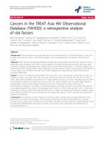
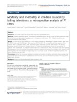
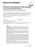
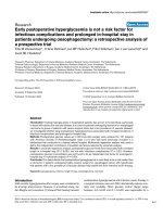
effect. Figures 1 and 2 show Kaplan–Meier survival curves
of TNM lymph node and tumor staging, respectively.
Discussion
In this study, the medical records of 69 Egyptian male
patients with breast cancer were analyzed with regard to
survival and its related possible prognostic factors. A
5-year OS rate of 46.6% can be considered low compared
with published data. Giordano et al. in a cohort of 2537
men with breast cancer obtained from the National Cancer
Institute’s Surveillance, Epidemiology, and End Results
(SEER) program using the registry 1973–1998 found a
5-year OS rate of 63% [2]. In a Turkish cohort of 86 male
patients treated over 37 years, Selcukbiricik and his coworkers reported a 65.8% 5-year OS rate [5]. In an Iranian
patient cohort of 64 patients, the 5-year OS rate was 66%
[6]. Moreover, O’Malley and her colleagues, in analyzing
the SEER program registry between 1973 and 1997 for
ethnic differences in OS of MBC, calculated 5-year OS
rates of 66% for whites, 57% for blacks, and 75% for men
of other races/ethnicities. Possible causes for our patients’
below-average overall survival may be the poorer quality
Table 3 Prognostic factors of survival in male breast
cancer patients in our Egyptian cohort
5 year
log
survival
Rank
percentage test
p value
Present
0%
1.533
0.216
Not
51%
Histopathological Invasive ductal
49.1%
type
undifferentiated 0%
1.221
0.269
Hormone
receptor status
Positive
50%
0.137
0.711
Negative
42.3%
Tumor size
T1c
52.9%
0.751
0.687
T2
45.9%
T3
50%
Lymph node
affection
N1
72.7%
N2
43.8%
N3
23.1%
Tumor grade
I
67%
II
50%
III
30%
Metastasis
*p < 0.05; statistically significant.
14.484 0.001*
10.372 0.03*
of care provided in terms of dose calculation and application for different chemotherapeutic agents, lack of HER2
status testing and hence treatment, stereotactic planning
and application of radiation, lack of a solid follow-up program for cancer patients, very poor patient compliance
both to treatment and to follow-up, and finally inadequate
general supportive care for cancer patients, compared
with Western standards. It may also be due to late-stage
diagnosis with a larger tumor burden, as all of our recruited patients had TNM stage T1c or beyond. Giordano
et al. reported tumor sizes in their cohort to be 1 to <2 cm
(T1c): 29.8%; 2 to <5 cm (T2): 39%; and ≥ 5 cm (T3): 5.3%
[2], whereas tumor sizes in our cohort on presentation
were T1c: 27.1%; T2: 68.5%; and T3: 3%. The 5-year OS
rates in Giordano’s cohort by tumor size were <2 cm:
74%; 2–5 cm: 53%; and >5 cm: 37% [2]; whereas in our
cohort, 5-year OS rates were T1c: 52.9%; T2: 45.9%; and
T3: 50%. Our patients had worse survival than their counterparts presenting with the same tumor sizes [2] except for
the two patients in our cohort who presented with T3 tumors, who had better survival rates.
We believe ethnic differences might have not played a
crucial role in this case, as the data from Iran and Turkey,
which both also presented data of Middle Eastern populations, had rather similar 5-year OS rates, even when compared with OS rates from developed countries.
Median age in our cohort was younger than in most
other published series. Giordano et al. reported a median
age of 67 years at diagnosis [2] whereas Baojiang reported a median age of 60 years in their series [7]. However, a case series of 42 Indian patients with MBC had a
median age of 56 years [8]; another report of 64 Iranian
patients with MBC had a mean age at diagnosis of
60.3 years [6]. Despite published data that indicated
advanced age to be a predictor of worse OS [2,9], our
findings did not bear this out. Although our median age
of 58 years is younger than the cut-off of 65 years used
in the abovementioned reports as a predictor of worse
prognosis, our patients did show worse OS. This may be
related to the quality of care provided, or to environmental or ethnic factors that are still unclear.
The only two prognostic factors that significantly affected survival in this cohort were lymph node status
and tumor grade. Tumor grading as a negative predictor
of OS seems to be controversial. Some authors found it
to have a significant negative effect on OS [2] whereas
others found no significant impact [5,6].
Similarly, some authors found significant negative effects on OS for tumor size [2,5], and others did not [6].
Interestingly, reports based on regional and national patient registries with large samples seem to show significant negative effects on OS by tumor size whereas
single-center retrospective reports with small samples
did not show such significant effects.
Soliman et al. BMC Cancer 2014, 14:227
/>
Page 4 of 5
Figure 1 Kaplan–Meier survival curve based on lymph node stage, showing 95% confidence interval (CI). p < 0.5 is considered
statistically significant.
HR expression was present in only 42.1% of our patients, which is remarkably lower than the 65–92% seen
in published series [6,8,10-14]. HR status did not significantly affect OS in our cohort, which concords with
other reports where HR positivity was more highly
expressed than in our patients [2,3,15]. Giordano and
coworkers reported a 5.7% negative estrogen receptor
status in their cohort, with a 5-year survival of 64%. HR
status did not, however, significantly affect survival, as
mentioned above [2]. These rates differ from what we
found in our cohort where HR-negative patients were 42.1%
of the cohort with a 42.3% 5-year OS in this subgroup.
Lack of HER2 testing and hence treatment in our cohort
may confound this absence of significant impact of HR
status on survival.
This study has some drawbacks. It is a retrospective
study with a small sample size. The incompleteness of the
data, with 13 out of 69 patients lost to follow-up, the lack
of HER2 receptor status as a standard of care, and the
missing data regarding progression-free interval in the
follow-up are all important flaws in this investigation.
However, this is one of a few studies of MBC in Middle
Eastern men and will be of help for future research. Moreover, it throws light on the differences between prognoses
and outcomes of patients in developed countries and those
in developing countries, which are probably the result of
differences in the quality of care between the two groups.
Conclusions
This study showed that lymph node status and tumor
grade are the most important predictors of OS for MBC
in Egyptian men, and that remarkably low expression of
HRs in MBC that did not have a significantly affect OS.
It also indicated a lower median age of incidence of
MBC in Egyptians than internationally reported data,
and with still worse OS. Further research is needed into
the factors that affect this disease.
Figure 2 Kaplan–Meier survival curve based on tumor stage,
showing 95% confidence interval (CI). p < 0.5 is considered
statistically significant.
Abbreviations
BMI: Body mass index; CI: Confidence interval; HER2: Human epidermal
growth factor receptor-2; HR: Hormone receptor; MBC: Male breast cancer;
OS: Overall survival; SEER: The National Cancer Institute’s Surveillance,
Epidemiology, and End Results program; SPSS: A software package for
statistical analysis of data; TNM: An organ-specific cancer staging system
adopted by the Union for International Cancer Control (UICC).
Soliman et al. BMC Cancer 2014, 14:227
/>
Competing interests
The authors declare no potential conflicts of interest.
Authors’ contributions
AS, AD, WE, AA, and BR shared in the conception and design of this study.
AS, WE, AA, and BR collected data for the study and prepared it for statistical
analysis. AS and BR analyzed the assembled data and interpreted it. AS, WE,
and BR contributed fully to manuscript writing, and AD and AA revised the
manuscript and prepared it for the final submission. All authors approved
the final form of the manuscript for submission.
Acknowledgment
This study was funded through an internal grant in the Department of
Surgical Oncology, Mansoura Cancer Center, through which AS, WE, and AA
benefited as external co-workers and AD and BR benefited as institutional
members. We thank Mr. Eslam Abou-Elwafa and Mr. Sherif M Shawer for their
contributions to this work. Mr. Shawer provided invaluable efforts in
acquiring and assembling data for this study. Mr. Abo-Elwafa shared in the
data analysis and interpretation, and in the literature search needed for the
manuscript writing.
Page 5 of 5
14. Arslan UY, Oksuzoglu B, Ozdemir N, Aksoy S, Alkis N, Gok A, Kaplan MA,
Gumus M, Berk V, Uncu D, Baykara M, Colak D, Uyetürk U, Türker I, Işıkdoğan
A: Outcome of non-metastatic male breast cancer: 118 patients.
Med Oncol 2012, 29(2):554–560.
15. Wang-Rodriguez J, Cross K, Gallagher S, Djahanban M, Armstrong JM,
Wiedner N, Shapiro DH: Male breast carcinoma: correlation of ER, PR,
Ki-67, Her2-Neu, and p53 with treatment and survival, a study of
65 cases. Mod Pathol 2002, 15(8):853–861.
doi:10.1186/1471-2407-14-227
Cite this article as: Soliman et al.: A retrospective analysis of survival and
prognostic factors of male breast cancer from a single center. BMC
Cancer 2014 14:227.
Author details
1
Department of Obstetrics and Gynecology, El-Shatby Maternity University
Hospital, University of Alexandria, Port Said Street, El Shatby, Alexandria
21526, Egypt. 2Department of Surgical Oncology, Mansoura Cancer Center,
Mansoura 35511, Egypt. 3Department of Nuclear Medicine, Mansoura Cancer
Center, Mansoura 35511, Egypt.
Received: 25 October 2013 Accepted: 25 March 2014
Published: 28 March 2014
References
1. Siegel R, DeSantis C, Virgo K, Stein K, Mariotto A, Smith T, Cooper D, Gansler
T, Lerro C, Fedewa S, Lin C, Leach C, Cannady RS, Cho H, Scoppa S, Hachey
M, Kirch R, Jemal A, Ward E: Cancer treatment and survivorship statistics,
2012. CA Canc J Clin 2012, 62(4):220–241.
2. Giordano SH, Cohen DS, Buzdar AU, Perkins G, Hortobagyi GN: Breast
carcinoma in men: a population-based study. Cancer 2004, 101(1):51–57.
3. Chen X, Liu X, Zhang L, Li S, Shi Y, Tong Z: Poorer survival of male breast
cancer compared with female breast cancer patients may be due to
biological differences. Jpn J Clin Oncol 2013, 43(10):954–963.
4. O'Malley CD, Prehn AW, Shema SJ, Glaser SL: Racial/ethnic differences in
survival rates in a population-based series of men with breast carcinoma.
Cancer 2002, 94(11):2836–2843.
5. Selcukbiricik F, Tural D, Aydogan F, Bese N, Buyukunal E, Serdengecti S:
Male breast cancer: 37-year data study at a single experience center in
Turkey. J Breast Canc 2013, 16(1):60–65.
6. Salehi A, Zeraati H, Mohammad K, Mahmoudi M, Talei AR, Ghaderi A,
Imanieh MH, Fotouhi A: Survival of male breast cancer in Fars, south of
Iran. Iran Med Crescent Med J 2011, 13(2):99–105.
7. Baojiang L, Tingting L, Gang L, Li Z: Male breast cancer: A retrospective
study comparing survival with female breast cancer. Oncol Lett 2012,
4(4):642–646.
8. Shah S, Bhattacharyya S, Gupta A, Ghosh A, Basak S: Male breast cancer: a
clinicopathologic study of 42 patients in eastern India. Indian J Surg
Oncol 2012, 3(3):245–249.
9. Tural D, Selcukbiricik F, Aydogan F, Bese N, Yetmen O, Ilvan S, Buyukunal E,
Serdengecti S: Male breast cancers behave differently in elderly patients.
Jpn J Clin Oncol 2013, 43(1):22–27.
10. Murphy CE, Carder PJ, Lansdown MR, Speirs V: Steroid hormone receptor
expression in male breast cancer. Eur J Surg Oncol 2006, 32(1):44–47.
11. Tan PH, Sng IT: Male breast cancer: a retrospective study with
immunohistochemical analysis of hormone receptor expression.
Pathology 1997, 29(1):2–6.
12. Anderson WF, Jatoi I, Tse J, Rosenberg PS: Male breast cancer: a
population-based comparison with female breast cancer. J Clin Oncol
2010, 28(2):232–239.
13. Sharif MA, Mamoon N, Arif A, Mushtaq S, Khadim MT: Histological and
immuno-histochemical study of male breast carcinoma in Northern
Pakistan. J Pak Med Assoc 2009, 59(2):67–71.
Submit your next manuscript to BioMed Central
and take full advantage of:
• Convenient online submission
• Thorough peer review
• No space constraints or color figure charges
• Immediate publication on acceptance
• Inclusion in PubMed, CAS, Scopus and Google Scholar
• Research which is freely available for redistribution
Submit your manuscript at
www.biomedcentral.com/submit