Clinical implications in the shift of syndecan-1 expression from the cell membrane to the cytoplasm in bladder cancer
Bạn đang xem bản rút gọn của tài liệu. Xem và tải ngay bản đầy đủ của tài liệu tại đây (1 MB, 7 trang )
Miyake et al. BMC Cancer 2014, 14:86
/>
RESEARCH ARTICLE
Open Access
Clinical implications in the shift of syndecan-1
expression from the cell membrane to the
cytoplasm in bladder cancer
Makito Miyake1, Adrienne Lawton2, Yunfeng Dai3, Myron Chang3, Lourdes Mengual4, Antonio Alcaraz4,
Steve Goodison1,5,6 and Charles J Rosser1,5,7*
Abstract
Background: To determine the diagnostic and prognostic capability of urinary and tumoral syndecan-1 (SDC-1)
levels in patients with cancer of the urinary bladder.
Methods: SDC-1 levels were quantitated by enzyme-linked immunosorbent assay (ELISA) in 308 subjects (102
cancer subjects and 206 non-cancer subjects) to assess its diagnostic capabilities in voided urine. The performance
of SDC-1 was evaluated using the area under the curve of a receiver operating characteristic curve. In addition,
immunohistochemical (IHC) staining assessed SDC-1 protein expression in 193 bladder specimens (185 cancer
subjects and 8 non-cancer subjects). Outcomes were correlated to SDC-1 levels.
Results: Mean urinary levels of SDC-1 did not differ between the cancer subjects and the non-cancer subjects,
however, the mean urinary levels of SDC-1 were reduced in high-grade compared to low-grade disease (p < 0.0001),
and in muscle invasive bladder cancer (MIBC) compared to non-muscle invasive bladder cancer (NMIBC) (p = 0.005).
Correspondingly, preliminary data note a shift from a membranous cellular localization of SDC-1 in normal tissue,
low-grade tumors and NMIBC, to a distinctly cytoplasmic localization in high-grade tumors and MIBC was observed in
tissue specimens.
Conclusion: Alone urinary SDC-1 may not be a diagnostic biomarker for bladder cancer, but its urinary levels and cellular
localization were associated with the differentiation status of patients with bladder tumors. Further studies are warranted
to define the potential role for SDC-1 in bladder cancer progression.
Keywords: Syndecan, Bladder, Cancer biomarker, Specificity
Background
Syndecan 1 (SDC-1) is one of four members of a transmembrane heparan sulfate proteoglycan family. SDC-1 is
the major syndecan expressed in epithelia, and it plays a
critical role in cellular processes including differentiation,
cell adhesion, migration and invasion, and angiogenesis
[1-3]. Functions have been ascribed to the extracellular
domain that carries glycosaminoglycan (GAG) side chains,
to the transmembrane domain and to the cytoplasmic domain that transduces signals from extracellular ligand
binding [3]. Altered SDC-1 expression has been reported
* Correspondence:
1
Cancer Research Institute, Orlando Health, Orlando, FL 32827, USA
5
Nonagen Bioscience Corp, Orlando, FL 32827, USA
Full list of author information is available at the end of the article
in a number of malignant tumor types and has been associated with differentiation status and survival [4-6].
Aaboe et al., identified SDC-1 as a bladder cancer (BCa)
biomarker using gene expression profiling [7]. Through
proteomic analyses of voided urines from BCa patients,
SDC-1 has also been identified as a potential diagnostic
biomarker [8]. However, in a subsequent multiplex biomarker study of 127 subjects, urinary SDC-1 protein could
not be confirmed to be significantly elevated in patients
with BCa [9]. The observed inconsistency as a diagnostic
biomarker may be related to the study cohorts employed
to date, but it may also be due in part to the transmembrane nature of SDC-1. Release of SDC-1 into the soluble
fraction of the urine is dependent on a number of factors
including: cellular turnover, aberrant processing in disease
© 2014 Miyake et al.; licensee BioMed Central Ltd. This is an Open Access article distributed under the terms of the Creative
Commons Attribution License ( which permits unrestricted use, distribution, and
reproduction in any medium, provided the original work is properly credited.
Miyake et al. BMC Cancer 2014, 14:86
/>
states, release by inflammation-associated shedding [10],
and a shift of expression from epithelial to stromal cells in
tumors [11].
Herein, we report further evaluation of the potential
utility of SDC-1 as a diagnostic and prognostic biomarker
in BCa by analysis of a large diverse test cohort through
enzyme-linked immunosorbent assay (ELISA), and the investigation of SDC-1 protein expression patterns in bladder tumors through immunohistochemical (IHC) analysis
of archival tissue specimens.
Methods
Urinary SDC-1 levels
After Institutional Review Board approval by MD Anderson
Cancer Center Orlando and Hospital Clínic of Barcelona
and written informed consent, voided urines were collected
into institutional tissue banks. From these tissue banks in
the Departments of Urology from Orlando Health and
Hospital Clínic of Barcelona, 308 voided urine samples and
associated clinical data were identified. The study cohort
consisted of 206 adult subjects with no active BCa or previous history of BCa (47 with voiding symptoms, 44 with
urolithiasis, 9 with gross hematuria, 14 with urinary tract
infection and 92 without any of the above diagnoses) and
102 subjects diagnosed with de novo urothelial carcinoma.
Median follow-up of the patients with BCa was 14 months.
In our cancer group and hematuria group, imaging of the
upper urinary tract and cystoscopy were performed. Furthermore, the histologic subtype, urothelial carcinoma, was
confirmed by histological examination of excised tissue in
the cancer group.
Voided urine samples were centrifuged to separate
the supernatant from the cellular pellet. The supernatant was decanted and aliquoted, and the urinary pellet was snap frozen. Both the supernatant and pellet
were stored at -80°C prior to analysis. Urine supernatant
protein concentration was determined using Pierce 660nm Protein Assay Kit (Thermo Fisher Scientific Inc.,
Waltham, MA, USA). The level of human SDC-1 (Cat#
ab46507 Abcam, Cambridge, MA, USA) was monitored in
urine samples using a commercial ELISA assay. The assay
was conducted according to the manufacturer’s instructions. A calibration curve was prepared using purified
standards for SDC-1. Curve fitting was accomplished by
either linear or four-parameter logistic regression following manufacturer’s instructions. Laboratory personnel
were blinded to final diagnosis.
Syndecan-1 expression in human bladder tumors
Under Institutional Review Board approval with a waiver
of consent, 185 bladder tumor paraffin blocks and eight
benign bladder paraffin blocks dating from 2002-2009
were identified in the pathologic archives of Orlando
Health Department of Pathology. The eight benign bladder
Page 2 of 7
paraffin blocks were from autopsy cases in which there
was no record of BCa, hematuria or tobacco use. Median follow-up of the patients was 18 months. All paraffin blocks were examined by H&E for histological
verification of urothelial carcinoma only histology. Paraffin blocks were cut 5 μm sections and placed on a
Superfrost Plus Microslide. Sections were deparaffinized
followed by antigen retrieval using citric acid buffer
(pH 6.0, 95°C for 20 min). Slides were treated with 1%
hydrogen peroxide in methanol to block endogenous
peroxidase activity. After 20 min blocking in phosphate
buffered saline (PBS) containing 1% bovine serum
albumin (BSA), slides were incubated overnight at 4°C
with anti-human SDC-1 antibody (mouse monoclonal–Abcam ab34164, dilution 1/400 in PBS containing
1% BSA). Next, slides were incubated with 2 μg/mL of biotinylated anti-mouse IgG secondary antibody (Vector
Laboratories, Burlingame, CA) for 30 min at room temperature. Subsequently, the sections were stained using
Standard Ultra-Sensitive ABC Peroxidase Staining kit
(Pierce/Thermo Fisher Scientific, San Jose, CA) and 3,
3′-diaminobenzidine (DAB; Vector Laboratories), counterstained by hematoxylin, dehydrated, and mounted
with a cover slide. Human liver tissue, known to stain
strongly for SDC-1, was used as a positive control and
omitting the primary antibody served as the negative
control. The above immunostaining for SDC-1 as well
as the interpretation of the immunostaining for SDC-1
were based on a previous report by Mukunyadzi, et al.
[12]. Briefly, the location of immunoreactivity (e.g.,
nuclear, cytoplasm, cell membrane, and stroma) was
noted. The sections were analyzed and staining assessed
using a semiquantitative grading system as follows:
negative (-), complete lack of staining or staining in
<10% of tumor cells; weak (+), staining in 10 to 20% of
tumor cells; mild (++), staining in 20 to 50% of tumor
cells; moderate (+++), staining in 50 to 70% of tumor
cells and; strong (++++), staining in >70% of tumor cells.
Using light microscopy, two investigators (MM and AL),
who were both blinded to patients’ data, interpreted immunostaining results. A third investigator (CJR) reviewed
discrepancies and rendered a final score.
Data analysis
The Wilcoxon rank sum test was used to determine the
association between urinary SDC-1 and BCa. Nonparametric receiver operating characteristic (ROC) curves
were plotted and the ability of the urinary SDC-1 biomarker to indicate BCa was estimated by calculating the
area under the ROC curves (AUC). The sensitivity and
specificity of the biomarker at the optimal cutoff value
was defined by calculating the Youden index [13]. The
agreement between interpreting SDC-1 immunohistochemistry results between the two investigators was analyzed
Miyake et al. BMC Cancer 2014, 14:86
/>
Page 3 of 7
Table 1 Demographic and clinicopathologic characteristics of 308 subjects comprising ELISA study cohort and 193
subjects comprising IHC study cohort
ELISA Cohort
Median Age (range, y)
Male: Female ratio
IHC Cohort
BCa (%) n = 102
Controls (%) n = 206
BCa (%) n = 185
Controls (%) n = 8
69 (20–93)
56 (18–89)
73 (30–94)
26 (21–43)
84 : 18
152 : 54
143 : 42
4:4
91 (89%)
135 (66%)
156 (84%)
N/A
5 (5%)
20 (10%)
8 (4%)
N/A
Race
White
African American
Other
6 (6%)
51 (24%)
19 (12%)
N/A
Positive FISH
40 / 74 (54%)
2/22 (9%)
N/A
N/A
Suspicious/positive cytology
37 / 94 (39%)
2/22 (9%)
N/A
N/A
Median follow-up (months)
14
4
18
N/A
Clinical stage
Tis
6 (6%)
17 (9%)
Ta
41 (40%)
45 (24%)
T1
14 (14%)
63 (34%)
≥T2
41 (40%)
60 (33%)
Tumor grade
Low
38 (37%)
27 (15%)
High
64 (63%)
158 (85%)
3.0
3.0
Median tumor size (cm)
IHC immunohistochemistry, BCa bladder cancer, N/A not available.
using kappa statistics with the strength of agreement
0.81-1.00 interpreted as almost perfect. The results are
presented as weighted kappa with 95% confidence interval (CI). Comparison of immunohistochemical distribution data was performed using Chi square test.
Disease-specific survival (DSS) curves were obtained
using the Kaplan-Meier method, and compared by the
log rank test for each prognostic variable [14]. Multivariate analysis was performed to identify independent
prognostic variables using a stepwise Cox proportional
hazards regression model. Statistical significance in this
study was set at p < 0.05 and all reported p values were
2-sided. All analyses were performed using SAS software version 5.00 (San Diego, CA).
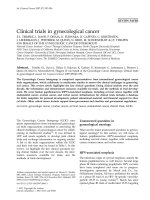
Figure 1 Urinary Syndecan-1 levels. Comparison of urinary concentrations of SDC-1 between the cancer and non-cancer groups. In the
box-and-whisker plot of urinary concentration of SDC-1, the central box represents the value from the lower to upper quartile. Significance (p < 0.05)
was assessed by the Wilcoxon rank sum test.
Miyake et al. BMC Cancer 2014, 14:86
/>
Page 4 of 7
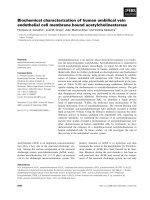
Figure 2 Expression of Syndecan-1 protein in human bladder tissue. a) Representative staining of benign bladder epithelium (left) and
cancerous bladder (right) showing membranous staining of epithelial cells. b) Representative staining of low-grade bladder cancer (left) and
high-grade bladder cancer (right). High-grade cancers were noted to have cytoplasmic staining while losing their membranous staining. c)
Representative staining of low pathologic stage (pTa) bladder cancer (left) and high pathologic stage (pT2) bladder cancer (right). All images were
captured at 400× magnification. Column bar graphs illustrate the population of subjects with SDC-1 membrane staining and SDC-1 cytoplasmic
staining in (d) benign bladder epithelium vs. non-muscle invasive bladder cancer (NMIBC) vs. muscle invasive bladder cancer (MIBC), (e) low-grade
tumor vs. high-grade tumor and (f), Ta-1 tumor vs. T2-4 tumor.
Results
Urinary SDC-1 ELISA
Characteristics of the study cohort of 308 subjects (102
subjects with active BCa and 206 subjects with no evidence of active BCa or a history of BCa) are presented
in the Table 1. The median urinary concentration of
SDC-1 was not significantly higher overall in subjects
with BCa compared to subjects without BCa (71.25 ng/
ml vs. 36.10 ng/ml, p = 0.23) (Figure 1). Neither did
SDC-1 levels differ among the groups that made up the
diverse control cohort (p = 0.562, data not shown). However, a difference in urinary SDC-1 level was noted between patients with tumors of differing grade and invasive
subtype. Specifically, low-grade bladder tumors were noted
to have higher median urinary SDC-1 levels compared to
high-grade bladder tumors (64.55 ng/ml vs. 26.1 ng/ml,
p < 0.0001), and non-muscle invasive bladder cancer
(NMIBC) had higher median urinary SDC-1 levels
compared to muscle invasive bladder cancer (MIBC)
(58.23 ng/ml vs. 28.53 ng/ml, p = 0.0049) (Figure 1).
Immunohistochemical staining of bladder tissue
specimens
Characteristics of the study cohort of 193 subjects (185
subjects with urothelia carcinoma histology only and 8
subjects with benign bladder histology) are presented in
the Table 1. The pathologists’ intra-observer agreement
on SDC-1 interpretation and scoring was ‘good’ with a
Miyake et al. BMC Cancer 2014, 14:86
/>
Page 5 of 7
reduced membranous expression of SDC-1 (Figure 2b).
In the same way, higher stage tumors (T2-4 vs. Ta-1,
p < 0.0001) were noted to have increased cytoplasmic
expression and reduced membranous expression of
SDC-1 (Figure 2c). Though the location of staining
changed from membranous to cytoplasmic amongst
high-grade and high stage tumors, immunostaining
grading, weak (+) to strong (++++), did not change,
illustrating a shift of the ubiquitously expressed SDC-1
from the cellular membrane in well-differentiated, low
stage tumors to the cytoplasm in poorly-differentiated,
higher stage tumors.
Analyses of prognostic parameters associated with
disease specific survival
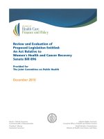
Univariate analysis revealed that NMIBC and membranous immnostaining for SDC-1 represent favourable prognostic factors associated with disease-specific survival
(DSS) (p < 0.0001 and p = 0.0004, respectively) (Figure 3).
However on multivariate analysis (Table 2), only MIBC
(hazard ratio [HR] = 21.1, 95% confidence interval [CI] =
4.24–105.1, p = 0.0001) proved to be an independent risk
factor for DSS, resulting in a significant reduction in
survival. Furthermore, MIBC was associated with a significant reduction in overall survival (HR = 9.60, CI:
2.59-35,5, p = 0.001).
Figure 3 Kaplan-Meier curves for disease-specific survival.
Disease-specific survival stratified by (a) membranous vs. cytoplasmic
SDC-1, (b) low-grade vs. high-grade and (c) non-muscle invasive bladder
cancer (NMIBC) vs. muscle invasive bladder cancer (MIBC). HR, hazard
ratio; 95% CI, 95% confidence interval.
noted kappa score of 0.64 (0.8–1.0, excellent; 0.6–0.8,
good; 0.4–0.6, moderate; 0–0.4, poor), 95% CI 0.61–0.68.
The percentage agreement was 82.0%. In normal tissue, as
well as low-grade disease and NMIBC, over 70% of SDC-1
immunostaining was located within the cellular membrane (Figure 2a) and was graded as moderate (+++) to
strong (++++). Minimal immunoreactivity was noted in
the stroma. Within bladder tumors, 55% of high-grade
tumors (compared to low-grade tumors, p < 0.0001)
were noted to have increased cytoplasmic expression and
Discussion
SDC-1 is expressed mainly in epithelial tissues, hence,
studies aiming to address its role in malignancies have
focused on carcinoma. In a number of malignancies, the
expression of SDC-1 correlates with tumor stage and
grade [15-18], but the association between SDC-1 status
and BCa has not been extensively studied. Other investigators have reported a positive correlation of SDC-1
with fibroblast growth factors (FGFRs) in bladder tumors, these factors are thought to be key molecules in
low-grade BCa [19]. Only Shimada et al., have investigated the biologic role of SDC-1 in human BCa cells. In
their study, the BCa cell lines, UMUC2 and UMUC3
had SDC-1 expression silenced by siRNA transfection,
which led to an induction of apoptosis in vitro and a reduction in mouse orthotopic bladder tumor growth [20].
To our knowledge, our study is the largest study to
date to evaluate SDC-1 in human bladder tumors both
in voided urine and in tumor sections. We used two
complimentary approaches to classify SDC-1 expression
in human bladder tumors. First, urinary SDC-1 levels
were monitored by ELISA in a cohort of 308 subjects.
While there was no difference in urinary SDC-1 levels
between BCa-bearing subjects and non-BCa bearing
subjects (p = 0.23), lower urinary levels of SDC-1 were
associated with the presence of high-grade tumors and/
or MIBC. The prognostic capability of SDC-1 in
Miyake et al. BMC Cancer 2014, 14:86
/>
Page 6 of 7
Table 2 Multivariate analysis of disease specific survival and overall survival
Disease-specific survival
Variables
N
HR
Overall survival
95% CI
p
HR
4.24–105.1
0.0001
9.60
95% CI
p
2.59–35.5
0.001
0.27–3.60
0.99
Stage
NMIBC
125
1
MIBC
60
21.10
Membrane
96
1
Cytoplasm
89
0.87
1
SDC-1 expression
1
0.21–3.60
0.85
0.99
HR Hazard ratio, 95% CI 95% confidence interval, NMIBC Non-muscle invasive bladder cancer, MIBC Muscle invasive bladder cancer.
predicting higher grade and higher stage disease prior to
patients undergoing cystoscopy and transurethral resection of bladder tumor has the potential to improve patients’ outcomes. Second, we determined the expression
pattern of SDC-1 protein in a cohort of 193 bladder tissue specimens. Though a difference in SDC-1 expression pattern was not seen between bladder tumors and
benign bladder histology, possibly due to the small sample size of the benign cohort, a significant shift in cellular localization of SDC-1 was associated with high-grade
tumors and MIBC. These tumors tended to lose the distinct membranous staining observed in normal urothelia.
The two complimentary approaches utilized in the current
study yielded similar inferences, i.e., more aggressive or
more advance BCa has less membrane bound SDC-1. If
less membrane bound SDC-1 is present in a tumor mass,
then it might be expected that less shed or released SDC-1
would be present in the soluble fraction of voided urine
from patients with more aggressive or advanced BCa.
Shifts in SDC-1 expression patterns have been alluded
to in previous reports, but none in BCa. A study by
Mennerich et al., described a shift of SDC-1 expression
from the epithelial component to the stromal component in solid tumors [11]. An observed overall increase
in tumor SDC-1 mRNA was demonstrated by in situ
hybridization and protein levels confirmed by immunohistochemistry in tumor-associated stromal cells in
breast, lung and colon carcinoma. We did not observe
this phenomenon in our study, the majority of SDC-1
expression was in the epithelial component of the bladder
tumors. The expression pattern shift that our analyses revealed was from distinctly membranous to diffusely cytoplasmic in high-grade and high-stage bladder tumors. This
association with disease progression suggests that the loss
of SDC-1 function at the cell-surface or cell membrane
and thus may facilitate cancer progression and the development of invasive and metastatic disease. Several studies
have shown the involvement of cell-surface SDC-1 in cellcell and cell-matrix adhesion, possibly through the regulation of integrin activities [21]. The loss of SDC-1 at the cell
surface by extracellular cleavage can decrease the strength
of tumor cell adhesion within the tissue architecture,
resulting in an increase in cellular motility. This in turn
may allow cancer cells to cross the basement membrane
and invade surrounding tissues as well as distant sites [11].
The loss of SDC-1 at the cell-surface could also occur
through a switch to translation of alternative, nonmembranous isoforms, or by aberrant processing in an advanced tumor. This concept exists for the well-known
tumor suppressor gene E-cadherin. Similar to SDC-1, cellsurface E-cadherin assists in cell adhesion and loss of
E-cadherin is associated with more aggressive BCa that possess a greater potential to invade and metastasize [22,23].
Though the present studies are quite intriguing, they only
elude to a biologic phenomenon which now must be further explored to a) report associated cellular and molecular
changes, b) confirm the ELISA and immunohistochemistry
results in a large cohort, c) determine which domain (cytoplasmic, transmembrane or extracellular) is shed in voided
urine and d) determine in addition to changes in location
in expression if there are changes in the quantity of SDC-1
expression between the various disease states. Furthermore,
the preliminary nature of our immunohistochemical results
should be confirmed in a larger cohort.
Conclusions
In summary, decreased urinary levels of SDC-1 in BCa
patients were associated with high-grade or high-stage
disease, and this phenomenon correlated with a shift of
SDC-1 protein cellular localization from the cellular
membrane to the cytoplasm in these high-grade and
high stage bladder tumors. On univariate analysis, loss
of membranous localization of SDC-1 was associated
with a significant reduction in DSS. This is the first report to describe specific SDC-1 expression changes as
being associated with more aggressive, lethal BCa. Further studies are underway to understand the role of
SDC-1 in BCa and to investigate the prognostic potential of SDC-1 monitoring in human bladder tumors.
Competing interests
Dr Goodison and Charles J. Rosser have a competing interest in that they are
officers of Nonagen Bioscience Corp, a small biotech company with an
interest to develop urinary biomarkers. No other authors possess a
competing interest.
Miyake et al. BMC Cancer 2014, 14:86
/>
Authors’ contributions
All authors have read and approved the final manuscript. MM, AL: acquisition
of data. YD, MC: statistical analysis. LM, AA: clinical samples, drafting of
manuscript. SG: study concept and design, drafting of manuscript. CJR: study
concept and design, drafting of manuscript, funding.
Acknowledgements
The authors are grateful to the 501 subjects who participated in this study.
This work was supported by research grants from Flight Attendant Medical
Research Institute (CJR), Florida Department of Health James and Esther King
Team Science Award 10KT-01 (CJR), Florida State James and Esther King
Biomedical Research Award Technology Transfer Feasibility 1KF06 (SG) and
National Cancer Institute RO1 CA116161 (SG). SG and CR are employees of
Nonagen Bioscience Corp.
Author details
1
Cancer Research Institute, Orlando Health, Orlando, FL 32827, USA.
2
Department of Pathology, Orlando Health, Orlando, FL 32806, USA.
3
Department of Biostatistics, The University of Florida, Gainesville, FL 32610,
USA. 4Laboratory and Department of Urology, Hospital Clínic, Universitat de
Barcelona, Barcelona, Spain. 5Nonagen Bioscience Corp, Orlando, FL 32827,
USA. 6Department of Health Sciences Research, Mayo Clinic, Jacksonville, FL
32224, USA. 7University of Hawaii Cancer Center, Clinical and Translational
Research Program, 701 Ilalo Street, Honolulu, HI 96813, USA.
Received: 3 April 2013 Accepted: 11 February 2014
Published: 13 February 2014
References
1. Couchman JR, Pataki CA: An introduction to proteoglycans and their
localization. J Histochem Cytochem 2012, 60(12):885–897.
2. McQuade KJ, Rapraeger AC: Syndecan-1 transmembrane and extracellular
domains have unique and distinct roles in cell spreading. J Biol Chem
2003, 278(47):46607–46615. Epub 2003 Sep 14.
3. Teng YH, Aquino RS, Park PW: Molecular functions of syndecan-1 in
disease. Matrix Biol 2012, 31(1):3–16. Epub 2011 Oct 18.
4. Conejo JR, Kleeff J, Koliopanos A, Matsuda K, Zhu ZW, Goecke H, Bicheng N,
Zimmermann A, Korc M, Friess H, Büchler MW: Syndecan-1 expression is
up-regulated in pancreatic but not in other gastrointestinal cancers. Int J
Cancer 2000, 88(1):12–20.
5. Contreras HR, Ledezma RA, Vergara J, Cifuentes F, Barra C, Cabello P, Gallegos I,
Morales B, Huidobro C, Castellón EA: The expression of syndecan-1 and -2 is
associated with Gleason score and epithelial-mesenchymal transition
markers, E-cadherin and beta-catenin, in prostate cancer. Urol Oncol 2010,
28(5):534–540. Epub 2009 May 17.
6. Shah L, Walter KL, Borczuk AC, Kawut SM, Sonett JR, Gorenstein LA,
Ginsburg ME, Steinglass KM, Powell CA: Expression of syndecan-1 and
expression of epidermal growth factor receptor are associated with
survival in patients with nonsmall cell lung carcinoma. Cancer 2004,
101(7):1632–1638.
7. Aaboe M, Marcussen N, Jensen KM, Thykjaer T, Dyrskjøt L, Orntoft TF: Gene
expression profiling of noninvasive primary urothelial tumours using
microarrays. Br J Cancer 2005, 93(10):1182–1190.
8. Yang N, Feng S, Shedden K, Xie X, Liu Y, Rosser CJ, Lubman DM, Goodison
S: Urinary glycoprotein biomarker discovery for bladder cancer detection
using LC/MS-MS and label-free quantification. Clin Cancer Res 2011,
17(10):3349–3359.
9. Urquidi V, Chang M, Dai Y, Kim J, Wolfson ED, Goodison S, Rosser CJ: IL-8 as
a urinary biomarker for the detection of bladder cancer. BMC Urol 2012,
12:12.
10. Pruessmeyer J, Martin C, Hess FM, Schwarz N, Schmidt S, Kogel T, Hoettecke N,
Schmidt B, Sechi A, Uhlig S, Ludwig A: A disintegrin and metalloproteinase
17 (ADAM17) mediates inflammation-induced shedding of syndecan-1
and -4 by lung epithelial cells. J Biol Chem 2010, 285(1):555–564.
11. Mennerich D, Vogel A, Klaman I, Dahl E, Lichtner RB, Rosenthal A, Pohlenz
HD, Thierauch KH, Sommer A: Shift of syndecan-1 expression from
epithelial to stromal cells during progression of solid tumours. Eur J
Cancer 2004, 40(9):1373–1382.
12. Mukunyadzi P, Liu K, Hanna EY, Suen JY, Fan CY: Induced expression of
syndecan-1 in the stroma of head and neck squamous cell carcinoma.
Mod Pathol 2003, 16(8):796–801.
Page 7 of 7
13. Fluss R, Faraggi D, Reiser B: Estimation of the Youden Index and its
associated cutoff point. Biom J 2005, 47:458–472.
14. Pepe MS, Feng Z, Janes H, Bossuyt PM, Potter JD: Pivotal evaluation of the
accuracy of a biomarker used for classification or prediction: standards
for study design. J Natl Cancer Inst 2008, 100:1432–1438.
15. Anttonen A, Kajanti M, Heikkilä P, Jalkanen M, Joensuu H: Syndecan-1
expression has prognostic significance in head and neck carcinoma.
Br J Cancer 1999, 79(3–4):558–564.
16. Altemeier WA, Schlesinger SY, Buell CA, Brauer R, Rapraeger AC, Parks WC,
Chen P: Transmembrane and extracellular domains of syndecan-1 have
distinct functions in regulating lung epithelial migration and adhesion.
J Biol Chem 2012, 287(42):34927–34935. doi:10.1074/jbc.M112.376814. Epub
2012 Aug 30.
17. Barbareschi M, Maisonneuve P, Aldovini D, Cangi MG, Pecciarini L, Angelo
Mauri F, Veronese S, Caffo O, Lucenti A, Palma PD, Galligioni E, Doglioni C:
High syndecan-1 expression in breast carcinoma is related to an
aggressive phenotype and to poorer prognosis. Cancer 2003,
98(3):474–483.
18. Lundin M, Nordling S, Lundin J, Isola J, Wiksten JP, Haglund C: Epithelial
syndecan-1 expression is associated with stage and grade in colorectal
cancer. Oncology 2005, 68(4-6):306–313. Epub 2005 Jul 12.
19. Marzioni D, Lorenzi T, Mazzucchelli R, Capparuccia L, Morroni M, Fiorini R,
Bracalenti C, Catalano A, David G, Castellucci M, Muzzonigro G, Montironi R:
Expression of basic fibroblast growth factor, its receptors and syndecans
in bladder cancer. Int J Immunopathol Pharmacol 2009, 22(3):627–638.
20. Shimada K, Nakamura M, De Velasco MA, Tanaka M, Ouji Y, Miyake M,
Fujimoto K, Hirao K, Konishi N: Role of syndecan-1 (CD138) in cell survival
of human urothelial carcinoma. Cancer Sci 2010, 101(1):155–160.
21. Chen P, Abacherli LE, Nadler ST, Wang Y, Li Q, et al: MMP7 shedding of
syndecan-1 facilitates re-epithelialization by affecting α2β1 integrin
activation. PLoS ONE 2009, 4(8):e6565.
22. Thoreson MA, Reynolds AB: Altered expression of the catenin p120 in
human cancer: implications for tumor progression. Differentiation 2002,
70(9–10):583–589.
23. Hu X, Ruan Y, Cheng F, Yu W, Zhang X, Larré S: p130Cas, E-cadherin and
β-catenin in human transitional cell carcinoma of the bladder: expression and clinicopathological significance. Int J Urol 2011, 18(9):630–637.
doi:10.1111/j.1442-2042.2011.02793.x. Epub 2011 Jun 14.
doi:10.1186/1471-2407-14-86
Cite this article as: Miyake et al.: Clinical implications in the shift of
syndecan-1 expression from the cell membrane to the cytoplasm in
bladder cancer. BMC Cancer 2014 14:86.
Submit your next manuscript to BioMed Central
and take full advantage of:
• Convenient online submission
• Thorough peer review
• No space constraints or color figure charges
• Immediate publication on acceptance
• Inclusion in PubMed, CAS, Scopus and Google Scholar
• Research which is freely available for redistribution
Submit your manuscript at
www.biomedcentral.com/submit