Decreased GATA5 mRNA expression associates with CpG island methylation and shortened recurrence-free survival in clear cell renal cell carcinoma
Bạn đang xem bản rút gọn của tài liệu. Xem và tải ngay bản đầy đủ của tài liệu tại đây (583.17 KB, 6 trang )
Peters et al. BMC Cancer 2014, 14:101
/>
RESEARCH ARTICLE
Open Access
Decreased GATA5 mRNA expression associates
with CpG island methylation and shortened
recurrence-free survival in clear cell renal cell
carcinoma
Inga Peters1, Natalia Dubrowinskaja1, Michael Kogosov1, Mahmoud Abbas2, Jörg Hennenlotter3, Christoph von Klot1,
Axel S Merseburger1, Arnulf Stenzl3, Ralph Scherer4, Markus A Kuczyk1 and Jürgen Serth1*
Abstract
Background: GATA-5, a zinc-finger transcription factor and member of the GATA family proteins 1–6, is known to
be involved in cellular differentiation. We recently found that tumor-specific hypermethylation of the GATA5 CpG
island (CGI) occurs in renal cell carcinoma (RCC) and is associated with an adverse clinical outcome. In this study,
we investigated whether epigenetic GATA5 alterations may result in changes in GATA5 mRNA expression levels and
correlate with the observed prognostic impact of epigenetic changes in GATA5 in RCC.
Methods: Quantitative real-time reverse-transcribed polymerase chain reaction was applied to measure relative
GATA5 mRNA expression levels in 135 kidney tissue samples, including 77 clear cell RCC (ccRCC) tissues and 58
paired adjacent normal renal tissue samples. Relative GATA5 expression levels were determined using the ΔΔCt
method and detection of three endogenous control genes then compared to previously measured values of relative methylation.
Results: The mean relative GATA5 mRNA expression level exhibited an approximately 31-fold reduction in tumor
specimens compared with corresponding normal tissues (p < 0.001, paired t-test). Decreased GATA5 mRNA expression was inversely correlated with increased GATA5 CGI methylation (p < 0.001) and was associated with shortened
recurrence-free survival in ccRCC patients (p = 0.023, hazard ratio = 0.25).
Conclusion: GATA5 mRNA expression is decreased in ccRCC, likely due to gene silencing by methylation of the
GATA5 CGI. Moreover, reduced GATA5 mRNA levels were associated with a poor clinical outcome, indicating a
possible role of GATA5 for the development of aggressive ccRCC phenotypes.
Keywords: GATA5, Renal cell carcinoma, mRNA, Prognosis, DNA methylation
Background
Renal cell carcinoma (RCC) is one of the top ten causes
of cancer deaths in industrialized countries, and its incidence has consistently increased during the past decades
[1]. Clear cell renal cell carcinomas (ccRCCs) are the
most frequently occurring histological entity, comprising
approximately 75% of all RCC.
* Correspondence:
1
Department of Urology and Urologic Oncology, Hannover Medical School,
Carl-Neuberg-Str.1, Hannover 30625, Germany
Full list of author information is available at the end of the article
As a member of the GATA family of transcription factors, GATA5 is known to be functionally involved in cellular lineage and cell differentiation during embryonic
development of the heart, lung, urogenital tract, and gut
epithelium [2]. Altered expression of GATA5 has been
associated with intestinal epithelial cell differentiation
[3]. GATA5 is assumed to be a selective transcriptional
regulator of mucin genes in gastrointestinal tissues [4],
and regulates the promoter of the sodium-hydrogen exchanger isoform 3 that is expressed in intestinal and
renal epithelium via Sp-family transcription factors [5].
© 2014 Peters et al.; licensee BioMed Central Ltd. This is an Open Access article distributed under the terms of the Creative
Commons Attribution License ( which permits unrestricted use, distribution, and
reproduction in any medium, provided the original work is properly credited.
Peters et al. BMC Cancer 2014, 14:101
/>
Of note, a previous study found GATA5 hypermethylation and associated epigenetic silencing to be involved in
carcinogenesis of gastric and colorectal cancers [6]. Epigenetic alterations of GATA5 were also described in
other tumor tissues and were linked to the development
of ovarian, lung, pancreatic, and esophageal cancer
[7-10]. In a recent study aimed at identifying new DNA
methylation targets in ccRCC, we detected for the first
time a tumor-specific hypermethylation of the GATA5
CpG island (CGI) in RCC [11]. Hypermethylation was
also associated with advanced disease and shortened
recurrence-free survival (RFS) of patients.
In this study, we asked whether mRNA expression
levels of GATA5 are reduced as suggested by our previous DNA methylation analysis, and if the mRNA levels
are associated with adverse clinical parameters, further
underlining the relevance of epigenetic GATA5 alterations in ccRCC carcinogenesis.
Material and methods
Page 2 of 6
Table 1 Clinicopathological parameters of ccRCC patients
Clinicopathological
parameters
n
Cases in total
77 100
Sex
RNA isolation, cDNA synthesis, and quantitative real-time
PCR analysis
Isolation of total RNA from tissue specimens and from
renal proximal tubular epithelial cells (RPTEC) as
controls, cDNA synthesis, and quantitative real-time
PCR analysis (qRT-PCR) were carried out as described
Female
29 38
Male
48 62
Median age, years
64
Median tumor size, cm
5.5
Primary tumor classification pT1
Lymph node status
Tissue specimens
One hundred and thirty-five kidney tissues, including 77
ccRCCs and 58 paired adjacent normal renal tissue samples, were included in this study. RCC tissues were obtained from open or laparoscopic nephrectomies and
partial resection. Paired tissue samples with adjacent
normal tissue (adN) were obtained from a subgroup of
our 77 ccRCCs cohort. Adjacent normal tissues, i.e.
morphologically normal kidney were isolated with minimum of 0.5 cm to 2 cm distant from the primary tumor
lesion. Samples were snap-frozen in liquid nitrogen immediately following surgery and stored at −80°C. Ethical
approval of the university ethical committee (Prof. H. D.
Tröger, Hannover Medical School, Carl-Neuberg-Str. 1,
Hannover, Germany) and informed consent from all patients were obtained. Localized disease was defined as
pT ≤ 2, lymph node involvement and metastasis negative
(N0/M0), and grading (G) G1 and 1–2, whereas advanced RCC was defined as pT ≥ 3, N1 and/or M1, and
G2-3 and G3. Patients with G2 were assigned to the
intermediate risk group and were not considered as a
parameter for low vs. high grade group comparisons.
Follow up data were available for 35 patients, and RFS
was defined as the interval up to the time that disease
progression could be detected by computer tomography
scans. Clinical and histopathological parameters are
summarized in Table 1.
%
Status of metastasis
6
8
pT1a
19 25
pT1b
14 18
pT2
3
4
pT3
2
3
pT3a
9
12
pT3b/c
22 29
pT4
0
0
not known
2
3
N0
68 88
N1
9
12
N2
0
0
M0
56 73
M1
21 27
G1
14 18
G1-2
8
G2
40 52
Grade
- Low risk
- Intermediate risk
- High risk
10
G2-3
5
G3
10 13
Localized disease
pT ≤ 2, N0, M0 and G1; G1-2
34 44
Advanced disease
pT ≥ 3 and/or N1, M1 or G2-3; G3 43 56
Paired samples
6
58 75
Abbreviations: ccRCC clear cell renal cell carcinoma, n number.
previously [12]. Duplicate measurements for qRT-PCR
analysis were performed using 384 sample plates, an automated liquid handling system (FasTrans, AnalyticJena,
Jena, Germany), and the ABI 7900 Fast Sequence Detection System as described previously [12]. Experimenters
were blinded to any patient clinicopathological or survival information. The TaqMan expression assays used
were GATA-5 (Hs00388359_m1), HPRT1 (Hs999999
09_m1), GUSB (Hs00939627_m1), and RPL13A (Hs0304
3885_g1) (all assays were from Life Technologies, Foster
City, CA, USA). HPRT1, GUSB, and RPL13A were included as endogenous references. The cDNA obtained
from RPTEC primary cell transcripts served as biological
controls. For each qRT-PCR run, blank and no-template
controls were included. Relative expression levels were
calculated using the delta-deltaCT (ΔΔCt) method
Peters et al. BMC Cancer 2014, 14:101
/>
[13,14], and the SDS 2.3 Manager and dataAssist V2.0
software (Life technologies) as described previously [12].
The endogenous controls, HPRT1, RPL13A and GUSB,
were combined by dataAssist V2.0 software and “arithmetic mean” was used as a method of normalization.
Statistics and survival analysis
Natural logarithms of relative expression (lnRQ) values
were used for statistical calculations. All statistics were
done using the statistical software, R 2.15.2 [15]. The
paired t-test was used for statistical analyses of expression differences in paired tumor and adjacent normal
tissues samples, whereas univariate Cox regression
models were used for statistical analysis of RFS. The
threshold for dichotomization of expression values was
calculated using the selected rank statistics of R package,
which provides the minimum p-value for log rank statistics [15]. P-values < 0.05 were considered to be statistically significant. The Kaplan-Meier method was used for
survival analyses.
Results
Page 3 of 6
Loss of GATA5 mRNA expression correlates with CGI
hypermethylation
We compared lnRQ expression values and natural logarithms relative methylation (lnRML) values of GATA5 in
all samples (Figure 2). Regression analysis revealed an inverse relationship between relative expression levels and
methylation of GATA5 (coefficient of regression = −0.41,
p < 0.001). The comparison of paired tissues, indicated by
solid lines, shows that high methylation values with concurrent low expression is frequently observed in tumor
tissues, whereas corresponding adN samples largely had
high expression levels and low methylation.
GATA5 mRNA expression is associated with tumor diameter
Logistic regression analysis for comparison of tumor
subgroups detected a significant difference in mRNA expression levels only for the tumor diameter (p = 0.02,
odds ratio = 0.63; 95% CI: 0.42–0.93) whereas other clinicopathological parameters like sex, gender, distant
metastasis, lymph node metastasis and tumor grade exhibited no statistically significant association.
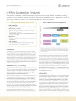
GATA5 mRNA expression is decreased in ccRCC
Loss of GATA5 mRNA expression is associated with
decreased recurrence-free survival
The analyses of relative GATA5 mRNA expression levels
revealed significantly decreased expression in tumor specimens (TU; mean lnRQ = −1.7; ±SD = 1.63) compared with
the corresponding adN (mean lnRQ = 1.73; ±SD = 1.32;
p < 0.001; paired t-test). Figure 1A illustrates the differences in expression values observed for paired tumor and
adN, indicating a strong reduction of up to 31-fold in the
expression levels, largely in tumor tissues. The comparison of the distribution of relative expression values between both tissue groups showed only a small overlap
(Figure 1B).
Cox regression survival analyses using a statistically calculated optimum cut off value for relative GATA5
mRNA expression (lnRQ = −3.52) showed that a lower
expression status was associated with increased risk for
shorter time to disease recurrence (p = 0.023, hazard
ratio (HR) = 0.25, 95% CI: 0.07–0.82; Table 2). Within
30 months, four out of five patients (80%) whose tumor
specimens demonstrated expression values below the
cut off value were identified with disease recurrence
(Figure 3). The status of localized and advanced disease
(p = 0.03, HR = 4.18; 95% CI: 1.15–15.2), status of
Figure 1 GATA5 mRNA expression in paired clear cell renal cell carcinoma and adjacent normal tissues. A) Comparison of the relative
GATA5 expression (RQ) values in adjacent normal (adN) and tumor (TU) tissues from ccRCC patients (p < 0.001). B) Scatterplot analysis illustrating
the distribution of relative expression values (RQ) observed for TU and adN in ccRCC specimens. Bold lines indicate the median of relative
expression values.
Peters et al. BMC Cancer 2014, 14:101
/>
Page 4 of 6
Figure 2 Association of GATA5 CGI methylation and relative
mRNA expression in tumor and adjacent normal tissues. Solid
lines connect the subgroup of paired tissues (tumor tissue = solid
triangle; adjacent normal = solid squares). The regression line
(dashed line) and 95% CI (grey shaded) are presented. Note that
tissues exhibiting concurrent high methylation and low mRNA
expression can be found in the upper left corner, whereas the
occurrence of low methylation and higher relative expression in
tissues is displayed in the lower right corner.
metastasis (p = 0.009, HR = 4.27; 95% CI: 1.43–12.8), and
tumor grade (p < 0.001, HR = 9.48; 95% CI: 2.92–30.8)
were also shown to be associated with RFS (Table 2).
Pairwise bivariate Cox regression analyses were first carried out to investigate whether an association between
expression status and clinicopathological parameters and
RFS exists. In bivariate statistical models, considering
the statuses of advanced disease, metastasis, and tumor
grade as covariates, we found in each case that GATA5
Table 2 Univariate statistical association of GATA5 mRNA
expression and clinicopathological parameters with
recurrence-free survival
Figure 3 Kaplan-Meier plot for illustrating recurrence-free
survival. The solid line shows the Kaplan-Meier curve for 5 patients
(pts.) with GATA5 mRNA expression lower than or equal to the cut off
of −3.52 (natural logarithm), indicating patients with a shortened
recurrence-free survival. The dashed line illustrates the Kaplan-Meier
curve for patients with mRNA expression levels above the cut off
including 30 pts. Disease progression of ccRCC within a period of
approximately two years was observed in seven cases with a low
relative mRNA expression level, whereas high GATA5 mRNA expression
phenotypes showed only four progression events within that time interval.
expression was not associated with RFS while mRNA
levels were detected as a significant parameter in bivariate
Cox regression models including the statuses of lymph
node metastasis, age and gender (Table 3). Moreover,
we carried out a multivariate analysis demonstrating
Table 3 Bivariate statistical association of GATA5 mRNA
expression and clinicopathology with recurrence-free
survival
p-value°
HR
95% CI
GATA5 mRNA expression
0.137
0.390
0.11-1.35
Localized vs Advanced
0.080
3.340
0.87-12.9
GATA5 mRNA expression
0.113
0.370
0.11-1.27
Status of metastasis
0.036
3.410
1.08-10.8
0.511
0.640
0.17-2.41
p-value°
HR
95% CI
GATA5 mRNA expression
GATA5 mRNA expression
0.023
0.25
0.07-0.82
Tumor grade
0.001
8.320
2.34-29.6
Localized vs. Advanced
0.030
4.18
1.15-15.2
GATA5 mRNA expression
0.032
0.260
0.08-0.90
Status of metastasis
0.009
4.27
1.43-12.8
Lymph node metastasis
0.657
1.420
0.29-6.77
Tumor grade
<0.001
9.48
2.92-30.8
GATA5 mRNA expression
0.035
0.270
0.08-0.91
Lymph node status
0.398
1.920
0.42-8.72
Age*
0.214
0.470
0.14-1.55
Age*
0.155
0.420
0.13-1.38
GATA5 mRNA expression
0.023
0.250
0.08-0.83
Gender
0.489
1.510
0.47-0.82
Gender
0.512
1.480
0.46-4.84
°Univariate Cox regression analysis.
*Dichotomization by the median of parameter.
bold numbers: p-value < 0.05 (statistically significant).
hazard ratio (HR).
confidence interval (CI).
°Bivariate Cox regression analysis.
*Dichotomization by the median of parameter.
bold numbers: p-value < 0.05 (statistically significant).
hazard ratio (HR).
confidence interval (CI).
Peters et al. BMC Cancer 2014, 14:101
/>
that mRNA expression of GATA5 (p = 0.12, HR = 0.32;
95% CI: 0.07–1.34) is not significantly associated with
RFS including the covariates status of advance disease
(p = 0.29, HR = 0.18; 95% CI: 0.01-4.41), status of metastasis (p = 0.14, HR = 6.60; 95% CI: 0.55–78.68), tumor grade
p = 0.002, HR = 0.09; 95% CI: 2.46–56.14, age (p = 0.36,
HR = 2.54; 95% CI: 0.05–2.85) and gender (p = 0.39, HR =
2.03; 95% CI: 0.39–10.38).
Discussion
Members of the GATA1-6 transcription factor family
contribute to stem cell differentiation in embryonic tissue, and GATA5 is involved in intestinal epithelial cell
differentiation in adults [3]. Moreover, previous analyses
found GATA5 hypermethylation in human malignancies
such as gastric and colorectal cancers and demonstrated
that epigenetic silencing of the gene occurred in various
human cancer cell lines, providing evidence that GATA5
alterations may represent epigenetic alterations of wider
relevance for carcinogenesis [4,6].
In a recent study aimed at identifying new DNA methylation targets in ccRCC, we detected tumor-specific
hypermethylation of the GATA5 CGI in RCC [11].
Hypermethylation was also associated with advanced
disease and shortened RFS of patients, which had not
been previously reported for any other human cancer.
Hypermethylation of GATA5 in RCC indicated that
the expression of GATA5 might be epigenetically silenced in tumor cells, leading to a biologically more aggressive tumor phenotype. In the current study, we
demonstrate that GATA5 mRNA expression is strongly
reduced in ccRCC and, moreover, that a subgroup of tissues shows a clear relationship between methylation of
the GATA5 CGI and reduced mRNA expression, indicating that epigenetic silencing of GATA5 occurs in a substantial fraction of ccRCC. A significant relationship
between GATA5 hypermethylation and reduced GATA5
mRNA expression within a human tissue, to the best of
our knowledge, has not been previously demonstrated.
Thus, our results support the notion that epigenetic
silencing due to DNA methylation is a relevant process
in RCC.
A subgroup of tissues showed only moderate reduction of expression, although GATA5 methylation was detectable, indicating that other biological mechanisms,
e.g. histone alterations, play a role in tumor development
in ccRCC. Additional functional investigations are required to clarify these aspects.
GATA5 methylation is associated with various clinicopathological parameters as well as RFS. Hence, we hypothesized that reduced mRNA expression levels in
ccRCC would also show an association with unfavorable
clinical parameters. Indeed, we found that decreased
GATA5 mRNA expression is associated with the diameter
Page 5 of 6
of tumors and RFS in univariate Cox regression analysis,
showing a hazard ratio of 0.25, which resembles the reciprocal hazard ratio observed for the corresponding methylation analysis.
Interestingly, a subset of patients with very low mRNA
expression levels also demonstrated a shortened
recurrence-free survival in univariate Cox regression
analysis. However, taking into account that only a small
number of tumors have been identified, these results require future extended evaluation studies including multivariate analyses.
Conclusion
Decreased GATA5 mRNA expression in ccRCC may be
caused by epigenetic silencing, and is likely associated
with a poor clinical outcome. Our results underline the
need for further functional studies to characterize the
interaction of GATA5 and cellular signaling in ccRCC
with respect to the observed changes in expression and
methylation levels, and its association with tumor
progression.
Competing interest
The authors’ declare that they have no competing interest.
Authors’ contributions
IP wrote the manuscript, prepared the figures and participated in the study
design. ND and MK carried out the methylation analyses and participated in
the sequence analyses. JH and MK assembled histopathological,
clinicopathological and survival data. JH performed isolation and
characterization of tissue samples and assembly of patients. MA evaluated
the histopathologies of given tissues. MK, CvK, AM and AS assisted with
general scientific discussion. JS identified the candidate promoter, conceived
of the study, developed the study design and analytical assays, constructed
and ran the clinical database, performed statistical analyses together with RS
and participated in manuscript preparation. All authors read and approved
the final manuscript.
Acknowledgements
We thank Christel Reese and Magrit Hepke for technical assistance.
Author details
Department of Urology and Urologic Oncology, Hannover Medical School,
Carl-Neuberg-Str.1, Hannover 30625, Germany. 2Department of Pathology,
Hannover Medical School, Hannover, Germany. 3Department of Urology,
Eberhard Karls University of Tuebingen, Tuebingen, Germany. 4Department of
Biometry, Hannover Medical School, Hannover, Germany.
1
Received: 18 August 2013 Accepted: 12 February 2014
Published: 17 February 2014
References
1. Jemal A, Bray F, Center MM, Ferlay J, Ward E, Forman D: Global cancer
statistics. CA Cancer J Clin 2011, 61(2):69–90.
2. Morrisey EE, Ip HS, Tang Z, Lu MM, Parmacek MS: GATA-5: a transcriptional
activator expressed in a novel temporally and spatially-restricted pattern
during embryonic development. Dev Biol 1997, 183(1):21–36.
3. Gao X, Sedgwick T, Shi YB, Evans T: Distinct functions are implicated for
the GATA-4, -5, and −6 transcription factors in the regulation of intestine
epithelial cell differentiation. Mol Cell Biol 1998, 18(5):2901–2911.
4. Ren CY, Akiyama Y, Miyake S, Yuasa Y: Transcription factor GATA-5
selectively up-regulates mucin gene expression. J Cancer Res Clin Oncol
2004, 130(5):245–252.
Peters et al. BMC Cancer 2014, 14:101
/>
5.
6.
7.
8.
9.
10.
11.
12.
13.
14.
15.
Page 6 of 6
Kiela PR, LeSueur J, Collins JF, Ghishan FK: Transcriptional regulation of the
rat NHE3 gene. Functional interactions between GATA-5 and Sp family
transcription factors. J Biol Chem 2003, 278(8):5659–5668.
Akiyama Y, Watkins N, Suzuki H, Jair K-W, van Engeland M, Esteller M, Sakai H,
Ren C-Y, Yuasa Y, Herman JG, et al: GATA-4 and GATA-5 transcription
factor genes and potential downstream antitumor target genes are
epigenetically silenced in colorectal and gastric cancer. Mol Cell Biol 2003,
23(23):8429–8439.
Wakana K, Akiyama Y, Aso T, Yuasa Y: Involvement of GATA-4/-5 transcription
factors in ovarian carcinogenesis. Cancer Lett 2006, 241(2):281–288.
Guo M, Akiyama Y, House MG, Hooker CM, Heath E, Gabrielson E, Yang SC,
Han Y, Baylin SB, Herman JG, et al: Hypermethylation of the GATA genes
in lung cancer. Clin Cancer Res 2004, 10(23):7917–7924.
Guo M, House MG, Akiyama Y, Qi Y, Capagna D, Harmon J, Baylin SB,
Brock MV, Herman JG: Hypermethylation of the GATA gene family in
esophageal cancer. Int J Cancer 2006, 119(9):2078–2083.
Fu B, Guo M, Wang S, Campagna D, Luo M, Herman JG, Iacobuzio-Donahue CA:
Evaluation of GATA-4 and GATA-5 methylation profiles in human pancreatic
cancers indicate promoter methylation patterns distinct from other human
tumor types. Cancer Biol Ther 2007, 6(10):1546–1552.
Peters I, Eggers H, Atschekzei F, Hennenlotter J, Waalkes S, Trankenschuh W,
Grosshennig A, Merseburger AS, Stenzl A, Kuczyk MA, et al: GATA5 CpG
island methylation in renal cell cancer: a potential biomarker for
metastasis and disease progression. BJU Int 2012, 110(2 Pt 2):E144–E152.
Waalkes S, Atschekzei F, Kramer MW, Hennenlotter J, Vetter G, Becker JU,
Stenzl A, Merseburger AS, Schrader AJ, Kuczyk MA, et al: Fibronectin 1
mRNA expression correlates with advanced disease in renal cancer.
BMC Cancer 2010, 10:503.
Livak KJ, Schmittgen TD: Analysis of relative gene expression data using
real-time quantitative PCR and the 2(−Delta Delta C(T)) Method.
Methods 2001, 25(4):402–408.
Schmittgen TD, Livak KJ: Analyzing real-time PCR data by the comparative
C(T) method. Nat Protoc 2008, 3(6):1101–1108.
Team RDC: R: A Language and Environment for Statistical Computing.
Vienna Austria; 2011. />
doi:10.1186/1471-2407-14-101
Cite this article as: Peters et al.: Decreased GATA5 mRNA expression
associates with CpG island methylation and shortened recurrence-free
survival in clear cell renal cell carcinoma. BMC Cancer 2014 14:101.
Submit your next manuscript to BioMed Central
and take full advantage of:
• Convenient online submission
• Thorough peer review
• No space constraints or color figure charges
• Immediate publication on acceptance
• Inclusion in PubMed, CAS, Scopus and Google Scholar
• Research which is freely available for redistribution
Submit your manuscript at
www.biomedcentral.com/submit