Retrospective analysis of 104 histologically proven adult brainstem gliomas: Clinical symptoms, therapeutic approaches and prognostic factors
Bạn đang xem bản rút gọn của tài liệu. Xem và tải ngay bản đầy đủ của tài liệu tại đây (366.73 KB, 8 trang )
Reithmeier et al. BMC Cancer 2014, 14:115
/>
RESEARCH ARTICLE
Open Access
Retrospective analysis of 104 histologically
proven adult brainstem gliomas: clinical
symptoms, therapeutic approaches and
prognostic factors
Thomas Reithmeier1,4*, Aanyo Kuzeawu2, Bettina Hentschel3, Markus Loeffler3, Michael Trippel4
and Guido Nikkhah4,5
Abstract
Background: Adult brainstem gliomas are rare primary brain tumors (<2% of gliomas). The goal of this study was
to analyze clinical, prognostic and therapeutic factors in a large series of histologically proven brainstem gliomas.
Methods: Between 1997 and 2007, 104 patients with a histologically proven brainstem glioma were retrospectively
analyzed. Data about clinical course of disease, neuropathological findings and therapeutic approaches were
analyzed.
Results: The median age at diagnosis was 41 years (range 18-89 years), median KPS before any operative procedure
was 80 (range 20-100) and median survival for the whole cohort was 18.8 months. Histopathological examinations
revealed 16 grade I, 31 grade II, 42 grade III and 14 grade IV gliomas. Grading was not possible in 1 patient.
Therapeutic concepts differed according to the histopathology of the disease. Median overall survival for grade
II tumors was 26.4 months, for grade III tumors 12.9 months and for grade IV tumors 9.8 months. On multivariate
analysis the relative risk to die increased with a KPS ≤ 70 by factor 6.7, with grade III/IV gliomas by the factor 1.8
and for age ≥ 40 by the factor 1.7. External beam radiation reduced the risk to die by factor 0.4.
Conclusion: Adult brainstem gliomas present with a wide variety of neurological symptoms and postoperative
radiation remains the cornerstone of therapy with no proven benefit of adding chemotherapy. Low KPS, age ≥ 40 and
higher tumor grade have a negative impact on overall survival.
Keywords: Brainstem glioma, Adult, Neuropathology, Stereotactic surgery
Background
Adult brainstem gliomas are a very rare (<2% of gliomas)
and poorly investigated disease. Recently several larger
series on patients with brainstem gliomas have been reported, however, these series were only partly based on a
histologically established diagnosis (Kesari [1], Landolfi
[2], Guillamo [3], Salmaggi [4]) or involved also other
pathologies than gliomas (Rachinger [5], Samadani [6]).
Importantly, Rachinger and colleagues recently stated
* Correspondence:
1
Department of Neurosurgery, Schwabing Academic Teaching Hospital,
Munich, Germany
4
Division of Stereotactic Neurosurgery, Department of General Neurosurgery,
University Freiburg – Medical Centre, Freiburg, Germany
Full list of author information is available at the end of the article
that intraaxial brainstem lesions with a radiological pattern of glioma represent a very heterogeneous tumour
group with completely different outcomes and that metastasis, lymphoma, inflammation and cavernoma could
be misinterpreted as a glioma by magnetic resonance
imaging (MRI).
The goal of this study was therefore to analyze clinical,
prognostic and therapeutic factors in the largest series of
histological proven brainstem glioma reported so far.
Methods
Patients and data collection
Between 1997 and 2007, 104 patients (age > 18 years)
with histologically proven gliomas of the brainstem
© 2014 Reithmeier et al.; licensee BioMed Central Ltd. This is an Open Access article distributed under the terms of the
Creative Commons Attribution License ( which permits unrestricted use,
distribution, and reproduction in any medium, provided the original work is properly credited.
Reithmeier et al. BMC Cancer 2014, 14:115
/>
were included in this study from five German centres
(Freiburg: n = 73, Tübingen: n = 12, Munich: n = 10,
Dresden: n = 7, Bonn: n = 2). The brainstem was subdivided in a superior (mesencephalon, crus cerebri and
lamina quadrigemina), middle (pons) and inferior part
(medulla oblongata). The tumor was defined as a brainstem glioma when more than 50% of the tumor involved
the brainstem and a histological diagnosis of a glioma
was available. This definition includes according to
Donaldson and Reith diffuse brainstem gliomas (brainstem involvement > 50%) as well as focal brainstem gliomas (brainstem involvement < 50%) and excludes
tumors which significantly involve areas adjacent to the
brainstem [7,8]. Data on clinical course of disease, neuroradiological imaging, therapeutic approaches and neuropathological findings were collected and analyzed with the
assistance of the central database.
Tissue samples were available from all patients either
by stereotactic biopsy or microsurgical tumor resection
and clinical follow-up information was collected on electronic case report forms in regular intervals. The local
ethical committees of the participating institutions (Freiburg,
Tübingen, Munich, Bonn, Dresden) enrolling patients approved the study.
The local neuropathologist of the corresponding university centre enrolling a patient performed neuropathological
diagnosis. Preoperative MRI examination was performed
by university neuroradiologist in 47 cases and by local radiologist of the admitting institution in 57 cases. Neuroradiological findings of the preoperative MRI were analyzed for
etiological classification. Central neuroradiological review
was not performed to depict daily clinical practice.
Statistical analysis
The association of clinical data was tested by χ2-test,
Fisher’s exact test and Kruskal-Wallis-test. Logrank test
was used to compare outcome data. Cox regression
models for OS were fitted to assess the impact of age (<40
vs. ≥ 40), WHO grading (grade I+II vs. grade III+IV), KPS
(≤70 vs. > 70) and initial treatment (no vs. external beam
radiation or radiochemotherapy). Data were analyzed by
IBM SPSS (Version 20.0.0) and StatXact-8 (Cytel Studio
Version 8.0.0).
Page 2 of 8
Table 1 Patient characteristics
All patients N = 104
Age (years)
Median (range)
40 (18 - 89)
Gender, n(%)
Male
61 (58.7%)
Female
43 (41.3%)
KPS (n = 71)
Median (Range)
80 (20 - 100)
≤ 70
26 (36.6%)
> 70
45 (63.4%)
Extend of resection
Stereotactical biopsy
93 (89.4%)
Microsurgical operation
11 (10.6%)
Histopathological WHO-diagnosis (n = 101)
Oligoastrocytoma II
1 (1.0%)
Anaplastic oligoastrocytoma III
1 (1.0%)
Ependymoma II
2 (2.0%)
Diffuse Astrocytoma II
23 (22.8%)
Anaplastic astrocytoma III
39 (38.6%)
Fibrillary astrocytoma
4 (4.0%)
Pilocytic astrocytoma
17 (16.8%)
Glioblastoma
14 (13.9%)
WHO grade (n = 103)
Low grade
47 (45.6%)
High grade
56 (54.4%)
First-line treatment (n = 101)
External beam radiation
45 (44.6%)
Radio-/Chemotherapy
22 (21.8%)
Interstitial radiosurgery
7 (6.9%)
Chemotherapy
4 (4.0%)
No tumor specific therapy
23 (22.8%)
more often male (p = 0.024) and older (p = 0.041) than
patients with low-grade tumors. Median follow up of the
whole population was 49.3 months. Median overall survival was 18.8 months with 95% CI from 11.2 to 26.3
months (1-year-OS-rate 60.9%, 2-year-OS-rate 44.1%,
and 5-years-OS-rate 34.0%).
Results
Patient population
Initial symptoms
Patient characteristics are given in Table 1. All patients
were adults and age ranged from 18 to 89 years (median
41 years). WHO grading was determined in 103 patients
(grade I glioma in 15.5%, grade II glioma in 30.1%, grade
III glioma in 40.8% and grade IV glioma in 13.6%). Median Karnofsky Performance Score (KPS) at diagnosis
was 80 (range 20–100). The male to female ratio was
58.7% to 41.3%. Patients with high-grade tumors were
There was a wide variety of symptoms and combination of
symptoms at the time of initial presentation. The most common presenting symptoms were sensory symptoms (29.8%),
symptoms of cranial nerves II, III, IV and VI (ophthalmological symptoms in 28.8%), impaired coordination (28.8%),
paresis (21.2%), pain (21.2%), gait ataxia (18.3%), dysarthria
and dysphagia (13.5%), signs of raised intracranial pressure
(12.5%), organic psycho-syndrome (7.7%), nausea and
Reithmeier et al. BMC Cancer 2014, 14:115
/>
vomiting (6.7%), myoclonus (2.9%), tinnitus or auditory
disturbances (1.9%), incontinence (1.9%). In 4.8% diagnosis of a brainstem glioma was an incidental finding.
40.4% of patients presented with one symptom, and
54.8% with a combination of up to 6 symptoms.
Neuroradiological imaging
Preoperative T1-weighted MR images were available in 95
patients, T2-weighted images in 32, FLAIR sequences in 7,
diffusion-weighted images in 5 patients and additional sequences were performed in 5 patients. Data about the location of the tumor were available in 99 cases. The tumor
was located solely in the inferior brainstem in 6 cases (6%),
in the middle brainstem in 33 cases (33%) and in the superior brainstem in 19 cases (19%). Two parts of the brainstem were involved in 41 cases (41%) with an infiltration of
the inferior/middle brainstem in 23 cases (23%) and of the
middle/superior brainstem in 18 cases (18%). Definitive
diagnosis of a glioma by neuroradiological imaging was
made in 41 cases, diagnosis of another disease was made in
7 cases and in 56 cases no conclusive diagnosis was
made. Most common differential diagnoses were lymphoma
(n = 4), inflammatory disease (n = 3), abscess (n = 3), metastasis (n = 2), demyelinating disease (n = 1), ependymoma
(n = 1), hemangioblastoma (n = 1) and infarction (n = 1).
Initial surgical procedure and complications
Tissue samples were obtained by stereotactic biopsy in
89.4% (93 patients). The majority of patients were operated in supine position with a frame-based stereotactic
system in local anesthesia by a frontal approach. A suboccipital approach was chosen in 2 patients. Mean duration of the operative procedure was 93 minutes and an
average of 7 probes was obtained.
In 10.6% (11 patients) a microsurgical operation was
performed. Mean duration of the operative procedure
based on 7 patients was 203 minutes, with a total resection in 2 patients, a subtotal resection in 2 patients, a
partial resection in 1 patient and a biopsy in 5 patients.
Data from one patient were missing.
The rate of postoperative complications was 11.8% (11
patients) in stereotactically biopsied patients. Severe
complications occurred in 2.2% and consisted of acute
coma and hemiparesis caused by occlusive hydrocephalus due to postoperative bleeding in one patient, and a
postoperative pontine bleeding combined with an infarction in one patient. Other complications occurred in
9.6% (9 patients). 1 patient developed an aggravation of
ptosis and double vision, in 1 patient a dysphagia, dysarthria and facial paresis occured, and 1 patient suffered
from singultus. One patient developed a focal epilepsy
and aggravation of preexisting hemiparesis and one patient an aggravation of dysarthria, dysphagia and
ataxia. One patient developed an intracranial abscess
Page 3 of 8
and one patient a liquor leckage. In two patients clinical asymptomatic postoperative hemorrhage was detected in the postoperative CCT scan.
The rate of perioperative complications in the microsurgically operated group was 36.4% (4 patients) and
consisted of 2 postoperative hemorrhages, 1 infection of
the bone flap and 1 respiratory insufficiency.
Histopathological results
103 of 104 tumors were graded according to the WHO
classification and in 1 patient grading was not possible
by the local universitary neuropathologist. 16 patients
had a grade I, 31 patients a grade II, 42 patients a grade
III and 14 patients a grade IV tumor.
Histopathological diagnosis according to the WHO classification was possible in 101 patients (Table 1). Anaplastic
pilocytic astrocytoma was diagnosed in two patients and
an astrocytoma without any further classification in another patient.
Treatment
Initial treatment
Initial treatments after surgery were chemotherapy, external beam radiation, interstitial radiosurgery with implantation of I-125 seed, a combination of radio- and
chemotherapy or a wait and see strategy. Information
about all treatments administered was available in 101 of
104 patients. In 23 patients a wait and see approach was
chosen, 22 patients received combined radiochemotherapy, 45 patients were treated with external beam radiation, 7 patients with interstitial radiosurgery and 4
patients with chemotherapy alone. Median overall survival for patients treated supportive was 4.3 months, for
patients who received external beam radiation was 26.4
months and for patients treated by radio-/chemotherapy
was 13.4 months. Therapeutic strategies differed between
the different WHO grades. Brachytherapy was performed
only in low-grade gliomas whereas radiochemotherapy
was predominantly given to patients with high-grade gliomas (17 patients with high grade gliomas vs. 5 patients
with low-grade gliomas; for details see Table 2).
Data on salvage treatment at progression were available
in 22 patients. Chemotherapy alone was performed in 11
patients. 6 patients received temozolomide, and 5 patients
received a combination of temozolomide and ACNU, temozolomide and PC, temozolomide and PCV, or PCV and
ACNU alone. Radiotherapy was performed in 8 patients, a
combination of radio- and chemotherapy in 1 patients, a
combination of brachytherapy and chemotherapy in 1 patient, and brachytherapy alone in 1 patient.
Influence of KPS and age on treatment decision
35.6% of patients over 40 years received supportive therapy compared to 15.6% of patients < 40 years (p = 0.069).
Reithmeier et al. BMC Cancer 2014, 14:115
/>
Page 4 of 8
Table 2 Therapeutic strategies according to WHO grade
WHO grade
Total
I
II
III
IV
External beam radiation
6 (40.0%)
16 (53.3%)
18 (43.9%)
4 (28.6%)
44 (44.0%)
Radio-/Chemotherapy
1 (6.7%)
4 (13.3%)
12 (29.3%)
5 (35.7%)
22 (22.0%)
Interstitial radiosurgery
4 (26.7%)
3 (10.0%)
-
-
7 (7.0%)
Chemotherapy
-
-
2 (4.9%)
2 (14.3%)
4 (4.0%)
Therapy
No tumor specific therapy
4 (26.7%)
7 (23.3%)
9 (22.0%)
3 (21.4%)
23 (23.0%)
Total
15 (100.0%)
30 (100.0%)
41 (100.0%)
14 (100.0%)
100 (100.0%)
No tumor-specific therapy was initiated in 54.2% of patients with a KPS ≤ 70 compared to 8.1% of patients with
a KPS > 70 (p<0.001).
group. Supportive care only was associated with an unfavourable outcome in both groups. In the high-grade
group the addition of chemotherapy to radiotherapy was
not associated with improved survival.
Overall survival
Median overall survival for the whole population was 18.8
months but differed significantly for the different WHO
grades. Median overall survival for grade II tumors was
26.2 months, for grade III tumors 12.9 months and for
grade IV tumors 9.8 months.
Treatment was associated with improved survival in
the high-grade glioma as well as in the low-grade glioma
Prognostic factors
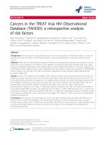
Grading correlated with survival (p = 0.003, Figure 1a).
In univariate analysis high-grade tumors had an about
two times increased relative risk (RR) for death related
to low-grade tumors.
Younger age was associated with improved overall survival. Median overall survival for patients < 40 years was
Figure 1 Prognostic factors: a) tumor grade; b) age, c) Karnofsky performance index, d) therapy related factors.
Reithmeier et al. BMC Cancer 2014, 14:115
/>
47.3 months whereas patients ≥ 40 years had a median
overall survival of 10.3 months (p = 0.006; Figure 1b) with
an increased risk for death by factor 2.
Karnofsky performance score at diagnosis was associated with prognosis: patients with a KPS > 70 had a median overall survival of 56.3 months whereas patients
with a KPS ≤ 70 had a median overall survival of 4.8
months (p<0.001, Figure 1c), with an increased risk of
death by factor 7.1.
Initial treatment (supportive care = no tumor-specific
therapy, external beam radiation, radiochemotherapy)
was also associated with overall survival on univariate
analysis: median overall survival for patients, who received no tumor-specific therapy was 4.3 months
whereas patients who were initially treated by external
beam radiation had a median overall survival of 26.4
months (external beam radiation vs. no tumor-specific
therapy, p<0.001) and patients treated with a combined
radiochemotherapy had a median overall survival of 13.4
months (radiochemotherapy vs. no tumor-specific therapy p = 0.003). There was no significant difference in
overall survival among patients who received external
beam radiation alone as opposed to radiochemotherapy
(p = 0.093, Figure 1d). External beam radiation or radiochemotherapy reduces the risk to die by factor 0.3 and
0.4.
To assess the independent impact of the above mentioned factors on overall survival a multivariate cox regression model was built. Karnofsky performance score
≤ 70 showed the strongest effect and increased the relative risk to die by factor 6.7, followed by therapy (radiochemotherapy reduced the relative risc to die by factor
0.3 and external beam radiation by factor 0.4). High
grade glioma increased the relative risc to die by the factor 1.8 and age ≥ 40 by 1.7. However on multivariate
analysis age and WHO grade did not reach statistical
significance (Table 3).
Discussion
Here we report one of the largest series of histopathologically proven gliomas of the brainstem. Patients presented with a wide variety of symptoms, and median
survival was only 18.8 months for the whole patient
population. The amount of neuroradiological differential
diagnoses confirmed the necessity of histopathological
evaluation. In multivariate analysis KPS ≤ 70, higher
tumor grade and age ≥ 40 were negative prognostic factors, whereas radiation therapy or radio-chemotherapy
improved prognosis.
Recently several large series about brainstem gliomas in
adults with median overall survival rates between 54 and
85 months have been published (Table 4). However diagnosis of a brainstem glioma in these series was mainly
based on neuroradiological imaging and confirmed by
Page 5 of 8
Table 3 Cox regression models to assess the impact of
age (<40 vs. ≥ 40), WHO grading (grade I + II vs. grade III
+ IV), KPS (≤70 vs. > 70) and initial treatment (no vs.
external beam radiation or radiochemotherapy) on the
relative risc to die
Relative risk
95% CI
p-value
0.8 to 3.4
0.143
2.9 to 15.8
< 0.001
0.9 to 3.6
0.104
Age (years)
< 40
1
≥ 40
1.7
KPS
> 70
1
≤ 70
6.7
WHO grade
Low
1
High
1.8
Treatment
No tumor specific therapy
1
External beam radiation
0.4
0.2 to 0.9
0.021
Radio-/Chemotherapy
0.3
0.1 to 0.9
0.041
histopathological examinations only in 13% - 67% of cases.
Rachinger found that in a series of 46 radiologically suspected brainstem gliomas histological examination confirmed a glioma in only 28 cases (61%) and revealed
metastasis in 15% (n = 7), lymphoma in 11% (n = 5), inflammatory disease in 4% (n = 2), cavernoma in 2% (n = 1)
and gliosis in 6% (n = 3). The authors pointed out, that
intra-axial brainstem lesions with a radiological pattern of
glioma represent a very heterogeneous tumor group with
completely different clinical outcomes [5]. Samadani published a meta-analysis of 293 brainstem biopsies in children and adults. Stereotactic biopsy was in 96% diagnostic
with a mortality rate of 0.3%, a transient morbidity rate of
4% and a permanent morbidity rate of 1%. Pathology
showed that half of the adult brainstem intrinsic lesions
were gliomas, 10% were metastases, and the remainders
were hematomas, vascular malformations, lymphomas,
demyelination, cysts, radiation necrosis, abscesses, vasculitis, infarcts, leukemia, cryptococcus, or granulomas.
Table 4 Literature overview of large series of adult brain
stem glioma: histological confirmation of diagnosis varied
between 13% and 100% (actual study)
Author
N
Histology
HGG
LGG
Median OS
Landolfi (1998)
23
3 (13.0%)
1 (33.3%)
2 (66.7%)
54.0 months
Salmaggi (2008)
34
20 (58.8%)
11 (55.0%) 9 (45.0%)
59.0 months
Guillamo (2001)
48
32 (67.0%)
15 (46.9%) 17 (53.1%) 64.8 months
Kesari (2008)
101 46 (45.5%)
31 (68.9%) 15 (31.1%) 85.0 months
Reithmeier (2013) 104 104* (100.0%) 46 (44.7%) 57 (55.3%) 18.8 months
*In one patient grading was not performed.
Reithmeier et al. BMC Cancer 2014, 14:115
/>
They pointed out that neuroradiological diagnosis of noncontrast-enhancing lesions of the brainstem as low grade
glioma is insufficient and that histopathological diagnosis
for non-enhancing brainstem lesions with a long duration
of symptoms revealed hematomas, arteriovenous malformations, lymphomas, demyelination, radiation necrosis
and infarction. Therefore the authors concluded that
stereotactic biopsy is indicated for both enhancing and
non-enhancing lesions of the brainstem [6].
Kickingereder et al. recently published a large meta
analysis of 1480 stereotactic biopsies for brainstem tumors and found a diagnostic success rate of 96.2%, an
overall morbidity rate of 7.8%, a permanent morbidity
rate of 1.7% and a mortality rate of 0.9% [9].
The clinically relevant postoperative morbidity of 9.7%
in our series is in line with the data of Kickengereder
but higher in comparison with figures from the metaanalysis of Samadani and colleagues [6]. The main difference between these two series was the homogenous
histopathology in our series which consisted only of gliomas with a proportion of high-grade tumors of nearly
50% as opposed to a broad variety of tumorous and
non-tumorous diseases in the latter series. Because of
the rich neovascularization of high-grade gliomas, the
risk of of postoperative hemorrhage or malignant brainedema is likely to be higher in malignant gliomas
compared to other pathologies and may explain the
differences in morbidity.
Several publication have discussed this issue with regard
to stereotactic brain biopsy for supratentorial lesions.
Bernstein suggested that biopsy of specific pathologies
(e.g. glioblastoma, lymphoma) may be associated with
an increased risk of either hemorrhage or severe
edema, due to the abnormal neovasculature of these
tumors [10]. Savin confirmed these data and identified
malignant glioma pathology to be associated with a 4-fold
increased risk of morbidity, especially from hemorrhage
[11]. However other authors found no association between
lesion pathology and complication rates [12]. A recent
study about complication of frame-based stereotactic biopsy in 622 cases identified an association between mortality and glioblastoma pathology and suggested that
abnormal tumor neovasculature of malignant glioma may
be the reason therefore [13].
Overall morbidity of stereotactic biopsy in brainstem
tumors ranges between 7.8% and 12% in larger series
and is therefore not distinctly higher compared to a
morbidity rate of 4.9% in general stereotactic brain biopsy, especially when considering the low rate of 1.7% of
permanent morbidity in stereotactic brainstem biopsy
[1,4,9,13].
Dellaretti and colleagues investigated the correlation
between magnetic resonance imaging findings and histological diagnosis of intrinsic brainstem lesions in adults
Page 6 of 8
in a series of 96 patients. Stereotactic biopsy established
a precise histological diagnosis in 92 patients which consisted of 63 diffuse brainstem gliomas, 19 other neoplastic
diseases (lymphomas, metastases, pilocytic astrocytomas,
craniopharyngioma, ganglioma) and 10 non-neoplastic lesions (inflammatory disease, ischemic lesion, fungal abscess, gliosis). Overall morbidity rate was 9% and one
patient died from exacerbated peritumoral edema. With
regard to neuroradiological features the diagnostic effect
of stereotactic biopsy was greater in patients with focal or
enhancing lesions shown by MRI in whom the diagnosis
of a diffuse gliomas was less frequent [14].
The value of additional imaging modalities to improve
non-invasive diagnostic accuracy by MR spectroscopy or
positron emission tomography is currently under investigation. However, Massager showed recently in a series of
30 brainstem gliomas that the integration of PET imaging
can not replace histological analysis as MRI combined
with PET data was only concordant with histological findings in 63% of cases [15].
The results of these studies are indicative that in adult
patients with lesions of the brainstem therapeutic decisions should be based on a histopathological examination due to the wide spectrum of differential diagnoses.
Therefore we included in our study only patients with
a histopathologically confirmed brainstem glioma to exclude a possible bias due to non-glioma lesions classified
as gliomas by MRI, which may have a significant better
prognosis. This and the high rate of malignant brain
stem gliomas of 44.7% might explain the distinct difference in median overall survival of 18.8 months in our
series in comparison to the actual literature of brainstem
gliomas. We found that median overall survival of
treated patients with HGG of the brainstem resembled
the overall survival data of patients with supratentorial
high grade gliomas. However median overall survival of
patients with grade II gliomas of the brainstem was significant shorter than in brainstem glioma series of Kesari
(26.4 months vs. 168 months) and in comparison to
supratentorial low grade gliomas (26.4 months vs. 7–8
years). Reasons for this difference might be the location
within a highly eloquent area, faster malignant transformation than in supratentorial gliomas for unknown
reasons or possible histopathological undergrading. It is
also notable that in Kesari’s series grade I brainstem gliomas had a significant shorter median overall survival of
83 months in comparison to grade II brainstem gliomas
with a median overall survival of 168 months. Inaccurate
neuroradiological diagnosis and subsequent undergrading may also explain this as up to 60% of high grade
brainstem gliomas show no contrast enhancement after
gadolinium application [6].
The location within a highly eloquent area may also
explain the finding that the percentage of glioblastoma is
Reithmeier et al. BMC Cancer 2014, 14:115
/>
significant lower in the brainstem compared to its supratentorial counterpart (12.5% vs 60-75%) [16] as brainstem gliomas may become clinically symptomatic very
early in the course of the disease. We also found that
pilocytic astrocytomas of the brainstem are surprisingly
not such a benign disease as supported by many authors
[17-19] with rapid progression especially in the first 20
months after diagnosis and stabilization in survival
thereafter (see Figure 1a). These data are also supported
by Stuer [20] who observed after a median follow-up of
55 months 30% tumor recurrence and 18% deaths. Another surprising result was the lack of pure oligodendroglial brain stem gliomas and the low proportion of
oligoastrocytic tumors. Interestingly in the series of
Guillamo 25% of biopsied brainstem gliomas were oligodendrocytic or mixed gliomas.
Established prognostic factors in the literature are age,
duration of symptoms, KPS, contrast enhancement, MRI
“necrosis”, histology and location in the pons and medulla, mainly based on univariate analysis [2,3,21,22].
We confirmed the strong prognostic impact of KPS and
age in multivariate analysis and the positive effect of radiation therapy as the cornerstone of therapeutic measures on overall survival (therapy reduces the risk of
death by a factor of 0.4). The value of chemotherapy in
low and high grade brainstem gliomas is still undefined.
Efficacy of different chemotherapeutic agents (temozolomide, nitrosureas or platinum based chemotherapeutic
protocols) is currently unproven. At relapse a wide variety of chemotherapeutic agents are used which included BCNU [1,2-bis(2-chloroethyl)-1-nitrosourea],
BCNU - procarbazine, CCNU [1-(2-chloroethyl)-3cyclohexyl-1-nitrosourea] -procarbazine-vincristine (PCV),
carboplatin, carboplatin-VP16, carboplatin-VP16-ifosfamid,
ifosfamid, procarbazine-VP16, temozolomide, CCNU, vincristine, irinotecan, cisplatin and temozolomide, ACNU
and procarbazine. Effectiveness of these protocols were
limited: Guillamo [3] reported a radiological response rate
of 7% three months after onset of chemotherapy and clinical improvement lasting longer than 6 months in 15% of
patients and Samlaggi [4] described a temporary clinical
and radiological stabilization in 22% of patients after
chemotherapy.
Alternative therapeutic strategies like interstitial radiosurgery with implantation of iodine-125 seeds and application of anti-angiogenic drugs like bevacizumab
have to be also considered in the therapeutic concept.
Mundinger treated in a series of 89 low grade brainstem
gliomas 55 patients with stereotactic brachytherapy. 29
patients received iodine-125 seeds with a 5 year survival rate of 54.8% and 26 patients received iridium
192 seed with a 5 year survival rate of 26.9% in comparison to 5 year survival rate of 14.7% in patients who
underwent only biopsy [23]. Ruge recently published a
Page 7 of 8
series of 47 patients with inoperable focal brainstem gliomas WHO grades I and II treated by stereotactic implantation of iodine-125 seeds with a 5-year overall
survival rate of 97.4 ±2.6% [24].
Survival rates of interstitial radiosurgery are therefore
at least comparable to external beam radiotherapy with
reported 5-year survival rates between 45%-58% [1,2]
and both methods should be evaluation against each
other in prospective randomized trials.
Reports about the use of antiangiogenic substances in
the literature are rare. Besides two case reports [25,26] a
small series of 3 patients [27] showed the effectivness of
bevacizumab as a salvage therapy for progressive brainstem gliomas with improvement of clinical condition, reduction of daily dexamethasone dosage and radiological
response.
Conclusion
Adult brain stem gliomas present with a wide variety of
neurological symptoms and neuroradiological differential
diagnoses. Stereotactic biopsy is the procedure of choice
to obtain a histopathological diagnosis. Prognosis of
high-grade gliomas resembles its supratentorial counterparts whereas low-grade gliomas of the brainstem have a
worse prognosis compared to the actual literature.
Cornerstone of therapy remains radiation and alternative
strategies like interstitial radiosurgery, chemotherapy or
antiangiogenic drugs need to be further explored, ideally
in the context of molecular profiling for common alterations such as 1p/19q codeletion, MGMT promoter
methylation and IDH mutation.
Competing interests
The authors declare that they have no competing interests.
Authors’ contribution
TR analyzed the data and created the first draft of the article, AK collected
and analyzed the data, MT and GN were involved in conception and design
of the study, GN critically reviewed the first draft, BH and ML performed
statistical analysis and interpretation of data, and all authors approved the
final draft.
Acknowledgement
We thank the Department of Neurosurgery of the Ludwig Maximilian
University Munich, the Department of Neurosurgery of the University of
Tübingen, the Department of Neurosurgery of the Technical University
Dresden and the Department of Neurosurgery of the University of Bonn for
enrolling patients into this study.
Funding
This work was supported by the German Cancer Aid providing a central data
base.
Author details
1
Department of Neurosurgery, Schwabing Academic Teaching Hospital,
Munich, Germany. 2Service de Neurochirurgie, Hopital Louis Pasteur, Colmar,
France. 3Institute for Medical Informatics, Statistics and Epidemiology,
University of Leipzig, Leipzig, Germany. 4Division of Stereotactic Neurosurgery,
Department of General Neurosurgery, University Freiburg – Medical Centre,
Freiburg, Germany. 5Department of Neurosurgery, University Hospital, Erlangen,
Germany.
Reithmeier et al. BMC Cancer 2014, 14:115
/>
Received: 13 June 2013 Accepted: 12 February 2014
Published: 21 February 2014
References
1. Kesari S, Kim RS, Markos V, Drappatz J, Wen PY, Pruitt AA: Prognostic
factors in adult brainstem gliomas: a multicenter, retrospective analysis
of 101 cases. J Neurooncol 2008, 88:175–183.
2. Landolfi JC, Thaler HT, DeAngelis LM: Adult brainstem gliomas.
Neurology 1998, 51:1136–1139.
3. Guillamo JS, Monjour A, Taillandier L, Devaux B, Varlet P, Haie-Meder C,
Defer GL, Maison P, Mazeron JJ, Cornu P, Delattre JY: Brainstem gliomas in
adults: prognostic factors and classification. Brain 2001, 124:2528–2539.
4. Salmaggi A, Fariselli L, Milanesi I, Lamperti E, Silvani A, Bizzi A, Maccagnano
E, Trevisan E, Laguzzi E, Rudá R, Boiardi A, Soffietti R, Associazione Italiana di
Neuro-oncologia: Natural history and management of brainstem gliomas
in adults. A retrospective Italian study. J Neurol 2008, 255:171–177.
5. Rachinger W, Grau S, Holtmannspötter M, Herms J, Tonn JC, Kreth FW:
Serial stereotactic biopsy of brainstem lesions in adults improves
diagnostic accuracy compared with MRI only. J Neurol Neurosurg
Psychiatry 2009, 80:1134–1139.
6. Samadani U, Judy KD: Stereotactic brainstem biopsy is indicated for the
diagnosis of a vast array of brainstem pathology. Stereotact Funct
Neurosurg 2003, 81:5–9.
7. Donaldson SS, Laningham J, Fisher PG: Advances toward an
understanding of brainstem gliomas. J Clin Oncol 2006, 24:1266.1272.
8. Reith W: Gehirn. In Diagnostische und Interventionelle Radiologie. Edited by
Vogl TJ, Reith W, Rummeny EJ. Berlin Heidelberg: Springer; 2011:61–272.
9. Kickingereder P, Willeit P, Simon T, Ruge MI: Diagnostic value and safety of
stereotactic biopsy of brainstem tumors: a systematic review and
meta-analysis of 1480 cases. Neurosurgery 2013, 72:873–881.
10. Bernstein M, Parrent AG: Complications of CT-guided stereotactic biopsy
of intra-axial brain lesions. J Neurosurg 1994, 81:165–168.
11. Sawin PD, Hitchon PW, Follett KA, Torner JC: Computed imaging-assisted
stereotactic brain biopsy: a risk analysis of 225 consecutive cases. Surg
Neurol 1998, 49:640–649.
12. Grossman R, Sadetzki S, Spiegelmann R, Ram Z: Haemorrhagic
complications and the incidence of asymptomatic bleeding associated
with stereotactic brain biopsies. Acta Neurochir (Wien) 2005, 147:627–631.
13. Kongkham PN, Knifed E, Tamber MS, Bernstein M: Complications in 622
cases of frame-based stereotactic biopsy, a decreasing procedure.
Can J Neurol Sci 2008, 35:9–84.
14. Dellaretti M, Touzet G, Reyns N, Dubois F, Gusmao S, Pereira JL, Blond S:
Correlation between magnetic resonance imaging findings and
histological diagnosis of intrinsic brainstem lesions in adults. Neuro Oncol
2012, 14:381–385.
15. Massager N, David P, Goldman S, Pirotte B, Wikler D, Salmon I, Nagy N,
Brotchi J, Levivier M: Combined magnetic resonsace imaging- and
positron emission tomography-guided stereotactic biopsy in brainstem
mass lesions: diagnostic yield in a series of 30 patients. J Neurosurg 2000,
93:951–957.
16. Ohgaki H, Kleihues P: Population-based studies on incidence, survival
rates, and genetic alterations in astrocytic and oligodendroglial gliomas.
J Neuropathol Exp Neurol 2005, 64:479–489.
17. Afra D, Muller W, Slowik F, Firsching R: Supratentorial lobar pilocytic
astrocytomas: report of 45 operated cases, including 9 recurrences.
Acta Neurochir (Wien) 1986, 81:90–93.
18. Brown PD, Buckner JC, O'Fallon JR, Iturria NL, Brown CA, O'Neill BP,
Scheithauer BW, Dinapoli RP, Arusell RM, Abrams RA, Curran WJ, Shaw EG,
North Central Cancer Treatment Group: Adult patients with supratentorial
pilocytic astrocytomas: a prospective multicenter clinical trial. Int J Radiat
Oncol Biol Phys 2004, 58:1153–1160.
19. Bell D, Chitnavis BP, Al-Sarraj S, Connor S, Sharr MM, Gullan RW: Pilocytic
astrocytoma of the adult—clinical features, radiological features and
management. Br J Neurosurg 2004, 18:613–616.
20. Stüer C, Vilz B, Majo Becker A, Schramm J, Simon M: Frequent recurrences
and progression in pilocytic astrocytoma in adults. Cancer 2007,
110:2799–2808.
21. Linstadt DE, Edwards MS, Prados M, Larson DA, Wara WM:
Hyperfractionated irradiation for adults with brainstem gliomas.
Int J Radiat Oncol Biol Phys 1991, 20:757–760.
Page 8 of 8
22. Gribsby PW, Garcia DM, Simpson JR, Fineberg BB, Schwartz HG: Prognostic
factors and results of therapy for adult thalamic and brainstem tumors.
Cancer 1989, 63:2124–2129.
23. Mundinger F, Braus DF, Kraus JK, Birg W: Long-term outcome of 89 lowgrade brain-stem gliomas after interstitial radiation therapy. J Neurosurg
1991, 75:740–746.
24. Ruge MI, Kickingereder P, Simon T, Treuer H, Sturm V: Stereotactic iodine125 brachytherapy for treatment of inoperable focal brainstem gliomas
of WHO grades I and II: feasibility and long-term outcome. J Neurooncol
2012, 109:273–283.
25. Torcuator R, Zuniga R, Loutfi R, Mikkelsen T: Bevacizumab and irinotecan
treatment for progressive diffuse brain stem glioma: case report.
J Neurooncol 2009, 93:409–412.
26. Raza S, Donach M: Bevacizumab in adult malignant brainstem gliomas.
J Neurooncol 2009, 95:299–300.
27. Reithmeier T, Lopez WO, Spehl TS, Nguyen T, Mader I, Nikkhah G, Pinsker
MO: Bevacizumab as salvage therapy for progressive brain stem gliomas.
Clin Neurol Neurosurg 2013, 115:165–169.
doi:10.1186/1471-2407-14-115
Cite this article as: Reithmeier et al.: Retrospective analysis of 104
histologically proven adult brainstem gliomas: clinical symptoms,
therapeutic approaches and prognostic factors. BMC Cancer 2014 14:115.
Submit your next manuscript to BioMed Central
and take full advantage of:
• Convenient online submission
• Thorough peer review
• No space constraints or color figure charges
• Immediate publication on acceptance
• Inclusion in PubMed, CAS, Scopus and Google Scholar
• Research which is freely available for redistribution
Submit your manuscript at
www.biomedcentral.com/submit