Dissecting the signaling pathways associated with the oncogenic activity of MLK3 P252H mutation
Bạn đang xem bản rút gọn của tài liệu. Xem và tải ngay bản đầy đủ của tài liệu tại đây (717.97 KB, 6 trang )
Velho et al. BMC Cancer 2014, 14:182
/>
RESEARCH ARTICLE
Open Access
Dissecting the signaling pathways associated with
the oncogenic activity of MLK3 P252H mutation
Sérgia Velho1, Ana Pinto1,2, Danilo Licastro3, Maria José Oliveira2, Filipa Sousa1, Elia Stupka4 and Raquel Seruca1,5*
Abstract
Background: MLK3 gene mutations were described to occur in about 20% of microsatellite unstable
gastrointestinal cancers and to harbor oncogenic activity. In particular, mutation P252H, located in the kinase
domain, was found to have a strong transforming potential, and to promote the growth of highly invasive tumors
when subcutaneously injected in nude mice. Nevertheless, the molecular mechanism underlying the oncogenic
activity of P252H mutant remained elusive.
Methods: In this work, we performed Illumina Whole Genome arrays on three biological replicas of human HEK293
cells stably transfected with the wild-type MLK3, the P252H mutation and with the empty vector (Mock) in order to
identify the putative signaling pathways associated with P252H mutation.
Results: Our microarray results showed that mutant MLK3 deregulates several important colorectal cancer- associated signaling pathways such as WNT, MAPK, NOTCH, TGF-beta and p53, helping to narrow down the number of
potential MLK3 targets responsible for its oncogenic effects. A more detailed analysis of the alterations affecting the
WNT signaling pathway revealed a down-regulation of molecules involved in the canonical pathway, such as DVL2,
LEF1, CCND1 and c-Myc, and an up-regulation of DKK, a well-known negative regulator of canonical WNT signaling,
in MLK3 mutant cells. Additionally, FZD6 and FZD10 genes, known to act as negative regulators of the canonical
WNT signaling cascade and as positive regulators of the planar cell polarity (PCP) pathway, a non-canonic WNT
pathway, were found to be up-regulated in P252H cells.
Conclusion: The results provide an overall view of the expression profile associated with mutant MLK3, and they
support the functional role of mutant MLK3 by showing a deregulation of several signaling pathways known to
play important roles in the development and progression of colorectal cancer. The results also suggest that mutant
MLK3 may be a novel modulator of WNT signaling, and pinpoint the activation of PCP pathway as a possible
mechanism underlying the invasive potential of MLK3 mutant cells.
Keywords: Colorectal cancer, MLK3, WNT pathway, MSI, Planar cell polarity
Background
Mixed-lineage kinase 3 (MLK3) belongs to a family of
seven different mammalian MLKs, that are clustered in
three subgroups accordingly with their structural similarities: the MLKs (MLK1, MLK2, MLK3, MLK4); the
dual-leucine-zipper-bearing kinases (Dlk and Lzk); and
zipper sterile-α-motif kinase (Zakα and Zakβ) [1].
MLK3 protein is composed by a Src-homology-3 (SH3)
domain, located at the amino terminus of the protein,
* Correspondence:
1
Instituto de Patologia e Imunologia Molecular da Universidade do Porto,
Rua Dr. Roberto Frias, 4200-465 Porto, Portugal
5
Medical Faculty of the University of Porto, Porto, Portugal
Full list of author information is available at the end of the article
followed by a kinase domain, a leucine-zipper region, a
Cdc42/RAC interacting binding motif (CRIB) and a
Proline/Serine/Threonine-rich (P/S/T-rich) carboxy terminal domain [1-4]. All these domains show a very high degree of homology between MLK members (MLK1-MLK4)
[1], except the carboxy-terminus P/S/T- rich domain which
is the less conserved region among MLK proteins [1].
MLK3 is a serine/threonine protein kinase that regulates the mitogen-activated protein kinase (MAPK)
pathway, activating ERK, p38 and JNK in response
to extracellular signals [1,5]. Additionally, functional
studies have demonstrated that overexpression of wildtype MLK3 leads to morphological transformation of
© 2014 Velho et al.; licensee BioMed Central Ltd. This is an Open Access article distributed under the terms of the Creative
Commons Attribution License ( which permits unrestricted use, distribution, and
reproduction in any medium, provided the original work is properly credited. The Creative Commons Public Domain
Dedication waiver ( applies to the data made available in this article,
unless otherwise stated.
Velho et al. BMC Cancer 2014, 14:182
/>
NIH 3T3 fibroblasts and growth in soft agar, a capacity
that is MEK/ERK dependent [6]. Further, MLK3 has
been demonstrated to function as a scaffolding protein,
involved in the formation of a multiprotein complex
containing MLK3/BRAF/RAF1 [1,5,7,8]. The formation of this complex was shown to be important for the
activation of wild-type BRAF and, consequently, to
the activation of ERK signaling [1,5,7,8]. Furthermore,
MLK3 was reported to be important for the proliferation of tumor cells, bearing either oncogenic KRAS or
neurofibromatosis-1 (NF1) or NF2 inactivating mutations [8]. In addition, the MLK3 signaling activation
was associated with an increase in the migratory and
invasive capacity of tumor cells in gastric [9], breast
[10-12], lung [13] and ovarian [14] cancers. All together, these observations implicate MLK3 as a cancerrelated gene although, until recently, nothing was
known about MLK3 gene deregulation in primary cancer tissues.
Our group reported the occurrence of MLK3 mutations
in microsatellite unstable (MSI) gastrointestinal tumors
(both sporadic and hereditary forms) in a frequency of
about 20%. Using in vitro transforming assays, we demonstrated that several MLK3 mutations affecting different domains of the protein had transforming potential when
compared to cells expressing the wild-type and the kinasedead forms of the protein [15]. These results were further
supported by in vivo studies in which one of the two most
transforming mutations (P252H – located in the kinase domain) was found to be tumorigenic and to give rise to
highly invasive tumors when subcutaneously injected in
nude mice. Thus, our previous work pointed mutant
MLK3 as a new oncogene in MSI gastrointestinal cancers,
however, the signaling pathways associated to its oncogenic
activity remained to be explored.
In this work, we aimed at identifying the signaling pathways associated to mutant MLK3, in particular the P252H
mutation. The reasons underlying the choice for this mutation were: (a) it was one of the most transforming mutations
previously analyzed; (b) showed high tumorigenic capacity
with strong invasive potential in in vivo studies; and (c) it
was located in the kinase domain which is an important domain for the regulation of downstream signaling pathways.
The results showed that P252H mutation interferes with
important colorectal cancer-associated signaling pathways
such as WNT, MAPK, NOTCH, TGF-β and P53.
Methods
cDNA constructs and mutagenesis
Wild-type MLK3 and mutant sequences were cloned
into pLENTID6/V5 directional TOPO (Invitrogen). Mutant
MLK3 P252H sequence was generated by site-directed mutagenesis using the MLK3 wild-type sequence cloned in
pLENTID6/V5 as template. pLENTID6/V5 empty vector
Page 2 of 6
(Mock) was obtained by the insertion of a small fragment
of cDNA in order to circularize the plasmid.
Cell lines
Human HEK293 were maintained in DMEM (Gibco,
Invitrogen) supplemented with 10% FBS and 1% penicillin–streptomycin (Gibco, Invitrogen), and were incubated in a humidified chamber with 5% CO2 at 37°C.
Transfections
For HEK293 stable transfections, ViraPower Lentiviral
Expression kit (Invitrogen) was used for the transduction
of the MLK3 wild-type and mutant P252H sequences
as well as the empty vector. Lentiviral transduction
was performed following the manufacturer’s instructions.
Transduced cells were selected by antibiotic resistance
(blasticidin, 12 μg/ml) (Gibco, Invitrogen). The expression levels of MLK3 in the different clones selected were
measured by western-blot.
RNA extraction and cDNA synthesis
Total RNA was isolated from cell lines using TriPure
Isolation Reagent (Roche Applied Science), following
manufacturer’s instructions. Complementary DNA was
synthesized from 1 μg of total RNA using SuperScriptII
Reverse Transcriptase (Invitrogen) and Random Primers
(Invitrogen).
Labeling and hybridization
Five hundred ng aliquots of RNA from the samples were
quality checked using the Agilent 2100 Byoanalyzer and
only samples above integrity quality number (RIN) 8
[16] were used and amplified according to the specifications of the Illumina® TotalPrep™ RNA Amplification Kit
(Ambion, Austin, TX, USA). The cRNA samples were
applied to the arrays of Sentrix® Human-v6 Expression
BeadChip (Illumina, San Diego, CA, USA) and hybridized
according to manufacturer's specifications. The Sentrix
BeadChips were scanned with the Illumina's Beadarray
system 500G Scanner (Illumina®).
Microarray data analysis
The signal intensity was extracted from the hybridization
images, background subtracted and normalized using
Illumina Inc. BeadStudio software version 3.3.7. The
data produced was checked against the Illumina internal
quality controls and loaded into the Bioconductor software [17,18]. To identify differentially expressed genes
based on a moderate t-test, the bioconductor Limma
package [19] was used. Genes were selected based on a
p-value cut-off (after adjustment) of p < 0.01 to control the
false discovery rate (FDR) [20]. To test the association of selected differentially expressed genes with KEGG pathways
[21], information provided in the Illumina annotation files
Velho et al. BMC Cancer 2014, 14:182
/>
Figure 1 (See legend on next page.)
Page 3 of 6
Velho et al. BMC Cancer 2014, 14:182
/>
Page 4 of 6
(See figure on previous page.)
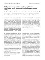
Figure 1 Expression profiling of biological triplicates of HEK293 cells transfected with mutant and wild-type MLK3. (a) Heatmap of the
445 genes which clearly classify the mutant vs the wild-type, selected on the basis of a 2 log-fold change in expression between cells transfected
with P252H mutant and wild-type forms of MLK3, and eliminating genes which were differentially expressed between the Mock and wild-type
transfection (red=down-regulated in P252H mutant MLK3, green=up-regulated in P252H mutant MLK3). (b) Illustrates a subset of the
heatmap, focusing on genes which are part of the colorectal cancer pathway which is significantly affected (p-value = 0.03). (c) Graphical
representation of the colorectal pathway (larger rounded rectangle) and relevant pathways contained within it (smaller rounded rectangles),
such as MAPK, WNT, TGF-β, NOTCH and p53, indicating differentially expressed genes (ovals) present in Figure 1, their relationships to each
other (solid line indicates a direct protein-protein interaction, T shaped ending for inhibition interactions, arrow ending for activation
interactions, and no ending for other known interactions), as well as the direction of the expression change (red = down-regulated in P252H
mutant MLK3 transfection, green = up-regulated in mutant MLK3). White Diamond shaped boxes indicate entire gene families, which are
significantly affected, genes within the families are shown as colored ovals. Long dashed lines indicate genes, which are present in multiple
pathways. (d) Graphical representation of Real-time PCR quantification of mutant vs wild-type MLK3 targets (LEF1, CCND1, BMP6 and
FZD10) selected from expression microarrays.
(Sentrix® Human-v6 Expression BeadChip) Hypergeometric
test available in the GOstats packages [22] was used. A
p-value cut off of 0.05 was considered. Microarray data can
be found at the GEO repository with the accession number
GSE54611.
50°C, 1 cycle of 10 minutes at 95°C, 40 cycles of 15 seconds at 95°C followed by 1 minute at 60°C in an ABI
Prism 7000 (Applied Biosystyems). Real-Time PCR
assays (absolute quantification) were performed in, at
least, three biological replicas.
Real-time PCR
Statistical analyses
One μl of cDNA was added to 10 μl Real-Time PCR
mixtures containing 1x TaqMan® Universal PCR Master
Mix, No AmpErase® UNG (Applied Biosystems) and
1x TaqMan® MGB specific probes and primers mix.
Taqman expression assays for BMP6 (Hs01099594_
m1), CCND1 (Hs00277039_m1), FZD10 (Hs00273077_
s1), LEF1 (Hs00212390_m1) were purchased from
Applied Biosystems. The eukaryotic 18S rRNA assay
(Hs99999901_s1; Applied Biosystems) was used as an
endogenous control gene. Standard TaqMan thermocycling conditions were used: 1 cycle of 2 minutes at
For statistical analyzes of in vitro transformation assays
a t-student test was used and p < 0.05 was taken as statistically significant. Specific statistical tests used for
microarray interpretation are embedded in the corresponding materials and methods section.
Results and discussion
MLK3 P252H mutation affects fundamental colorectal
cancer-associated pathways
In order to assess the effect of a tumorigenic MLK3 mutant
on a genome-wide level, we performed microarray-based
Table 1 KEGG Pathways significantly affected by mutant MLK3
KEGG
p-value
ID
970
Odds
Exp count
Count
Size
Term
Ratio
0.000
4.669
5
16
35
Aminoacyl-tRNA biosynthesis
290
0.001
7.682
2
7
12
Valine, leucine and isoleucine biosynthesis
5120
0.001
2.477
11
21
68
Epithelial cell signaling in Helicobacter pylori infection
251
0.004
3.184
5
11
30
Glutamate metabolism
630
0.009
4.693
2
6
13
Glyoxylate and dicarboxylate metabolism
4115
0.013
1.984
11
18
68
p53 signaling pathway
330
0.014
2.517
5
11
35
Arginine and proline metabolism
3020
0.018
2.923
4
8
23
RNA polymerase
5020
0.026
2.948
3
7
20
Parkinson's disease
670
0.027
3.283
2
6
16
One carbon pool by folate
5210
0.03
1.721
13
20
84
Colorectal cancer
600
0.031
2.155
6
11
39
Sphingolipid metabolism
260
0.038
1.995
7
12
45
Glycine, serine and threonine metabolism
5040
0.041
2.241
5
9
31
Huntington's disease
5110
0.044
2.01
6
11
41
Cholera - infection
Velho et al. BMC Cancer 2014, 14:182
/>
expression profiling experiments. We used Illumina Whole
Genome arrays on three biological replicas of human
HEK293 cells stably transfected with the wild-type MLK3,
with the MLK3 P252H mutation and with the empty vector
(Mock). The expression profiles were obtained by comparing all biological replicas from each transfection experiment. Colorectal cancer cell lines were not used in this
experiment, as proteins from the MAPK pathway are frequent mutation targets in this type of cancer, and would
most likely interfere with the interpretation of the results.
The expression profiles were compared to identify genes
that were differentially expressed at least 2 log-fold (FDR <
0.01) between wild-type and Mock, as well as between
P252H and Mock. A final set of 445 genes was identified
which showed significant differential expression only between P252H and wild-type and not between wild-type and
Mock (Figure 1a). The most statistically significantly differentially expressed genes are displayed in Figure 1b. The
genes identified were significantly enriched (p < 0.05)
in several KEGG pathways (Table 1) involved in overall
biosynthesis processes, as well as in response in disease
relevant processes. Interestingly, the colorectal pathway, which encompasses several relevant pathways
such as WNT, MAPK, NOTCH, TGF-β and P53,
was significantly over-represented (Figure 1c). These
signaling pathways are crucial to maintain intestinal
epithelium homeostasis by balancing the rate of proliferation, apoptosis, and differentiation along the crypt-villus
axis, and their de-regulation is commonly associated to
colorectal cancer initiation and progression [23]. Corroborating our results, a recent study using both in vivo and
in vitro approaches showed that MLK3 signaling is important in intestinal mucosal healing and epithelial cell motility
[24], therefore implicating MLK3 signaling in the maintenance of intestinal epithelial homeostasis.
In order to further validate the microarray data, a set
of differentially expressed genes (LEF1, CCND1, FZD10,
and BMP6) were selected for validation by real-time
PCR in HEK293 cells stably expressing MLK3 wild-type
or MLK3 P252H. The results obtained with the microarrays were validated for all genes tested (Figure 1d).
Of particular interest are the alterations that mutant
MLK3 induces in the WNT pathway. Activation of canonical WNT signaling through WNT/β-catenin cascade
has traditionally been regarded as a critical player in
colorectal tumorigenesis [25]. More recently, accumulating evidence supports a role for the non-canonical
WNT planar cell polarity (PCP) pathway, a signaling
cascade involved in the polarization of cells during tissue remodeling, and cell adhesion and motility, in cancer progression, invasion, metastasis, and angiogenesis
[26-28]. A more detailed analyzes of our microarray
data showed that the expression of several molecular
components of the canonical pathway, such as DVL2,
Page 5 of 6
LEF1, CCND1 and c-MYC were down-regulated in
MLK3 mutant cells, and the expression of DKK, a wellknown negative regulator of canonical WNT signaling
[29], was up-regulated (Figure 1c and d). On the other
hand, genes encoding two WNT receptors known to act as
negative regulators of the canonical WNT/β-catenin signaling cascade and as positive regulators of the PCP pathway,
FZD6 and FZD10, were found to be up-regulated in P252H
cells (Figure 1c and d). Taken together, the down-regulation
of DVL2, LEF1, CCND1 and c-MYC, and the up-regulation
of DKK and FZD receptors suggest a role of mutant MLK3
as a molecular switch between canonic and non-canonic
WNT signaling. In accordance, it was recently reported that
MLK3 reduces the expression of β-catenin/TCF downstream targets by promoting the interaction between βcatenin and KLF4, a known repressor of β-catenin/TCF
transcriptional activity [30]. Furthermore, in accordance
with a role of PCP in colorectal cancer, FZD10 was
recently demonstrated to be up-regulated in colorectal
cancers and matched liver metastases, and its overexpression was associated with the activation of noncanonical WNT pathway [31,32].
Conclusion
In conclusion, our results provide an overall view of
the expression profile associated with mutant MLK3,
and they support the functional role of mutant MLK3
by showing a deregulation of several signaling pathways known to play important roles in the development and progression of colorectal cancer. The results
also suggest that mutant MLK3 may be a novel modulator of WNT signaling, and pinpoint the activation of
the PCP pathway as a possible mechanism underlying
the invasive potential of MLK3 mutant cells. Nevertheless, further studies are required in order to validate
this hypothesis in a panel of gastrointestinal cell lines
and human primary tumors, to determine if the altered
signaling pathways are common to other MLK3 mutations, and to investigate the role of mutant MLK3 in
the context of mutant KRAS and BRAF genes.
Abbreviations
MLK: Mixed lineage kinase; WNT: Wingless type; BMP: Bone morphogenetic
protein; TGF-β: Transforming growth factor β; MSI: Microsatellite instability;
PCP: Planar cell polarity; DKK: Dickkopf; FZD: Frizelled; DVL2: Dishevelled;
LEF1: Lymphoid enhancer binding factor 1; CCND1: Cyclin D1; KLF4: Krupfellike factor; c-MYC: v-myc avian myelocytomatosis viral oncogene homolog;
TCF: T cell-specific transcription factor; PCR: Polymerase chain reaction;
MEK: MAP kinase-ERK kinase; ERK: Extracellular signal-regulated kinase;
MAPK: Mitogen-activated protein kinase; JNK: c-Jun amino-terminal kinase;
BRAF: v-raf murine sarcoma viral oncogene homolog B; KRAS: Kirsten rat
sarcoma viral oncogene homolog; Dlk: Dual-leucine-zipper-bearing kinase;
Lzk: Leucine-zipper-bearing kinase; Zak: Zipper sterile-α-motif kinase;
SH3: Src-homology-3.
Competing interests
The authors declare that they have no competing interests.
Velho et al. BMC Cancer 2014, 14:182
/>
Authors’ contributions
SV contributed to the acquisition, analysis and interpretation of data and
drafted the manuscript; AP participated in the acquisition, analysis and
interpretation of data; DL contributed to the acquisition, analysis and
interpretation of data; MJO was responsible for the conception and design
of the experimental system and critically reviewed the manuscript; FS was
involved in the acquisition, analysis and interpretation of data; ES
contributed for the conception and design of the experimental system and
critically reviewed the manuscript; RS contributed to the conception and
design of the project and experimental system, interpretation of data, and
was responsible for the final approval of the version to be published.
Acknowledgements
This work was supported by Grants from The Portuguese Foundation for
Science and Technology (FCT) (Project PTDC/SAU-OBD/68310/2006), the
Portuguese Ministry for Science and Education, by FEDER- European Fund
for the Regional Development and the Programs COMPETE- Programa
Operacional de Fatores de Competitividade (POFC) and PEst-C/SAU/LA0002/
2013. MJ Oliveira was supported by a Investigator FCT Grant and SV by a
Post-doctoral fellowship (SFRH/BPD/69089/2010) from the Portuguese
Foundation for Science and Technology (FCT).
Author details
1
Instituto de Patologia e Imunologia Molecular da Universidade do Porto,
Rua Dr. Roberto Frias, 4200-465 Porto, Portugal. 2New Therapies Group,
INEB-Institute for Biomedical Engineering, Porto, Portugal. 3CBM S.c.r.l. AREA
SCIENCE PARK, Trieste, Italy. 4Center for Translational Genomics and
Bioinformatics, San Raffaele Scientific Institute, Milan, Italy. 5Medical Faculty of
the University of Porto, Porto, Portugal.
Received: 23 September 2013 Accepted: 25 February 2014
Published: 14 March 2014
References
1. Gallo KA, Johnson GL: Mixed-lineage kinase control of JNK and p38 MAPK
pathways. Nat Rev Mol Cell Biol 2002, 3(9):663–672.
2. Burbelo PD, Drechsel D, Hall A: A conserved binding motif defines numerous
candidate target proteins for both Cdc42 and Rac GTPases. J Biol Chem 1995,
270(49):29071–29074.
3. Gallo KA, Mark MR, Scadden DT, Wang Z, Gu Q, Godowski PJ: Identification
and characterization of SPRK, a novel src-homology 3 domaincontaining proline-rich kinase with serine/threonine kinase activity. J Biol
Chem 1994, 269(21):15092–15100.
4. Ing Y, Leung I, Heng H, Tsui L, Lassam N: MLK-3: identification of a widelyexpressed protein kinase bearing an SH3 domain and a leucine zipper-basic
region domain. Oncogene 1994, 9(6):1745–1750.
5. Chadee D, Kyriakis J: A novel role for mixed lineage kinase 3 (MLK3) in BRaf activation and cell proliferation. Cell Cycle 2004, 3(10):1227–1229.
6. Hartkamp J, Troppmair J, Rapp UR: The JNK/SAPK activator mixed lineage
kinase 3 (MLK3) transforms NIH 3T3 cells in a MEK-dependent fashion.
Cancer Res 1999, 59(9):2195–2202.
7. Chadee DN, Kyriakis JM: MLK3 is required for mitogen activation of B-Raf,
ERK and cell proliferation. Nat Cell Biol 2004, 6(8):770–776.
8. Chadee DN, Xu D, Hung G, Andalibi A, Lim DJ, Luo Z, Gutmann DH, Kyriakis
JM: Mixed-lineage kinase 3 regulates B-Raf through maintenance of the
B-Raf/Raf-1 complex and inhibition by the NF2 tumor suppressor protein.
Proc Natl Acad Sci U S A 2006, 103(12):4463–4468.
9. Mishra P, Senthivinayagam S, Rangasamy V, Sondarva G, Rana B: Mixed
lineage kinase-3/JNK1 axis promotes migration of human gastric cancer
cells following gastrin stimulation. Mol Endocrinol 2010, 24(3):598–607.
10. Chen J, Gallo KA: MLK3 regulates paxillin phosphorylation in chemokinemediated breast cancer cell migration and invasion to drive metastasis.
Cancer Res 2012, 72(16):4130–4140.
11. Chen J, Miller EM, Gallo KA: MLK3 is critical for breast cancer cell
migration and promotes a malignant phenotype in mammary epithelial
cells. Oncogene 2010, 29(31):4399–4411.
12. Cronan MR, Nakamura K, Johnson NL, Granger DA, Cuevas BD, Wang JG,
Mackman N, Scott JE, Dohlman HG, Johnson GL: Defining MAP3 kinases
required for MDA-MB-231 cell tumor growth and metastasis. Oncogene
2012, 31(34):3889–3900.
Page 6 of 6
13. Chien ST, Lin SS, Wang CK, Lee YB, Chen KS, Fong Y, Shih YW: Acacetin
inhibits the invasion and migration of human non-small cell lung
cancer A549 cells by suppressing the p38alpha MAPK signaling pathway.
Mol Cell Biochem 2011, 350(1–2):135–148.
14. Zhan Y, Abi Saab WF, Modi N, Stewart AM, Liu J, Chadee DN: Mixed lineage
kinase 3 is required for matrix metalloproteinase expression and
invasion in ovarian cancer cells. Exp Cell Res 2012, 318(14):1641–1648.
15. Velho S, Oliveira C, Paredes J, Sousa S, Leite M, Matos P, Milanezi F, Ribeiro
AS, Mendes N, Licastro D, Karhu A, Oliveira MJ, Ligtenberg M, Hamelin R,
Carneiro F, Lindblom A, Peltomaki P, Castedo S, Schwartz S Jr, Jordan P,
Aaltonen LA, Hofstra RM, Suriano G, Stupka E, Fialho AM, Seruca R: Mixed
lineage kinase 3 gene mutations in mismatch repair deficient
gastrointestinal tumours. Hum Mol Genet 2010, 19(4):697–706.
16. Schroeder A, Mueller O, Stocker S, Salowsky R, Leiber M, Gassmann M,
Lightfoot S, Menzel W, Granzow M, Ragg T: The RIN: an RNA integrity
number for assigning integrity values to RNA measurements. BMC Mol
Biol 2006, 7(1):3.
17. Gentleman R, Carey V, Bates D, Bolstad B, Dettling M, Dudoit S, Ellis B,
Gautier L, Ge Y, Gentry J, Hornik K, Hothorn T, Huber W, Iacus S, Irizarry R,
Leisch F, Li C, Maechler M, Rossini A, Sawitzki G, Smith C, Smyth G, Tierney
L, Yang J, Zhang J: Bioconductor: open software development for
computational biology and bioinformatics. Genome Biol 2004, 5(10):R80.
18. R Development Core Team: R: A language and environment for statistical
computing. Vienna, Austria: R Foundation for Statistical Computing; 2008.
19. Smyth G: Limma: linear models for microarray data. New York: Springer; 2005.
20. Benjamini Y, Hochberg Y: Controlling the false discovery rate: a practical and
powerful approach to multiple testing. J R Stat Soc B 1995, 57:289–300.
21. Liu T, Lin C, Falcon S, Zhang J, MacDonald J: Kegg: a data package
containing annotation data for KEGG. R package version 2.2.0. 2007.
22. Gentleman R, Falcon S: Gostats: tools for manipulating GO and
microarrays. R package version 2.6.0. 2007:.
23. Radtke F, Clevers H, Riccio O: From gut homeostasis to cancer. Curr Mol
Med 2006, 6(3):275–289.
24. Kovalenko PL, Kunovska L, Chen J, Gallo KA, Basson MD: Loss of MLK3
signaling impedes ulcer healing by modulating MAPK signaling in
mouse intestinal mucosa. Am J Physiol Gastrointest Liver Physiol 2012,
303(8):G951–G960.
25. de Lau W, Barker N, Clevers H: WNT signaling in the normal intestine and
colorectal cancer. Front Biosci 2007, 12:471–491.
26. Naz G, Pasternack SM, Perrin C, Mattheisen M, Refke M, Khan S, Gul A,
Simons M, Ahmad W, Betz RC: FZD6 encoding the Wnt receptor frizzled
6 is mutated in autosomal-recessive nail dysplasia. Br J Dermatol 2012,
166(5):1088–1094.
27. Seifert JR, Mlodzik M: Frizzled/PCP signalling: a conserved mechanism
regulating cell polarity and directed motility. Nat Rev Genet 2007,
8(2):126–138.
28. Wang Y: Wnt/Planar cell polarity signaling: a new paradigm for cancer
therapy. Mol Cancer Therap 2009, 8(8):2103–2109.
29. Rao TP, Kuhl M: An updated overview on Wnt signaling pathways: a prelude
for more. Circ Res 2010, 106(12):1798–1806.
30. Thylur RP, Senthivinayagam S, Campbell EM, Rangasamy V, Thorenoor N,
Sondarva G, Mehrotra S, Mishra P, Zook E, Le PT, Rana B: Mixed lineage
kinase 3 modulates beta-catenin signaling in cancer cells. J Biol Chem
2011, 286(43):37470–37482.
31. Fukukawa C, Nagayama S, Tsunoda T, Toguchida J, Nakamura Y, Katagiri T:
Activation of the non-canonical Dvl-Rac1-JNK pathway by Frizzled
homologue 10 in human synovial sarcoma. Oncogene 2009, 28(8):1110–1120.
32. Nagayama S, Yamada E, Kohno Y, Aoyama T, Fukukawa C, Kubo H,
Watanabe G, Katagiri T, Nakamura Y, Sakai Y, Toguchida J: Inverse
correlation of the up-regulation of FZD10 expression and the activation
of beta-catenin in synchronous colorectal tumors. Cancer Sci 2009,
100(3):405–412.
doi:10.1186/1471-2407-14-182
Cite this article as: Velho et al.: Dissecting the signaling pathways
associated with the oncogenic activity of MLK3 P252H mutation. BMC
Cancer 2014 14:182.