The novel IGF-IR/Akt–dependent anticancer activities of glucosamine
Bạn đang xem bản rút gọn của tài liệu. Xem và tải ngay bản đầy đủ của tài liệu tại đây (1.2 MB, 12 trang )
Song et al. BMC Cancer 2014, 14:31
/>
RESEARCH ARTICLE
Open Access
The novel IGF-IR/Akt–dependent anticancer
activities of glucosamine
Ki-Hoon Song1, Ju-Hee Kang1,2, Jong-Kyu Woo5, Jeong-Seok Nam3, Hye-Young Min4, Ho-Young Lee4,
Soo-Youl Kim1 and Seung-Hyun Oh5,6*
Abstract
Background: Recent studies have shown that glucosamine inhibits the proliferation of various human cancer cell
lines and downregulates the activity of COX-2, HIF-1α, p70S6K, and transglutaminase 2. Because the IGF-1R/Akt
pathway is a common upstream regulator of p70S6K, HIF-1α, and COX-2, we hypothesized that glucosamine inhibits
cancer cell proliferation through this pathway.
Methods: We used various in vitro assays including flow cytometry assays, small interfering RNA (siRNA)
transfection, western blot analysis, MTT (3-(4,5-dimethylthiazol-2-yl)-2,5-diphenyltetrazolium bromide) assays, reverse
transcription-polymerase chain reaction, and in vivo xenograft mouse model to confirm anticancer activities of
glucosamine and to investigate the molecular mechanism.
Results: We found that glucosamine inhibited the growth of human non-small cell lung cancer (NSCLC) cells and
negatively regulated the expression of IGF-1R and phosphorylation of Akt. Glucosamine decreased the stability of
IGF-1R and induced its proteasomal degradation by increasing the levels of abnormal glycosylation on IGF-1R.
Moreover, picropodophyllin, a selective inhibitor of IGF-1R, and the IGF-1R blocking antibody IMC-A12 induced
significant cell growth inhibition in glucosamine-sensitive, but not glucosamine-resistant cell lines. Using in vivo
xenograft model, we confirmed that glucosamine prohibits primary tumor growth through reducing IGF-1R
signalling and increasing ER-stress.
Conclusions: Taken together, our results suggest that targeting the IGF-1R/Akt pathway with glucosamine may be
an effective therapeutic strategy for treating some type of cancer.
Keywords: Glucosamine, Anticancer agent, IGF-1R, Akt, Glycosylation, ER-stress
Background
Since the effect of glucosamine as an inhibitor of tumor
growth was first reported by Quastel and Cantero, [1]
many in vitro studies have shown that it interferes with
the glycoslyation of glycoproteins, [2,3] decreases the rate
of glycolysis and fructolysis, [4,5] and changes the component ratio of nucleotides in various carcinoma cell lines
[6,7]. Results of a recent study indicated that glucosamine
induces G1 cell-cycle arrest in mesangial cells and human
cancer cells through a mechanism involving decreased
expression of cyclin D1 and increased expression of
* Correspondence:
5
Gachon Institute of Pharmaceutical Science, Gachon University, Incheon
406-840, Republic of Korea
6
College of Pharmacy, Gachon University, 7-45 Songdo-dong, Yeonsu-gu,
Incheon 406-840, Republic of Korea
Full list of author information is available at the end of the article
p21Waf1/Cip1, which are positive and negative regulators of
cell cycle progression, respectively [8,9].
The PI3K/Akt pathway is often overactivated in various
types of cancer cells. PI3K/Akt can transmit signals from
RTKs and G-protein-coupled receptors that are activated
by growth factors or cytokines; therefore, the PI3K/Akt
signal transduction pathway regulates multiple cellular
functions, including transcription, translation, and cell
proliferation, cell cycle progression, and survival [10-12].
Although the RTK-mediated signal transduction pathways
overlap, PI3K-mediated activation of Akt specifically contributes to the anti-apoptotic activity of IGF-1R.
Recent studies have demonstrated that target proteins
of glucosamine may exist in cancer cells [13-16]. Glucosamine inhibits the growth of cancer cells by downregulating the phosphorylation of p70S6K, a regulator of
© 2014 Song et al.; licensee BioMed Central Ltd. This is an Open Access article distributed under the terms of the Creative
Commons Attribution License ( which permits unrestricted use, distribution, and
reproduction in any medium, provided the original work is properly cited. The Creative Commons Public Domain Dedication
waiver ( applies to the data made available in this article, unless otherwise
stated.
Song et al. BMC Cancer 2014, 14:31
/>
protein translation [15]. In addition, glucosamine inhibits HIF-1α by inhibiting protein translation through
the reduction of phosphorylated p70S6K levels [16].
Jang et al. reported that glucosamine hydrochloride inhibits N-glycosylation of COX-2 and enhances COX-2
protein turnover [13]. Finally, glucosamine induces
NF-κB inactivation by inhibiting transglutaminase 2
(TGase 2) activity [14]. Together, these studies suggest
that glucosamine has potential as an anticancer drug,
although its mechanism of action remains poorly understood [17]. Thus, we tested whether the IGF-1R/PI3K/
Akt pathway, upstream of p70S6K and COX-2, is target
of glucosamine. We also investigated the molecular
mechanisms underlying the anticancer activity of glucosamine in NSCLC cells.
Methods
Cell lines and materials
Human NSCLC cell lines A549, H226B, H1299, and
H460 were purchased from the American Type Culture
Collection (Manassas, VA, USA).
The HA-Akt1 (T308D/S473D) expression vector was
kindly provided by Dr. Gordon Mills (The University of
Texas MD Anderson Cancer Center). The H226B-Babe
cells were produced by infecting H226B NSCLC cells with
a pBabe retroviral control vector. The H226B-Akt1-DD
cells that possess a constitutively active form of Akt were
produced by infecting H226B with a pBabe-HA-Akt1-DD
construct harboring mutations that change Ser473 and
Thr308 to aspartic acids. The H226B-Akt2-DD and The
H226B-Akt3-DD cells were kindly provided by Dr. HoYoung Lee (College of Pharmacy, Seoul National University, Seoul, Republic of Korea).
D-(+)-Glucosamine hydrochloride, MG132, and tunicamycin (TN) were purchased from Sigma-Aldrich (St Louis,
MO, USA). Antibodies against pIGF-1R, pAkt, pERK1/2,
Akt, PTEN, PARP, PDI, IRE1α, ATF4, GRP78, CHOP, and
a/β-tubulin were purchased from Cell Signaling Technology (Beverly, MA, USA). Antibodies against IGF-1R,
COX-2, CDK2, CDK4, and β- ACTIN were purchased
from Santa Cruz Biotechnology, Inc. (Santa Cruz, CA,
USA), and the antibody against TGase 2 was obtained
from Thermo Fisher Scientific, Inc. (Fremont, CA, USA).
Page 2 of 12
was measured using caliper and calculated according to
the formula (L x W2)/2. All of the mice were sacrificed
on Day 71, and tumor tissues were isolated from them.
The results were represented as the mean of tumor
volumes (n = 10) with SEM.
siRNA transfection
For RNA interference, A549, H1299, and H460 cells transfected with 40 nM siRNA. Double-stranded siRNAs designed to target IGF-1R (5′-CUG ACA UGG GCC UUU
AAG A-3′), and a scrambled non-targeting siRNA were
synthesized by Bionner (Seoul, South Korea). Cells were
transfected with siRNAs using Lipofectamine reagent
(Invitrogen, CA, USA) according to the manufacturer’s
protocol.
Semiquantitative RT-PCR
First strand cDNA was synthesized from 2 μg of extracted
RNA using M-MLV reverse transcriptase (Invitrogen).
RT-PCR was carried out with gene-specific primers for
IGF-1R, COX-2, XBP1, GRP78, CHOP, ATF4, GAPDH,
and β-ACTIN (Table 1). Primers amplifying a region of
β-ACTIN or GAPDH were used as an internal control.
Western blot analysis
Preparation of whole-cell lysates from cancer cells, electrophoresis, and membrane transfer were performed as
previously described [18]. The membranes were then
incubated overnight at 4°C with primary antibodies in
TBS-T containing 5% bovine serum albumin. Membranes
were washed with TBS-T and then incubated with an
Table 1 Primer sequences used for RT-PCR
Gene
Primer sequence (5′ - 3′)
IGF-1R
F 5′-ACG CCA ATA AGT TCG TCC AC-3′
R 5′-TCC ATC CTT GAG GGA CTC AG-3′
COX-2
F 5′-ATC TTT GGG GAG ACC ATG GTA GA-3′
R 5′-ACT GAA TTG AGG CAG TGT TGA TG-3′
XBP1
F 5′-TTA CGA GAG AAA ACT CAT GGC C-3′
R 5′-GGG TCC AAG TTG TCC AGA ATG C-3′
GRP78
F 5′-GGT ACA TTT GAT CTG ACT G-3′
R 5′-CAC TTC ACT AGA GTT TGC TG-3′
Xenograft mouse tumor model
All animal experimental procedures were approved by
Institutional Animal Care and Use Committee (IACUC)
of National Cancer Center in Republic of Korea. To confirm antitumor effect of glucosamine in animal, we used
xenograft tumor model. A549 cells (5 x 10⁶ cells) were
subcutaneously injected into flank region of BALB/c
nude mice. After cancer cell injection, glucosamine
(500 mg/kg body weight/day) was administered intrapenitorially to immuoncompromised mice. Tumor volume
CHOP
F 5′-CTT CAC TAC TCT TGA CCC TGC AT-3′
R 5′-ATG TGC ACT GGA GAT TGC TT-3′
ATF4
F 5′-GTT CTC CAG CGA CAA GGC TA-3′
R 5′-ATC CTC CTT GCT GTT GTT GG-3′
GAPDH
F 5′-GGT GAA GGT CGG TGT GAA CGG ATT T-3′
R 5′-ATT GCC AAA GTT GTC ATG GAT GAC C-3′
β-ACTIN
F 5′-GTG GGG CGC CCC AGG CAC CA-3′
R 5′-CTC CTT AAT GTC ACG CAC GAT TTC-3′
Song et al. BMC Cancer 2014, 14:31
/>
appropriate horseradish peroxidase-conjugated secondary
antibody in 5% skim milk for 1 hour at room temperature.
Cell proliferation analysis
To determine the effects of glucosamine on the proliferation of various cancer cell lines, cells were seeded in
96-well plates (3,000 cells/well). On the following day, the
medium was replaced with medium containing glucosamine, picropodophyllin, and A12 at the desired concentrations. After incubation for an additional 2 days, MTT
assay was performed according to standard procedures.
The bars represent SD of results.
Cell cycle analysis
For the cell cycle analysis, three human NSCLC cell lines
were treated with the indicated concentration of glucosamine. Floating and attached cells were fixed in 70% ethanol for 1 hour at 4°C. After centrifugation, the cell pellet
was washed twice with phosphate-buffered saline (PBS)
and stained with propidium iodide (PI) containing RNase
A (40 μg/ml) for 30 minutes at 4°C in the dark. The total
cellular DNA content of each cancer cell line was quantified by flow cytometry.
Apoptosis analysis
To analyze the number of apoptotic cells after 2 days of
glucosamine treatment, A549, H1299, and H460 cells were
harvested and washed twice with PBS on ice. The cells
were resuspended in 1 X binding buffer containing 5 μl
fluorescein isothiocyanate (FITC)-conjugated Annexin V
and 5 μl PI. Apoptotic events were detected by flow
cytometry at 488 nm and 633 nm using the FITC Annexin
V apoptosis detection kit I (BD Pharmingen, San Jose,
CA). All procedures were carried out according to the
manufacturer’s instructions.
Immunohistochemistry
Primary tumors from PBS or glucosamine treated animals
were embedded in paraffin depending on the application.
The 5 μm tumor tissue sections were prepared for immunohistochemistry. Paraffin sections were incubated overnight at 4°C with primary antibody against anti-phosphoAkt (Cell Signaling Technology; 1 : 100 dilution) and then
processed for avidin – biotin immunohistochemistry according to the manufacturer’s instructions (Vector Laboratories, Burlingame, CA). These sections counterstained
with hematoxylin and eosin-Y (H&E). Immunohistochemical analysis were performed as previously described [18].
Results
Glucosamine induces cell cycle arrest and apoptosis in
NSCLC cells
Previous studies have reported that glucosamine inhibits
cell growth [15,16] and cell-cycle progression [8,9,19]
Page 3 of 12
and induces apoptosis [20] in various cell lines. We
therefore investigated whether the anti-cancer effect of
glucosamine was associated with cell growth, cell-cycle
arrest and apoptosis in NSCLC cell lines. Glucosamine
reduced the proliferation of all four NSCLC cell lines,
but the extent of the inhibition differed among NSCLC
cell lines (Figure 1A). Flow cytometric analysis indicated
that glucosamine induced cell-cycle arrest at the G0/G1
phase in a dose-dependent manner (Figure 1B) and that
glucosamine induced apoptosis in A549, H226B, H1299,
and H460 NSCLC cell lines (Figure 1C and Additional
file 1: Figure S1). Consistent with the results of the cell
proliferation assay, in the cell cycle and apoptosis analyses, the A549 and H226B cells had a more significant
response to glucosamine than the others.
In addition, expression of cleaved poly-(ADP-ribose)
polymerase (PARP), a marker for apoptosis, was high in
A549 and H226B cells and low in H460 cells (Figure 1D).
Treatment with 5 mM glucosamine reduced the expression of both CDK4 and CDK2 in A549 and H226B cells
and that of CDK4 only in H1299 cells. In contrast, the
levels of CDK4 and CDK2 were not obviously changed
in H460 cells (Figure 1D). These findings suggest that
the glucosamine-mediated growth inhibition of NSCLC
cells is associated with the induction of cell-cycle arrest
and apoptosis.
The basal expression levels of TGase 2 and COX-2 proteins
in NSCLC cells are not correlated with glucosamine
sensitivity
We investigated the expression levels of TGase 2 and
COX-2 proteins that were previously identified as major
targets of glucosamine. Expression of TGase 2 was
markedly higher in A549 and H1299 cells than in H460
and H226B cells. We also found that A549 and H460
cell lines showed a high basal level of COX-2 expression,
whereas COX-2 expression was not detected in H1299
cells (Additional file 2: Figure S2). Therefore, the basal
TGase 2 and COX-2 levels in the NSCLC cell lines were
not correlated with glucosamine sensitivity.
Glucosamine suppresses activation of Akt by reducing
IGF-1R expression in cell lines that have an
IGF-1R-dependent Akt activation pathway
Because we observed that glucosamine downregulated
CDK4 expression in NSCLC cells (Figure 1D) and a previous report showed that the PI3K/Akt pathway affects
CDK4 expression [21], we tested the effect of glucosamine
on the IGF-1R/Akt signaling pathway. Glucosamine reduced the IGF-1R and pAkt levels in A549 and H460 cell
lines in a dose-dependent manner (Figure 2A). Moreover,
activation of both pIGF-1R and pAkt by IGF-1 was downregulated by glucosamine (Figure 2B). These results
*
40
20
0
0
0.5
1
2
*
40
20
0
0
5
0.5
1
2
60
40
20
0
5
0
0.5
1
H226B
H1299
G0/G1
S
G2/M
Sub G1
G0/G1
100
20
40
20
10 2
10 4
10 4
10
10 3
10 1
10 2
–
+
10 2
103
104
H1299
–
–
+
10 0
10 1
H460
–
+
Glucosamine (5 mM)
PARP
cleaved-PARP
CDK4
CDK2
β-actin
Figure 1 (See legend on next page.)
10 1
10 2
10 3
10 4
19.9%
0
10
10 1
10 0
3
10
10
1
H226B
+
10 4
0
100
Annexin V
A549
10 3
22.5%
2
10
1
10
10 3
10
10 2
10 1
10
10
2
10
2
10
1
10
10 1
10 0
3
3
10
10
4
10 3
59.6%
10.6%
10 2
10
10 2
10 1
10 1
0
10
10 0
20
H460
9.9%
0
10 0
10 4
Sub G1
0
1
2
5
Glucosamine (mM)
3
3
10
2
10
1
10
0
10
10 3
40.8%
G2/M
0
10
10
4
10
3
10
2
10
1
10
0
10
10 2
PI
10 1
S
40
H1299
13.7%
5
60
0
1
2
5
Glucosamine (mM)
H226B
17.2%
2
80
0
A549
10 0
G0/G1
Sub G1
60
4
C
1
100
0
1
2
5
Glucosamine (mM)
0
1
2
5
Glucosamine (mM)
0.5
H460
80
0
0
Glucosamine (mM)
Cell population (%)
40
20
10
20
60
40
10
40
80
*
10 2
60
G2/M
*
60
0
100
Cell population (%)
Cell population (%)
80
S
*
80
10 1
Sub G1
10
G2/M
10
S
100
5
A549
0
D
2
Glucosamine (mM)
G0/G1
GluN
(5 mM)
*
Glucosamine (mM)
100
Cell population (%)
60
80
*
Glucosamine (mM)
B
Control
*
Cell Proliferation (% Control)
60
*
*
4
*
80
100
H460
120
10 0
80
100
H1299
120
4
*
*
120
10 2
10 3
10 4
10
100
H226B
Cell Proliferation (% Control)
A549
120
Page 4 of 12
4
Cell Proliferation (% Control)
A
Cell Proliferation (% Control)
Song et al. BMC Cancer 2014, 14:31
/>
10 0
10 1
10 2
10 3
10 4
Song et al. BMC Cancer 2014, 14:31
/>
Page 5 of 12
(See figure on previous page.)
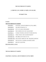
Figure 1 Effect of glucosamine on cell growth, cell cycle arrest, and apoptosis in NSCLC cells. (A) MTT growth assay of A549, H1299, and
H460 cells. *P < 0.05 compared with glucosamine 0 mM. (B) NSCLC cells were treated with glucosamine for 2 days and then stained with PI for
cell-cycle analysis. (C) Apoptosis/necrosis was determined by Annexin V-FITC and PI staining. (D) The protein levels of CDK2, CDK4, PARP, and
cleaved PARP.
Similarly, pAkt expression was completely abolished in
cells cotreated with glucosamine and siIGF-1R (Figure 2D).
We next performed MTT assay on NSCLC cells to
determine whether a combination of siIGF-1R and glucosamine inhibits cell proliferation more efficiently than
either agent alone. As expected, IGF-1R knockdown
enhanced the glucosamine-induced inhibition of cell
growth in the A549 cell line but not the H460 cell
line in which siIGF-1R did not affect the pAkt level
(Figure 2E). Thus, we concluded that glucosamine
demonstrate that glucosamine effectively inhibits IGF-1R/
Akt signal transduction.
All cell lines showed a dose-dependent decrease in
IGF-1R expression, but there was a significant reduction
in pAkt expression in the A549 and H226B cell lines
(Figure 2C). To confirm that glucosamine inhibited the
IGF-1R/Akt signaling pathway, we also carried out small
interfering RNA (siRNA) transfection studies. IGF-1R
expression was completely abolished following treatment
of siIGF-1R–transfected A549 cells with glucosamine.
B
A
Glucosamine 5 mM
Glucosamine 1 mM
C
12
C
24
C
C
48
12
C
24
C
48
Time (h)
–
+
+
+
IGF-1 (50 ng/ml)
–
–
1
5
Glucosamine (mM)
IGF-1R
pIGF-IR
pAkt
IGF-IR
Akt
pAkt
COX-2
Akt
Tubulin
COX-2
Tubulin
C
D
0
1
H1299
A549
H226B
5
0
1
5
0
1
H460
5
0
1
5
GlcN (mM)
–
+
–
–
+
–
siScr
–
–
–
–
+
–
–
+
–
+
+
+
siIGF-1R
GlcN (1 mM)
IGF-1R
IGF-1R
PTEN
pAkt
pAkt
Akt
Akt
Tubulin
COX-2
Cell Proliferation (% Control)
E
120
A549
*
siSCR
*
100
siIGF-1R
*
80
60
*
40
20
0
0
1
2
5
GlcN (mM)
Cell Proliferation (% Control)
Tubulin
H460
120
siSCR
100
siIGF-1R
80
60
40
20
0
0
1
2
5
GlcN (mM)
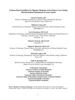
Figure 2 Glucosamine down-regulates the IGF-1R kinase-dependent Akt pathway in NSCLC cells. (A) Time- and dose-dependent effects of
glucosamine in A549 cells. (B) A549 cells were pretreated with glucosamine for 1 day and then activated with IGF-1 for 15 minutes. (C) The effect
of glucosamine on different NSCLC cell lines. (D) A549 cells transfected with siIGF-1R or scrambled non-targeting siRNA (siSCR) 2 days prior to a
1-day treatment with 1 mM glucosamine. (E) Cell proliferation assay in NSCLC cells transfected with siIGF-1R and treated with the indicated doses
of glucosamine for 2 days. * P < 0.05 compared with siSCR-transfected cells.
Song et al. BMC Cancer 2014, 14:31
/>
Page 6 of 12
inhibits the proliferation of NSCLC cells by reducing
the expression of IGF-1R, and the extent of the glucosamine-induced reduction in the pAkt level is associated
with the anticancer effect of glucosamine.
Glucosamine and other IGF-1R–targeting agents have
similar effects in glucosamine-sensitive and -resistant cell
lines
We hypothesized that if glucosamine acts as an IGF-1Rspecific inhibitor, siIGF-1R and other agents that inhibit
IGF-1R will exhibit anticancer effects similar to those
induced by glucosamine in the A549 and H460 cell lines.
Thus, we investigated whether molecules inhibiting IGF1R also reduce the pAkt level and inhibit cell proliferation in these cell lines. First, siIGF-1R dramatically
reduced the IGF-1R level in A549 and H460 cells but
A549
H460
0.5
siRNA
SCR IGF-1R SCR IGF-1R
IGF-1R
pAkt
Akt
H460
0.8
siSCR
siIGF-1R
0.4
*
*
0.3
0.2
siSCR
siIGF-1R
O.D. (562 nm)
A549
O.D. (562 nm)
A
only partially reduced the pAkt level in the A549 cell
line. In addition, an antisense oligonucleotide targeting
IGF-1R only inhibited the growth of the A549 cells
(Figure 3A). In addition to siIGF-1R, picropodophyllin
(PPP), an IGF-1R-specific small-molecule inhibitor, reduced the levels of pIGF-1R and pAkt and inhibited the
growth of A549 cells more efficiently than that of H460
cells (Figure 3B).
One of IGF-1R blocking antibodies, A12, binds directly
to IGF-1R and promotes its internalization and degradation [22]. A12 significantly reduced the level of IGF-1R
in both A549 and H460 cells (Figure 3C). The pAkt levels
were dramatically reduced in the A549 cell line but only
slightly reduced in the H460 cell line. In concordance with
these results, A12 reduced the proliferation of A549 cells
but had no effect on the growth of H460 cells (Figure 3C).
0.6
0.4
0.2
0.1
β-actin
0
0
1
2
3
1
4
A549
0
2
H460
5
0
2
5
PPP (µM)
pIGF-1R
IGF-1R
pAkt
Akt
Cell Proliferation (% Control)
B
2
3
4
Days
Days
A549
120
H460
*
100
80
*
*
*
0.5
1
60
40
20
0
0
0.1
0.2
PPP (µM)
A549
0
2
H460
10
0
2
10
A12 (µg/ml)
IGF-1R
pAkt
Akt
Tubulin
Cell Proliferation (% Control)
C
A549
120
H460
*
*
*
*
1
2
5
10
100
80
60
40
20
0
0
A12 (µg/ml)
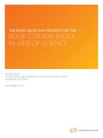
Figure 3 The inhibitory effect of glucosamine and IGF-1R targeting agents on the IGF-1R/Akt pathway. (A) The effect of knocking down
IGF-1R on the pAkt level and cell growth. (B and C) Western blotting (left) and MTT assay (right) of A549 and H460 cells treated with PPP (B) and
A12 (C) at the concentrations indicated. *P < 0.05 denotes significant differences between the conditions indicated.
Song et al. BMC Cancer 2014, 14:31
/>
Page 7 of 12
of H226B-Akt1-DD cells (Figure 4A). Under normal
culture conditions (10% FBS), 10 mM glucosamine reduced the viability of H226B-Babe and H226B-Akt1-DD
cells to 22.1% ± 3.6% and 32.9% ± 3.7% viable cells,
respectively. Interestingly, the changes in cell viability
were more pronounced in the cells treated under normal
culture conditions than in those grown in media containing 1% FBS (Figure 4B). PARP cleavage also was not
detected in H226B-Akt1-DD cells exposed to glucosamine
(Figure 4C), and we detected only minimal induction of
PARP cleavage in H226B-Babe cells treated with 10 and
20 mM glucosamine. These results suggest that constitutive activation of Akt1 may inhibit the anti-proliferative
effect of glucosamine.
These results suggest that IGF-1R is one of the major protein targets of glucosamine in various types of cancer cells
that have an IGF-1R-dependent Akt signal transduction
pathway.
Constitutive activation of Akt1 alleviates the growthinhibitory effect of glucosamine in H226B human NSCLC
cells
To evaluate whether constitutive activation of Akt isoforms alters the anti-proliferative effect of glucosamine,
H226B-Babe and H226B-Akt1-DD cells were treated
with various concentrations of glucosamine for 3 days.
Glucosamine effectively suppressed the proliferation of
H226B-Babe cells and, to a lesser extent, the proliferation
Cell Proliferation (% Control)
A
120
*
H226B-Babe cells
*
100
H226B-Akt1-DD cells
*
*
H226B-Akt2-DD cells
80
*
H226B-Akt3-DD cells
*
60
*
*
40
20
0
0.5
1
2
5
10
Glucosamine (mM)
1 % FBS
100
10 % FBS
H226B-Babe cells
H226B-Akt1-DD cells
80
*
60
40
*
20
0
0.5
1
2.5
5
Cell Proliferation (% Control)
Cell Proliferation (% Control)
B
100
H226B-Babe cells
*
80
60
*
*
40
20
0
0.5
10
H226B-Babe
0
10
20
1
2.5
5
10
Glucosamine (mM)
Glucosamine (mM)
C
H226B-Akt1-DD cells
*
H226B-Akt1-DD
0
10
20
Glucosamine (mM)
PARP
cleaved-PARP
β-actin
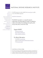
Figure 4 The role of Akt1 in the glucosamine-induced regulation of cell proliferation in H226B cells. (A) The indicated cells were
incubated with 0.5 ~ 10 mM glucosamine for 3 days and the MTT assay was performed. (B) An MTT assay was carried out under low and high
serum conditions. *P < 0.05 compared with H226B-Babe cells. (C) Adherent and floating cells were analyzed using western blotting.
Song et al. BMC Cancer 2014, 14:31
/>
Page 8 of 12
Glucosamine affects the stability of IGF-1R in a posttranslational modification and proteasome-dependent
manner
We next investigated whether the suppression of IGF-1R/
Akt signal transduction by glucosamine occurred at the
transcriptional and/or translational level. First, we observed that glucosamine treatment did not change the
levels of either IGF-1R or COX-2 mRNA (Figure 5A).
These findings led us to examine whether the observed
decrease in the IGF-1R protein level following exposure to
glucosamine was associated with the stability of the IGF1R protein. The proteasome inhibitor MG132 restored the
IGF-1R level in cells treated with 1 mM glucosamine but
not in cells treated with 5 mM glucosamine. In contrast,
pAkt expression was fully rescued in cells treated with
1 mM glucosamine (Figure 5B). A previous study reported
that glucosamine accelerated the proteasome-dependent
degradation of only the higher molecular weight species of
COX-2; [13] however, our results showed that both higher
and lower molecular weight species of COX-2 were restored when cells were treated with either 1 mM or 5 mM
glucosamine. In addition to COX-2, the molecular mass of
prototype IGF-1R (pro-IGF-1R) was also reduced by glucosamine in a dose-dependent manner (Figure 5B). We
next explored whether the glucosamine-induced decrease
in the level of IGF-1R protein involved the translation
A
B
–
+
Glucosamine (5 mM)
–
–
1
1
5
5
Glucosamine (mM)
–
+
–
+
–
+
MG132 (25 µM)
pro-IGF-IR
IGF-1R
COX-2
IGF-1R
-actin
pAkt
Akt
COX-2
Tubulin
C
D
1
5
–
1
5
GlcN (mM)
–
1
5
–
–
–
–
4h 4h 4h 16h 16h 16h CHX (100 µg/ml)
–
GlcN (mM)
TN (µg/ml)
1
0.1 0.5
2
5
1
pro-IGF-1R
pro-IGF-1R
IGF-1R
IGF-1R
pAkt
COX-2
Akt
Tubulin
COX-2
Tubulin
E
F
–
–
+
+
–
–
GlcN (1 mM)
–
1
5
–
GlcN (mM)
–
–
–
–
+
+
TN (0.5 µg/ml)
–
–
–
+
TN (0.5 µg/ml)
–
+
–
+
–
+
MG132 (25 µM)
pro-IGF-1R
PDI
IRE1α
IGF-1R
ATF4
COX-2
GRP78
Tubulin
CHOP
Tubulin
Figure 5 Glucosamine inhibits IGF-1R at the post-translational level. (A) Semiquantitative RT-PCR analysis of IGF-1R and COX-2 mRNA levels
in A549 cells. (B) A549 cells were treated with the indicated doses of glucosamine and the proteasome inhibitor MG132 for 1 day. (C) A549 cells
were pretreated with the indicated concentrations of glucosamine for 16 hours and then with cycloheximide for 2 hours. (D) A549 cells were
treated with the indicated concentrations of glucosamine or tunicamycin for 6 hours. (E) A549 cells were treated with 1 mM glucosamine or
0.5 μg/ml tunicamycin for 12 hours in the absence or presence of 25 μM MG132. (F) Western blot analysis of the expression of various ER
stress- and unfolded protein response-related genes was performed using cell lysates from harvested A549 cells.
Song et al. BMC Cancer 2014, 14:31
/>
process. Cycloheximide, a ribosomal inhibitor, inhibited
the de novo biosynthesis of pro-IGF-1R in A549 cells, and
glucosamine did not affect the IGF-1R, pAkt, and COX-2
levels (Figure 5C). These findings collectively suggest that
glucosamine may induce the hypoglycosylation of proIGF-1R and COX-2 and facilitate their degradation at the
post-translational level.
Besides, more recent studies have shown that glucosamine inhibits N-glycosylation of certain proteins including
COX-2, glucose transporter1, and a lipoprotein apo-B-100
[13,23,24]. Therefore, we next tested whether glucosamine
induces abnormal N-glycosylation of pro-IGF-1R protein.
As shown in Figure 5D, glucosamine treatment obviously
prevented pro-IGF-1R glycosylation in concentration
dependent manner, resulting in low molecular mass of
that. Tunicamycin (TN), the protein N-glycosylation inhibitor, was used as a positive control to confirm the effect
of glucosamine on pro-IGF-1R N-glycosylation. We next
challenged whether glucosamine-induced abnormal glycosylation of pro-IGF-1R protein is recovered by MG132. As
depicted in Figure 5E, reduction of pro-IGF-1R molecular
mass by glucosamine was remarkably restored by treating
A549 cells with MG132. In addition, previous studies have
elucidated that glucosamine caused endoplasmic reticulum (ER) stress and activated a series of signaling pathway termed the unfolded protein response (UPR) [3,25].
We also confirmed that glucosamine induces ER stress
and activates the UPR through changing of various
marker genes including spliced XBP1, PDI, IRE1α ATF4,
GRP78, and CHOP (Figure 5F and Additional file 3:
Figure S3). Overall, these data demonstrate that glucosamine negatively affects IGF-1R and COX-2 protein stability through a proteasome-dependent pathway, and the
production of hypoglycosylated pro-IGF-1R by glucosamine is associated with this pathway.
Glucosamine suppress primary tumor growth in vivo
To determine whether glucosamine inhibits primary tumor initiation and growth in vivo, glucosamine-sensitive
A549 cells were injected subcutaneously into immunocompromised mice. After injection, tumor bearing mice
were treated with PBS or glucosamine intraperitoneally.
As shown in Figure 6A, glucosamine significantly decreased subcutaneous tumor growth. Although glucosamine did not totally suppress the tumor growth,
outgrowth of tumor mass was effectively reduced by glucosamine treatment (Figure 6B). pIGF-1R level significantly was decreased in glucosamine treated tumor tissues
compared with PBS treated samples (Figure 6C). We
found that glucosamine treated primary tumor showed
moderately reduced pAkt level (Figure 6E), although the
difference in western blot analysis was not significant
(Figure 6C). In addition, RT-PCR data showed that glucosamine induced ER-stress in tumor tissue (Figure 6D).
Page 9 of 12
These findings suggest that glucosamine can inhibit primary tumor formation in vivo as well as cell proliferation
in vitro through restraining the IGF-1R/Akt signaling by
glucosamine-induced ER-stress.
Discussion
In this study, we showed that glucosamine effectively inhibits IGF-1R-mediated Akt signal transduction in various human carcinoma cell lines by both suppressing
IGF-1-induced IGF-1R activation and reducing IGF-1R
protein stability.
Some investigators have reported that glucosamine induces cell-cycle arrest at the G0/G1 phase in human
cancer cells [26,27]. These studies have shown that this
phenomenon is mediated by decreased cyclin D1 expression and increased p21waf1/cip1 expression. Here, we
showed that in three NSCLC cell lines, glucosamine
could also downregulate CDK4 and CDK2 expression
and that the extent of the glucosamine-mediated inhibition of these proteins reflected the proportion of cells
arrested in the G0/G1 phase (Figure 1B and D). In
addition, Lee et al. reported that expression of CDK4 is
associated with PI3K/Akt and that the PI3K inhibitor
LY294002 decreases the CDK4 level in corneal endothelial cells [21]. In our study, the pAkt level was more effectively reduced by glucosamine in A549 and H226B
cells, which exhibited more significant decreases in CDK
levels than either the H1299 or H460 cells (Figure 2C
and Figure 1D). Similarly, because pAkt is a positive
regulator of cell survival and anti-apoptotic events, the
glucosamine-induced increase in apoptosis and cleaved
PARP were more evident in A549 cells than in the other
cell lines (Figure 1C and D).
Interestingly, we found no significant differences in the
total pAkt level in primary tumors derived from glucosamine treated animal although pIGF-1R level was significantly decreased (Figure 6C). However, we also confirmed
the decrease of Akt activation in glucosamine treated
tissue using histology analysis (Figure 6E). These conflicting findings were probably resulted from drug delivery
system. Until 40 days after injection, glucosamine definitely reduced the primary tumor growth. However, after
that, the effect of glucosamine was decreased (Figure 6A)
and COX-2 level rather increased in the tumor mass
(Figure 6C). According to tumor mass getting bigger and
bigger, it is difficult that glucosamine could not penetrate
into primary tumor. Thus, insufficient dose of glucosamine may partially decrease pAkt level. In addition, the
complex tumor microenvironment and the presence of
multiple redundant survival pathways at the primary
tumor site may reduce the effect of glucosamine and compensate the pAkt activation.
Exogenous glucosamine can be transported across the
hydrophobic cell membrane through facilitative glucose
Song et al. BMC Cancer 2014, 14:31
/>
Page 10 of 12
A
B
P = 0.0018
P = 0.0018
C
D
–
–
–
–
–
+
+
+
+
Glucosamine
pIGF-1R
IGF-1R
pAkt
–
–
–
–
+
+
+
+
Glucosamine
Unspliced XBP1
Spliced XBP1
CHOP
b-actin
Akt
COX-2
Tubulin
E
Control
Glucosamine
pAkt
Figure 6 Glucosamine represses primary tumor growth in vivo. (A) Xenograft experiment for anticancer activity of glucosamine was performed.
A549 cells were injected subcutaneously into the BALB/c nude mice. (B) Distribution histogram represents the individual signals of mice in each group at
day 71. (C - D) Tumor tissues extracted from each mice were used for western blotting (C) and RT-PCR (D). (E) Immunohistochemistry of phosphorylated
Akt (pAkt). Bars, 50 μm.
transporters (GLUTs) [8,28]. Once inside the cell, glucosamine is converted to UDP-N-acetylglucosamine
(UDP-GlcNAc), the substrate for O-GlcNAc modification, through the hexosamine pathway [8]. UDP-GlcNAc
covalently modifies cytosolic and nuclear proteins, influencing the stability, localization, enzymatic activity,
protein-protein interactions, and phosphorylation status
of target proteins [29]. Thus, intact O-GlcNAc modification is very important for proper cell cycle progression,
and increasing O-GlcNAc modification of cell cycle
regulators using the O-GlcNAcase inhibitor PUGNAc
causes growth inhibition that is similar to G2/M arrest
[30]. Alteration of glycosylation on the surface of target
proteins following glucosamine treatment could be responsible for the decreased CDK2 and CDK4 expression
and increased cell cycle arrest. O-GlcNAc modification
can also affect the protein turnover rate, and modified
proteins are subject to proteasome degradation [31]. We
demonstrated that glucosamine influenced the IGF-1R
protein stability and facilitated its proteasomal degradation
(Figure 5B and C); therefore, the reduction in the IGF1R half-life following glucosamine treatment may result
from the alteration of glycoconjugate structures through
O-linked glycosylation. Recent studies have shown that
glucosamine inhibits COX-2 N-glycosylation and increases
the COX-2 turnover rate [13]. In this study, similar effects
were also observed for the IGF-1R prototype, which
showed a reduced molecular mass (Figure 5).
Although our results suggest that glucosamine effectively inhibits cell proliferation and tumor growth in A549,
some of NSCLCs such as H460 relatively show glucosamine-resistant phenotype. Because the IGF-1R/Akt signaling axis includes many signal regulators, such as PI3K,
PTEN, and p53, that can influence the pAkt level, [32-35]
reduction of pAkt by glucosamine could affect each cell
line differently. Therefore, we will investigate whether mutation status of these genes affect glucosamine sensitivity
in various types of cancer cell including NSCLCs.
Song et al. BMC Cancer 2014, 14:31
/>
Conclusions
In summary, our results indicate that glucosamine is an
effective inhibitor of the IGF-1R/Akt pathway. The findings of the present study provide evidence supporting
the value of glucosamine as an effective and non-toxic
IGF-1R blocking agent for cancer therapeutics.
Additional files
Additional file 1: Figure S1. Differential effect of glucosamine-induced
apoptosis in A549 and H1299 cells. A549 and H1299 cells were treated
with 1mM glucosamine for 48 hrs. Cells were stained with Annexin
V-FITC and PI and then analyzed by flow cytometry. Results shown are
representative of three independent experiments.
Additional file 2: Figure S2. Relationship between glucosamineinduced growth inhibition and TGase 2 expression in NSCLC cells. NSCLC
cells were grown in RPMI medium 1640 containing 10% FBS for 2 days.
Western blot analysis of the expression of TGase 2 and COX-2 was
performed using cell lysates from harvested cells.
Additional file 3: Figure S3. Glucosamine induce ER stress- and unfolded
protein response. A549 cells were treated with glucosamine and tunicamycin
for 6 hours. RT-PCR analysis was conducted with indicated gene specific
primers.
Page 11 of 12
5.
6.
7.
8.
9.
10.
11.
12.
13.
14.
Competing interests
The authors declare that they have no competing interests.
15.
Authors’ contributions
All authors participated in design of the study. K-HS, J-HK, and H-YM performed
the experimental work and wrote the manuscript. J-KW, J-SN, H-YL, and S-YK
contributed to data analysis and interpretation. S-HO conceived of the study,
participated in the experimental design, and helped to draft the manuscript.
All authors read and approved the final manuscript.
Acknowledgment
This work was supported by National Research Foundation grant to Seung-Hyun
Oh (20110030678), and National Research Foundation grant to S-Y Kim funded
by the Korean Government (MEST) (No. 2011–0027248) in Republic of Korea.
Author details
1
Research Institute, National Cancer Center, Goyang-si, Gyeonggi-do 410-769,
Republic of Korea. 2Department of Food and Nutrition, College of Human
Ecology, Chung-Ang University, Ansung, Gyeonggi-do, Republic of Korea.
3
Laboratory of Tumor Suppressor, Lee Gil Ya Cancer and Diabetes Institute,
Gachon University, Incheon 406-840, Republic of Korea. 4College of
Pharmacy, Seoul National University, Seoul 151-742, Republic of Korea.
5
Gachon Institute of Pharmaceutical Science, Gachon University, Incheon
406-840, Republic of Korea. 6College of Pharmacy, Gachon University, 7-45
Songdo-dong, Yeonsu-gu, Incheon 406-840, Republic of Korea.
16.
17.
18.
19.
20.
21.
Received: 3 September 2013 Accepted: 15 January 2014
Published: 20 January 2014
22.
References
1. Quastel JH, Cantero A: Inhibition of tumour growth by D-glucosamine.
Nature 1953, 171(4345):252–254.
2. Koch HU, Schwarz RT, Scholtissek C: Glucosamine itself mediates
reversible inhibition of protein glycosylation. A study of glucosamine
metabolism at inhibitory concentrations in influenza-virus-infected cells.
Eur J Biochem 1979, 94(2):515–522.
3. Morin MJ, Porter CW, McKernan P, Bernacki RJ: The biochemical and
ultrastructural effects of tunicamycin and D-glucosamine in L1210
leukemic cells. J Cell Physiol 1983, 114(2):162–172.
4. Yushok WD: Inhibition of glucolysis and fructolysis of Krebs 2 ascites
carcinoma cells by chemical agents. Cancer Res 1958, 18(8 Part 2):379–389.
23.
24.
25.
26.
Sukeno T, Kikuchi H, Saeki H, Tsuiki S: Transformation of glucosamine to
glycogen and lactate by ascites tumor cells. Biochim Biophys Acta 1971,
244(1):19–29.
Bekesi JG, Winzler RJ: The effect of D-glucosamine on the adenine and
uridine nucleotides of sarcoma 180 ascites tumor cells. J Biol Chem 1969,
244(20):5663–5668.
Bosmann HB: Inhibition of protein, glycoprotein, ribonucleic acid and
deoxyribonucleic acid synthesis by D-glucosamine and other sugars in
mouse leukemic cells L5178Y and selective inhibition in SV-3 T3
compared with 3 T3 cells. Biochim Biophys Acta 1971, 240(1):74–93.
Masson E, Lagarde M, Wiernsperger N, El Bawab S: Hyperglycemia and
glucosamine-induced mesangial cell cycle arrest and hypertrophy:
common or independent mechanisms? IUBMB Life 2006,
58(7):381–388.
Ju Y, Yu A, Sun X, Wu D, Zhang H: Glucosamine, a naturally occurring
amino monosaccharide, inhibits A549 and H446 cell proliferation by
blocking G1/S transition. Mol Med Rep 2013, 8(3):794–798.
Vivanco I, Sawyers CL: The phosphatidylinositol 3-Kinase AKT pathway in
human cancer. Nat Rev Cancer 2002, 2(7):489–501.
Cantley LC: The phosphoinositide 3-kinase pathway. Science 2002,
296(5573):1655–1657.
Franke TF: PI3K/Akt: getting it right matters. Oncogene 2008,
27(50):6473–6488.
Jang BC, Sung SH, Park JG, Park JW, Bae JH, Shin DH, Park GY, Han SB,
Suh SI: Glucosamine hydrochloride specifically inhibits COX-2 by
preventing COX-2 N-glycosylation and by increasing COX-2 protein
turnover in a proteasome-dependent manner. J Biol Chem 2007,
282(38):27622–27632.
Kim DS, Park KS, Jeong KC, Lee BI, Lee CH, Kim SY: Glucosamine is an
effective chemo-sensitizer via transglutaminase 2 inhibition. Cancer Lett
2009, 273(2):243–249.
Oh HJ, Lee JS, Song DK, Shin DH, Jang BC, Suh SI, Park JW, Suh MH,
Baek WK: D-glucosamine inhibits proliferation of human cancer cells
through inhibition of p70S6K. Biochem Biophys Res Commun 2007,
360(4):840–845.
Park JY, Park JW, Suh SI, Baek WK: D-glucosamine down-regulates HIF-1alpha
through inhibition of protein translation in DU145 prostate cancer cells.
Biochem Biophys Res Commun 2009, 382(1):96–101.
Kantor ED, Lampe JW, Peters U, Shen DD, Vaughan TL, White E: Use of
glucosamine and chondroitin supplements and risk of colorectal cancer.
Cancer Causes Control 2013, 24(6):1137–1146.
Han JY, Oh SH, Morgillo F, Myers JN, Kim E, Hong WK, Lee HY: Hypoxiainducible factor 1alpha and antiangiogenic activity of farnesyltransferase
inhibitor SCH66336 in human aerodigestive tract cancer. J Natl Cancer
Inst 2005, 97(17):1272–1286.
Krug E, Zweibaum A, Schulz-Holstege C, Keppler D: D-glucosamine-induced
changes in nucleotide metabolism and growth of colon-carcinoma cells
in culture. Biochem J 1984, 217(3):701–708.
Wang Z, Liang R, Huang GS, Piao Y, Zhang YQ, Wang AQ, Dong BX,
Feng JL, Yang GR, Guo Y: Glucosamine sulfate-induced apoptosis in
chronic myelogenous leukemia K562 cells is associated with
translocation of cathepsin D and downregulation of Bcl-xL. Apoptosis
2006, 11(10):1851–1860.
Lee HT, Kay EP: Regulatory role of PI 3-kinase on expression of Cdk4 and
p27, nuclear localization of Cdk4, and phosphorylation of p27 in corneal
endothelial cells. Invest Ophthalmol Vis Sci 2003, 44(4):1521–1528.
Yeh J, Litz J, Hauck P, Ludwig DL, Krystal GW: Selective inhibition of SCLC
growth by the A12 anti-IGF-1R monoclonal antibody correlates with
inhibition of Akt. Lung Cancer 2008, 60(2):166–174.
Davidson MB, Hunt K, Fernandez-Mejia C: The hexosamine biosynthetic
pathway and glucose-induced down regulation of glucose transport in
L6 myotubes. Biochim Biophys Acta 1994, 1201(1):113–117.
Qiu W, Su Q, Rutledge AC, Zhang J, Adeli K: Glucosamine-induced
endoplasmic reticulum stress attenuates apolipoprotein B100 synthesis
via PERK signaling. J Lipid Res 2009, 50(9):1814–1823.
Werstuck GH, Khan MI, Femia G, Kim AJ, Tedesco V, Trigatti B, Shi Y:
Glucosamine-induced endoplasmic reticulum dysfunction is associated
with accelerated atherosclerosis in a hyperglycemic mouse model.
Diabetes 2006, 55(1):93–101.
Tomida A, Suzuki H, Kim HD, Tsuruo T: Glucose-regulated stresses cause
decreased expression of cyclin D1 and hypophosphorylation of
Song et al. BMC Cancer 2014, 14:31
/>
27.
28.
29.
30.
31.
32.
33.
34.
35.
Page 12 of 12
retinoblastoma protein in human cancer cells. Oncogene 1996,
13(12):2699–2705.
Zhang L, Liu WS, Han BQ, Peng YF, Wang DF: Antitumor activities of
D-glucosamine and its derivatives. J Zhejiang Univ Sci B 2006, 7(8):608–614.
Calvo MB, Figueroa A, Pulido EG, Campelo RG, Aparicio LA: Potential role of
sugar transporters in cancer and their relationship with anticancer
therapy. Int J Endocrinol 2010, 2010:1–14.
Zachara NE, Hart GW: The emerging significance of O-GlcNAc in cellular
regulation. Chem Rev 2002, 102(2):431–438.
Slawson C, Zachara NE, Vosseller K, Cheung WD, Lane MD, Hart GW:
Perturbations in O-linked beta-N-acetylglucosamine protein modification
cause severe defects in mitotic progression and cytokinesis. J Biol Chem
2005, 280(38):32944–32956.
Han I, Kudlow JE: Reduced O glycosylation of Sp1 is associated with
increased proteasome susceptibility. Mol Cell Biol 1997, 17(5):2550–2558.
Yuan TL, Cantley LC: PI3K pathway alterations in cancer: variations on a
theme. Oncogene 2008, 27(41):5497–5510.
Berglind H, Pawitan Y, Kato S, Ishioka C, Soussi T: Analysis of p53
mutation status in human cancer cell lines: a paradigm for cell line
cross-contamination. Cancer Biol Ther 2008, 7(5):699–708.
Sun SY, Yue P, Mao L, Dawson MI, Shroot B, Lamph WW, Heyman RA,
Chandraratna RA, Shudo K, Hong WK, et al: Identification of receptorselective retinoids that are potent inhibitors of the growth of human
head and neck squamous cell carcinoma cells. Clin Cancer Res 2000,
6(4):1563–1573.
Ouyang X, Wang X, Xu K, Jin DY, Cheung AL, Tsao SW, Wong YC: Effect of
p53 on centrosome amplification in prostate cancer cells.
Biochim Biophys Acta 2001, 1541(3):212–220.
doi:10.1186/1471-2407-14-31
Cite this article as: Song et al.: The novel IGF-IR/Akt–dependent
anticancer activities of glucosamine. BMC Cancer 2014 14:31.
Submit your next manuscript to BioMed Central
and take full advantage of:
• Convenient online submission
• Thorough peer review
• No space constraints or color figure charges
• Immediate publication on acceptance
• Inclusion in PubMed, CAS, Scopus and Google Scholar
• Research which is freely available for redistribution
Submit your manuscript at
www.biomedcentral.com/submit