High prevalence of Y-box protein-1/p18 fragment in plasma of patients with malignancies of different origin
Bạn đang xem bản rút gọn của tài liệu. Xem và tải ngay bản đầy đủ của tài liệu tại đây (1.14 MB, 10 trang )
Tacke et al. BMC Cancer 2014, 14:33
/>
RESEARCH ARTICLE
Open Access
High prevalence of Y-box protein-1/p18 fragment
in plasma of patients with malignancies of
different origin
Frank Tacke1†, Oliver Galm2†, Nicolas Kanig3†, Eray Yagmur4, Sabine Brandt3, Jonathan A Lindquist3,
Christiane S Eberhardt3, Ute Raffetseder5 and Peter R Mertens3*
Abstract
Background: Expression of the cold shock protein Y-box protein 1 (YB-1) is associated with deleterious outcome in
various malignant diseases. Our group recently showed that the detection of an 18 kDa YB-1 fragment (YB-1/p18)
in human plasma identifies patients with malignant diseases. We now tested the prevalence, clinical, and diagnostic
value of YB-1/p18 detection in common tumors.
Methods: A newly established monoclonal YB-1 antibody was used to detect YB-1/p18 by immunoblotting in
plasma samples from 151 unselected tumor patients, alongside established tumor markers and various diagnostic
measures, during evaluation for a cancerous disease and in follow-up studies after therapeutic interventions.
Results: Circulating YB-1/p18 was detected in 78% of patients having a tumor disease. YB-1/p18 positivity was
highly prevalent in all examined malignancies, including lung cancer (32/37; 87%), breast cancer (7/10; 70%), cancer
of unknown primary (CUP; 5/5, 100%) or hematological malignancies (42/62; 68%). Positivity for YB-1/p18 was
independent of other routine laboratory parameters, tumor stage, or histology. In comparison to 13 established
tumor markers (cancer antigens 15–3, 19–9, 72–4, and 125; carcinoembryonic antigen; cytokeratin fragments 21–1;
neuron-specific enolase; alpha-fetoprotein; beta-2-microglobulin; squamous cell carcinoma antigen; thymidine kinase;
tissue polypeptide antigen; pro-gastrin-releasing peptide), YB-1/p18 detection within serum samples was the most
sensitive general parameter identifying malignant disorders. YB-1/p18 concentrations altered during therapeutic
interventions, but did not predict prognosis.
Conclusions: Plasma YB-1/p18 detection has a high specific prevalence in malignancies, thereby providing a novel tool
for cancer screening independent of the tumor origin.
Keywords: Cold shock proteins, Cancer disease, Serum biomarker, Cancer screening, Prognosis, YB-1
Background
Cold shock proteins are evolutionarily conserved, and
share a so-called cold shock domain [1,2]. In humans,
three members of the protein family have been described,
denoted DNA-binding protein A (DbpA) (also called
zona occludens 1-associated nucleic acid binding protein
(ZONAB) or cold shock domain A (CSDA)), DbpB (Y-box
protein-1, YB-1), and DbpC (Contrin). Whereas initial
* Correspondence:
†
Equal contributors
3
Department of Nephrology and Hypertension, Diabetes and Endocrinology,
Otto-von-Guericke University Magdeburg, Leipziger Str. 44, 39120
Magdeburg, Germany
Full list of author information is available at the end of the article
studies dealt with the transcriptional activities of cold
shock proteins, i.e. their association with the DNA promoter elements of various target genes, it has become
clear that cold shock proteins also associate with mRNA
and thereby influence the half-life of mRNA as well
as affect pre-mRNA splicing [3]. Transcription rates of
proliferation-associated genes are upregulated by YB-1,
e.g. DNA-polymerase-α, epidermal growth-factor receptor,
platelet-derived growth factor, and matrix metalloproteinase-2 [1,2]. A pivotal role of YB-1 in cancerogenesis has
been proposed by several groups, which has been substantiated by its interplay with c-Myc expression in multiple
myeloma, as well as p53 function/signaling in malignant
© 2014 Tacke et al.; licensee BioMed Central Ltd. This is an open access article distributed under the terms of the Creative
Commons Attribution License ( which permits unrestricted use, distribution, and
reproduction in any medium, provided the original work is properly cited.
Tacke et al. BMC Cancer 2014, 14:33
/>
melanoma [4,5]. YB-1 facilitates the binding of wild type
p53 to DNA motifs, however not of mutated p53, and
thereby represses cell death-associated fas gene transcription. The first hint for the participation of cold shock
proteins in cancerogenesis and the promotion of metastasis formation has been described in breast cancer disease,
as YB-1 expression correlates with cell transformation
and confers aggressive tumor growth [6,7]. The overexpression of (mostly nuclear) YB-1 has been associated
with poor outcome, e.g. early relapses and aggressive
tumor growth, in several tumor entities (summarized
in [2]). For instance, nuclear YB-1 expression in tumor
tissue from patients with non-small cell lung cancer
were associated with disease progression, proliferation
markers and prognosis [8-10].
Cold shock proteins may also be actively secreted by
both transformed and non-transformed cells following
challenge with cytokines (e.g. PDGF-B, TGF-β) or exposure to oxidative stress [11]. YB-1 lacks an N-terminal
signal peptide motif and therefore its secretion is
regulated similar to that of other leaderless proteins,
including interleukin-1β, high mobility group box protein (HMGB1) and macrophage migratory inhibitory
factor (MIF). In addition to the full-length YB-1 protein,
we have also detected protein fragments in conditioned
cell culture medium [11]. In a recent pilot study, we
were able to demonstrate that the detection of YB-1/p18
fragment was able to identify patients with malignancies
in a well defined cohort of patients with chronic liver
diseases awaiting liver transplantation [12]. The band
at 18 kDa was identified as the truncated cold-shock
domain with peptides corresponding to aa81-137 of the
YB-1 protein [12]. In contrast, YB-1/p18 was almost
undetectable in human plasma from healthy volunteers,
patients with inflammatory diseases, renal, or hepatic
failure. We therefore hypothesized that YB-1/p18 detection
might represent a novel, yet unrecognized characteristic of
patients with malignancies. In order to test this hypothesis,
we have conducted the current study in which we tested
the prevalence, clinical, and diagnostic value of YB-1/
p18 detection in common tumor entities using a novel
immunoblotting system.
Page 2 of 10
University Hospital Aachen, Germany. Only patients with
a histologically confirmed diagnosis of a malignancy were
included. Concomitant diseases, routine laboratory tests,
tumor staging, and current treatment as well as treatment
history were recorded. Blood samples were collected in
EDTA plasma separator tubes, and plasma was stored
at −80°C. In 42/151 patients, samples were also obtained
during follow-up visits (median 3 samples from different
time-points). All patients were followed for at least
12 months after inclusion in the study to assess the
predictive value of YB-1/p18 and other tumor markers
on survival.
YB-1 immunoblotting
Human plasma (0.5 μl diluted 1:10 with ddH2O) was
separated on 12.5% SDS-polyacrylamide gels and transferred to nitrocellulose membranes. Following blocking
for 1 h with 2.5% milk in Tris-buffered saline the membranes were incubated with primary antibody, monoclonal
anti-YB-1 ([13], Portugal (II 2C-5)/ Biotin 1.3 g/ml Lot 1
A1 biotinylated, 1:1000) overnight at 4°C. After extensive
washing with TBST, peroxidase-conjugated Streptavidin
(Dianova, 1:10,000) was incubated for 1 h at room temperature. Detection was performed with the ECL system
(Amersham).
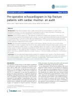
On each blot, one sample obtained from a patient with
metastasized small cell lung cancer that was strongly
YB-1/p18 positive was run in parallel as a positive control
(Figure 1). The immunoblots were performed and analyzed
by a scientist blinded to the origin of the samples. YB-1/
p18 signals were quantified by densitometry (NIH imager)
Methods
Patients
The study protocol was approved by the local ethics
committee and conducted in accordance with the ethical
standards laid down in the 1974 Declaration of Helsinki
(ethics committee of the University Hospital Aachen,
RWTH-University, Aachen, Germany, reference number
EK 107/05). Written informed consent was obtained from
each participant. Plasma samples were obtained from
consecutive patients with various malignant disorders
presenting to the Outpatient Cancer Clinic at the
Figure 1 YB-1/p18 detection in human plasma. Human plasma
was blotted onto a nitrocellulose membrane and YB-1 detected by
immunoblotting using a novel monoclonal antibody. Patients with
malignant disorders often displayed an additional signal at 18 kDa
(“YB-1/p18”), and its intensity was quantified by densitometry. A
positive reference sample was run on each blot (T168). Samples with
an 18-kD band ≥0.45 were considered to be YB-1/p18 positive.
Tacke et al. BMC Cancer 2014, 14:33
/>
and compared to the positive control, which was assigned
the optical density of ′1.0′. The relative optical density
of the samples was calculated and signals ≥0.45 were
considered to be YB-1/p18 positive (Figure 1), as previously
described [12].
Other tumor markers
Established tumor markers were assessed using the manufacturers’ protocols with reference cut-off values recommended by the manufacturer and validated with internal
controls at the Department of Clinical Chemistry and
Pathobiochemistry, University Hospital Aachen, Germany.
The following assays were used: from Roche, Mannheim,
Germany: cancer antigen 125 (CA 125, reference <36 kU/L);
carcinoembryonic antigen (CEA, <5 μg/L; CA 15–3, <26
kU/L; CA 19–9, <38 kU/L; CA 72–4, <7 kU/L; cytokeratin
fragments 21–1 (CYFRA 21–1, <3.4 μg/L; neuron-specific
enolase (NSE), <14 μg/L; alpha-fetoprotein (AFP), <10 μg/L;
Dade Behring, Marburg, Germany: beta-2-microglobulin
Page 3 of 10
(β2-micro), <1.8 mg/L; Abbott, Wiesbaden, Germany: squamous cell carcinoma antigen (SCC), <1.6 μg/L; DiaSorin S.
p.A., Saluggia, Italy: thymidine kinase (TK), <6.8 U/L,
tissue polypeptide antigen (TPA), <92 U/L; IBL, Hamburg,
Germany: pro-gastrin-releasing peptide (PGRP), <46 ng/L.
Due to limited sample availability, these tumor markers
were detected in subgroups of the whole cohort.
Statistics
Results were reported as median and range, and differences between groups were assessed by Mann–Whitney
U-test, Kruskal-Wallis-ANOVA, or chi-square-test [14].
The prognostic value of the variables was tested by univariate and multivariate analyses in the Cox regression model.
Kaplan Meier curves were plotted to display the impact on
survival [15]. The manufacturers’ reference intervals for
other tumor markers were used as the cut-offs discriminating tumor marker positivity and negativity, respectively. All
statistical analyses were performed using SPSS.
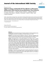
Figure 2 High prevalence of circulating YB-1/p18 in patients with various malignancies. (A) YB-1/p18 was previously measured in healthy
volunteers (n = 60), patients with inflammatory or renal disease (n = 60), and patients with chronic liver disease (n = 91) with a low rate of YB-1/
p18 positivity in patients without malignancies. In contrast, almost all patients with metastatic gastrointestinal tumors (n = 16) tested YB-1/p18
positive. (B) YB-1/p18 was detected in plasma samples of patients with malignant diseases with a rate of 62.5% (gastrointestinal [GI] tumors) to
100%. CUP, cancer of unknown primary site. (C) Cancer patients with or without detection of plasma YB-1/p18 did not differ with respect to their
white-blood cell count (WBC), C-reactive protein (CRP) or lactate dehydrogenase (LDH); P > 0.05, not significant, U-Test. The box-and-whiskers
plots display the median, quartiles, range and extreme values. The whiskers extend from the minimum to the maximum value excluding outside
(>1.5 times upper/lower quartile, open circle) and “far out” (>3 time upper/lower quartile, asterisks) values.
Tacke et al. BMC Cancer 2014, 14:33
/>
Results
Page 4 of 10
Table 1 Patient characteristics and tumor entities
Circulating YB-1/p18 is frequently detected in various
malignant diseases
Our group has recently shown that a YB-1/p18 fragment,
detected by immunoblotting with a novel biotinylated
monoclonal antibody (Figure 1), can be found in the plasma
of patients with malignancies. In a prior study from our
group, none of the healthy volunteers (0/60) tested positive
for YB-1/p18, whereas 88% of patients with metastatic
gastrointestinal tumors (14/16) had detectable plasma
YB-1/p18 levels (Figure 2A) [12]. In two cohorts of patients
without overt malignancies, but with inflammatory or renal
diseases, as well as patients with chronic liver disease,
YB-1/p18 positivity was detected in approximately 15% of
cases (Figure 2A) [12]. In order to assess the prevalence of
YB-1/p18 in malignant diseases, 151 patients (56% male,
44% female, median age 65 years, range 19–84 years) with
various malignancies were included in this study (Table 1).
The different etiologies of the malignancies are given in
Table 1, the stage of remission, tumor staging, and current
therapy are listed in Table 2. YB-1/p18 was detected in
plasma samples of 77.5% (117/151) of all patients.
There was no difference between male (79.8% positive)
and female (74.6%) patients (P > 0.05, not significant,
Chi-square test, Table 3). Furthermore, the patient’s age had
no influence on YB-1/p18 positivity either (not shown).
Among the different etiologies, the vast majority of
tested patients with lung cancer (32/37, 86.5%), breast
cancer (7/10, 70%), urogenital tumors (8/8, 100%), cancer
of unknown primary site (5/5, 100%), and other solid
tumors (13/13, 100%) had detectable plasma YB-1/p18
levels (Figure 2B). Patients with gastrointestinal tumors
(10/16, 62.5%) and hematological malignancies (42/62,
67.7%) had a lower prevalence of plasma YB-1/p18 levels
above the defined threshold. Within the group of patients
with hematological malignancies (Table 3), lymphoma
(21/25, 84%) or multiple myeloma (10/13, 77%) were
more often associated with plasma YB-1/p18 than acute
or chronic leukemia (5/9, 56%) or myelodyplastic disorders
(2/10, 20%). In contrast, in patients with lung cancer,
urogenital carcinomas, or other solid tumors, the histological subtypes did not differ with respect to the YB-1/
p18 result (Table 3).
We also analyzed if patients with or without detectable
plasma YB-1/p18 levels varied in their laboratory or other
clinical characteristics. As shown in Figure 2C, parameters
indicating inflammation or (general) tumor load, such as
white blood cell count (WBC, median 7.9 G/L in positive
vs. 8.3 in negative patients), C-reactive protein (CRP,
median 14 versus 8 mg/L), or lactate dehydrogenase (LDH,
median 216 versus 234 U/L) did not significantly differ
between YB-1/p18 positive and negative patients (P > 0.05,
U-test). Furthermore, parameters associated with liver
or renal function deterioration did not display significant
Age [years]
n
%
Median
Range
All patients
151
100
64.8
18.5- 84.4
Male
84
55.6
Female
67
44.4
37
24.5
62.8
43.9- 83.5
Adeno
15
9.9
Small cell
10
6.6
Squamous
10
6.6
Other non small cell
2
1.3
10
6.6
55.9
34.9- 71.3
9
90
67.5
42.3- 84.4
64.2
18.5- 83.2
64.4
38.4- 81.7
Malignancy
Group I: lung cancer
Group II: breast cancer
Ductal
Lobular
1
10
16
10.6
Stomach cancer
3
2.0
Colorectal cancer
7
4.6
Group III: gastrointestinal
Other
6
4.0
62
41.1
Acute myeloid leukemia
7
4.6
Chronic myelogenous leukemia
2
1.3
Hodgkin’s lymphoma
5
3.3
Non-Hodgkin’s lymphoma
13
8.6
Other lymphoma
7
4.6
Multiple myeloma
13
8.6
Idiopathic thrombocytopenia
5
3.3
Myelodysplastic syndrome
10
6.6
Group IV: hematological
Group V: urogenital cancers
8
5.3
Ovarian cancer
2
1.3
Testicular cancer
2
1.3
Prostate cancer
1
0.7
Urinary tract cancer
3
2.0
Group VI: CUP
5
3.3
64.7
41.2- 80.6
Group VII: other solid tumors
13
8.6
64.1
30.7- 73.5
differences either (data not shown). There was no difference in tumor stage, metastases, or co-morbidities (such
as chronic heart failure, arterial hypertension, coronary
artery disease, chronic obstructive pulmonary disease,
diabetes, renal insufficiency, etc.) between YB-1/p18 seropositive and -negative patients in the total cohort.
YB-1/p18 is a more sensitive diagnostic biomarker for
cancer than established tumor markers
A panel of 13 established tumor markers was assessed
alongside YB-1/p18 in the cohort of patients with malignancies, namely CA 125, CEA, CA 15–3, CA 19–9, CA
Tacke et al. BMC Cancer 2014, 14:33
/>
Page 5 of 10
Table 2 Stage of remission, therapy and metastases at study entry
All patients
Group I:
lung cancer
Group II:
breast cancer
Group III:
GI tumors
Group IV:
hematol.
Group V:
uro-genital
Group VI: CUP
Group VII:
others
n (%)
n (%)
n (%)
n (%)
n (%)
n (%)
n (%)
n (%)
27 (27.3)
1 (1.1)
9 (9.1)
45 (45.5)
5 (5)
5 (5)
7 (7)
Stage of remission
ID
99 (65.6)
CR
2 (1.3)
0
0
0
2 (100)
0
0
0
PR
4 (2.6)
1 (25)
0
0
2 (50)
1(25)
0
0
NC
2 (1.3)
1 (50)
0
0
1 (50)
0
0
0
PD
44 (29.1)
8 (18.2)
9 (20.5)
7 (15.9)
12 (27.3)
2 (4.5)
0
6 (13.6)
CNS
16 (10.6)
8 (50)
0
1 (6.3)
3 (18.7)
2 (12.5)
0
2 (12.5)
Bone
32 (21.2)
9 (45)
6 (18.8)
1 (3.1)
8 (25)
3 (9.4)
2 (6.3)
3 (9.4)
Liver
17 (11.3)
2 (11.8)
2 (11.8)
8 (47.0)
3 (17.6)
2 (11.8)
0
0
Lymphatic
85 (56.3)
21 (24.1)
7 (8)
11 (12.6)
33 (37.9)
5 (5.7)
3 (3.4)
5 (5.7)
Metastases
Therapy (at study entry)
Chemo
18 (11.9)
4 (22.2)
1 (5.6)
1 (5.6)
4 (22.2)
2 (11.1)
1 (5.6)
5 (27.7)
Radiation
3 (2)
2 (66.7)
0
0
0
0
0
1 (33.3)
Hormone
3 (2)
0
1 (33.3)
0
2 (66.7)
0
0
0
ID, initial diagnosis; CR, complete remission; PR, partial remission; NC, no change; PD, progressive disease.
72–4, CYFRA 21–1, NSE, AFP, β2-microglobulin, SCC,
thymidine kinase, TPA, and PGRP. YB-1/p18-positivity
was not statistically linked to positivity of any of these
parameters (cross-table analysis and Chi-square-tests, data
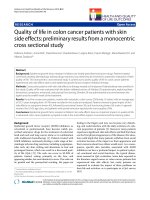
not shown). For the total cohort, YB-1/p18 was the
marker with the highest sensitivity in detecting malignancies (78% positive, Figure 3A). CA 125 (59%) and
β2-microglobulin (74%) also tested positive in the majority
of cancer patients, whereas all other markers remained
negative in at least half of the patient cohort (Figure 3A).
When the different tumor entities were analyzed separately, YB-1/p18 positivity was the most frequently detected
tumor marker in patients with lung cancer (Figure 3B),
urogenital tumors, CUP syndrome, breast cancer (Figure 3C),
and the mixed group of patients with other solid tumors
(Figure 3D, n = 13) that included very heterogeneous
entities such as tumors of the CNS (n = 3), nasopharyngeal
tumors (n = 3), or sarcomas (n = 2). Figure 3B and D
display the next most sensitive tumor markers for these
subgroups, emphasizing that none of the other markers
had a similar overall sensitivity comparable to YB-1/p18.
In most cases, such as in lung cancer, YB-1/p18 positivity
was independent of the histological subtype (Figure 4).
Importantly, some tumor markers had a higher sensitivity than YB-1/p18 in distinct subgroups of patients. For
instance, CA 15–3 was more sensitive, while NSE or TPA
tested equally sensitive in patients with breast cancer
when compared to YB-1/p18 (Figure 3C). In a prior study,
we had reported AFP to be more sensitive in detecting
hepatocellular carcinoma than YB-1/p18 [12].
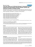
YB-1/p18 detection in plasma varies during therapy, but
has limited potential to predict prognosis
One important clinical value of established tumor markers
is their correlation in individual patients with the effectiveness of treatment. In our cohort, we followed 42 out of
the 151 patients during the course of therapy. As shown
in Figure 5 for an individual patient with small cell lung
cancer subsequent to chemotherapy, YB-1/p18 detection
in plasma was altered, indicating a response to therapy.
Concentrations of other tumor markers that were found
positive were also lower after response to therapy. However, truly quantitative assessments of tumor markers
were superior to measurement of YB-1/p18 intensity in
immunoblotting for predicting individual response to
cancer therapy (data not shown).
Based on the association with histological expression of
YB-1 by tumor tissue and unfavorable prognosis (summarized in [2]) we tested whether positivity for YB-1/p18
in serum or other tumor markers were associated with
survival. Using Cox regression analysis, YB-1/p18 seropositivity did not predict survival; similar to most other
tumor markers (Figure 6). Only positivity for CA 72–4 and
TPA was associated with poor prognosis within the cohort
of tumor patients, indicating that the tumor markers that
were specifically found in subtypes of cancers had a higher
probability to have additional prognostic value.
Discussion
In this study, we set out to test the prevalence and relevance of YB-1/p18 seropositivity in patients diagnosed
Tacke et al. BMC Cancer 2014, 14:33
/>
Page 6 of 10
Table 3 Positivity for YB-1/p18 detection in human serum
samples of patients with different malignant disorders
n
YB-1/p18 positivity n/N (%)
All patients
151
117/151 (77.5)
Male
84
67/84 (79.8)
Female
67
50/67 (74.6)
37
32/37 (86.5)
15
12/15 (80)
Malignancy
Group I: lung cancer
Adeno
Small cell
10
9/10 (90)
Squamous
10
9/10 (90)
Non small cell
2
2/2 (100)
10
7/10 (70)
Ductal
9
6/9 (66.7)
Lobular
1
1/1 (100)
Group II: breast cancer
Group III: gastrointestinal
16
10/16 (62.5)
Stomach cancer
3
2/3 (66.7)
Colorectal cancer
7
4/7 (57.1)
Other
6
4/6 (66.7)
62
42/62 (67.7)
7
4/7 (57.1)
Group IV: hematological
Acute myeloid leukemia
Chronic myelogenous leukemia
2
1/2 (50)
Hodgkin’s lymphoma
5
4/5 (85.7)
Non-Hodgkin’s lymphoma
13
11/13 (84.6)
Other lymphoma
7
6/7 (85.7)
Multiple myeloma
13
10/13 (77)
Idiopathic thrombocytopenia
5
4/5 (76.9)
Myelodysplastic syndrome
10
2/10 (20)
8
8/8 (100)
Group V: urogenital cancers
Ovarian cancer
2
2/2 (100)
Testicular cancer
2
2/2 (100)
Prostate cancer
1
1/1 (100)
Urinary tract cancer
3
3/3 (100)
Group VI: CUP
5
5/5 (100)
Group VII: other solid tumors
13
13/13 (100)
with different cancerous and leukemic/hematological
diseases. Our study cohort was comprised of an unselected heterogeneous group of patients with malignant
diseases of various entities and at various stages of
disease (e.g., early and advanced, before or during
chemotherapy etc.). The main finding of the study is a
high positivity rate of the YB-1/p18 in plasma samples
of cancer patients. Unexpectedly, this positivity is
found irrespective of the underlying cancer provenience and other clinical co-variables that were tested.
Such an universal positivity is difficult to understand,
given that most cancers derive from specific genetic or
epigenetic defects with ensuing alterations of the cellular genome and cancer cell environment [16]. For all
the other tested tumor markers, the sensitivity and
specificity rates were lower.
The finding of universal seropositivity for circulating
YB-1/p18 fragments may be explained by similar widespread YB-1 positivity in tissue samples of cancer patients
[2,4,17], emphasizing that YB-1 dysregulation is a common
feature found in tumor tissue. So far, immunostaining
and analysis of the subcellular distribution of YB-1 from
tumor tissue has been used to correlate and predict poor
prognosis, especially with nuclear protein accumulation
in breast cancer patients [2]. Our findings of circulating
YB-1/p18 fragments in plasma of patients with cancer
now indicates that dysregulated YB-1 may be prone to be
released as fragments into the circulation, which would
allow its easy use as a non-invasive disease marker. Similar
observations are currently gathered for many micro-RNA
(miRNA) in cancerous diseases, as the dysregulation of
distinct key miRNA in tumor tissue can be associated with
elevated levels of circulating miRNAs in cancer patients
[18]. Nevertheless, the profile of circulating miRNAs
appears very specific for different types of malignancies
or non-malignant diseases [19], while YB-1/p18 detection
did not allow us to distinguish between different tumor
entities.
It is important to emphasize that YB-1/p18 detection
appeared relatively specific for malignant diseases and
had a high sensitivity in various malignancies, but its
detection, unlike many established tumor markers, was
not clearly related to the disease stage or prognosis.
The reason for this might be the semi-quantitative nature
of our immunoblotting assay. The threshold for YB-1/
p18 seropositivity was optimized for sensitivity [12], and
immunoblotting quantification against a positive control
certainly did not allow linear-range quantification. In fact,
in individual patients with several longitudinal YB-1/p18
measurements, there was a moderate association between
the quantification of the signal with the response or failure
to therapy. Due to the relatively small number of patients
with distinct hematological malignancies and the semiquantitative nature of the immunoblotting method, we
were unable to detect a clear association between the
number of circulating blasts and the intensity of p18
bands. Thus, a different, more quantitative technique
for YB-1/p18 fragment measurement is highly warranted
to better estimate the prognostic value of YB-1/p18 in
cancerous disease. The development of such an ELISA
system, however, is hampered by the fact that currently
most antibodies detect not only the p18 fragment, but also
full-length YB-1 [13].
It is currently unclear, whether circulating YB-1/p18
fragments are functionally active in patients with malignancies. Inside tumor cells, YB-1 has been shown to fulfill
Tacke et al. BMC Cancer 2014, 14:33
/>
Page 7 of 10
Figure 3 Comparison of YB-1/p18 with established tumor markers in patients with various malignancies. (A) Positivity of YB-1/p18 and
other available tumor markers for the total cohort of patients with malignant disorders. (B-D) Positivity of YB-1/p18 in comparison to the most
sensitive of the other tested markers for patients with lung cancer (B), breast cancer (C), or other solid tumors (D). The number of patients in
which the respective markers were assessed is given. Abbreviations are used as in the main text. Cut-off values for the established tumor markers
are given in the Methods’ section.
critical cellular functions, such as the transcriptional upregulation of proliferation-associated and downregulation
of apoptosis-related genes or induction of drug-transporter genes (like MDR-1) involved in chemoresistance
[6,20]. Data from our own laboratories indicated that
also extracellular YB-1 may be involved in tumor progression, since adding recombinant YB-1 protein to cancer
cell-lines in vitro revealed profound pro-mitogenic
effects suggesting that secreted YB-1 or its fragments
could act as a tumor growth-promoting factor [11].
Further studies are needed to define the exact functions
of circulating full-length YB-1 compared to p18 fragments and to define the exact cellular source (tumor vs.
stromal cells) of YB-1/p18 in patients with malignant
disorders.
Nevertheless, our study demonstrated that circulating
YB-1/p18 is highly prevalent in cancer patients and
reasonably specific in distinguishing malignant versus
non-malignant disorders. One of the challenges in the
era of ‘personalized medicine’ is the early detection of
cancer, and many biomarkers have failed to be used
for screening purposes in clinical practice, even for the
most common tumor entities, such as breast or lung
cancer [21]. The data provided by our study suggest
that it might be highly valuable to incorporate YB-1/
p18 measurement into cancer screening approaches.
Tacke et al. BMC Cancer 2014, 14:33
/>
Page 8 of 10
Figure 4 YB-1/p18, β2-microglobulin, and NSE in lung cancer patients. Comparison of YB-1/p18 positivity (data from original immunoblotting
of serum, relative optical density [rel. OD] of the 18 kDa signal) with individual serum concentrations of β2-microglobulin and NSE (reference interval
given in gray). Histological classification of the tumor is given.
Figure 5 YB-1/p18 during therapy in a patient with small cell lung cancer. Serum samples were obtained from a 45-year old male with small
cell lung cancer at diagnosis (lane 1) and during chemotherapy with cisplatin/etoposide (lanes 2–3), initially with good response. He was found
to progress in month 3 (lane 4), and then was treated with epirubicine, cyclophosphamide, and vincristine (lanes 5–7). Simultaneous measurements of
NSE and PGRP are displayed. NSE and PGRP were the only two positive parameters of 13 tumor markers initially tested in this patient. The relative
optical density (rel. OD) of the YB-1/p18 signal in immunoblotting is given.
Tacke et al. BMC Cancer 2014, 14:33
/>
Page 9 of 10
Figure 6 Prognostic value of YB-1/p18 positivity in comparison to established tumor markers. Kaplan-Meier curves are depicted to display
survival in patients with positive or negative tumor markers, p-values from Cox regression analyses are given; n.s., not significant. The number of
patients in which the respective tumor marker has been assessed is given in the figure. Except for CA 72–4 and TPA, that had predictive value for
patients’ survival, tumor markers were not related to the prognosis.
However, it is important to point out that our study
comprised a heterogeneous group of patients with different tumor entities and stages. Due to the relatively
small patient numbers in the subgroups, our study does
not allow to draw specific conclusions for individual
tumor entities, e.g. on associations with staging, prognosis
or treatment response. Prospective trials with large cohorts are warranted to confirm that circulating YB-1/
p18 fragments might be suitable as a general biomarker
for the presence of malignant disorders and to assess
its potential specific value in distinct tumor entities as
a prognostic marker.
Conclusions
The detection of cold shock proteins, especially of
YB-1, by immunostaining in tumor tissue of cancer
patients has been related to adverse outcome. Our study
now demonstrated that a 18 kD secreted form of YB1, termed YB-1/p18, carries potential as a circulating
biomarker in oncology. By using a novel immunoblotting assay for YB-1/p18 for analyzing YB-1/p18 in
plasma of 151 unselected patients with various malignancies, circulating YB-1/p18 had a higher prevalence
compared to other established tumor markers and was
associated to therapy response in longitudinal assessments. Unlike ‘traditional’ entity-specific cancer biomarkers, YB-1/p18 was largely independent from the
histological subtype or stage of disease progression and
did not predict the individual prognosis. These data
indicate that YB-1/p18 fragments in human plasma
may therefore have exceptional potential as a cancer
screening marker.
Tacke et al. BMC Cancer 2014, 14:33
/>
Abbreviations
AFP: Alpha-fetoprotein; β2-micro: Beta-2-microglobulin; CA 125: Cancer
antigen 125; CA19-9: Carbohydrate antigen; CEA: Carcinoembryonic antigen;
CRP: C-reactive protein; CSD: Cold shock domain; CYFRA 21–1: Cytokeratin
fragments 21–1; CUP: Cancer of unknown primary; Dbp: DNA-binding
protein; HMGB: High mobility group box protein; LDH: Lactate dehydrogenase;
MDR: Multiple drug resistence; miRNA: micro-RNA; NSE: Neuron-specific enolase;
PGRP: Pro-gastrin-releasing peptide; PSA: Prostate-specific antigen; ROC: Receiver
operating characteristic; SCC: Squamous cell carcinoma antigen; TGF: Transforming
growth factor; TK: Thymidine kinase; TPA: Tissue polypeptide antigen; WBC: White
blood cell count; YB-1: Y-box protein-1; YB-1/p18: YB-1 protein fragment p18.
Page 10 of 10
8.
9.
10.
Competing interests
None of the authors declares competing financial or non-financial interests.
11.
Authors’ contributions
NK and CSE conducted YB-1 immunoblots. NK, OG and EY performed patient
recruitment and provided samples. SB, JAL and UR provided experimental tools
and assisted in experiments. FT, NK and PRM designed the study, analyzed data
and wrote the manuscript. All authors read and approved the final manuscript.
12.
Acknowledgments
The study has been funded by Sonderforschungsbereiche (SFB) 854 and
TRR57, German Research Foundation (DFG ME1365/7-1 to PRM, Ta 434/2-1
to FT), Fritz Bender Stiftung (to UR) and the Interdisciplinary Centre for
Clinical Research (IZKF) within the Faculty of Medicine at the RWTH Aachen
University (to FT).
Author details
1
Medical Clinic III, University Hospital RWTH-Aachen, Pauwelsstrasse 30,
52074 Aachen, Germany. 2Medical Clinic IV, University Hospital
RWTH-Aachen, Pauwelsstrasse 30, 52074 Aachen, Germany. 3Department of
Nephrology and Hypertension, Diabetes and Endocrinology,
Otto-von-Guericke University Magdeburg, Leipziger Str. 44, 39120
Magdeburg, Germany. 4Medical Care Center, Dr. Stein and colleagues,
41061 Mönchengladbach, Germany. 5Medical Clinic II, University Hospital
RWTH-Aachen, Pauwelsstrasse 30, 52074 Aachen, Germany.
Received: 25 March 2013 Accepted: 31 October 2013
Published: 20 January 2014
References
1. Brandt S, Raffetseder U, Djudjaj S, Schreiter A, Kadereit B, Michele M, Pabst M,
Zhu C, Mertens PR: Cold shock Y-box protein-1 participates in signaling
circuits with auto-regulatory activities. Eur J Cell Biol 2011, 91(6–7):464–471.
2. Lasham A, Print CG, Woolley AG, Dunn SE, Braithwaite AW: YB-1:
oncoprotein, prognostic marker and therapeutic target? Biochem J 2013,
449(1):11–23.
3. Raffetseder U, Frye B, Rauen T, Jurchott K, Royer HD, Jansen PL, Mertens PR:
Splicing factor SRp30c interaction with Y-box protein-1 confers nuclear
YB-1 shuttling and alternative splice site selection. J Biol Chem 2003, 278
(20):18241–18248.
4. Bommert KS, Effenberger M, Leich E, Kuspert M, Murphy D, Langer C, Moll R,
Janz S, Mottok A, Weissbach S, et al: The feed-forward loop between YB-1
and MYC is essential for multiple myeloma cell survival. Leukemia 2012,
27(2):441–450.
5. Sinnberg T, Sauer B, Holm P, Spangler B, Kuphal S, Bosserhoff A, Schittek B:
MAPK and PI3K/AKT mediated YB-1 activation promotes melanoma cell
proliferation which is counteracted by an autoregulatory loop. Exp
Dermatol 2012, 21(4):265–270.
6. Bargou RC, Jurchott K, Wagener C, Bergmann S, Metzner S, Bommert K,
Mapara MY, Winzer KJ, Dietel M, Dorken B, et al: Nuclear localization and
increased levels of transcription factor YB-1 in primary human breast
cancers are associated with intrinsic MDR1 gene expression. Nat Med
1997, 3(4):447–450.
7. Bergmann S, Royer-Pokora B, Fietze E, Jurchott K, Hildebrandt B, Trost D,
Leenders F, Claude JC, Theuring F, Bargou R, et al: YB-1 provokes breast
cancer through the induction of chromosomal instability that emerges
from mitotic failure and centrosome amplification. Cancer Res 2005,
65(10):4078–4087.
13.
14.
15.
16.
17.
18.
19.
20.
21.
Hyogotani A, Ito K, Yoshida K, Izumi H, Kohno K, Amano J: Association of
nuclear YB-1 localization with lung resistance-related protein and
epidermal growth factor receptor expression in lung cancer. Clin Lung
Cancer 2012, 13(5):375–384.
Shibahara K, Sugio K, Osaki T, Uchiumi T, Maehara Y, Kohno K, Yasumoto K,
Sugimachi K, Kuwano M: Nuclear expression of the Y-box binding protein,
YB-1, as a novel marker of disease progression in non-small cell lung
cancer. Clin Cancer Res 2001, 7(10):3151–3155.
Yoshimatsu T, Uramoto H, Oyama T, Yashima Y, Gu C, Morita M, Sugio K,
Kohno K, Yasumoto K: Y-box-binding protein-1 expression is not correlated
with p53 expression but with proliferating cell nuclear antigen expression
in non-small cell lung cancer. Anticancer Res 2005, 25(5):3437–3443.
Frye BC, Halfter S, Djudjaj S, Muehlenberg P, Weber S, Raffetseder U, En-Nia A,
Knott H, Baron JM, Dooley S, et al: Y-box protein-1 is actively secreted
through a non-classical pathway and acts as an extracellular mitogen.
EMBO reports 2009, 10(7):783–789.
Tacke F, Kanig N, En-Nia A, Kaehne T, Eberhardt CS, Shpacovitch V, Trautwein C,
Mertens PR: Y-box protein-1/p18 fragment identifies malignancies in
patients with chronic liver disease. BMC Cancer 2011, 11:185.
Dahl E, En-Nia A, Wiesmann F, Krings R, Djudjaj S, Breuer E, Fuchs T, Wild PJ,
Hartmann A, Dunn SE, et al: Nuclear detection of Y-box protein-1 (YB-1)
closely associates with progesterone receptor negativity and is a strong
adverse survival factor in human breast cancer. BMC Cancer 2009, 9:410.
Yagmur E, Trautwein C, Gressner AM, Tacke F: Resistin serum levels are
associated with insulin resistance, disease severity, clinical complications,
and prognosis in patients with chronic liver diseases. Am J Gastroenterol
2006, 101(6):1244–1252.
Koch A, Voigt S, Kruschinski C, Sanson E, Duckers H, Horn A, Yagmur E,
Zimmermann H, Trautwein C, Tacke F: Circulating soluble urokinase
plasminogen activator receptor is stably elevated during the first week
of treatment in the intensive care unit and predicts mortality in critically
ill patients. Critical care 2011, 15(1):R63.
Rizki A, Weaver VM, Lee SY, Rozenberg GI, Chin K, Myers CA, Bascom JL,
Mott JD, Semeiks JR, Grate LR, et al: A human breast cell model of
preinvasive to invasive transition. Cancer Res 2008, 68(5):1378–1387.
Janz M, Harbeck N, Dettmar P, Berger U, Schmidt A, Jurchott K, Schmitt M,
Royer HD: Y-box factor YB-1 predicts drug resistance and patient outcome
in breast cancer independent of clinically relevant tumor biologic factors
HER2, uPA and PAI-1. Int J Cancer 2002, 97(3):278–282.
Mo MH, Chen L, Fu Y, Wang W, Fu SW: Cell-free circulating miRNA
biomarkers in cancer. J Cancer 2012, 3:432–448.
Roderburg C, Mollnow T, Bongaerts B, Elfimova N, Vargas Cardenas D,
Berger K, Zimmermann H, Koch A, Vucur M, Luedde M, et al: Micro-RNA
profiling in human serum reveals compartment-specific roles of miR-571
and miR-652 in liver cirrhosis. PloS one 2012, 7(3):e32999.
Oda Y, Ohishi Y, Saito T, Hinoshita E, Uchiumi T, Kinukawa N, Iwamoto Y,
Kohno K, Kuwano M, Tsuneyoshi M: Nuclear expression of Y-box-binding
protein-1 correlates with P-glycoprotein and topoisomerase II alpha
expression, and with poor prognosis in synovial sarcoma. J Pathol 2003,
199(2):251–258.
Schmalfuss F, Kolominsky-Rabas PL: Personalized medicine in screening
for malignant disease: a review of methods and applications. Biomarker
Insights 2013, 8:9–14.
doi:10.1186/1471-2407-14-33
Cite this article as: Tacke et al.: High prevalence of Y-box protein-1/p18
fragment in plasma of patients with malignancies of different origin.
BMC Cancer 2014 14:33.