MicroRNA-302b suppresses cell proliferation by targeting EGFR in human hepatocellular carcinoma SMMC-7721 cells
Bạn đang xem bản rút gọn của tài liệu. Xem và tải ngay bản đầy đủ của tài liệu tại đây (1.31 MB, 9 trang )
Wang et al. BMC Cancer 2013, 13:448
/>
RESEARCH ARTICLE
Open Access
MicroRNA-302b suppresses cell proliferation by
targeting EGFR in human hepatocellular
carcinoma SMMC-7721 cells
Lumin Wang1†, Jiayi Yao1†, Xin Shi2, Lili Hu1, Zongfang Li3, Tusheng Song1 and Chen Huang1,4,5*
Abstract
Background: MicroRNAs are regulators that can play an essential role in tumorigenesis. Although miR-302 families
have been suggested to be tumor repressors in human cancer, the mechanism by which they suppress tumor
development remains to be defined. In this study, we discover that miR302b suppresses tumor proliferation may
due to directly targeting EGFR in human hepatocellular carcinoma (HCC).
Methods: QRT-PCR was used to assess miR-302b and EGFR expression in 27 pairs of clinical hepatocellular
carcinoma tissues and their corresponding adjacent nontumorous liver tissues. MTT, colony formation,
immunofluorescence staining, and cell cycle assays were used to examine the tumor suppressor role of miR302b in
cell proliferation. Luciferase assays were performed to assess the EGFR was a novel target of miR-302b. Western blot
assay was used to validate the protein expression level.
Results: We demonstrated that miR-302b was frequently down-regulated, whereas EGFR was up-regulated in 27
pairs of clinical HCC and non-tumorous counterparts. The dual-luciferase reporter assays revealed that EGFR was a
novel target of miR-302b. Re-expression of miR-302b resulted in the inhibition of proliferation in hepatocellular
carcinoma SMMC-7721 cells. The silencing of EGFR by miR-302b or siEGFR led to down-regulation of proliferationrelated proteins, such as AKT2, CCND1, and CDK2.
Conclusion: miR-302b suppresses HCC growth may due to targeting the EGFR/AKT2/CCND1 pathway.
Keywords: miR-302b, Hepatocellular carcinoma, EGFR, Proliferation, Cell cycle
Background
Hepatocelluar carcinoma (HCC) is the third leading
cause of cancer-related deaths worldwide, and the burden of this devastating cancer is expected to increase
further in the coming years [1]. Due to the difficulty of
effectively diagnosing HCC at its early stage, only about
10 to 20% of patients with hepatocellular carcinoma are
currently eligible for surgical intervention [2-6]. Therefore, elucidating the molecular mechanisms involved in
HCC is essential for developing cancer prevention
* Correspondence:
†
Equal contributors
1
Department of Genetics and Molecular Biology, Xi’an Jiaotong University
Health Science Center, No.76 West Yanta Road, Xi’an, Shaanxi 710061, P.R.
China
4
Key Laboratory of Environment and Genes Related to Diseases, Xi’an
Jiaotong University Health Science Center, Xi’an, Shaanxi, China
Full list of author information is available at the end of the article
strategies and possible guiding disease management in
the clinic.
Accumulating evidence suggests that microRNAs
(miRNAs) are involved in the initiation and progression
of HCC [7]. First, the 22nt noncoding miRNAs act as
key regulators of various fundamental biological processes, such as development, differentiation, apoptosis,
and cell proliferation, in which common pathways are
shared with cancer [8-11]. Second, bioinformation analyses estimate that miRNAs may regulate as much as
30% of the human protein coding genes, including oncogenes and tumor suppressors, suggesting that these
small RNAs may act to coordinate the interplay between
complex signal transduction pathways [12]. Third, increasing evidence shows that the expression of miRNAs
is remarkably deregulated in cancer due to multiple epigenetic and genomic alterations. Fourth, several miRNAs
© 2013 Wang et al.; licensee BioMed Central Ltd. This is an Open Access article distributed under the terms of the Creative
Commons Attribution License ( which permits unrestricted use, distribution, and
reproduction in any medium, provided the original work is properly cited.
Wang et al. BMC Cancer 2013, 13:448
/>
themselves have been demonstrated to serve as tumor
suppressor genes or oncogenes in tumors [13-15].
The miR-302 family consists of four highly-homologous
miRNA members, which are transcribed together as a
noncoding RNA cluster containing mir-302b, mir-302c,
mir-302a, mir-302d, and mir-367 in a 5′-to-3′ direction
[16]. To date, miR-302 s have been proven to posttranscriptionally regulate CCND1 and CDK4, therefore
affecting cell cycle progression. Other studies have demonstrated the tumor suppressive activity of miR-302 in
human pluripotent stem cell by both the CCNE-CDK2
and CCND-CDK4/6 pathways in G1-S cell cycle transition. Although miR-302 has been suggested to have tumor
suppressor potential, the present studies focused on the
self-renewal and proliferation properties of miR-302b in
the stemness maintenance of embryonic stem cells (ESCs)
or tumor stem cell properties in advanced cancer cells
[17,18]. So, the relationship between miR-320b and cancers needs to be investigated further.
In this research, we analyzed the miR-302b targets by
bioinformatics software, and found that miR-302b can
target EGFR. Next, we found that miR-302b was frequently down-regulated in HCC tissues and cells. Further, in vitro experiments proved that the re-expression
of miR-302b inhibited HCC proliferation dramatically,
and arrested the HCC cell cycle at the G1/S phase. The
dual-luciferase reporter assays further demonstrated that
EGFR was a novel target of miR-302b. The silencing of
EGFR by miR-302b or siEGFR led to the downregulation of cell-cycle related proteins, such as AKT2,
CCND1, and CDK2, strongly suggesting that miR-302b
suppresses the growth of SMMC-7721 cells by targeting
EGFR involved the EGFR/AKT2/CCND1 pathway.
Methods
Page 2 of 9
Plasmid constructions
pcDNA™6.2-GW/EmGFP-miR vector (Invitrogen) was
used to construct vectors of re-expression miR-302b.
First, we inserted EcoRI and HindIII sites into the MCS
of the vector. Then, the miR-302b was chemically synthesized and cloned into pcDNA™6.2-GW/EmGFP-miR
vector between the EcoRI and HindIII sites. RegRNA
(A Regulatory RNA Motifs and Elements Finder http://
regrna.mbc.nctu.edu.tw/), TargetScan (getscan.
org/) and DIANA ( />were used for gene-related specified microRNA prediction. Through bioinformatics analysis, we got the predicted fragment of targeted gene (EGFR), which was
associated with miR302b. Specified fragments of EGFR
were chemically synthesized, and are shown in supporting
Table 1. The luciferase-UTR reporter constructions
were generated by introducing the Wt/Mut-EGFR 3′UTR, carrying a putative miR-302b binding site into
pmirGLO Dual-Luciferase miRNA Target Expression
vector (Promega) between the XhoI and SacI sites.
Table 1 Primers and oligonucleotides used in this work
Name
Sequence (5′-3′)
Pri-miR-302b-S
5′-AATTCGCTCCCTTCAACTTTAACATGGAAGTGCTTTC
TGTGACTTTAAAAGTAAGTGCTTCCATGTTTTAGTAGG
AGTA-3′
Pri-miR-302b-A
5′-AGCTTACTCCTACTAAAACATGGAAGCACTTACTTTT
AAAGTCACAGAAAGCACTTCCATGTTAAAGTTGAAGG
GAGCG-3′
EGFR 3′UTR-S
5′-CAAGAAGCTTGCTGGTAGCACTTGC- 3′
EGFR 3′UTR-A
5′-TCGAGCAAGTGCTACCAGCAAGCTTCTTGAGCT- 3′.
EGFR 3′UTR-MS
5′-CAAGAAGCTTGCTGGCAGCGTTTGC-3′
EGFR 3′UTR-MA
5′-TCGAGCAAACGCTGCCAGCAAGCTTCTTGAGCT-3′
siRNA-ctrl-S
5′-ACCGAACGTGTCACGT-3′
Cell lines and tissue specimens
siRNA-ctrl-A
5′-ACGTGACACGTTCGGAGAATT-3′
Bel7402, SMMC-7721, HepG2, Hep3B, and HL-7702
cells were maintained in 1640 medium (1640, PAA
Laboratories GmbH, Pasching, Austria), supplemented
with 10% fetal bovine serum (FBS, PAA Laboratories
GmbH, Pasching, Austria). Cells were maintained at
37°C in a humidified chamber with 95% air and 5%
CO2. 27 paired HCCs and adjacent non-tumor liver
tissues were collected from patients undergoing resection of HCC at the Hepatobiliary Surgery Department
of the First Affiliated Hospital of Xi’an Jiaotong University, P.R. China. No local or systemic treatment
had been conducted before operation. Tissue samples
were immediately snap frozen in liquid nitrogen until
RNA extraction. Both tumor and non-tumor tissues
were histologically confirmed. Informed consent was
obtained from each patient and was approved by the
Institute Research Ethics Committee at the Cancer
Center, Xi’an Jiaotong University.
siEGFR-S
5′-AACACAGTGGAGCGAATTCCT-3′
siEGFR-A
5′-AGGAATTCGCTCCACTGTGTT-3′
miR-302b RT
5′-TGCTTAAGTGCTTCCATGTT-3′
miR-302b-F
5′-ATCCAGTGCGTGTCGTG-3′
miR-302b-R
5′-TGCTTAAGTGCTTCCATGTT-3′
Inhibitor-ctrl
5′-TGACTGTACTGACTCGACTG-3′
MiR-302b
inhibitor
5′ -TGATTTTGTACCTTCTGGAAT-3
EGFR-F
5′-GCCTTGACTGAGGACAGCA-3′
EGFR-R
5′-TTTGGGAACGGACTGGTTTA-3′
β-actin-F
5′-CGGGAAGCTTGTCATCAATGG-3′
β-actin-R
5′-GGCAGTGATGGCATGGACTG-3′
U6 RT
5′-GTCGTATCCAGTGCAGGGTCCGAGGTGCACTGGATA
CGACAAAATATGG-3′
U6-F
5′-TGCGGGTGCTCGCTTCGGCAGC-3′
U6-R
5′ CCAGTGCAGGGTCCGAGGT 3′
Wang et al. BMC Cancer 2013, 13:448
/>
Quantitative real-time PCR
Total RNA was extracted using Trizol solution
(Invitrogen, USA) according to the manufacturer’s
protocol, and RNAse-free DNase was used to remove
DNA contamination. Total RNA concentration and
quantity were assessed using a DNA/Protein Analyzer
(GeneQuant pro RNA/DNA). cDNA was synthesized
from RNA, using a PrimeScript™ RT reagent Kit
(TaKaRa). The special primer was used to synthesize
miR-302b cDNA, which is shown in Table 1. The cDNA
specimens were amplified using an SYBR Premix Ex
Taq™ II (TaKaRa). The polymerase chain reaction (PCR)
primers used are shown in Table 1. PCR amplification
was performed on the IQ5 Optical System real-time
PCR machine. β-actin and U6 were used to normalize
mRNA and miRNA respectively. Relative quantification
of mRNA expression levels was determined using the
relative standard curve method according to the manufacturer’s instructions (Bio-Rad).
MTT assay
The cells were seeded into 96-well plates at a density of
1 × 10⁵ cells/well with 100 uL of 1640, supplemented
with 10% fetal bovine serum without antibiotics for 24 h.
Thereafter, 0.2 ug of the miR-302b ctrl (empty vector),
miR-302b expression vector, siEGFR or siRNA-ctrl oligonucleotide in 25 μl of 1640 and 0.5 μl of lipofectamine
2000 (Invitrogen, USA) in 25 μl of 1640 were preincubated for 5 min at room temperature, respectively, and
then mixed together and incubated for additional
25 min at room temperature. After the addition of 50 μl
of 1640, the entire mixture was added to the well, and
the cells were further cultivated for an additional
1–3 days. Cell viability was assessed using the 3-(4, 5dimethyl-2-thiazolyl)-2,5-diphenyl-2H-tetrazolium bromide
(MTT) assay on FLUOstar OPTIMA (BMG). Each experiment contained three replicates and was repeated at least
twice. The data were summarized as mean ± s.d.
Western blot
The culture of SMMC-7721 cells and the transfection of
miR-302b expression vector, miR-ctrl, siEGFR, and
siRNA-ctrl were performed as above. All RNA transfections were performed at a final concentration of 100 nM
unless otherwise indicated. SMMC-7721 cells were lysed
using RIPA buffer, supplemented with protease inhibitor
(invitrogen). Protein concentration was estimated by
quantitative analyzer (GeneQuant pro RNA/DNA). Protein was then separated with a 8% to 10% SDS-PAGE
(Invitrogen), transferred to a nitrocellulose membrane, incubated with the EGFR, pAKT2, AKT2, CCND1, CDK2,
p27, and β-actin antibodies (Bioworld, diluted 1/500).
After washed three times with TBST, the membrane was
incubated with a goat anti-rabbit antibody (Bioworld,
Page 3 of 9
diluted 1/5000). Relative protein expression was then
normalized to β-actin levels in each sample.
Immunofluorescence microscopy
To determine the effect of miR-302b/siEGFR on cell proliferation, we also performed immunofluorescence staining using the Ki-67 antibody (Millipore, diluted 1/100).
Plasmid miR-302b or siEGFR was transfected into
SMMC-7721 cells using Lipofectamine 2000 (Invitrogen
Co., Carlsbad, CA, USA) into SMMC-7721 cells, miR-ctrl
and siRNA-ctrl as respective controls. After 48 h, transfected SMMC-7721 cells were fixed with 4% formaldehyde
for 20 min, then incubated with 0.5% Triton X-100. AntiKi-67 antibody (Bioworld, 1:100) was used for immunofluorescence staining. After washed three times with PBS,
the cells were incubated with a goat anti-mouse antibody
(Millipore, diluted 1:500), and measured by immunofluorescence microscopy.
Dual luciferase assay
PmirGLO-EGFR-3′UTR-wt vector or pmirGLO-EGFR3′UTR-mut vector were co-transfected with miR-302b
or miR-ctrl into SMMC-7721 cells using lipofectamine
2000 (Invitrogen). Then, reporter gene assays were performed 24 h and 48 h post-transfection using the Dual
luciferase Reporter assay system (Promega) according to
the manufacturer’s protocol. The normalized firefly luciferase activity was obtained by firefly luciferase activity/
Renilla luciferase activity. All experiments were performed at least three times.
Colony assay
Post-transfected SMMC-7721 cells were resuspended
and seeded onto 12-well plates at a density of 2000 cells/
well, incubated two weeks later, and then were stained
with 0.5% crystal violet for 30 min. Excess dye was
rinsed off twice with PBS. The pictures were obtained by
using computer software (Bio-Rad quantity one).
Cell cycle analysis
The SMMC-7721 cells were transfected with miR-302b
re-expression vector, miR-ctrl, siEGFR or siRNA-ctrl.
Cells were harvested by trypsinization, and 1 × 10⁶ cells
were used for analysis after 24 h, 48 h, and 72 h. The
cells were washed in PBS and fixed in ice-cold ethanol
overnight at 4°C. The cells were then washed in PBS and
incubated in 1 ml staining solution (20 ug/ml propidium
iodide and 10 U/ml RNaseA) for 30 min at room
temperature. Cell cycle distributions were assayed by
fluorescence-activated cell sorting using a flow cytometer (FACSort; Becton).
Wang et al. BMC Cancer 2013, 13:448
/>
Statistical analysis
Each experiment was repeated at least three times.
Numerical data were presented as mean ± s.d.. Unless
indicated, the differences between the two groups were
analyzed using a Student’s t-test (two-tailed). All statistical analyses were performed using SPSS13.0 software
(SPSS, Chicago, IL, USA). The linear correlation coefficient (Pearson’s r) was calculated to estimate the correlation between miR-302b values and EGFR levels in the
matched HCC tumor specimens.
Results
MiR-302b is low-expressed and EGFR is high-expressed in
HCC tissue samples and HCC cells
To validate the tumor suppressor role of miR-302b in clinical hepatoma, we analyzed the expression of miR-302b in
27 pairs of clinical HCCs and adjacent nontumorous liver
tissues using quantitative real-time PCR (qRT–PCR) and
normalized to an endogenous control (U6 RNA). Among
Page 4 of 9
the 27 pairs of clinical tissues, down-regulation of miR302b was observed in 22 (81%) HCC samples compared
with their adjacent nontumorous liver tissues, whereas
up-regulation of EGFR at mRNA level was found in 21
(78%) HCC tissues compared with adjacent nontumorous
counterparts (Figure 1A and B). Moreover, we found that
miR-302b was down-regulated in examined HCC cells
compared with normal hepatocytes (HL-7702 cells)
(Figure 1C). Furthermore, the protein levels of EGFR were
up-regulated in four paired tissues (Figure 1D) and in four
hepatoma cells compared with adjacent nontumorous
liver tissues and normal hepatic cells (Figure 1E). The
results suggested that the reduced miR-302b expression
and increased EGFR expression were frequent events in
human HCC tissues.
MiR-302b targets at EGFR
We searched for miR-302b target genes using three
computer-aided miRNA target prediction programs,
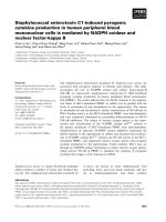
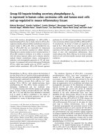
Figure 1 Dysregulated miR-302b/EGFR in hepatocarcinoma tissues and cells. A- qRT–PCR analysis of miR-302b expression in 27 paired HCC
tissues and their corresponding adjacent nontumorous livers. The expression of miR-302b was normalized to U6 snRNA. The data are reported as
mean ± s.d. (*P < 0.05, Student’s t-test). B- qRT–PCR analysis of EGFR expression in 27 paired primary HCC tissues and their corresponding adjacent
nontumorous liver tissues. The expression of EGFR was normalized to β-actin (*P < 0.05, Student’s t-test). C- qRT–PCR analysis of miR-302b
expression in normal hepatocytes (HL-7702 cells) and HCC cells (Bel7402, Hep3B, HepG2, SMMC-7721 cells), (**P < 0.01, *P < 0.05, Student’s t-test).
D- Western blot analysis of EGFR expression in four pairs of HCC tissues and their corresponding nontumorous liver tissues, with β-actin as an
internal control. The intensity for each band was quantified. The value under each lane indicates the EGFR expression in four tumor tissues
compared with their corresponding nontumorous liver tissues. E- Western blot analysis of EGFR expression in hepatocytes, and hepatoma cells,
with β-actin as an internal control. The value under each lane is represented the intensity ratio between hepatoma cells and hepatocytes.
Wang et al. BMC Cancer 2013, 13:448
/>
RegRNA, DIANA and TargetScan. As shown in
Figure 2A, there is a miR-302b-binding site at 42594284nt of the EGFR 3′ UTR. Comparing the human
sequence with interspecies homology, we found that the
miR-302b targeting sequence was highly conserved
among different species. To determine whether EGFR was
a direct target of miR-302b, we constructed pmirGLOEGFR-3′UTR-wt and pmirGLO-EGFR-3′UTR-mut. Later,
we have co-transfected miR-302b or miR-ctrl with pmir
GLO-EGFR-3′UTR-wt or pmirGLO-EGFR-3′UTR-mut
into SMMC-7721 cells. The results showed that miR-302b
obviously suppressed the firefly luciferase activity of
pmirGLO-EGFR-3′UTR-wt at 24 and 48 h, compared
with miR-ctrl (Figure 2B). In addition, we proved that the
re-expression of miR-302b did not affect the mRNA
expression of EGFR (P > 0.05), but could suppress EGFR
at the protein level (50%). Meanwhile, after transfected
miR-302b inhibitor into SMMC-7721 cells, the expression
of EGFR at mRNA levels did not change. However, transfection of miR-302b inhibitor can increase the expression
of EGFR at protein level (Figure 2C), suggesting that
Page 5 of 9
miR302b inhibit EGFR expression at translational level
but not transcription level in SMMC-7721 cells. Interestingly, as shown in Figure 2D, miR-302b expression level
in vivo was inversely-correlated with EGFR mRNA
expression level, which was verified by Pearson’s correlation coefficient test, suggesting that miR-302b may relate
to EGFR mRNA expression level. Taken together, our data
demonstrated that miR-302b targeted at EFGR and
suppressed its expression at translation level in SMMC7721 cells.
The miR-302b inhibited the growth of SMMC-7721 cells
through targeting EGFR
To examine the effects of miR-302b on the growth of
SMMC-7721 cells through targeting EGFR, we designed
the siRNA for EGFR (siEGFR), which induced 50%
decrease of EGFR expression both at the mRNA and
protein levels in SMMC-7721 cells. At the same time,
we transfected miR-302b into SMMC-7721 cells and
observed a thirty-fold increase of the miR-302b expression (Figure 3A). MTT assay showed that miR-302b
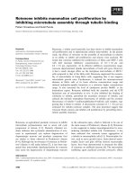
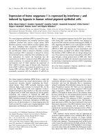
Figure 2 MiR-302b targets at EGFR. A- miR-302b-binding site at 4259–4284 nt of the EGFR 3′ UTR is predicted to be evolutionarily conserved
across different species. B- 24 and 48 h after transfection, luciferase assay in SMMC-7721 cells. 25-bp regions (wt) miR-302b binding sites in the
EGFR 3′ UTR was cloned into pmirGLO Dual-Luciferase miRNA Target Expression vector. Identical constructs mutation was generated. Either miRctrl or miR-302b was co-transfected with pmirGLO-EGFR-3′ UTR-wt or pmirGLO-EGFR-3′ UTR–mut into SMMC-7721 cells and luciferase activity
assayed after 24 h and 48 h. The normalized firefly luciferase activity was obtained by firefly luciferase activity/Renilla luciferase activity. Luciferase
activity of reporter gene (EGFR 3′ UTR-wt) displayed a significant decrease by transfecting miR-302b. These experiments were performed in
triplicate, and the results are shown as the mean ± s.d. (**P < 0.01 Student’s t-test). C- EGFR mRNA and protein expression levels measured by
qRT-PCR and western blot 48 h after transfecting with miR-ctrl, miR-302b, inhibitor-ctrl or miR-302b-inhibitor. The intensity for each band was
quantified. The value under each lane indicates the expression level of EGFR, which is represented by the intensity ratio between miR-302b or
inhibitor and miR-ctrl or inhibitor-ctrl groups. D- Inverse correlation between miR-302b and EGFR expression in HCC tissues. Expression of EGFR
analyzed by qRT–PCR and normalized to β-actin. The miR-302b expression was examined by qRT–PCR analysis and normalized to U6 expression.
Statistical analysis was performed using Pearson’s correlation coefficient (r = −0.48, *P < 0.05).
Wang et al. BMC Cancer 2013, 13:448
/>
Page 6 of 9
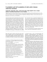
Figure 3 Overexpression of miR-302b and knockdown of miR-302b-target gene EGFR decrease hepatoma cell growth and induce G1/S
arrest in vitro. A- qRT-PCR analysis of miR-302b in SMMC-7721 cells transfected with miR-302b over-expression construct and miR-ctrl (left).
qRT–PCR (upper portion) and western blot analysis (lower portion) were performed to determine the expression level of EGFR after transfection
of siEGFR and siRNA-ctrl. (*P < 0.05, **P < 0.01, Student’s t-test) (right). B- The effects of miR-302b or siEGFR on SMMC-7721 cell proliferation were
determined by MTT assay at 24 h, 48 h, and 72 h after transfecting of miR-302b over-expression construct or siEGFR, with miR-ctrl or siRNA-ctrl as
the respective controls (*P < 0.05, **P <0.01, Student’s t-test). C- Representative results of colony formation of SMMC-7721 after transfection of
miR-302b re-expression or siEGFR. D- Cell cycles determined in SMMC-7721 cells after transfection of miR-302b over-expression construct or
siEGFR at 24 h, 48 h, 72 h, with miR-ctrl or siRNA-ctrl as the respective controls. Histogram indicates the percentage of cells at G0–G1, S, and
G2–M cell-cycle phases (*P < 0.05, Student’s t-test). E- Expression of Ki-67 was verified by immunofluorescence staining after transfecting with
miR-ctrl, miR-302b, siRNA-ctrl, or siEGFR. Merged pictures are overlays of both Ki-67 red signals and nuclear staining by DAPI (blue).
overexpression resulted in the suppression of the
SMMC-7721cells growth at 48 and 72 h, which was in
accord with the effect of siEGFR (Figure 3B). To further
examine the inhibitory role of miR-302b and siEGFR in
SMMC-7721 cells, colony formation assay was employed.
Notably, miR-302b/siEGFR–transfected cells displayed
fewer and smaller colonies compared with their respective
controls (Figure 3C). Moreover, miR-302b and siEGFR
suppressed cell proliferation at the G0/G1 phase at 24 h,
48 h and 72 h time points (Figure 3D). Finally, to determine the growth fraction of HCC cells after overexpression of miR-302b/siEGFR, we performed Ki-67
immunofluorescence staining. The signal of Ki-67 in the
miR-302b/siEGFR–transfected SMMC-7721 cells was
visibly low compared with that of the cells transfected
with their respective controls (Figure 3E). These findings
demonstrated that the effect of miR-302b re-expression
on cell proliferation was consistent with that of siEGFR
on SMMC-7721 cells, suggesting that miR-302b may
inhibit the growth of SMMC-7721 cells through targeting EGFR.
MiR-302b inhibits cell proliferation through EGFR-dependent
cell cycle regulation
AKT is the key molecule in the signaling pathway, which
is regulated by EGFR. Abnormal expression of EGFR leads
to a change of AKT expression [19,20]. The re-expression
of miR-302b reduced the expression of AKT2, pAKT2,
and its downstream gene CCND1, CDK2, and up-regulated CDK inhibitor p27 in SMMC-7721 cells (Figure 4A).
Similar results were proved by the treatment of siEGFR
(Figure 4B), suggesting that miR-302b may suppress the
growth of SMMC-7721 cells by targeting the EGFR/
AKT2/CCND1 signaling pathway.
Discussion
HCC is a primary lethal neoplasm of the liver and the
third cause of cancer-related deaths worldwide [21].
However, its underlying molecular mechanism remains
largely unknown. In the past ten years, microRNAs
(miRNAs) have been found to be involved in the initiation and progression of HCC. According to its tumorigenesis function, miRNAs can be divided in two classes:
Wang et al. BMC Cancer 2013, 13:448
/>
Page 7 of 9
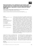
Figure 4 MiR-302b inhibits cell proliferation through EGFR-dependent cell cycle regulation. A- Western blot results of miR-302b-targeted
EGFR and non-direct targeted cell cycle regulation proteins in SMMC-7721 cells after transfection of miR-302b over-expression construct and miRctrl. β-actin was used as an internal control. B- Protein level of EGFR and relative cell cycle regulation proteins were measured by western blot
48 h after transfection of siEGFR and siRNA-ctrl into SMMC-7721 cells. The expression of the proliferation-related gene was normalized to β-actin.
The intensity for each band was quantified. The value under each lane indicates the relative expression level of the proliferation-related gene,
which is represented by the intensity ratio between miR-302b and miR-ctrl or inhibitor and inhibitor-ctrl groups.
oncogenes and tumor suppressor genes [22]. Many
oncogenic miRNAs, such as miR-221 and miR-222, are
involved in sustaining proliferative signaling, resisting
growth suppression and apoptosis, enabling immortality,
prompting angiogenesis, invasion and metastasis, evading and so on [23-28], whereas tumor suppressor
miRNAs are involved in the reverse processes. Let-7
family and miR-101, as potential tumor suppressors,
were markedly decreased in HCC cells [29,30]. Recent
studies proved that the miR-302-367 cluster is downregulated in cervical cancer cells and gastric adenocarcinoma [31,32]. Our study showed that the expression of
the miR-302b was frequently down-regulated in clinical
HCC tissues and in SMMC-7721 cells (Figure 1). Thus,
we supposed that miR-302b might be a novel tumorsuppressor miRNA.
Human epidermal growth factor receptor (EGFR/HER/
ErbB) family of tyrosine kinases plays a major role in the
etiology and progression of many carcinomas, including
HCC. Increased expression of EGFR/HER1 occurs frequently in different human tumor types, and is involved in
the early stages of human hepatocarcinogenesis [33,34]. In
our study, increased expression of EGFR was observed in
the HCC samples and HCC cells (Figure 1D and E). Overexpression of EGFR is also related to the gene amplification of EGFR and deficiency of EGFR targeting miRNA.
There seemed to be a negative correlation between the
expression of EGFR and that of miR-302b in HCC tissues
(Figure 1A and B), implying that EGFR might be a novel
target of miR-302b. Further bio-information analysis
showed that there was a miR-302b-binding site at 4259–
4284 nt of the EGFR 3′ UTR. The dual-luciferase reporter
assays demonstrated that miR-302b targeted directly to
EGFR through the suppression of translation (Figure 2B).
In this research, we examine the relationship between
miR-302b and EGFR at both of the transcription level and
translational level, in which miR-302b was verified to
silence EGFR at translational level from in vitro and
in vivo clinical samples. At the transcription level, we
tested relationship between miR-302b and EGFR by using
Pearson’s correlation coefficient test in 27 paired HCC
tissues and found that they have inverse correlation in
mRNA level (Figure 2D). Whereas in SMMC-7721 cell
lines, the correlation between miR-302b and EGFR didn’t
show significant difference (Figure 2C), but it exhibited
the correlation trend, which were consistent with the
results of that in HCC tissues.
EGFR induces activation of the Ras/Raf/MEK/MAPK
pathway through either Grb2 or Shc adaptor proteins, and
that of PI3K/AKT/CCND1 pathway by recruitment of the
p85 regulatory subunit to the activated receptors [35]. The
activation of EGFR/AKT/NF-kB/CCND1 survival signaling pathway has been certified in cholesteatoma epithelium [36]. Function of dominant negative EGFR shows
Wang et al. BMC Cancer 2013, 13:448
/>
that dominant negative EGFR induces G0/G1 arrest by decreasing the expression of phosphorylated retinoblastoma
protein, phosphorylated GSK-3β, CCND1, and by increasing expression of p21 and p27 in human gastric cancer
cells SGC-7901 and NCI-N87 [37]. AKT2 is essential for
progressing from the G0/G1 to the S-phase by activating
the positive regulator of G1/S transition, including
CCND1, CCND2, and CCNE1, during cell cycle progression [38]. CCND1, as a AKT2 downstream gene, is
expressed in the G1 phase of the cell cycle, together with
its CDK partner, CDK2. p27, as a CDK inhibitor, could be
combined with CCND1-CDK2 complex to restrain CDK2
activity [39]. Our results showed that miR-302b may inhibit the growth of SMMC-7721 cells through targeting
EGFR, and that the cell cycle progression was arrested at
the G0/G1-phase (Figure 3). At the same time, the expression of AKT2 was down-regulated, and CCND1 and
CDK2 were reduced by miR-302b, while the expression of
CDK inhibitor p27 was up-regulated (Figure 4). A few of
the miR-302b targets have been found, including AKT1,
CCNA, CDK2, CCND1/D2, and BMI-1 [40]. These genes
are involved in the regulation of the cell cycle. In order to
prove the biological effects of miR-302b on inhibition of
EGFR, siEGFR was used. The results showed that the
effect of miR-302b re-expression on the cell proliferation
was consistent with that of siEGFR in SMMC-7721cells,
suggesting that miR-302b may suppress the growth of
SMMC-7721 cells by targeting the EGFR/AKT2/CCND1
signaling pathway.
Conclusions
In conclusion, the dysregulation of miR-302b is a frequent
event in human hepatocarcinoma. The high-expression of
EGFR is related to the down-regulation of miR-302b in
HCC. The re-expression of miR-302b suppresses the
growth of hepatoma cells may due to targeting the EGFR/
AKT2/CCND1 pathway, suggesting that miR-302b may
be an effector in gene therapy of HCC.
Competing interests
The authors declare that they have no competing interests.
Authors’ contributions
CH and JYY developed the hypotheses. LMW and LLH executed the
experiments. JYY, TSS and ZFL provided the experimental facilities and
research funds. LMW, JYY, XS, and CH wrote and revised the paper.
All authors read and approved the final manuscript.
Acknowledgments
This work was funded by The Key Science and Technology Major
Program of Shaanxi Province, China (2010ZDKG-50); The National Natural
Science Foundation of China (31100921); The Fundamental Research
Funds for the Central Universities (08142006); The National Natural
Science Foundation of China (81171398); and The Program for
Changjiang Scholars and Innovative Research Team in University
(PCSIRT: 1171).
Page 8 of 9
Author details
1
Department of Genetics and Molecular Biology, Xi’an Jiaotong University
Health Science Center, No.76 West Yanta Road, Xi’an, Shaanxi 710061, P.R.
China. 2Xi’an IV People’s Hospital, Xi’an, Shaanxi, P.R. China. 3Engineering
Research Center of Biotherapy and Translational Medicine of Shaanxi
Province, Xi’an, Shaanxi, P.R. China. 4Key Laboratory of Environment and
Genes Related to Diseases, Xi’an Jiaotong University Health Science Center,
Xi’an, Shaanxi, China. 5Cardiovascular Research Center, Xi’an Jiaotong
University Health Science Center, Xi’an, Shaanxi, P.R. China.
Received: 26 April 2013 Accepted: 26 September 2013
Published: 2 October 2013
References
1. El-Serag HB, Rudolph KL: Hepatocellular carcinoma: epidemiology and
molecular carcinogenesis. Gastroenterology 2007, 132:2557–2576.
2. Singal AG, Marrero JA: Recent advances in the treatment of
hepatocellular carcinoma. Curr Opin Gastroenterol 2010, 26:189–195.
3. Murray CJ, Lopez AD: Mortality by cause for eight regions of the world:
Global Burden of Disease Study. Lancet 1997, 349:1269–1276.
4. Schafer DF, Sorrell MF: Hepatocellular carcinoma. Lancet 1999,
353:1253–1257.
5. Makuuchi M, Takayama T, Kubota K, Kimura W, Midorikawa Y, Miyagawa S,
Kawasaki S: Hepatic resection for hepatocellular carcinoma - Japanese
experience. Hepatogastroenterology 1998, 45:1267–1274.
6. Fan ST, Lo CM, Liu CL, Lam CM, Yuen WK, Yeung C, Wong J: Hepatectomy
for hepatocellular carcinoma: toward zero hospital deaths. Ann Surg
1999, 229:322–330.
7. Faloppi L, Scartozzi M, Maccaroni E, Di Pietro Paolo M, Berardi R, Del Prete
M, Cascinu S: Evolving strategies for the treatment of hepatocellular
carcinoma: from clinical-guided to molecularly-tailored therapeutic
options. Cancer Treat Rev 2011, 37:169–177.
8. Wiemer EA: The role of microRNAs in cancer: no small matter. Eur J
Cancer 2007, 43:1529–1544.
9. Wienholds E, Plasterk RH: MicroRNA function in animal development.
FEBS Lett 2005, 579:5911–5922.
10. Liu W, Mao SY, Zhu WY: Impact of tiny miRNAs on cancers. World J
Gastroenterol 2007, 13:497–502.
11. Oakley EJ, Van Zant G: Unraveling the complex regulation of stem cells:
implications for aging and cancer. Leukemia 2007, 21:612–621.
12. Zamore PD, Haley B: Ribo-gnome: the big world of small RNAs.
Science 2005, 309:1519–1524.
13. Papagiannakopoulos T, Shapiro A, Kosik KS: MicroRNA-21 targets a
network of key tumor-suppressive pathways in glioblastoma cells.
Cancer Res 2008, 68:8164–8172.
14. Sachdeva M, Mo YY: MicroRNA-145 suppresses cell invasion and
metastasis by directly targeting mucin 1. Cancer Res 2010, 70:378–387.
15. Zhang S, Shan C, Kong G, Du Y, Ye L, Zhang X: MicroRNA-520e suppresses
growth of hepatoma cells by targeting the NF-kappaB-inducing kinase
(NIK). Oncogene 2012, 31:3607–3620.
16. Lin SL, Chang DC, Chang-Lin S, Lin CH, Wu DT, Chen DT, Ying SY: Mir-302
reprograms human skin cancer cells into a pluripotent ES-cell-like state.
RNA 2008, 14:2115–2124.
17. Barroso-delJesus A, Romero-Lopez C, Lucena-Aguilar G, Melen GJ, Sanchez
L, Ligero G, Berzal-Herranz A, Menendez P: Embryonic stem cell-specific
miR302-367 cluster: human gene structure and functional
characterization of its core promoter. Mol Cell Biol 2008, 28:6609–6619.
18. Card DA, Hebbar PB, Li L, Trotter KW, Komatsu Y, Mishina Y, Archer TK:
Oct4/Sox2-regulated miR-302 targets cyclin D1 in human embryonic
stem cells. Mol Cell Biol 2008, 28:6426–6438.
19. Hsu CH, Gao M, Chen CL, Yeh PY, Cheng AL: Inhibitors of epidermoid
growth factor receptor suppress cell growth and enhance
chemosensitivity of nasopharyngeal cancer cells in vitro. Oncology 2005,
68:538–547.
20. Sebastian S, Settleman J, Reshkin SJ, Azzariti A, Bellizzi A, Paradiso A: The
complexity of targeting EGFR signalling in cancer: from expression to
turnover. Biochimi Biophys Acta 2006, 1766:120–139.
21. El-Serag HB: Hepatocellular carcinoma. N Engl J Med 2011, 365:1118–1127.
22. Zhang B, Pan X, Cobb GP, et al: microRNAs as oncogenes and tumor
suppressors. Dev Biol 2007, 302:1–12.
Wang et al. BMC Cancer 2013, 13:448
/>
Page 9 of 9
23. Hanahan D, Weinberg RA: Hallmarks of cancer: the next generation.
Cell 2011, 144:646–674.
24. Lee SK, Calin GA: Non-coding RNAs and cancer: new paradigms in
oncology. Discov Med 2011, 11:245–254.
25. Negrini M, Nicoloso MS, Calin GA: MicroRNAs and cancer–new paradigms
in molecular oncology. Curr Opin Cell Biol 2009, 21:470–479.
26. Ruan K, Fang X, Ouyang G: MicroRNAs: novel regulators in the hallmarks
of human cancer. Cancer Lett 2009, 285:116–126.
27. Ross SA, Davis CD: MicroRNA, nutrition, and cancer prevention.
Adv Nutr 2011, 2:472–485.
28. Li Y, Kong D, Wang Z, Sarkar FH: Regulation of microRNAs by natural
agents: an emerging field in chemoprevention and chemotherapy
research. Pharml Res 2010, 27:1027–1041.
29. Zhu XM, Wu LJ, Xu J, Yang R, Wu FS: Let-7c microRNA expression and
clinical significance in hepatocellular carcinoma. J Int Med Res 2011,
39:2323–2329.
30. Xu Y, An Y, Wang Y, Zhang C, Zhang H, Huang C, Jiang H, Wang X, Li X:
miR-101 inhibits autophagy and enhances cisplatin-induced apoptosis in
hepatocellular carcinoma cells. Oncol Rep 2013, 29:2019–2024.
31. Cai N, Wang YD, Zheng PS: The microRNA-302-367 cluster suppresses the
proliferation of cervical carcinoma cells through the novel target AKT1.
RNA 2013, 19:85–95.
32. Khalili M, Sadeghizadeh M, Ghorbanian K, Malekzadeh R, Vasei M, Mowla SJ:
Down-regulation of miR-302b, an ESC-specific microRNA, in Gastric
Adenocarcinoma. Cell J 2012, 13:251–258.
33. Prakash I, Mathur RP, Kar P, Ranga S, Talib VH: Comparative evaluation of
cell proliferative indices and epidermal growth factor receptor
expression in gastric carcinoma. Indian J Pathol Microbiol 1997,
40:481–490.
34. Moghbeli M, Abbaszadegan MR, Farshchian M, Montazer M, Raeisossadati R,
Abdollahi A, Forghanifard MM: Association of PYGO2 and EGFR in
esophageal squamous cell carcinoma. Med Oncol 2013, 30:516.
35. Soltoff SP, Carraway KL 3rd, Prigent SA, Gullick WG, Cantley LC: ErbB3 is
involved in activation of phosphatidylinositol 3-kinase by epidermal
growth factor. Mol Cell Biol 1994, 14:3550–3558.
36. Liu W, Yin T, Ren J, Li L, Xiao Z, Chen X, Xie D: Activation of the EGFR/Akt/
NF-kappaB/cyclinD1 survival signaling pathway in human cholesteatoma
epithelium. Eur Arch Otorhinolaryngol 2013, 13:2403–2406.
37. Liao G, Wang Z, Zhang N, Dong P: Dominant Negative Epidermal Growth
Factor Receptor Inhibits Growth of Human Gastric Cancer Cells by
Inducing Cell Cycle Arrest and Apoptosis. Cancer Biother Radiopharm 2013,
28:450–480.
38. Testa JR, Bellacosa A: AKT plays a central role in tumorigenesis.
Proc Natl Acad Sci USA 2001, 98:10983–10985.
39. Shi C, Yu L, Yang F, Yan J, Zeng H: A novel organoselenium compound
induces cell cycle arrest and apoptosis in prostate cancer cell lines.
Biochem Biophys Res Commun 2003, 309:578–583.
40. Lin SL, Chang DC, Ying SY, Leu D, Wu DT: MicroRNA miR-302 inhibits the
tumorigenecity of human pluripotent stem cells by coordinate
suppression of the CDK2 and CDK4/6 cell cycle pathways. Cancer Res
2010, 70:9473–9482.
doi:10.1186/1471-2407-13-448
Cite this article as: Wang et al.: MicroRNA-302b suppresses cell
proliferation by targeting EGFR in human hepatocellular carcinoma
SMMC-7721 cells. BMC Cancer 2013 13:448.
Submit your next manuscript to BioMed Central
and take full advantage of:
• Convenient online submission
• Thorough peer review
• No space constraints or color figure charges
• Immediate publication on acceptance
• Inclusion in PubMed, CAS, Scopus and Google Scholar
• Research which is freely available for redistribution
Submit your manuscript at
www.biomedcentral.com/submit