Clinical significance of L-type amino acid transporter 1 expression as a prognostic marker and potential of new targeting therapy in biliary tract cancer
Bạn đang xem bản rút gọn của tài liệu. Xem và tải ngay bản đầy đủ của tài liệu tại đây (1.25 MB, 12 trang )
Kaira et al. BMC Cancer 2013, 13:482
/>
RESEARCH ARTICLE
Open Access
Clinical significance of L-type amino acid
transporter 1 expression as a prognostic marker
and potential of new targeting therapy in biliary
tract cancer
Kyoichi Kaira1,11,12*, Yutaka Sunose2†, Yasuhiro Ohshima3*†, Noriko S Ishioka3, Kazuhisa Arakawa4, Tetsushi Ogawa4,
Noriaki Sunaga1, Kimihiro Shimizu2, Hideyuki Tominaga5, Noboru Oriuchi6,7, Hideaki Itoh8, Shushi Nagamori9,
Yoshikatsu Kanai9, Aiko Yamaguchi10, Atsuki Segawa11, Munenori Ide11, Masatomo Mori1, Tetsunari Oyama11
and Izumi Takeyoshi2
Abstract
Background: The expression of L-type amino acid transporter 1 (LAT1) has been described to play essential roles in
tumor cell growth and survival. However, it remains unclear about the clinicopathological significance of LAT1
expression in biliary tract cancer. This study was conducted to determine biological significance of LAT1 expression
and investigate whether LAT1 could be a prognostic biomarker for biliary tract cancer.
Methods: A total of 139 consecutive patients with resected pathologic stage I-IV biliary tract adenocarcinoma were
retrospectively reviewed. Tumor specimens were stained by immunohistochemistry for LAT1, Ki-67, microvessel density
determined by CD34, and p53; and prognosis of patients was correlated. Biological significance of LAT1 expression was
investigated by in vitro and in vivo experiments with LAT inhibitor, 2-aminobicyclo-(2,2,1)-heptane-2-carboxylic acid
(BCH) using cholangiocarcinoma cell line.
Results: In total patients, high LAT1 expressions were recognized in 64.0%. The expression of LAT1 was closely
correlated with lymphatic metastases, cell proliferation and angiogenesis, and was a significant indicator for predicting
poor outcome after surgery. LAT1 expression was a significant independent predictor by multivariate analysis. Both
in vitro and in vivo preliminary experiments indicated that BCH significantly suppressed growth of the tumor and
yielded an additive therapeutic efficacy to gemcitabine and 5-FU.
Conclusions: High expression of LAT1 is a promising pathological marker to predict the outcome in patients with
biliary tract adenocarcinoma. Inhibition of LAT1 may be an effective targeted therapy for this distressing disease.
Keywords: LAT1, Biliary tract cancer, Amino acid transporter, Prognostic factor, BCH
* Correspondence: ;
†
Equal contributors
1
Department of Medicine and Molecular Science, Gunma University Graduate
School of Medicine, Showa-machi, Maebashi, Gunma, Japan
3
Medical Radioisotope Application Group, Quantum Beam Science Directorate,
Japan Atomic Energy Agency, Watanuki, 370-1292 Takasaki, Gunma, Japan
Full list of author information is available at the end of the article
© 2013 Kaira et al.; licensee BioMed Central Ltd. This is an open access article distributed under the terms of the Creative
Commons Attribution License ( which permits unrestricted use, distribution, and
reproduction in any medium, provided the original work is properly cited.
Kaira et al. BMC Cancer 2013, 13:482
/>
Background
Biliary tract cancer is a relatively uncommon malignant
neoplasm and is one of the aggressive malignancy with
poor prognosis [1]. Gallbladder carcinoma and extrahepatic
bile ducts carcinoma (cholangiocarcinoma) are the most
common biliary tract cancer and cholangiocarcinoma is
classified into intrahepatic and extrahepatic disease
according to its anatomical location within the biliary tree
[2]. Surgical resection remains the only potentially curative therapeutic option, however, more than half of
patients present with unresectable disease. Even if curative
resection can be performed, the 5-year overall survival is
20-32% for intrahepatic cholangiocarcinoma, 30-42% for
hilar cholangiocarcinoma, and 18-54% for distal cholangiocarcinoma [3-5]. Although many patients may receive adjuvant chemotherapy to improve chance of cure,
there is no established standard chemotherapy. In advanced biliary tract cancer, combination chemotherapy
with gemcitabine and a platinum-based agent is regarded
as a standard treatment, however, the prognosis after
treatment remains dismal [6]. To date, the patients with
biliary tract cancer lack a survival benefit if treated with
chemotherapy or radiation therapy. Thus, we need a new
effective therapy to improve the survival of patients. To
improve the outcome of therapy, therefore, clinical
markers that can predict response to the specific therapy
and the prognosis should be established.
Amino acid transporters are essential for growth and
proliferation of normal cells as well as transformed cells
[7,8]. L-type amino acid transporter 1 (LAT1) is one of
the L-type amino acid transporters, and transports large
neutral amino acids such as leucine, isoleucine, valine,
phenylalanine, tyrosine, tryptophan, methionine and histidine [9,10]. LAT1 requires covalent association with
the heavy chain of 4F2 cell surface antigen (CD98) for
its functional expression in plasma membrane [9]. LAT1
has been closely associated with cancerous or proliferative cells, and previous studies have shown LAT1 to be
highly expressed in proliferating tissues, many tumor cell
lines and primary human tumors [10-17]. In human
tumor tissues, LAT1 expression has a close relationship
with cell proliferation, angiogenesis and cell cycle regulator [18,19]. Recently, the expression of LAT1 has been
described to be a significant factor indicating a poor outcome in various human cancers [12-17]. Moreover, the
potential of targeting therapy for LAT1 had been suggested in tumor cell lines by the inhibition of LAT1
using 2-aminobicyclo-(2,2,1)-heptane-2-carboxylic acid
(BCH) [20,21]. However, it remains unknown whether
LAT1 expression has a clinical and pathological significance in patients with biliary tract cancer.
In the present study, we examined LAT1 expression in
the resected tissue specimens to evaluate the clinicopathological and prognostic significance of LAT1 in
Page 2 of 12
patients with biliary tract cancer. LAT1 expression was
correlated with pathological biomarkers such as cellular
proliferation, cell cycle regulator (p53) and angiogenesis.
In addition, in vitro and in vivo animal studies were
performed to investigate the potential of LAT1 as a
therapeutic biomarker in a novel targeting therapy.
Methods
Patients
We analyzed 157 consecutive patients with biliary tract
adenocarcinoma who underwent surgical resection at
Gunma University Hospital and Maebashi Red Cross
Hospital between September 2000 and October 2011.
Ten patients who received induction chemotherapy or
radiation therapy were excluded. In all cases, magnetic
resonance cholangiopancreatography (MRCP) and endoscopic retrograde cholangiopancreatography (ERCP)
were performed before surgical resection, and pancreatic
ductal adenocarcinoma and ampullary carcinoma were
excluded from the study. The specimens from eight patients were not available. All surgical specimens were
reviewed and classified according to the WHO classification by an experienced pathologist who was unaware of
clinical or imaging findings. Patients with pathological
diagnosis other than adenocarcinoma were excluded. In
total, 139 patients were analyzed in the study. The study
population consisted of patients with extrahepatic
cholangiocarcinoma (EHCC), intrahepatic cholangiocarcinoma (IHCC) and gallbladder carcinoma (GB).
Pathologic tumor-node-metastasis (TNM) stages were
established using the International System for Staging
bile duct cancer adopted by the American Joint Committee on Cancer and the Union Internationale Centre le
Cancer [22].
We also analyzed a control group of 16 patients with
surgically resected benign biliary tract lesions. Immunohistochemical staining of samples from these 16 patients was
performed and compared with that of biliary tract cancer.
The pathological diagnosis of the control group was as
follows: 6 patients with cholesterol polyp, 4 patients with
hyperplastic polyp, 3 patients with xanthogranulomatous
chlecystitis and 3 patients with adenomyomatosis. This
study was approved by the institutional review board of
Gunma University Hospital (ethical committee for clinical
studies-Gunma University faculty of Medicine) and written
informed consent was obtained from all of the patients or
their families who participated to this study.
Immunohistochemical staining
LAT1 expression was determined by immunohistochemical staining with LAT1 antibody (2 mg/mL, anti-human
monoclonal mouse antibody, 4A2, provided by Dr H.
Endou [J-Pharma, Tokyo, Japan], dilution; 1:3200). The
production and characterization of the LAT1 antibody
Kaira et al. BMC Cancer 2013, 13:482
/>
has previously been described [15]. The detailed protocol for immunostaining was published elsewhere [16].
The LAT1 expression score was assessed by the extent
of staining as follows: 1, ≤ 10% of tumor area stained; 2,
11-25% stained; 3, 26-50% stained; and 4, ≥51% stained.
The tumors in which stained tumor cells were scored as
3 or 4 were defined as high expression.
For CD34, Ki-67 and p53, immunohistochemical staining was performed according to the procedures described
in previous reports [23,24]. The following antibodies
were used: mouse monoclonal antibodies against CD34
(Nichirei, Tokyo, Japan, 1:800 dilution), Ki-67 (Dako,
Glostrup, Denmark, 1:40 dilution), and p53 (D07; Dako,
1:50 dilution). The number of CD34-positive vessels was
counted in four selected hot spots in a x 400 field
(0.26 mm2 field area). Microvessel density (MVD) was defined as the mean count of microvessels per 0.26 mm2
field area. The median number of CD34-positive vessels
was evaluated, and the tumors in which stained tumor
cells made up more than each median value were defined
as high expression. For p53, microscopic examination for
the nuclear reaction product was performed and scored,
and p53 expression in greater than 10% of tumor cells
was defined as positive expression [24]. For, Ki-67, a
highly cellular area of the immunostained sections was
evaluated. All epithelial cells with nuclear staining of any
intensity were defined as high expression. Approximately
1000 nuclei were counted on each slide. Proliferative activity was assessed as the percentage of Ki-67-stained nuclei (Ki-67 labeling index) in the sample. The median
value of the Ki-67 labeling index was evaluated, and the
tumor cells with greater than the median value were defined as high expression. The sections were assessed using
a light microscopy in a blinded fashion by at least two of
the authors.
Biochemical materials
Dulbecco’s modified Eagle’s medium (DMEM), penicillin
and streptomycin were purchased from WAKO Pure
Chemical Industries (Osaka, Japan). BCH was obtained
from NARD Institute (Hyogo, Japan). 3-[4,5-dimethyl-2thiazolyl]-2,5-diphenyl-2H-tetrazolium bromide (MTT)
were purchased from Dojindo Laboratories (Kumamoto,
Japan). All other chemicals used were of the highest purity available.
Cell culture
A human cholangiocarcinoma cell lines, HuCCT1
(JCRB0425), OZ (JCRB1032), and HuH28 (JCRB0426)
were purchased from the Health Science Research Resources Bank (Osaka, Japan) [25-27], and routinely
maintained in DMEM containing 10% heat-inactivated
fetal bovine serum (AusGeneX, Loganholme, QLD,
Australia), penicillin (100 units/ml), streptomycin
Page 3 of 12
(100 μg/ml) and L-glutamine (2 mM) at 37°C in 5%
CO2, 95% air.
Expression of LAT mRNA in cholangiocarcinoma
Previously, 4 subtypes of L-type amino acid transporter
(LAT1-4) have been identified [8,23-30]. Realtime RTPCR analysis was performed to determine the expression
of LAT1, LAT2, LAT3, and LAT4 mRNA in cholangiocarcinoma cell line. Total RNA was isolated from
HuCCT1 cells using a Fast Pure RNA kit (Takara Bio,
Shiga, Japan). The first-strand complement DNA was synthesized from 0.5 μg of total RNA with PrimeScript
Reverse Transcriptase (Takara Bio). The sequences of specific primers were shown in Additional file 1: Table S1
(online only). The realtime PCR analysis was performed
by first incubating each complement DNA sample with
the primers (0.5 μM each) and Thunderbird SYBR qPCR
Mix (Toyobo, Osaka, Japan). Amplification was carried
out for 40 cycles (95°C for 15 s, 60°C for 30 s) with PikoReal thermal cycler (Thermo Fisher Scientific, Waltham,
MA). The data was analyzed according to 2-ΔΔC
method
T
(internal control: β-actin, calibrator: LAT1).
Suppression of cell proliferation with LAT1 inhibition
Cells were plated at a concentration of 1 x 10³ cells/well
in 96-well plates and incubated in the growth medium
for 24 h. At first, in order to determine the effect of
LAT1 inhibition on cholangiocarcinoma, HuCCT1 cells
were treated with BCH (0.1, 1, 2, 3, 5, 10, 20, 30, or
100 mM) and incubated for 6 days. Next, the effect of
LAT1 inhibition on the antitumor activity of gemcitabine (GEM, Eli Lilly, Indianapolis, IN) or 5fluorouracil (5-FU, Kyowa Hakko Kirin, Shizuoka, Japan)
was evaluated. Cells were incubated for 6 days with
GEM (10, 20, 50 or 100 nM) or 5-FU (1, 10, or 100 μM)
in a presence or absence of 10 mM BCH. Then, cells
were incubated with 0.5 mg/ml MTT for 4 h at 37°C.
The resulting formazan was solubilized, and the absorbance was read at 590 nm with a microtiter plate reader
(Vmax; Molecular Devices, Sunnyvale, CA).
Suppression of amino acid uptake into cells with LAT1
inhibition
Inhibition of amino acid transport by BCH was examined
using [14C]L-leucine (Perkin-Elmer Life Sciences, Boston,
MA), one of the substrates of LATs [31]. HuCCT1 cells
(1.0 x 10⁵ cells/well) were plated in the 24-well plates and
incubated in the growth medium for 24 h. After the incubation, the cells were washed three times with sodiumfree Hunk’s balanced salt solution (Na+-free HBSS;
137 mM choline chloride, 5.3 mM KC1, 1.3 mM CaCl2,
0.49 mM MgCl2, 0.41 mM MgSO4, 0.35 mM K2HPO4,
0.44 mM KH2PO4, 4.2 mM KHCO3, 5.6 mM D-glucose
(pH 7.4)). The cells were incubated in Na+-free HBSS
Kaira et al. BMC Cancer 2013, 13:482
/>
containing various concentration of BCH (0.01, 0.03, 0.1,
0.3, 1, or 3 mM) for 10 min at 37°C, and then, the supernatant was replaced by Na+-free HBSS containing 1 μM
[14C]L-leucine and BCH with the same concentration
(0.01, 0.03, 0.1, 0.3, 1, or 3 mM). At 1 min after treatment
with [14C]L-leucine, uptake was terminated by removing
the uptake solution followed by washing three times with
ice-cold Na+-free HBSS. Cells were solubilized with 0.1 N
NaOH, and radioactivity was measured by liquid scintillation spectrometry (AccuFLEX LSC-7200, Hitachi Aloka
Medical, Tokyo, Japan).
Immunoblotting
Cells were dissolved in sample buffer (25% glycerin,
1% SDS, 62.5 mM Tris-Cl, 10 mM dithiothreitol) and
incubated at 65°C (LAT1) or 95°C (CD98 and β-actin) for
15 min. Aliquots of samples containing 40 μg of protein
were analyzed by 10% SDS-polyacrylamide gel electrophoresis and transferred onto a polyvinylidene difluoride
membrane. Blots were incubated at 4°C overnight in
10 mM Tris–HCl, 100 mM NaCl, 0.1% Tween 20, pH 7.5
(TBST), with 5% skim milk and then with rabbit antiLAT1 C-terminus antibody (1:5,000) [32], rabbit antiCD98 antibody (1:200; Santa Cruz Biotechnology) or
rabbit anti-actin antibody (1:1,000; Cell Signaling Technology, Beverly, MA) at 4°C overnight. After having been
washed with TBST, the blots were incubated with goat
horseradish peroxidase conjugated anti-rabbit IgG antibody (1:20,000; Cell Signaling Technology) for 1.5 h at
room temperature. The blots were further washed with
TBST, and specific proteins were visualized by using enhanced chemiluminescence western blotting detection reagents (GE Healthcare, Piscataway, NJ).
Anti-tumor effect of LAT1 inhibition
Five-week-old male BALB⁄ c nude mice were purchased
from CLEA Japan (Tokyo, Japan). The animals were
cared for and treated in accordance with the guidelines
of the animal care and experimentation committee at
our facility. HuCCT1 cells (1 x 10⁷ cells) were inoculated
s.c. into the flank of the mice. After inoculation, the longer and shorter diameters of the tumor were measured
with caliper and tumor volume was calculated by the
following formula: Tumor volume (mm3) = longer diameter x (shorter diameter)2 / 2. After tumor volumes had
reached approximately 100 mm3, the mice were divided
into control group and treatment group (n = 10). Saline
or BCH (200 mg/kg) was intravenously administered
once daily from the day of grouping (day 0) for 14 days.
Tumor volume and body weight were measured two or
three times a week for 42 days. No animals were excluded and no animals died due to toxicity.
To evaluate the effect of BCH on the tumor glucose
metabolism, positron emission tomography (PET)
Page 4 of 12
imaging of tumor-bearing mice was performed with [18F]
fluoro-2-deoxyglucose (18F-FDG) using an animal PET
scanner (Inveon, Siemens, Knoxville, TN). 18F was produced using a cyclotron (CYPRIS HM-18, Sumitomo
Heavy Industries, Tokyo, Japan) and 18F-FDG was synthesized in our facility. Mice for PET imaging were randomly
selected from treatment group and control. Before imaging, mice were fasted for 8 h and had free access to
water. 18F-FDG (10 MBq) was administered intravenously
into mice followed by 10 min data acquisition at 2 h after
the administration. Mice were maintained under isoflurane anesthesia during the administration, uptake
period and PET scan. For analysis of the image, region of
interest (ROI) was drawn around the edge of the tumor
activity using ASIPro VM (CTI Concorde Microsystems,
Knoxville, TN). The maximum and median activities were
recorded. Standardized uptake value (SUV) was used to
evaluate glucose metabolism of the tumor. SUV was calculated as follows: SUV = ROI activity (kBq/ml) / injected
dose (MBq) x body weight (kg). SUV max and SUV 50%
were compared between BCH-treated mice and control
mice.
Statistical analysis
Probability values of <0.05 indicated a statistically significant difference. Results are expressed as mean ± SEM.
The significance of difference was determined by Student’s
t-test. The correlation between different variables was analyzed using the nonparametric Spearman’s rank test. The
Kaplan-Meier method was used to estimate survival as a
function of time, and survival differences were analyzed
by the log-rank test. Overall survival (OS) was determined
as the time from tumor resection to death from any cause.
Progression-free survival (PFS) was defined as the time
between tumor resection and the first disease progression
or death. Multivariate analyses were performed using
stepwise Cox proportional hazards model to identify independent prognostic factors. Statistical analysis was performed using GraphPad Prism 4 software (Graph Pad
Software, San Diego, CA, USA) and JMP 8 (SAS, Institute
Inc., Cary, NC, USA) for Windows.
Results
Patient’s demographics
One hundred thirty-nine patients with biliary tract adenocarcinoma were analyzed (EHCC, n = 89; GB, n = 30; and
IHCC, n = 20). Clinicopathologic results stratified by
tumor location are listed in Table 1. The age of the patients ranged from 42 to 86 years, and the median age
was 71 years. Most tumors (n = 126, 90.6%) were pathological stages I to III. Fifty-one patients had received
postoperative adjuvant chemotherapy with GEM, S-1
(Taiho Pharmaceutical Co., Ltd, Tokyo, Japan) or oral
administration of tegafur (a fluorouracil derivative
Kaira et al. BMC Cancer 2013, 13:482
/>
Page 5 of 12
Table 1 Patient’s characteristics and pathological findings
Characteristic
All patients (n = 139)
No. of patients
Median age (years)
%
EHCC (n = 89)
No. of patients
71
GB (n = 30)
%
No. of patients
71
IHCC (n = 20)
%
No. of patients
74
Control (n = 16)
%
No. of patients
64
%
62
Male
86
61.8 65
73.0 8
26.7 13
65.0
NA
R0 resection
67
48.2 38
42.7 19
47.4 10
50.0
NA
Poorly differentiated
33
23.7 18
20.2 3
10.0 12
60.0
NA
Lymphatic permeation
111
79.8 74
83.1 23
76.7 14
70.0
NA
Vascular invasion
93
66.9 66
74.1 16
53.3 12
60.0
NA
Lymph node metastases 62
44.6 41
46.1 11
36.7 10
50.0
NA
40.0 5
25.0
NA
UICC p-stage
1
40
28.7 23
25.8 12
2
70
50.4 49
55.1 12
40.0 9
45.0
3
16
11.5 8
9.0
16.7 3
15.0
4
13
9.4
10.1 1
3.3
3
15.0
32
23.0 13
14.6 18
60.0 1
5.0
NA
Adjuvant chemotherapy 51
36.7 34
38.2 8
26.7 9
45.0
NA
CEA (high)
65
46.7 46
51.7 12
40.0 7
35.0
NA
CA19-9 (high)
66
47.5 44
49.4 12
40.0 10
50.0
LAT1 (high)
89
64.0 59
66.3 18
60.0 12
60.0
0
0.0%
Ki-67 (high)
62
44.6 43
48.3 13
43.3 6
30.0
0
0.0%
CD34 (high)
69
49.6 46
51.7 14
46.7 9
45.0
0
0.0%
p53 (positive)
71
51.1 44
49.4 20
66.7 7
35.0
0
0.0%
Papillary morphology
9
5
NA
Abbreviation: EHCC Extrahepatic cholangiocarcinoma, GB Gallbladder carcinoma, IHCC Intrahepatic cholangiocarcinoma, UICC International union against cancer,
p-stage Pathological stage, CEA Carcinoembryonic antigen, LAT1 L-type amino acid transporter 1, NA Not applicable.
drug). Intraoperative therapy was not performed on any
patients. The day of surgery was considered the starting
day for measuring postoperative survival. A median
follow-up duration for all patients was 18.6 months
(range, 3.0 to 110.3 months).
Immunohistochemical analysis
The immunohistochemical analysis was performed on
the 139 primary lesions with cholangiocarcinoma and 16
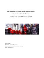
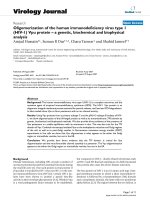
resected lesions with biliary benign diseases. Figure 1
represents the immunohistochemical staining of LAT1
expression. LAT1 immunostaining was detected in carcinoma cells in tumor tissues and localized predominantly on their plasma membrane. All positive cells
revealed strong membranous LAT1 immunostaining.
Cytoplasmic staining was rarely evident. The high expression rate and average scoring of LAT1 were compared according to tumor location (Additional file 2:
Figure 1 Immunohistochemical staining of tissue from a 79-years old man with extrahepatic cholangiocarcinoma (A) and a 66-years
old woman with Xanthogranulomatous chlecystitis as control group (B). Immunostaining of LAT1 demonstrates a membranous
immunostaining pattern in cholangiocarcinoma, but there was no evidence of LAT1 staining in xanthogranulomatous chlecystitis.
Kaira et al. BMC Cancer 2013, 13:482
/>
Page 6 of 12
Table S2, online only). In total patients, the high expression rate and average scoring of LAT1 were recognized
in 64.0% and 2.7 ± 0.9, respectively.
Based on the results of analysis on cholangiocarcinoma, cutoff points for high CD34 expression and
high Ki-67 labeling index were defined as follows. The
median number of CD34-positive vessels was 21 (range,
4–52), and the value of 21 was chosen as a cutoff point.
The median value of the Ki-67 labeling index was 35%
(range, 2–76), and the value of 35% was chosen as cutoff
point. Positive expression of p53 was recognized in
51.1% (71/139). Table 1 shows the expression status of
these biomarkers according to tumor location. Rate of
high expression or positivity in these biomarkers was
significantly higher in cholangiocarcinoma than in biliary
benign lesions (Table 1). Patient’s demographics according to LAT1 expression status are listed in Table 2. The
expression of LAT1 was significantly associated with
lymphatic permeation, vascular invasion, lymph node
metastasis, CA19-9, Ki-67, and MVD.
Correlation between LAT1 expression and other biomarkers
Analysis with Spearman’s rank correlation revealed that
LAT1 expression was significantly correlated with Ki-67
and CD34 in all tumor location except CD34 in IHCC
(Additional file 3: Table S3, online only).
Univariate and multivariate survival analysis
In all patients, the 5-year survival rate and median survival time (MST) for OS were 35.6% and 1073 days, respectively, and the 3-year survival rate and MST for PFS
was 45.1% and 840 days, respectively. Because of a postoperative recurrence, 39 patients received systemic
chemotherapy using GEM or S-1. Table 3 shows the univariate and multivariate analysis in all patients (n = 139).
Univariate analysis revealed that significant variables for
OS were resected status, tumor differentiation, lymphatic
permeation, vascular invasion, lymph nodes metastasis,
LAT1, and Ki-67. Significant prognostic markers for PFS
by the univariate analysis included resected status, tumor
differentiation, lymphatic permeation, vascular invasion,
lymph node metastasis, tumor stage, and LAT1. According to the results of univariate log-rank test, we screened
prognostic factors with cut-off of p < 0.05. Multivariate
analysis confirmed that lymphatic permeation and a high
LAT1 expression, lymphatic permeation and Ki-67 were
independent prognostic factors for predicting poor OS,
Table 2 Patient’s demographics according to LAT1 expression status
All patient (n = 139)
Parameter
Age
Gender
Tumor size(mm)
Resection status
Pathological
differentiation
Lymphatic
permeation
Extrahepatic CC (n = 89)
Gallbladder carcinoma
(n = 30)
Intrahepatic CC (n = 20)
High
Low
High
Low
High
Low
High
Low
(n = 89) (n = 50) p-value (n = 59) (n = 30) p-value (n = 18) (n = 12) p-value (n = 12) (n = 8) p-value
≤65 / >65
24 / 65
11 / 39
0.549
17 / 42
4 / 26
0.121
3 / 15
2 / 10
>0.999
5/7
5/3
0.649
M/F
55 / 34
31 / 19
>0.999
43 / 16
22 / 8
>0.999
4 / 14
4/8
0.677
8/4
5/3
>0.999
≤35 / >35
48 / 41
28 / 22
0.862
34 / 25
22 / 8
0.170
11 / 7
10 / 2
0.248
4/8
4/4
0.647
R0 / R1
42 / 47
25 / 25
0.859
25 / 34
13 / 17
>0.999
12 / 6
8/4
>0.999
5/7
5/3
0.649
WD or MD
/ PD
67 / 22
39 / 11
0.572
46 / 13
25 / 5
0.780
16 / 2
11 / 1
0.377
5/7
3/5
>0.999
Yes / No
78 / 11
33 / 17
0.003
53 / 6
9 / 21
<0.001
16 / 2
7/5
0.459
9/3
5/3
0.642
Vascular invasion
Yes / No
68 / 21
25 / 25
0.002
49 / 10
17 / 13
0.011
11 / 7
4/8
0.263
8/4
4/4
0.647
Lymph node
metastasis
Yes / No
51 / 38
11 / 39
<0.001
33 / 26
8 / 22
0.013
9/9
2 / 10
0.121
9/3
1/7
0.019
I or II / III or 64 / 25
IV
45 / 5
0.098
43 / 16
27 / 3
0.099
12 / 6
12 / 0
0.056
9/3
6/2
>0.999
Disease staging
Papillary pattern
Yes / No
18 / 71
14 / 36
0.302
8 / 51
5 / 25
0.755
10 / 8
8/4
0.708
0 / 12
1/7
0.400
Adjuvant
chemotherapy
Yes / No
40 / 49
11 / 39
0.009
28 / 31
6 / 24
0.012
7 / 11
1 / 11
0.099
5/7
4/4
>0.999
≤2.1 / >2.1
45 / 44
29 / 21
0.479
26 / 33
13 / 17
>0.999
11 / 7
7/5
>0.999
8/4
5/3
>0.999
≤45.1 /
>45.1
32 / 57
37 / 13
<0.001
22 / 37
23 / 7
<0.001
9/9
9/3
0.259
5/7
5/3
0.649
High / Low
47 / 42
15 / 35
0.012
33 / 26
10 / 20
0.072
10 / 8
3/9
0.141
4/8
2/6
>0.999
P/N
48 / 41
23 / 27
0.383
32 / 27
12 / 18
0.263
12 / 6
8/4
>0.999
4/8
3/5
>0.999
High / Low
51 / 38
18 / 32
0.021
34 / 25
12 / 18
0.124
11 / 7
3/9
0.071
6/6
3/5
0.669
CEA
CA19-9
Ki-67
p53
CD34
Abbreviation: LAT1 L-type amino acid transporter 1, CC Cholangiocarcinoma, M / F Male / Female, CEA Carcinoembryonic antigen, WD or MD / PD Well
differentiated or moderate differentiated / poorly differentiated, P /N Positive / Negative, Bold numbers Statistically significant difference.
Kaira et al. BMC Cancer 2013, 13:482
/>
Page 7 of 12
Table 3 Univariate and multivariate analysis in overall survival and progression-free survival
Overall survival
Variable
5-year
survival rate
(%)
Progression-free survival
p-value
p-value
Hazard
(univariate) (multivariate) ratio
95% CI
3-year
survival rate
(%)
p-value
p-value
Hazard
(univariate) (multivariate) ratio
95% CI
Anatomical
locations
EHCC
38.1
IHCC
28.0
0.837
28.2
48.3
GB
34.5
45.6
0.395
Age
≤65 yr
39.9
≻65 yr
27.5
0.095
48.4
0.707
54.7
Gender
Male
30.2
Female
33.5
0.267
49.7
58.6
Resection
R0
42.5
R1 or R2
29.8
0.026
0.075
1.300
Poorly
0.974 to
1.752
64.2
0.016
0.310
1.154
0.881 to
1.593
47.3
0.017
0.251
0.190
0.729 to
1.307
55.2
7.8
0.008
0.845
0.971
39.6
Lymphatic
permeation
1.057 to
7.629
Yes
19.9
No
79.9
0.002
0.036
2.555
0.939
20.5
No
58.7
0.011
20.5
44.0
<0.001
0.016
3.139
0.468 to
1.939
1.073 to
5.057
40.2
<0.001
0.031
2.244
74.8
0.977
Negative
44.9
0.862
Lymph nodes
metastasis
Positive
1.212 to
10.72
81.7
Vascular
invasion
Yes
0.041
0.552 to
1.706
0.935
0.517 to
4.534
41.3
0.003
0.454
1.507
0.003
0.088
1.685
63.2
Tumor stage
I or II
38.2
III or IV
29.5
0.051
60.1
20.3
Low
59.3
<0.001
0.013
2.414
29.6
Low
36.2
1.196 to
5.321
41.1
0.005
71.3
Ki-67
High
0.021
0.038
1.781
1.030 to
3.093
49.2
0.192
53.6
p53
Positive
21.3
Negative
38.1
0.922 to
2.980
26.8
LAT1
High
0.875 to
1.535
40.1
Tumor
differentiation
Well or
moderate
0.634
0.119
54.1
51.6
0.831
0.242
1.449
0.785 to
2.837
Kaira et al. BMC Cancer 2013, 13:482
/>
Page 8 of 12
Table 3 Univariate and multivariate analysis in overall survival and progression-free survival (Continued)
CD34
High
28.6
Low
42.8
0.349
51.1
0.696
52.2
Abbreviation: 95% CI 95% confidence interval, EHCC Extrahepatic cholangiocarcinoma, IHCC Intrahepatic cholangiocarcinoma, GB Gallbladder carcinoma,
CEA Carcinoembryonic antigen, LAT1 L-type amino acid transporter 1, Bold numbers Statistically significant difference.
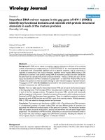
and lymphatic permeation and vascular invasion for poor
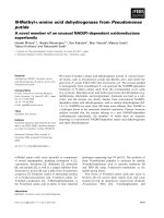
PFS. Figure 2 shows the Kaplan-Meier survival curve in
patients with high and low for LAT1 expression.
Expression of LAT1 and CD98 in human
cholangiocarcinoma cell lines
As shown in Additional file 4: Figure S1 (online only),
both LAT1 and CD98 were expressed in all three human
cholangiocarcinoma cell lines, HuCCT1, OZ, and
HuH28. The expression level of LAT1 in OZ was lower
than that of the other cell lines. HuCCT1 cell was used
in the following experiments because of its higher expression of LAT1 and tumorigenesis in nude mice.
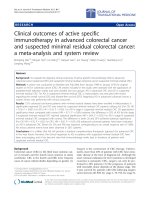
LAT inhibition suppresses cellular amino acid transport
and proliferation through LAT1
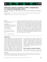
The cellular uptake of [14C]L-leucine was measured in a
presence of various concentrations of BCH, and was
inhibited concentration-dependently by the treatment
with BCH (Figure 3A). Expression profile of LAT1-4 in
HuCCT1 examined by realtime RT-PCR showed that
the expression of LAT1 was extremely higher than the
other LATs (Figure 3B). These results indicate that
BCH inhibits amino acid transport through LAT1 in
HuCCT1 cells. Furthermore, BCH decreased number of
cells concentration-dependently (Figure 3C), indicating
that BCH could inhibit proliferation of HuCCT1 cells
through inhibition of amino acid uptake.
LAT inhibition enhances anti-tumor activity of GEM and
5-FU
As shown in Figure 3D and E, combination of BCH with
chemotherapeutic agents decreased number of HuCCT1
cells. Cytotoxicity of GEM and 5-FU was significantly
enhanced in combination with 10 mM BCH, indicating
additive effect of LAT inhibitor on anti-tumor activity of
GEM and 5-FU in HuCCT1.
LAT inhibition suppresses growth of xenografts in nude
mice
Anti-tumor activity of BCH on cholangiocarcinoma was
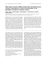
examined in vivo using HuCCT1-bearing mice. Daily administration of BCH (200 mg/kg) for 14 days caused statistically significant delay in the tumor growth up to 3
weeks after the completion of dosing (Figure 4A). There
was no change in the body weight by the treatment with
BCH (Figure 4B). Anti-tumor effect of BCH was also
monitored using 18F-FDG PET to determine the decrease
in the metabolism of the tumor. SUV max and SUV 50%
of 18F-FDG were decreased at day 17 and increased thereafter in BCH-treated mice (Figure 4C).
Discussion
This is the first study to elucidate the clinicopathologic
significance of LAT1 expression in patients with biliary
tract cancer. The expression of LAT1 in the tumor specimens was closely correlated with lymphatic metastases,
cell proliferation, and angiogenesis; and was a significant
Figure 2 Outcomes after surgical resection shown by Kaplan-Meier analysis of overall survival (OS) and progression-free survival (PFS)
according to LAT1 and CD98 expression. A statistically significant difference in OS (A) and PFS (B) was observed between patients with high
and low LAT1 expression.
Kaira et al. BMC Cancer 2013, 13:482
/>
Page 9 of 12
Figure 3 Effect of LAT inhibition on in vitro cellular proliferation and anti-tumor activity of GEM and 5-FU: (A) BCH inhibits [14C]L-leucine
uptake concentration-dependently in HuCCT1 cells (n = 4). Ordinate shows a percentage of [14C]L-leucine uptake in the absence of BCH as a
control. (B) Expression of LAT1, LAT2, LAT3, and LAT4 mRNA in HuCCT1 cells (n = 4). Ordinate shows relative quantity of mRNA calibrated by LAT1
mRNA. (C) BCH decreases number of HuCCT1 cells concentration-dependently (n = 4). Ordinate shows number of cells in a percentage of control
(without BCH). Addition of 10 mM BCH enhances anti-tumor effect of GEM (D) and 5-FU (E) on HuCCT1 cells. Ordinate shows number of cells in
a percentage of control (n = 4). A statistically significant difference from the control is indicated by *** (P < 0.001).
indicator for predicting poor outcome after surgical resection. Therefore, a high LAT1 expression may play an
important role on the growth of biliary tract cancer. No
anatomic site-related differences were observed for
LAT1. Results of our preliminary experiments indicated
that the inhibition of LAT1 had significant anti-tumor
effect on cholangiocarcinoma with acceptable toxicity
and yielded an additive therapeutic efficacy to GEM and
5-FU. Our data suggests that LAT1 inhibition suppresses
the growth of biliary tract cancer and LAT1 could be a
potential target for locally advanced or metastatic biliary
tract cancer.
Recently, two studies have exhibited the significance of
LAT1 expression as a prognostic predictor in pancreatic
cancer [33,34]. In pancreatic cancer, LAT1 was highly
expressed in 52.6% [33]. In biliary tract cancer, the ratio
of high LAT1 expression yielded a similar tendency
among all anatomic site (EHCC, IHCC, and GB). These
results indicate that the expression of LAT1 is higher in
biliary tract cancer than pancreatic cancer. The LAT1
expression is variable in human cancers, and relatively
low in adenocarcinoma, for example, 29% in pulmonary
adenocarcinoma [12], 22% in prostate cancer [15], 43%
in breast cancer [17], and 43% in gastric cancer [16].
LAT1 seemed to be expressed at higher level in biliary
tract adenocarcinoma than in adenocarcinoma of the
other organs. Therefore, LAT1 may play a crucial role in
enhancing the cell proliferation and tumor growth in biliary tract cancer.
Recently, we had evaluated the protein expression of
LAT1 by immunohistochemistry in patients with pulmonary neuroendocrine tumors [35]. Our data indicated that
the expression of LAT1 tended to increase from lowgrade to high-grade malignancies. Moreover, we confirmed the different expression of LAT1 between pancreatic cancer and pancreatic adenoma, showing that LAT1
expression was not observed in pancreatic adenoma,
whereas LAT1 was highly expressed in pancreatic cancer
[33]. Previous experimental data also demonstrated that
LAT1 is overexpressed in tumor cells and LAT2 is dominantly expressed in normal cells [9,10]. In the protein expression level of human tissue specimens, there was no
evidence of LAT1 expression in normal tissues. Thus, we
believe that LAT1 is tumor-specific amino acid transporter and has a potential target of cancer therapeutics.
This study investigated the therapeutic potential of
LAT1 inhibition in cholangiocarcinoma. We found that
BCH as a competitive LAT inhibitor suppressed
Kaira et al. BMC Cancer 2013, 13:482
/>
Page 10 of 12
Figure 4 In vivo anti-tumor effect of LAT inhibition on cholangiocarcinoma xenograft. (A) Intravenous administration of BCH shows delay
in the growth of HuCCT1 tumor (n = 10). A statistically significant difference from the control is indicated by * (P < 0.05), ** (P < 0.01),
and *** (P < 0.001). (B) Changes in the body weight of HuCCT1 tumor-bearing mice after administration of BCH (n = 10). (C) Representative
coronal section of 18F-FDG PET images of HuCCT1-bearing mice at 2 h after 18F-FDG injection. PET imaging was performed at indicated day after
the day of grouping (n = 2). The calibration bar is shown at right-side of images. SUV max and SUV 50% are shown below the images.
proliferation of cholangiocarcinoma cells and yielded an
additive therapeutic efficacy to GEM and 5-FU in vitro.
Moreover, in vivo experiment demonstrated significant
growth suppression of tumor with acceptable toxicity.
Recent reports also showed that the inhibition of LAT
activity by BCH resulted in the suppression of cell proliferation in various cancers [9,13,19,20]. Nawashiro et al.
showed that BCH reduced mortality of C6 gliomabearing rat model, and suggested that LAT1 inhibitors
could be an effective therapeutic option for high-grade
gliomas [14]. Kim et al. reported that BCH could lead
to apoptosis by inducing intracellular depletion of
amino acids required for the growth of cancer cells [20].
Liu et al. described that BCH induced apoptosis without
affecting DNA synthesis in proliferating vascular
smooth muscle cells, whereas it had no effect on quiescent smooth muscle cells. Therefore, the inhibition of
LAT1 gives rise to growth inhibition effects of highly
proliferative cells that require increased amino acid metabolism [36]. Another proposed mechanism of action
is cell cycle arrest at G1 phase by the inhibition of
LAT1 [37]. However, there is no established explanation
regarding the in vivo anti-tumor effect of LAT1 inhibitor, although there are two preclinical studies investigating the potential of LAT1 inhibitor in tumor xenografts
(glioma [13] and cholangiocarcinoma [current study]).
Further in vivo study is warranted to evaluate whether a
combination of GEM plus LAT1 inhibitor is effective
for biliary tract cancer xenograft compared to GEM
alone as seen in the current in vitro study that has been
demonstrating effect of GEM plus BCH.
A recent systemic review has suggested that p53 mutation, cyclins, proliferation indices (Ki-67), mucins, CA199, and CEA have potential as prognostic predictors in
Kaira et al. BMC Cancer 2013, 13:482
/>
Page 11 of 12
cholangiocarcinoma [38], however, there is no targeting
therapy for these molecules at present. Recently, antiepidermal growth factor receptor (EGFR) agents,
mitogen-activated protein kinase/extracellular-signal regulated kinase (MEK) inhibitors, and anti-angiogenic agents
have been thought to be the promising targeted agents for
biliary tract cancer [39]. However, the results of clinical
trials indicated no therapeutic efficacy to improve the survival of patients with advanced biliary tract cancer [39].
content; and KK, YO, TO, MM and IT have given final approval of the version
to be published. All authors read and approved the final manuscript.
Conclusion
In conclusion, high expression of LAT1 plays an important role in enhancing tumor growth and cell proliferation and is a promising pathological marker for
predicting poor prognosis in patients with biliary tract
cancer. The inhibition of LAT significantly suppressed
the growth of cholangiocarcinoma, and anti-tumor efficacy of GEM and 5-FU was augmented in combination
with LAT inhibitor. Since the LAT1 expression is a significant prognostic marker and LAT1 inhibition probably has anti-tumor efficacy, molecular targeting drug
that selectively inhibit LAT1 will aid in the promising
therapeutic strategy for bile duct cancer.
Author details
1
Department of Medicine and Molecular Science, Gunma University Graduate
School of Medicine, Showa-machi, Maebashi, Gunma, Japan. 2Department of
Thoracic and Visceral Surgery, Gunma University Graduate School of
Medicine, Showa-machi, Maebashi, Gunma, Japan. 3Medical Radioisotope
Application Group, Quantum Beam Science Directorate, Japan Atomic
Energy Agency, Watanuki, 370-1292 Takasaki, Gunma, Japan. 4Department of
Surgery, Maebashi Red Cross Hospital, Asahi-cho, Maebashi, Gunma, Japan.
5
Department of Molecular Imaging, Gunma University Graduate School of
Medicine, Showa-machi, Maebashi, Gunma, Japan. 6Department of
Diagnostic Radiology and Nuclear Medicine, Gunma University Graduate
School of Medicine, Showa-machi, Maebashi, Gunma, Japan. 7Department of
Radiology, Saku Central Hospital, Usuda, Saku, Nagano, Japan. 8Department
of Pathology, Maebashi Red Cross Hospital, Asahi-cho, Maebashi, Gunma,
Japan. 9Division of Bio-system Pharmacology, Department of Pharmacology,
Graduate School of Medicine, Osaka University, Osaka, Japan. 10Department
of Bioimaging Information Analysis, Gunma University Graduate School of
Medicine, Showa-machi, Maebashi, Gunma, Japan. 11Department of
Diagnostic Pathology, Gunma University Graduate School of Medicine,
Showa-machi, Maebashi, Gunma, Japan. 12Oncology Center, Gunma
University Hospital Showa-machi, 371-8511 Maebashi, Gunma, Japan.
Additional files
Additional file 1: Table S1. Primers for realtime RT-PCR used in the
present study.
Additional file 2: Table S2. Comparison of percentage of high
expression and average score of LAT1.
Additional file 3: Table S3. Correlation between LAT1 expression and
various biomarkers.
Additional file 4: Figure S1. Expression of LAT1 and CD98 in human
cholangiocarcinoma cell lines (HuCCT1, OZ and HuH28). Representative
images from three independent experiments are shown. β-actin was
shown as a control.
Abbreviations
LAT1: L-type amino acid transporter 1; BCH: 2-aminobicyclo-(2,2,1)-heptane-2carboxylic acid; MRCP: Magnetic resonance cholangiopancreatography;
ERCP: Endoscopic retrograde cholangiopancreatography; EHCC: Extrahepatic
cholangiocarcinoma; IHCC: Intrahepatic cholangiocarcinoma; GB: Gallbladder
carcinoma; TNM: Pathologic tumor-node-metastasis; MVD: Microvessel
density; DMEM: Dulbecco’s modified Eagle’s medium; MTT: 3-[4,5-dimethyl-2thiazolyl]-2,5-diphenyl-2H-tetrazolium bromide; GEM: Gemcitabine;
5-FU: 5-fluorouracil; 18F-FDG: [18F]fluoro-2-deoxyglucose; PET: Positron
emission tomography; ROI: Region of interest; SUV: Standardized uptake
value; OS: Overall survival; PFS: Progression-free survival; MST: Median survival
time; EGFR: Epidermal growth factor receptor; MEK: Mitogen-activated
protein kinase/extracellular-signal regulated kinase.
Competing interests
We, all authors, have no financial or personal relationships with other people
or organizations that could inappropriately influence our work. The authors
declare that they have no competing interests.
Authors’ contributions
KK, YS, YO, NSI, HT, NO and KA have made substantial contributions to
conception and design, or acquisition of data, or analysis and interpretation
of data; KK, YO, YK, TO, KS, NS, HI, SN, AY, AS and MI have been involved in
drafting the manuscript or revising it critically for important intellectual
Acknowledgements
This work was supported in part by Grant 23591750 (K. K) and 25461801
(N.O) from the Ministry of Education, Culture, Sports, Science and
Technology, Japan, and National Hospital Organization Policy Based Medical
Services. We thank Ms. Masako Saito for their technical assistance of
immunohistochemical analysis. Advanced research for medical products
Mining Programme of the National Institute of Biomedical Innovation
(NIBIO).
Received: 27 April 2013 Accepted: 25 September 2013
Published: 16 October 2013
References
1. Khan SA, Taylor-Robinson SD, Toledano MB, Beck A, Elliott P, Thomas HC:
Changing international trends in mortality rates for liver, biliary and
pancreatic tumors. J Hepatol 2002, 37:806–813.
2. Nakeeb A, Pitt HA, Sohn TA, Coleman J, Abrams RA, Piantadosi S, Hruban
RH, Lillemoe KD, Yeo CJ, Cameron JL: Cholangiocarcinoma. A spectrum of
intrahepatic, perihilar, and distal tumors. Ann Surg 1996, 22:463–475.
3. Nakagohri T, Kinoshita T, Konishi M, Takahashi S, Gotohda N: Surgical
outcome and prognostic factors in intrahepatic cholangiocarcinoma.
World J Surg 2008, 32:2675–2680.
4. Hirano S, Kondo S, Tanaka E, Shichinohe T, Tsuchikawa T, Kato K,
Matsumoto J, Kawasaki R: Outcome of surgical treatment of hilar
cholangiocarcinoma: a special reference to postoperative morbidity and
mortality. J Hepatobiliary Pancreat Sci 2010, 17:455–462.
5. Sakamoto Y, Kosuge T, Shimada K, Sano T, Ojima H, Yamamoto J, Yamasaki S,
Takayama T, Makuuchi M: Prognostic factors of surgical resection in middle
and distal bile duct cancer: an analysis of 55 patients concerning the
significance of ductal and radial margins. Surgery 2005, 37:396–402.
6. Valle J, Wasan H, Palmer DH, Cunningham D, Anthoney A, Maraveyas A,
Madhusudan S, Iveson T, Hughes S, Pereira SP, Roughton M, Bridgewater J:
ABC-02 trial investigators: cisplatin plus gemcitabine versus gemcitabine
for biliary tract cancer. N Engl J Med 2010, 362:1273–1281.
7. Christensen HN: Role of amino acid transport and countertransport in
nutrition and metabolism. Physiol Rev 1990, 70:43–77.
8. McGivan JD, Pastor-Anglada M: Regulatory and molecular aspects of
mammalian amino acid transport. Biochem J 1994, 299:321–334.
9. Kanai Y, Segawa H, Miyamoto K, Uchino H, Takeda E, Endou H: Expression
cloning and characterization of a transporter for large neutral amino
acids activated by the heavy chain of 4F2 antigen (CD98). J Biol Chem
1998, 273:23629–23632.
10. Yanagida O, Kanai Y, Chairoungdua A, Kim DK, Segawa H, Nii T, Cha SH,
Matsuo H, Fukushima J, Fukasawa Y, Tani Y, Taketani Y, Uchino H, Kim JY,
Inatomi J, Okayasu I, Miyamoto K, Takeda E, Goya T, Endou H: Human
Kaira et al. BMC Cancer 2013, 13:482
/>
11.
12.
13.
14.
15.
16.
17.
18.
19.
20.
21.
22.
23.
24.
25.
26.
27.
28.
29.
L-type amino acid transporter 1 (LAT 1): characterization of function and
expression in tumor cell lines. Biochim Biophys Acta 2001, 1514:291–302.
Kobayashi H, Ishii Y, Takayama T: Expression of L-type amino acid transporter
1 (LAT1) in esophageal carcinoma. J Surg Oncol 2005, 90:233–238.
Kaira K, Oriuchi N, Imai H, Shimizu K, Yanagitani N, Sunaga N, Hisada T,
Tanaka S, Ishizuka T, Kanai Y, Endou H, Nakajima T, Mori M: Prognostic
significance of L-type amino acid transporter 1 expression in resectable
stage I-III nonsmall cell lung cancer. Br J Cancer 2008, 98:742–748.
Nakanishi K, Ogata S, Matsuo H, Kanai Y, Endou H, Hiroi S, Tominaga S, Aida S,
Kasamatsu H, Kawai T: Expression of LAT1 predicts risk of progression of
transitional cell carcinoma of the upper urinary tract. Virchows Arch 2007,
451:681–690.
Nawashiro H, Otani N, Shinomiya N, Fukui S, Ooigawa H, Shima K, Matsuo H,
Kanai Y, Endou H: L-type amino acid transporter 1 as a potential molecular
target in human astrocytic tumors. Int J Cancer 2006, 119:484–492.
Sakata T, Ferdous G, Tsuruta T, Satoh T, Baba S, Muto T, Ueno A, Kanai Y,
Endou H, Okayasu I: L-type amino acid transporter 1 as a novel biomarker
for high-grade malignancy in prostate cancer. Pathol Int 2009, 59:7–18.
Ichinoe M, Mikami T, Yoshida T, Igawa I, Tsuruta T, Nakada N, Anzai N,
Suzuki Y, Endou H, Okayasu I: High expression of L-type amino-acid
transporter 1 (LAT1) in gastric carcinomas: comparison with
non-cancerous lesions. Pathol Int 2011, 61:281–289.
Furuya M, Horiguchi J, Nakajima H, Kanai Y, Oyama T: Correlation of L-type
amino acid transporter 1 and CD98 expression with triple negative
breast cancer prognosis. Cancer Sci 2012, 103:382–389.
Kaira K, Oriuchi N, Takahashi T, Nakagawa K, Ohde Y, Okumura T, Murakami H,
Shukuya T, Kenmotsu H, Naito T, Kanai Y, Endo M, Kondo H, Nakajima T,
Yamamoto N: LAT1 expression is closely associated with hypoxic markers
and mTOR in resected non-small cell lung cancer. Am J Transl Res 2011,
3:468–478.
Kaira K, Oriuchi N, Takahashi T, Nakagawa K, Ohde Y, Okumura T, Murakami H,
Shukuya T, Kenmotsu H, Naito T, Kanai Y, Endo M, Kondo H, Nakajima T,
Yamamoto N: L-type amino acid transporter 1 (LAT1) expression in
malignant pleural mesothelioma. Anticancer Res 2011, 31:4075–4082.
Kim CS, Cho SH, Chun HS, Lee SY, Endou H, Kanai Y, Kim do K: BCH, an
inhibitor of system L amino acid transporters, induces apoptosis in
cancer cells. Biol Pharm Bull 2008, 31:1096–1100.
Imai H, Kaira K, Oriuchi N, Shimizu K, Tominaga H, Yanagitani N, Sunaga N,
Ishizuka T, Nagamori S, Promchan K, Nakajima T, Yamamoto N, Mori M,
Kanai Y: Inhibition of L-type amino acid transporter 1 has antitumor
activity in non-small cell lung cancer. Anticancer Res 2010, 30:4819–4828.
Sobin LH, Gospodarowicz MK, Wittekind C (Eds): International Union Against
Cancer (UICC) TNM Classification of Malignant Tumors. 7th edition. Oxford,
UK: Wiley-Blackwell; 2009.
Kaira K, Oriuchi N, Imai H, Shimizu K, Yanagitani N, Sunaga N, Hisada T,
Kawashima O, Kamide Y, Ishizuka T, Kanai Y, Nakajima T, Mori M: CD98
expression is associated with poor prognosis in resected non-small-cell lung
cancer with lymph node metastases. Ann Surg Oncol 2009, 16:3473–3581.
Kaira K, Endo M, Abe M, Nakagawa K, Ohde Y, Okumura T, Takahashi T,
Murakami H, Tsuya A, Nakamura Y, Naito T, Hayashi I, Serizawa M, Koh Y,
Hanaoka H, Tominaga H, Oriuchi N, Kondo H, Nakajima T, Yamamoto N:
Biologic correlation of 2-[18F]-fluoro-2-deoxy-D-glucose uptake on
positron emission tomography in thymic epithelial tumors. J Clin Oncol
2010, 28:3746–3753.
Miyagiwa M, Ichida T, Tokiwa T, Sato J, Sasaki H: A new human
cholangiocellular carcinoma cell line (HuCC-T1) producing carbohydrate
antigen 19/9 in serum-free medium. In Vitro Cell Dev Biol 1989, 25:503–510.
Homma S, Nagamori S, Fujise K, Yamazaki K, Hasumura S, Sujino H,
Matsuura T, Shimizu K, Kameda H, Takaki K: Human bile duct carcinoma
cell line producing abundant mucin in vitro. Gastroenterol Jpn 1987,
22:474–479.
Kusaka Y, Tokiwa T, Sato J: Establishment and characterization of a cell
line from a human cholangiocellular carcinoma. Res Exp Med (Berl) 1988,
188:367–375.
Segawa H, Fukasawa Y, Miyamoto K, Takeda E, Endou H, Kanai Y:
Identification and functional characterization of a Na + −independent
neutral amino acid transporter with broad substrate selectivity. J Biol
Chem 1999, 274:19745–19751.
Babu E, Kanai Y, Chairoungdua A, Kim DK, Iribe Y, Tangtrongsup S, Jutabha P,
Li Y, Ahmed N, Sakamoto S, Anzai N, Nagamori S, Endou H: Identification of a
Page 12 of 12
30.
31.
32.
33.
34.
35.
36.
37.
38.
39.
novel system L amino acid transporter structurally distinct from
heterodimeric amino acid transporters. J Biol Chem 2003, 273:43838–43845.
Bodoy S, Martín L, Zorzano A, Palacín M, Estévez R, Bertran J: Identification
of LAT4, a novel amino acid transporter with system L activity. J Biol
Chem 2005, 280:12002–12011.
Kim DK, Kanai Y, Choi HW, Tangtrongsup S, Chairoungdua A, Babu E,
Tachampa K, Anzai N, Iribe Y, Endou H: Characterization of the system L
amino acid transporter in T24 human bladder carcinoma cells.
Biochim Biophys Acta 2002, 1565:112–121.
Morimoto E, Kanai Y, Kim do K, Chairoungdua A, Choi HW, Wempe MF,
Anzai N, Endou H: Establishment and characterization of mammalian cell
lines stably expressing human L-type amino acid transporters.
J Pharmacol Sci 2008, 108:505–516.
Kaira K, Sunose Y, Arakawa K, Ogawa T, Sunaga N, Shimizu K, Tominaga H,
Oriuchi N, Itoh H, Nagamori S, Kanai Y, Segawa A, Furuya M, Mori M, Oyama T,
Takeyoshi I: Prognostic significance of L-type amino-acid transporter 1
expression in surgically resected pancreatic cancer. Br J Cancer 2012,
107:632–638.
Yanagisawa N, Ichinoe M, Mikami T, Nakada N, Hana K, Koizumi W, Endou H,
Okayasu I: High expression of L-type amino acid transporter 1 (LAT1)
predicts poor prognosis in pancreatic ductal adenocarcinomas. J Clin
Pathol 2012, 65:1019–1023.
Kaira K, Oriuchi N, Imai H, Shimizu K, Yanagitani N, Sunaga N, Hisada T,
Kawashima O, Iijima H, Ishizuka T, Kanai Y, Endou H, Nakajima T, Mori M:
Expression of L-type amino acid transporter 1 (LAT1) in neuroendocrine
tumors of the lung. Pathol Res Pract 2008, 204:553–561.
Liu XM, Reyna SV, Ensenat D, Peyton KJ, Wang H, Schafer AI, Durante W:
Platelet-derived growth factor stimulates LAT1 gene expression in
vascular smooth muscle: role in cell growth. FASEB J 2004, 18:768–770.
Kim CS, Moon IS, Park JH, Shin WC, Chun HS, Lee SY, Kook JK, Kim HJ, Park JC,
Endou H, Kanai Y, Lee BK, Kim do K: Inhibition of L-type amino acid
transporter modulates the expression of cell cycle regulatory factors in KB
oral cancer cells. Biol Pharm Bull 2010, 33:1117–1121.
Briggs CD, Neal CP, Mann CD, Steward WP, Manson MM, Berry DP:
Prognostic molecular markers in cholangiocarcinoma: a systemic review.
Eur J Cancer 2009, 45:33–47.
Faris JE, Zhu AX: Targeted therapy for biliary tract cancers. J Hepatobiliary
Pancreat Sci 2012. Feb 09. [Epub ahead of print].
doi:10.1186/1471-2407-13-482
Cite this article as: Kaira et al.: Clinical significance of L-type amino acid
transporter 1 expression as a prognostic marker and potential of new
targeting therapy in biliary tract cancer. BMC Cancer 2013 13:482.
Submit your next manuscript to BioMed Central
and take full advantage of:
• Convenient online submission
• Thorough peer review
• No space constraints or color figure charges
• Immediate publication on acceptance
• Inclusion in PubMed, CAS, Scopus and Google Scholar
• Research which is freely available for redistribution
Submit your manuscript at
www.biomedcentral.com/submit