BRCA1 promoter hypermethylation, 53BP1 protein expression and PARP-1 activity as biomarkers of DNA repair deficit in breast cancer
Bạn đang xem bản rút gọn của tài liệu. Xem và tải ngay bản đầy đủ của tài liệu tại đây (368.01 KB, 11 trang )
Jacot et al. BMC Cancer 2013, 13:523
/>
RESEARCH ARTICLE
Open Access
BRCA1 promoter hypermethylation, 53BP1 protein
expression and PARP-1 activity as biomarkers of
DNA repair deficit in breast cancer
William Jacot1,2*, Simon Thezenas3, Romain Senal4, Cathy Viglianti4, Anne-Claire Laberenne4, Evelyne Lopez-Crapez2,4,
Frédéric Bibeau2,5, Jean-Pierre Bleuse3, Gilles Romieu1 and Pierre-Jean Lamy2,4
Abstract
Background: Poly(adenosine diphosphate–ribose) polymerase 1 (PARP-1) and the balance between BRCA1 and
53BP1 play a key role in the DNA repair and cell stress response. PARP inhibitors show promising clinical activity in
metastatic triple negative (TN) or BRCA-mutated breast cancer. However, a comprehensive analysis of PARP-1
activity, BRCA1 promoter methylation and 53BP1 expression in tumours without known BRCA1 mutation has not yet
been carried out.
Methods: We investigated cytosolic PARP-1 activity, 53BP1 protein levels and BRCA1 promoter methylation in 155
surgical breast tumour samples from patients without familial breast cancer history or known BRCA1 mutations who
were treated between January 2006 and November 2009 and evaluated their statistical association with classical
predictive and prognostic factors.
Results: The mitotic count score was the only parameter clearly associated with PARP-1 activity. BRCA1 promoter
hypermethylation (15.4% of all cancers) was significantly associated with uPA and PAI-1 levels, tumour grade, mitotic
count score, hormone receptor and HER2 negative status and TN profile (29% of TN tumours showed BRCA1
promoter hypermethylation compared to 5% of grade II-III hormone receptor-positive/HER2-negative and 2% of
HER2-positive tumours). No statistical association was found between BRCA1 promoter hypermethylation and PARP-1
activity. High 53BP1 protein levels correlated with lymph node positivity, hormone receptor positivity, molecular
grouping, unmethylated BRCA1 promoter and PARP-1 activity. In TN tumours, BRCA1 promoter methylation was
only marginally associated with age, PARP-1 activity was not associated with any of the tested clinico-pathological
factors and high 53BP1 protein levels were significantly associated with lymph node positivity. Only 3 of the 14 TN
tumours with BRCA1 promoter hypermethylation presented high 53BP1 protein levels.
Conclusions: Breast cancers that harbour simultaneously high 53BP1 protein level and BRCA1 promoter
hypermethylation and are the putative target population of drugs targeting DNA repair appear to be restricted to
a small subgroup of TN tumours.
Keywords: Breast cancer, PARP-1, 53BP1, BRCA, Methylation
* Correspondence:
1
Department of Medical Oncology, Montpellier Cancer Institute, Montpellier,
France
2
Translational Research Unit, Montpellier Cancer Institute, 208 rue des Apothicaires,
34298 Montpellier Cedex 5, France
Full list of author information is available at the end of the article
© 2013 Jacot et al.; licensee BioMed Central Ltd. This is an open access article distributed under the terms of the Creative
Commons Attribution License ( which permits unrestricted use, distribution, and
reproduction in any medium, provided the original work is properly cited.
Jacot et al. BMC Cancer 2013, 13:523
/>
Background
Up-regulation of their DNA repair capacity represents a
common mechanism used by cancer cells to survive
DNA-damaging therapy [1]. Lack of efficient DNA repair
by simultaneous loss or inhibition of two DNA repair
pathways causes synthetic lethality and cell death, thus
representing an attractive approach for cancer therapy
[2]. For instance, BRCA-deficient cancer cells, in which
DNA double strand break repair (DSB) by homologous
recombination is deficient [3,4], are particular sensitive
to treatment with inhibitors of Poly(ADP-ribose) (PAR)
polymerase 1 (PARP-1), a nuclear enzyme that recognizes
and facilitates repair of DNA damage induced by oxidation, alkylation and ionizing radiation [2,5-7], showing reduced clonogenic survival and DNA DSB repair
defects [8,9]. Moreover, the persistent single-strand
breaks (SSB) formed upon PARP-1 inhibition cannot
be repaired effectively in the absence of functional BRCA1
or BRCA2, resulting in accumulation of chromosomal abnormalities, cell cycle arrest and apoptosis [8,9]. Thus,
PARP-1 may be an important target for BRCA-deficient
breast cancer chemotherapy [8-11], as emphasized also by
the clinical activity of the PARP inhibitor (PARPi) olaparib
in patients with BRCA-mutated breast cancer [3]. Upregulation of PARP-1 expression and activity has been
observed in a variety of human tumours [12,13]. In
breast cancer, PARP-1 up-regulation has been associated with decreased survival [14] and triple-negative
(TN) cancers (breast tumours in which estrogen receptor [ER], progesterone receptor [PR] and human epidermal growth factor receptor 2 [HER2] are not expressed)
[15]. None of these studies considered PARP-1 activity
together with BRCA1 functional status, except in the
case of BRCA1-mutated cancers, which represent only
around 5% of all breast cancers [16-18]. Loss of BRCA1
nuclear expression correlates with high tumour grade
(p < 0.025) and ER-negative tumours. Absence or reduced
BRCA1 expression in tumours without BRCA1 mutations
appears linked to hypermethylation of the BRCA1 promoter region [19], a condition reported in 9.1–37% of
sporadic breast cancers and associated with infiltrating
ductal type, high (grade II-III) tumour grade, ER negativity, basal markers expression, younger age at diagnosis,
low BRCA1 mRNA expression and marked reduction or
loss of BRCA1 protein expression [19-25]. Thus, BRCA1
promoter hypermethylation could be a marker of BRCA1
deficiency in the absence of BRCA1 mutation, as these
two events appears mutually exclusive [24].
Some conditions, such as a loss of P53 binding protein
1 (53BP1, a protein involved in DNA damage checkpoint
activation and DNA repair), could allow cells to tolerate
BRCA1 deficiency. 53BP1 localizes to sites of DNA DSBs,
promotes non-homologous end joining (NHEJ)-mediated
repair and checkpoint activation and inhibits homologous
Page 2 of 11
recombination [26-29]. As BRCA1 promotes homologous
recombination, it might counteract 53BP1 effect [30,31].
Thus, the balance between 53BP1 and BRCA1 regulates
the competition between the NHEJ and homologous recombination pathways in DNA DSB repair [32]. In BRCA1
mutant/inactivated cells, repair by homologous recombination is defective and the error-prone NHEJ predominates,
resulting in high sensitivity to DNA-damaging agents and
PARPi. However, when both BRCA1 and 53BP1 are lost,
repair by homologous recombination is restored and the
sensitivity to DNA damaging agents is reduced, leading to
resistance to cis-platinum and PARPi in BRCA1-deficient
cells, suggesting a critical role of 53BP1 in cancer cells
in which BRCA1 is mutated or epigenetically silenced
[30-33]. Reduced 53BP1 expression has been reported
in sporadic basal-like, TN and BRCA-mutated breast
cancers [30]. It thus appears important to simultaneously
evaluate 53BP1 status and BRCA1 mutation/promoter
methylation to precisely estimate homologous recombination functionality in breast tumours.
Many PARPi are presently in pre-clinical or clinical
development, preferentially for patients with BRCAdeficient tumours or TN breast cancers, due to the overrepresentation of this breast cancer subtype in patients
with BRCA mutations. However, there is no validated
screening test to identify the patients who may receive the
most benefit from PARPi. Recent data show that most of
the non-BRCA-mutated TN breast cancers do not benefit
from such drugs, while some non-TN BRCA-mutated
tumours could respond to PARPi [34]. Moreover, two
different groups [35,36] recently reported that breast
cancers with epigenetically silenced BRCA1 are sensitive
to PARPi monotherapy, providing robust evidence to
support the use of PARPi in the treatment of selected
sporadic BRCA1-inactivated breast cancers. A comprehensive analysis of the PARP-1/BRCA1/53BP1 factors
of DNA repair in the different breast cancer subtypes
could enable this selection and promote the use of these
compounds outside the TN subtype.
Here, we comprehensively and simultaneously evaluated the BRCA1/53BP1/PARP-1 repair network in three
groups (HER2-positive, grade II-III hormone receptor
[HR]-positive/HER2-negative and TN) of sporadic breast
cancers (n = 155) from patients without familial breast
cancer history or known BRCA1 mutations to identify
tumour population(s) with a theoretically high susceptibility to PARPi.
Methods
Patients and tumour samples
This is a retrospective monocentric study using samples
from a research-dedicated tumour biobank (cytosol and
DNA samples). A total of 556 consecutive patients with
breast cancer referred to the Montpellier Cancer Institute
Jacot et al. BMC Cancer 2013, 13:523
/>
between January 2006 and November 2009 were prospectively entered in the biobank database. The DNA
collection was created using frozen, histologically
proven and macro-dissected invasive breast cancer
specimens that were primarily handled for uPA/PAI-1
testing [37]. Tumour samples dedicated to the molecular
analysis were selected based on the immediate diagnosis
by using frozen sections. Additional tumour tissue samples were then chosen after the definitive histological diagnosis (with quantification of the percentage of tumour
cells) and grade assessment after fixation. This could be
possible because frozen and formalin-fixed tumour tissue
samples were selected from the same tumour areas. Only
samples that contained at least 50% of tumour cells were
used for uPA/PAI-1 testing. ER and PR protein expression
was assessed by IHC using the anti-ER (clone 6 F11,
1:100, Leica Biosystems, United Kingdom) or anti-PR
(clone PgR636, 1:400, Dako, Denmark) mouse monoclonal
antibodies respectively. Tumours were considered as ERand PR-positive when more than 10% of tumour cells were
stained by immunohistochemistry (IHC). HER2 protein
expression was assessed by IHC using the A485 monoclonal antibody (Dako, Denmark). Breast cancers with HER2
scores of 0 and 1+ were considered negative. Gene amplification was evaluated in HER2 2+ tumours using FISH or
CISH. HER2 3+ tumours were considered as positive.
Grade scoring, using the Scarf, Bloom and Richardson
scoring method, modified as proposed by Elston and Ellis
[38], was performed to score all tumours. For this study,
155 sporadic breast tumours from patients without familial breast cancer history or known BRCA1 mutations were
selected. Tumours were classified in three groups (grade
II-III HR-positive/HER2-negative, n = 57; HER2-positive,
n = 50; or TN, n = 48) that were matched for age, T and N
status. This study was reviewed and approved by the
Montpellier Cancer Institute Review Board. All patients
gave their written, informed consent. Although this was
not a prognostic study, it followed the REMARK guidelines to enable future evaluation of the prognostic impact
of the evaluated factors [39].
Tissue processing and DNA extraction
Each frozen tumour tissue sample was pulverized in liquid
nitrogen with a grinder (Cryobroyeur-2000P Automatique,
Rivoire, Montpellier, France) and then homogenized with
a Polytron homogenizer (Glen Mills, Clifton, NJ) using a
Triton buffer/tissue ratio of 10:1 (vol/wt; Triton buffer 1%,
2 mL 10% Triton X-100 in 18 mL of Tris -buffered Saline
[TBS, 50 mM Tris, 150 mM NaCl], pH 8.5) [37]. Homogenates were centrifuged at 10000 × g for 15 minutes. The
supernatants were used to prepare cytosols and the total
protein content was quantified using the Pierce assay
(BCA Protein Assay Kit, Pierce Biotechnology, Rockford,
IL) as previously described [37]. Total genomic DNA was
Page 3 of 11
extracted from the pellets using the QIAamp DNA Mini
Kit (Qiagen GmbH, Hilden, Germany) according to the
manufacturer’s protocol. DNA yield and purity were
assessed using the Nanodrop (Thermo Fisher Scientific,
Waltham, USA) by measuring the absorbance at 260 nm
and 280 nm. All samples had a 260/280 nm ratio higher
than 1.7. DNA was stored at −20°C in TE buffer (10 mM
Tris and 0.5 mM EDTA, pH 7.6).
PARP-1 activity
The Trevigen HT Universal 96-well PARP Assay Kit (HT
Universal Colorimetric PARP Assay Kit with Histonecoated Strip Wells, Trevigen, Gaithersburg, MD, USA)
assesses cytosolic PARP-1 activity by measuring the
incorporation of biotinylated poly(ADP-ribose) onto histone proteins in a 96-well strip format. 50 μl of 1× PARP
Buffer was added to rehydrate the histone-coated wells for
30 minutes and then removed. The PARP-HSA standard
was used to obtain a standard curve with an activity range
from 1 mU to 1 U. Cytosol samples were diluted in PARP
Buffer in order to contain at least 20 μg of protein and
25 μL were added in each well. Then, 25 μl of 1× PARP
Cocktail (obtained by diluting 25 μL of 10× PARP Cocktail
and 25 μL of 10× Activated DNA in 1× PARP buffer) were
added to each well and incubated at room temperature for
60 minutes. After two washes with 200 μL 1× PBS + 0.1%
Triton X-100 and two washes with 200 μL 1× PBS, 50 μL
of 1× Strep-HRP was added and incubated at room
temperature for 60 minutes. Wells were washed as before
and 50 μL of pre-warmed TACS-Sapphire substrate was
added and incubated in the dark at room temperature for
15 minutes. Reactions were stopped with 50 μL 0.2 M
HCl. Absorbance was read at 450 nm and the concentration values of the diluted samples were calculated
from the standard curves and expressed in U/mL. PARP-1
activity was normalized to the protein concentration and
results were expressed in U/mg of protein (U/mgP).
BRAC1 promoter methylation status
DNA methylation patterns at the CpG islands of the
BRCA1 promoter were assessed using a methylationspecific PCR assay [40]. This method distinguishes
unmethylated and methylated alleles on the basis of sequence changes following bisulphite treatment of DNA
that converts only unmethylated cytosines to uracil.
Bisulphite treatment was performed using the EpiTect
Bisulfite Kit (QIAGEN GmbH, Hilden, Germany). PCRs
were performed on an Eppendorf Mastercycler® apparatus (Eppendorf, Hamburg, Germany) with the EpiTect
MSP-PCR Kit (QIAGEN GmbH, Hilden, Germany) and
specific primers designed for methylated or unmethylated
BRCA1 DNA sequences [40]. EpiTect PCR Control DNA
Set (Qiagen Hindel, Germany) containing both bisulfite converted methylated and unmethylated DNA and
Jacot et al. BMC Cancer 2013, 13:523
/>
unconverted unmethylated DNA were also added as
MS-PCR controls. Seven μL of each PCR product was
loaded directly onto 1% agarose + 3% Nusieve GTG
agarose gel, stained with 1 μL/10 ml SYBR® Safe DNA
gel stain and visualized under UV light.
53BP1 protein quantification
53BP1 concentration in the tumour cytosol samples was
determined using the TP53BP1 ELISA kit (Cusabio,
Wuhan, Hubei Province 430223, P.R.China). Protein
concentration in cytosols ranged from 0.5 to 20 mg/mL.
For 53BP1 quantification 100 μL of pure cytosol were
used for each sample. 100 μl of each sample and standards
were incubated at 37°C for 2 hours to allow binding of
53BP1 to the immobilized anti-TP53BP1 antibody. After
removal of unbound material without washing, each well
was incubated at 37°C with 100 μL of a biotin-conjugated
antibody specific for TP53BP1 for one hour. After three
washes, avidin-conjugated Horseradish Peroxidase (HRP)
was added at 37°C for one hour. Following a wash to remove any unbound avidin-HRP, 90 μl of TMB substrate
solution was added for 30 min. 50 μl of Stop Solution was
added into each well and absorbance was read at 450 nm
with an MRX spectrophotometer (Dynatech laboratories).
The range of standardization goes from 6.25 pg/ml to
400 pg/ml with a limit of detection of 2 pg/ml. 53BP1
levels were standardized to the total protein content and
results expressed in pg/mgP.
Statistical methods
In this monocentric retrospective study, our main goal
was to evaluate the correlations of clinico-pathological
features with PARP-1 activity, 53BP1 expression and
BRCA1 promoter hypermethylation. Categorical variables (all parameters precluding their concomitant use
in adjuvant decision making) were reported by means of
contingency tables. To investigate the association of
classical clinico-pathological parameters with PARP-1
activity, 53BP1 protein level and BRCA1 gene promoter
methylation, univariate analyses were performed for
categorical variables using the Pearson’s chi-square test or
the Fisher’s exact test when applicable. For continuous
variables, medians and ranges were computed. The nonparametric Kruskal-Wallis test or the Mann Whitney test
were used, as appropriate, to evaluate significant differences between groups of interest. Spearman’s correlation
was performed to investigate the strength of the relationship between pairs of variables. The Kaplan-Meier method
was used to estimate the survival rates from the date of
surgery until the date of the event of interest. Median
survivals were presented with 95% confidence interval
(95% CI). For OS, the event was death whatever the
cause. Patients lost to follow-up were censored at the
date of the last documented visit. For RFS, the event
Page 4 of 11
was recurrence. Patients alive at the last follow-up without recurrence were censored at the last follow-up date.
Patients who died without recurrence were censored at
the date of death. All p values reported are two-sided
and the significance level was set at 5% (p < 0.05). Statistical analysis was performed using the STATA 11 software
(Stata Corporation, College Station, TX).
Results
Patients’ and tumours’ characteristics
A total of 155 patients with breast cancers that were
classified in three molecular (HER2-positive, HR-positive /
HER2-negative and TN) groups were selected for this
study. The median age was 54 years (range 29–75 years).
The main clinico-pathological characteristics of the population are summarized in Table 1. As only one tumour
was classified as grade I and tubule formation score 1 and
none as nuclear pleomorphism score 1, tumours with
grade I and II and tubule formation scores 1 and 2 were
grouped for statistical analyses.
PARP-1 activity
The mean PARP-1 activity (U/mg of cytosolic protein)
was 12.2 (standard deviation: 17.02), with a median of 7.0
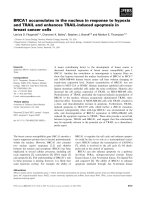
(range: 1.0 to 114.2). No significant difference was observed in the three tumour groups concerning PARP-1 activity. Only the mitotic count score was clearly correlated
with PARP-1 activity, using either the mean (p = 0.007),
median (Figure 1 and Table 1) or the upper quartile limit
(p = 0.03) as cut-off values. In addition, grade significantly
(p = 0.02) correlated with PARP-1 activity using the mean
as cut-off value. Using the mitotic count score as a continuous variable, a weak correlation was found between
the number of mitoses and PARP-1 cytosolic activity
(Spearman correlation coefficient: 0.234, p = 0.003).
BRCA1 promoter methylation
Bisulphite treatment was successfully performed for all
samples. BRCA1 promoter hypermethylation was detected in 18 tumours (Additional file 1: Table S1) and
was significantly associated with the TN status. Indeed,
in 29% (14/48) of TN breast tumours BRCA1 promoter
was hypermethylated compared to 5% (3/57) of HRpositive/HER2-negative and 2% (1/50) of HER2-positive
tumours (Table 1). BRCA1 promoter hypermethylation
was significantly associated also with uPA and PAI-1
levels, grade and mitotic count score and ER-, PR- or
HER2-negative status. No statistical association was
found between BRCA1 promoter hypermethylation and
PARP-1 cytosolic activity.
53BP1 protein expression level
53BP1 protein expression could not be determined in
three tumours, due to insufficient amount of biological
Jacot et al. BMC Cancer 2013, 13:523
/>
Page 5 of 11
Table 1 Patients and tumours characteristics
BRCA1 methylation status
PARP activity (Low < 7 U / mg Protein,
High ≥ 7 U / mg Protein)
Patients’ characteristics
N (%)
Low n (%)
High n (%)
Age at diagnosis (years)
Median (range)
p
Mean ± SD
0.92
p
Methylated
n (%)
Not Methylated
n (%)
0.83
p
0.17
54 (29–75)
≤ 54
80 (51.6%)
38 (52.1%)
42 (51.2%)
11.7 (13)
12 (66.7%)
68 (49.6%)
> 54
75 (48.4%)
35 (47.9%)
40 (48.8%)
12.7 (20.5)
6 (33.3%)
69 (50.4%)
Pre-menopausal
69 (44.5%)
33 (45.2%)
36 (43.9%)
9 (50.0%)
60 (43.8%)
Post-menopausal
86 (55.5%)
40 (54.8%)
46 (56.1%)
9 (50.0%)
77 (56.2%)
Menopausal status
0.87
T classification
0.54
12.5 (13.8)
11.9 (19.3)
0.67
0.62
0.60
0.76
T1
76 (49%)
36 (49.3%)
40 (48.8%)
14 (21.1)
9 (50.0%)
67 (48.9%)
T2
75 (48.4%)
36 (49.3%)
39 (47.6%)
10.4 (12)
9 (50.0%)
66 (48.2%)
T3-4
4 (2.6%)
1 (1.4%)
3 (3.7%)
0
4 (2.9%)
N classification
10 (3.7)
0.75
0.8
0.71
N0
106 (68.4%)
49 (67.1%)
57 (69.5%)
12.4 (18.1)
13 (72.2%)
93 (67.9%)
N+
49 (31.6%)
24 (32.9%)
25 (30.5%)
11.8 (14.6)
5 (27.8%)
44 (32.1%)
Ductal
120 (77.4%)
53 (72.6%)
67 (81.7%)
15 (83.3%)
105 (76.6%)
Histology
0.27
0.3
13.1 (18.3)
0.53
Lobular
9 (5.8%)
4 (5.5%)
5 (6.1%)
7.8 (8.3)
0
9 (6.6%)
Other
26 (16.8%)
16 (21.9%)
10 (12.2%)
9.6 (12.5)
3 (16.7%)
23 (16.8%)
I / II
1 (0.6%) / 51 (32.9%)
28 (38.4%)
24 (29.3%)
2 (11.1%)
50 (36.5%)
III
103 (66.5%)
45 (61.6%)
58 (70.7%)
16 (88.9%)
87 (63.5%)
Grade
0.23
Mitotic count score
0.02
9.2 (12.8)
13.7 (18.7)
0.04
0.03
0.003
0.04
1
31 (20%)
20 (27.4%)
11 (13.4%)
8.3 (13.6)
1 (5.6%)
30 (21.9%)
2
62 (40%)
30 (41.1%)
32 (39.0%)
10.8 (16.2)
5 (27.8%)
57 (41.6%)
3
62 (40%)
23 (31.5%)
39 (47.6%)
12 (66.7%)
50 (36.5%)
ER
15.6 (18.8)
0.88
0.66
0.001
Positive
88 (56.8%)
41 (56.2%)
47 (57.3%)
12.1 (16.9)
4 (22.2%)
84 (61.3%)
Negative
67 (43.2%)
32 (43.8%)
35 (42.7%)
12.4 (17.3)
14 (77.8%)
53 (38.7%)
Positive
59 (38.1%)
28 (38.4%)
31 (37.8%)
2 (11.1%)
57 (41.6%)
Negative
96 (61.9%)
45 (61.6%)
51 (62.2%)
16 (88.9%)
80 (58.4%)
PR
0.94
HER2
0.75
11.5 (14.3)
12.6 (18.6)
0.62
0.01
0.5
0.01
Positive
50 (32.3%)
25 (34.2%)
25 (30.5%)
9.7 (11.3)
1 (5.6%)
49 (35.8%)
Negative
105 (67.7%)
48 (65.8%)
57 (69.5%)
13.4 (19.1)
17 (94.4%)
88 (64.2%)
HER2+
50 (32.3%)
25 (34.2%)
25 (30.5%)
1 (5.6%)
49 (35.8%)
HR+/HER2-
57 (36.7%)
25 (34.2%)
32 (39.0%)
13.4 (19.9)
3 (16.7%)
54 (39.4%)
Triple negative
48 (31%)
23 (31.5%)
25 (30.5%)
13.4 (18.3)
14 (77.8%)
34 (24.8%)
High
97 (62.6%)
43 (58.9%)
54 (65.9%)
12.9 (17)
16 (88.9%)
81 (59.1%)
Low
58 (37.4%)
30 (41.1%)
28 (34.1%)
11 (17.2)
2 (11.1%)
56 (40.9%)
Molecular profile grouping
0.81
uPA level ( ≥3)
0.68
9.7 (11.3)
0.37
<0.001
0.3
0.01
Jacot et al. BMC Cancer 2013, 13:523
/>
Page 6 of 11
Table 1 Patients and tumours characteristics (Continued)
PAI-1 level ( ≥14)
0.32
0.42
0.03
High
112 (72.3%)
50 (68.5%)
62 (75.6%)
11.4 (12.4)
17 (94.4%)
95 (69.3%)
Low
43 (27.7%)
23 (31.5%)
20 (24.4%)
14.2 (25.5)
1 (5.6%)
42 (30.7%)
PARP Activity
0.82
≤ 2.6 U/mg Prot
39 (25.2%)
-
-
-
-
-
4 (22.2%)
35 (25.5%)
2.7 - 7
42 (27.1%)
-
-
-
-
-
6 (33.3%)
36 (26.3%)
7.1 - 14
37 (23.9%)
-
-
-
-
-
3 (16.7%)
34 (24.8%)
> 14
37 (23.9%)
-
-
-
-
-
5 (27.8%)
32 (23.4%)
sample. The mean 53BP1 protein level in the remaining
152 tumours was 12.5 pg/mgP (median: 9.6 pg/mgP;
range: 2.0-93.0 pg/mgP) (Table 2). High (≥9.6 pg/mgP)
53BP1 levels correlated with molecular grouping (63.2% of
HR-positive/HER2-negative vs. 47.9% of HER2-positive
and 36.2% of TN tumours, p = 0.022), lymph node positivity (43.3% of N0 vs. 64.6% of N1+ tumours, p = 0.015), ER
positivity (59.8% of ER-positive vs. 36.9% of ER-negative
tumours, p = 0.005), PR positivity (62.7% of PR-positive vs.
41.9% of PR-negative cancers, p = 0.013), unmethylated
BRCA1 promoter (53% of unmethylated vs. 27.8% of
methylated cancers, p = 0.045) and PARP-1 activity
(60.8% of tumours with high (≥7 U/mg Prot) PARP-1
activity vs. 38.4% of tumours with low (<7 U/mg Prot)
PARP-1 activity, p = 0.006 using PARP-1 median value
as a cut-off; p = 0.048 categorizing PARP-1 values as
quartiles [Table 2]). No correlation was found between
PARP-1 activity and 53BP1 levels using continuous
variables (Additional file 2: Figure S1). Both high 53BP1
levels and BRCA1 promoter hypermethylation were
observed in three TN tumours and two non-TN tumours
(Additional file 1: Table S1).
The BRCA1 / 53BP1/ PARP-1 pathway in triple negative
breast cancers
BRCA1 promoter methylation status, 53BP1 protein
levels and PARP-1 activity in the 48 TN breast cancers
and their clinico-pathologically data are presented in
Additional file 3: Table S2. In this group, only age was
almost negatively associated with BRCA1 promoter
methylation (83.3% of cancers were unmethylated in patients >54 vs. 58.3% in patients ≤54 years, p = 0.057).
PARP-1 activity was not associated with any of the
tested clinico-pathological features. High 53BP1 levels
were significantly associated with lymph node positivity
(24.2% of N0 vs. 64.3% of N1+ cancer, p = 0.009). The association of high 53BP1 and PAI-1 protein levels was almost significant (43.2% of cancers with high vs. 10% of
cancer with low PAI-1 protein levels, p = 0.052). Only
three of the 14 tumours with BRCA1 promoter
Figure 1 Correlation between PARP cytosolic level (logarithmic scale) and the mitotic count score.
Jacot et al. BMC Cancer 2013, 13:523
/>
Page 7 of 11
Table 2 53BP1 protein expression level and correlation with clinico-pathological parameters
53BP1 protein expression level (Low < 9.6 U / mg Protein, High ≥9.6 pg/mg Protein)
Patients’ characteristics
N (%)
Low n (%)
High n (%)
Age at diagnosis (years)
p
1
Median (range)
54 (29–75)
≤ 54
80 (52.6%)
40 (52.6%)
40 (52.6%)
> 54
72 (47.4%)
36 (47.4%)
36 (47.5%)
Menopausal status
0.87
Pre-menopausal
69 (44.7%)
34 (44.7%)
35 (46.1%)
Post-menopausal
83 (55.3%)
42 (55.3%)
41 (53.9%)
T1
75 (49.3%)
37 (48.7%)
38 (50%)
T2
73 (48%)
38 (50%)
35 (46.1%)
T3-4
4 (2.6%)
1 (1.3%)
3 (3.9%)
N0
104 (68.4%)
59 (77.6%)
45 (59.2%)
N+
48 (31.6%)
17 (22.4%)
31 (40.8%)
T classification
0.57
N classification
0.015
Histology
0.44
Ductal
117 (77%)
58 (76.4%)
59 (77.6%)
Lobular
9 (5.9%)
3 (3.9%)
6 (7.9%)
Other
26 (17.1%)
15 (19.7%)
11 (14.5%)
Grade
0.46
I / II
1 (0.7%) / 50 (32.9%)
23 (30.3%)
28 (36.8%)
III
101 (66.4%)
53 (69.7%)
48 (63.2%)
Positive
87 (57.2%)
35 (46.1%)
52 (68.4%)
Negative
65 (42.8%)
41 (53.9%)
24 (31.6%)
ER
0.005
PR
0.013
Positive
59 (38.8%)
22 (28.9%)
37 (48.7%)
Negative
93 (61.2%)
54 (71.1%)
39 (51.3%)
Positive
48 (31.6%)
25 (32.9%)
23 (30.3%)
Negative
104 (68.4%)
51 (67.1%)
53 (69.7%)
HER2
0.73
Molecular profile grouping
0.022
HER2+
48 (31.6%)
25 (32.9%)
23 (30.3%)
HR+/HER2-
57 (37.5%)
21 (27.6%)
36 (47.4%)
Triple Negative
47 (30.9%)
30 (39.5%)
17 (22.4%)
BRCA1 methylation Status
0.045
Methylated
18 (11.8%)
13 (17.1%)
5 (6.6%)
Not Methylated
134 (88.2%)
63 (82.9%)
71 (93.4%)
39 (25.6%)
21 (27.6%)
18 (23.8%)
PARP activity
≤ 2.6 U/mg Prot
0.048
2.7 - 7
41 (27%)
27 (35.5%)
14 (18.4%)
7.1 - 14
36 (23.7%)
14 (18.4%)
22 (28.9%)
> 14
36 (23.7%)
14 (18.4%)
22 (28.9%)
Jacot et al. BMC Cancer 2013, 13:523
/>
hypermethylation had high 53BP1 protein levels. No
clinico-pathological criterion could specifically identify
this breast cancer population.
As recommendations regarding ER and PR cut-offs are
not clearly established worldwide, we used an alternative,
North American, 1% cut-off to define ER and PR positivity /
negativity. Using this 1% threshold, the results were not
significantly modified even if 2 TN cases were reclassified
as HR+/HER2- cases using this alternative cut-off (2 cases
with a 1-9% ER status, none for PR status).
Survival analyses
Survival data were updated on June 10, 2012. At this time,
after a median follow-up of 43.6 months (range 1.9 –
75.7 months), only 2 cancer-related deaths and 2 relapses
were recorded (2 TN patients). The median 3-year OS and
RFS were 0.986 (95% CI 0.954 - 0.999) and 0.986 (95% CI
0.954 - 0.999), respectively. This low number of relapses
and deaths could be explained by a relatively brief followup, altogether with the fact that most of the tumours were
small (pT1) and/or node negative tumours. In addition,
considering the TN population, nearly all of the patients
of this study received adjuvant chemotherapy. Even if the
2 events occurred in the TN population, the low number
of events precludes a statistically robust analysis.
Discussion
This study reports a comprehensive analysis of the BRCA/
53BP1/PARP-1 factors of DNA repair in the largest cohort
of patients with sporadic breast cancer to date. Clinical
studies are currently under way to evaluate the efficacy of
PARPi in patients with TN breast cancer. However, triple
negativity alone does not appear to be a good surrogate
marker for PARPi clinical sensitivity [34] as important biological differences exist within this group of tumours.
Moreover, it is important to know whether sub-population
of HR-positive and HER2-positive patients might also be
eligible for such therapy.
We found that PARP-1 activity correlated only with
the mitotic count score, without statistical association
with BRCA1 promoter hypermethylation. Using IHC,
von Minckwitz et al. retrospectively evaluated the predictive and prognostic value of cytoplasmic (cPARP) and
nuclear PARP (nPARP) expression in 638 pre-treatment
biopsies from neoadjuvant anthracycline/taxane-treated
patients [13]. High cPARP expression was significantly
correlated with non-lobular histology, undifferentiated
grade, positive nodal and negative HR status, but not
with the HER2 status. Expression of cPARP was high in
35.5% of TN tumours, 24.6% of HER2-positive tumours
and 18.0% of HR-positive/HER2-negative tumours. High
cPARP expression was predictive of the achievement of
pathologic complete response, particularly in HR-positive
and HER2-negative tumours, and was a negative, but not
Page 8 of 11
independent prognostic factor of disease-free and overall
survival. No correlation was found for nPARP expression.
Ozretic et al. [41] investigated PARP expression in breast
cancers with BRCA1 (n = 66) or BRCA2 (n = 27) mutations and in 53 sporadic breast cancers. Although they
used the same PARP antibody described by von Minckwitz
et al. [13], they did not observe significant cPARP
staining. Conversely, nPARP expression was significantly
increased in cancers with BRCA1 or BRCA2 mutations
compared to sporadic tumours. No significant increase in
nPARP expression was observed in the few sporadic TN
breast cancers of their cohort. Their results suggest that
nPARP and not cPARP expression is associated with
BRCA-dependent DNA repair deficiency. However, their
results cannot be extrapolated to the whole population of
sporadic TN breast tumours due to the limited sample
size. The results of the study by Rojo et al. [14] are consistent with the findings by Ozteric et al. They quantitatively evaluated nPARP-1 expression using a specific IHC
signal intensity scanning assay in a range of normal to malignant breast lesions, including 330 patients treated for
early breast cancer. nPARP-1 was overexpressed in about
a third of ductal carcinoma in situ and infiltrating breast
cancers and was associated with higher tumour grade, ERnegative tumours and TN phenotype. In this study, Ki-67
staining was used instead of mitotic count. As discrepancies are common between these two methods of proliferation evaluation, [42] a parallel cannot be drawn between
this study and our results on this variable. Finally, multivariate analysis (median follow-up time: 4.8 years) indicated that nPARP-1 overexpression was an independent
prognostic factor for both disease-free (HR 10.05; 95% CI
5.42–10.66) and overall survival (HR 1.82; 95% CI 1.322.52) [14]. These discordant results regarding the association of PARP quantification by IHC with prognosis could
be linked to the fact that the IHC assay used for PARP determination detects both active and catalytically inactive,
auto-modified PARP and not only functionally active
PARP like in our study. However, to date, the question of
the better way to evaluate tumoral PARP-1 activity (functional cytosolic assay as in our study, or morphological
test such as IHC) is still open [13,14,41].
In our series, BRCA1 promoter hypermethylation was
found in 18 tumours and was significantly associated
with a more aggressive clinico-biological profile and
with triple negativity. Indeed, in 29% of TN tumours
BRCA1 promoter was hypermethylated compared to
5% of HR-positive/HER2-negative and 2% of HER2positive tumours, consistent with the 36.7% reported
by Veek et al. in 68 non-inherited TN breast cancers
[36]. Altogether, these results suggest that the analysis
of BRCA1 hypermethylation could be included in the
current and prospective PARPi clinical trials as a potential
predictive biomarker. Wei et al. found a strong correlation
Jacot et al. BMC Cancer 2013, 13:523
/>
between ER promoter and BRCA1 promoter methylation,
suggesting a higher frequency of BRCA1 methylation in
HR-negative breast cancers (no information was available
on the HER2 status of these tumours) [43]. In the study
evaluating the clinical impact of BRCA1 promoter methylation in 135 Bulgarian HR-positive and HR-negative patients, Krasteva et al. reported that hypermethylation was
present in 17.04% of the cases. Surprisingly, patients with
BRCA1 promoter hypermethylation displayed favourable
clinical status as their tumours were smaller in size, lacked
p53 gene mutations and were of lobular type [44]. BRCA1
promoter methylation was not significantly associated
with ER, PR and HER2 status; however an evaluation
of its association with the TN status was not reported.
The presence of BRCA1 promoter hypermethylation
was not significantly associated with better overall survival (HR = 0.47, p = 0.2). No clear explanation of these
discrepancies compared to other publications was proposed by the authors. No conclusion could be issued in
our present study regarding the impact of these biomarkers status on survival, considering the relatively
brief median follow-up of our population. However, this
information will be studied later, after a significantly
longer follow-up, allowing the occurrence of more
events. Finally, we show that 53BP1 protein expression
levels was significantly correlated with molecular grouping (63.2% of HR-positive/HER2-negative vs. 47.9% of
HER2-positive and 36.2% of TN tumours) and unmethylated BRCA1 promoter (53% of unmethylated vs.
27.8% of methylated cancers). Regarding definition of
ER and PR positivity, recommendations regarding ER
and PR cut-offs are not clearly established worldwide.
We used in this study an European 10% cut-off to consider positive or negative ER and PR status [45]. This 10%
cut-off can be considered as a standard of care in many
countries. The 1% cut-off can be considered as another,
North American, standard. However, our results were not
significantly modified by the use of this 1% threshold for
ER (2 TN cases) and PR positivity (no TN cases), and thus
cannot be explained by the use of one or another ER/PR
positivity threshold.
Conclusions
In our study, the association of BRCA1 promoter methylation and high 53BP1 protein levels was a rare event, even
in the TN group. As this association appears to be the best
situation to predict PARPi clinical activity (because loss of
53BP1 leads to partial restoration of homologous recombination and resistance to PARPi) [33] these results pledge
for a strict selection of the target population of future
trials involving these agents, and could, at the same
time, explain the negative results of previous trials that
did not include such strict selection [46]. A retrospective
analysis of BRCA1 promoter methylation and 53BP1
Page 9 of 11
protein levels in the patients enrolled in such trials
could help confirm the predictive impact of this tumour
profile. In addition, evaluation of the 53BP1 protein
levels in cases harbouring deleterious mutations in
other less common homologous recombination genes
with moderate penetrance, such as PALB2 [47,48], need
to be performed, as well as determination of the PALB2
methylation status of this gene in PALB2 non-mutated
cases, as PALB2-deficient cells appears to be sensitive to
PARPi [49].
Additional files
Additional file 1: Table S1. Patients and Tumours Characteristics of the
18 breast cancers with BRCA1 promoter methylation.
Additional file 2: Figure S1. Correlation between PARP-1 activity and
53BP1 levels.
Additional file 3: Table S2. Patients and Tumours Characteristics of the
48 triple negative breast cancers.
Abbreviations
53BP1: P53 binding protein 1; BRCA1: Breast cancer type 1 susceptibility
protein; CISH: Chromogenic in situ hybridization; DNA: DeoxyriboNucleic
acid; DSB: Double strand break; EDTA: EthyleneDiamineTetraacetic acid;
ER: Estrogen receptor; FISH: Fluorescent in situ hybridization; Grade: Scarf,
bloom and Richardson scoring method, modified as proposed by Elston and
Ellis; HER2: Human epidermal growth factor receptor 2; HR: Hormone
receptors; IHC: ImmunoHistoChemistry; NHEJ: Non-homologous end joining;
PAI-1: Plasminogen activator inhibitor type 1; PARP-1: Poly(ADP-Ribose)
polymerase 1; PARPi: PARP inhibitor; PCR: Polymerase chain reaction;
PR: Progesterone receptor; SSB: Single-strand breaks; TN: Triple negative
breast cancers; uPA: urokinase-type plasminogen activator.
Competing interests
The authors declare that they have no competing interests.
Authors’ contributions
WJ participated in the conception and design of the study, provided study
patients and material, collected, assembled and interpreted the data and
drafted the manuscript. ST participated in the conception and design of the
study, participated in the design of the study and performed the statistical
analysis. RS collected and assembled the data and carried out the assays. CV
collected and assembled the data and participated in the assays. ACL
collected and assembled the data and carried out the assays. ELC collected
and assembled the data and carried out the assays. FB provided study
patients and material, collected and assembled the data. JPB participated in
the conception and design of the study and helped to draft the manuscript.
GR participated in the conception and design of the study, provided study
patients and material and helped to draft the manuscript. PJL participated in
the conception and design of the study, provided study material, participated
in the assays, collected, assembled and interpreted the data and helped to
draft the manuscript. All authors read and approved the final manuscript.
Acknowledgments
The authors want to thank Martine Pascal and Isabelle Pantel for their
technical assistance.
This study was supported by a research grant from Sanofi-Aventis France.
Part of this study was presented at the 2012 and 2013 AACR Annual Meetings.
Author details
1
Department of Medical Oncology, Montpellier Cancer Institute, Montpellier,
France. 2Translational Research Unit, Montpellier Cancer Institute, 208 rue des
Apothicaires, 34298 Montpellier Cedex 5, France. 3Department of Biostatistics,
Montpellier Cancer Institute, Montpellier, France. 4Department of Biology and
Oncogenetic, Montpellier Cancer Institute, Montpellier, France. 5Department of
Pathology, Montpellier Cancer Institute, Montpellier, France.
Jacot et al. BMC Cancer 2013, 13:523
/>
Received: 28 June 2013 Accepted: 23 October 2013
Published: 5 November 2013
References
1. Liu X, Palma J, Kinders R, Shi Y, Donawho C, Ellis PA, Rodriguez LE, ColonLopez M, Saltarelli M, LeBlond D, et al: An enzyme-linked immunosorbent
poly(ADP-ribose) polymerase biomarker assay for clinical trials of PARP
inhibitors. Anal Biochem 2008, 381(2):240–247.
2. Helleday T, Petermann E, Lundin C, Hodgson B, Sharma RA: DNA repair
pathways as targets for cancer therapy. Nat Rev Cancer 2008, 8(3):193–204.
3. Tutt A, Robson M, Garber JE, Domchek SM, Audeh MW, Weitzel JN,
Friedlander M, Arun B, Loman N, Schmutzler RK, et al: Oral poly(ADPribose) polymerase inhibitor olaparib in patients with BRCA1 or BRCA2
mutations and advanced breast cancer: a proof-of-concept trial. Lancet
2010, 376(9737):235–244.
4. Wooster R, Weber BL: Breast and ovarian cancer. N Engl J Med 2003,
348(23):2339–2347.
5. D’Amours D, Desnoyers S, D’Silva I, Poirier GG: Poly(ADP-ribosyl)ation
reactions in the regulation of nuclear functions. Biochem J 1999,
342(Pt 2):249–268.
6. Schreiber V, Dantzer F, Ame JC, de Murcia G: Poly(ADP-ribose): novel
functions for an old molecule. Nat Rev Mol Cell Biol 2006, 7(7):517–528.
7. Ame JC, Spenlehauer C, de Murcia G: The PARP superfamily. Bioessays
2004, 26(8):882–893.
8. Bryant HE, Schultz N, Thomas HD, Parker KM, Flower D, Lopez E, Kyle S,
Meuth M, Curtin NJ, Helleday T: Specific killing of BRCA2-deficient
tumours with inhibitors of poly(ADP-ribose) polymerase. Nature 2005,
434(7035):913–917.
9. Farmer H, McCabe N, Lord CJ, Tutt AN, Johnson DA, Richardson TB,
Santarosa M, Dillon KJ, Hickson I, Knights C, et al: Targeting the DNA repair
defect in BRCA mutant cells as a therapeutic strategy. Nature 2005,
434(7035):917–921.
10. Curtin NJ: PARP inhibitors for cancer therapy. Expert Rev Mol Med 2005,
7(4):1–20.
11. Curtin N: Therapeutic potential of drugs to modulate DNA repair in
cancer. Expert Opin Ther Targets 2007, 11(6):783–799.
12. Domagala P, Huzarski T, Lubinski J, Gugala K, Domagala W: PARP-1
expression in breast cancer including BRCA1-associated, triple negative
and basal-like tumors: possible implications for PARP-1 inhibitor therapy.
Breast Cancer Res Treat 2011, 127(3):861–869.
13. von Minckwitz G, Muller BM, Loibl S, Budczies J, Hanusch C, Darb-Esfahani S,
Hilfrich J, Weiss E, Huober J, Blohmer JU, et al: Cytoplasmic poly(adenosine
diphosphate-ribose) polymerase expression is predictive and prognostic
in patients with breast cancer treated with neoadjuvant chemotherapy.
J Clin Oncol 2011, 29(16):2150–2157.
14. Rojo F, Garcia-Parra J, Zazo S, Tusquets I, Ferrer-Lozano J, Menendez S, Eroles P,
Chamizo C, Servitja S, Ramirez-Merino N, et al: Nuclear PARP-1 protein
overexpression is associated with poor overall survival in early breast
cancer. Ann Oncol 2012, 23(5):1156–1164.
15. Tuma RS: PARP inhibitors: will the new class of drugs match the hype?
J Natl Cancer Inst 2009, 101(18):1230–1232.
16. Blackwood MA, Weber BL: BRCA1 and BRCA2: from molecular genetics to
clinical medicine. J Clin Oncol 1998, 16(5):1969–1977.
17. Miki Y, Katagiri T, Kasumi F, Yoshimoto T, Nakamura Y: Mutation analysis in
the BRCA2 gene in primary breast cancers. Nat Genet 1996, 13(2):245–247.
18. Miki Y, Swensen J, Shattuck-Eidens D, Futreal PA, Harshman K, Tavtigian S,
Liu Q, Cochran C, Bennett LM, Ding W, et al: A strong candidate for the
breast and ovarian cancer susceptibility gene BRCA1. Science 1994,
266(5182):66–71.
19. Bal A, Verma S, Joshi K, Singla A, Thakur R, Arora S, Singh G: BRCA1methylated sporadic breast cancers are BRCA-like in showing a basal
phenotype and absence of ER expression. Virchows Arch 2012,
461(3):305–312.
20. Birgisdottir V, Stefansson OA, Bodvarsdottir SK, Hilmarsdottir H, Jonasson JG,
Eyfjord JE: Epigenetic silencing and deletion of the BRCA1 gene in
sporadic breast cancer. Breast Cancer Res 2006, 8(4):R38.
21. Lips EH, Mulder L, Hannemann J, Laddach N, Vrancken Peeters MT, van de
Vijver MJ, Wesseling J, Nederlof PM, Rodenhuis S: Indicators of
homologous recombination deficiency in breast cancer and association
with response to neoadjuvant chemotherapy. Ann Oncol 2011,
22(4):870–876.
Page 10 of 11
22. Mirza S, Sharma G, Prasad CP, Parshad R, Srivastava A, Gupta SD, Ralhan R:
Promoter hypermethylation of TMS1, BRCA1, ERalpha and PRB in serum
and tumor DNA of invasive ductal breast carcinoma patients. Life Sci
2007, 81(4):280–287.
23. Wei M, Grushko TA, Dignam J, Hagos F, Nanda R, Sveen L, Xu J, Fackenthal J,
Tretiakova M, Das S, et al: BRCA1 promoter methylation in sporadic breast
cancer is associated with reduced BRCA1 copy number and chromosome
17 aneusomy. Cancer Res 2005, 65(23):10692–10699.
24. Lips EH, Mulder L, Oonk A, van der Kolk LE, Hogervorst FB, Imholz AL,
Wesseling J, Rodenhuis S, Nederlof PM: Triple-negative breast cancer:
BRCAness and concordance of clinical features with BRCA1-mutation
carriers. Br J Cancer 2013, 108(10):2172–2177.
25. Birkbak NJ, Wang ZC, Kim JY, Eklund AC, Li Q, Tian R, Bowman-Colin C, Li Y,
Greene-Colozzi A, Iglehart JD, et al: Telomeric allelic imbalance indicates
defective DNA repair and sensitivity to DNA-damaging agents. Cancer
Discov 2012, 2(4):366–375.
26. Wang B, Matsuoka S, Carpenter PB, Elledge SJ: 53BP1, a mediator of the
DNA damage checkpoint. Science 2002, 298(5597):1435–1438.
27. Ward I, Kim JE, Minn K, Chini CC, Mer G, Chen J: The tandem BRCT domain
of 53BP1 is not required for its repair function. J Biol Chem 2006,
281(50):38472–38477.
28. FitzGerald JE, Grenon M, Lowndes NF: 53BP1: function and mechanisms of
focal recruitment. Biochem Soc Trans 2009, 37(Pt 4):897–904.
29. Huo Q, Yang Q: P53-binding protein 1: a new player for tumorigenesis
and a new target for breast cancer treatment. Med Hypotheses 2011,
77(3):359–363.
30. Bouwman P, Aly A, Escandell JM, Pieterse M, Bartkova J, van der Gulden H,
Hiddingh S, Thanasoula M, Kulkarni A, Yang Q, et al: 53BP1 loss rescues
BRCA1 deficiency and is associated with triple-negative and BRCAmutated breast cancers. Nat Struct Mol Biol 2010, 17(6):688–695.
31. Bunting SF, Callen E, Wong N, Chen HT, Polato F, Gunn A, Bothmer A,
Feldhahn N, Fernandez-Capetillo O, Cao L, et al: 53BP1 inhibits homologous recombination in Brca1-deficient cells by blocking resection of
DNA breaks. Cell 2010, 141(2):243–254.
32. Aly A, Ganesan S: BRCA1, PARP, and 53BP1: conditional synthetic lethality
and synthetic viability. J Mol Cell Biol 2011, 3(1):66–74.
33. Jaspers JE, Kersbergen A, Boon U, Sol W, van Deemter L, Zander SA, Drost R,
Wientjens E, Ji J, Aly A, et al: Loss of 53BP1 causes PARP inhibitor resistance
in Brca1-mutated mouse mammary tumors. Cancer Discov 2013, 3(1):68–81.
34. Gelmon KA, Tischkowitz M, Mackay H, Swenerton K, Robidoux A, Tonkin K,
Hirte H, Huntsman D, Clemons M, Gilks B, et al: Olaparib in patients with
recurrent high-grade serous or poorly differentiated ovarian carcinoma
or triple-negative breast cancer: a phase 2, multicentre, open-label,
non-randomised study. Lancet Oncol 2011, 12(9):852–861.
35. Drew Y, Mulligan EA, Vong WT, Thomas HD, Kahn S, Kyle S, Mukhopadhyay
A, Los G, Hostomsky Z, Plummer ER, et al: Therapeutic potential of poly
(ADP-ribose) polymerase inhibitor AG014699 in human cancers with
mutated or methylated BRCA1 or BRCA2. J Natl Cancer Inst 2011,
103(4):334–346.
36. Veeck J, Ropero S, Setien F, Gonzalez-Suarez E, Osorio A, Benitez J, Herman JG,
Esteller M: BRCA1 CpG island hypermethylation predicts sensitivity to
poly(adenosine diphosphate)-ribose polymerase inhibitors. J Clin Oncol
2010, 28(29):e563–564. author reply e565-566.
37. Lamy PJ, Verjat T, Servanton AC, Paye M, Leissner P, Mougin B: Urokinasetype plasminogen activator and plasminogen activator inhibitor type-1
mRNA assessment in breast cancer by means of NASBA: correlation with
protein expression. Am J Clin Pathol 2007, 128(3):404–413.
38. Elston CW, Ellis IO: Pathological prognostic factors in breast cancer. I. The
value of histological grade in breast cancer: experience from a large
study with long-term follow-up. Histopathology 1991, 19(5):403–410.
39. Mallett S, Timmer A, Sauerbrei W, Altman DG: Reporting of prognostic
studies of tumour markers: a review of published articles in relation to
REMARK guidelines. Br J Cancer 2010, 102(1):173–180.
40. Esteller M, Silva JM, Dominguez G, Bonilla F, Matias-Guiu X, Lerma E,
Bussaglia E, Prat J, Harkes IC, Repasky EA, et al: Promoter hypermethylation
and BRCA1 inactivation in sporadic breast and ovarian tumors. J Natl Cancer
Inst 2000, 92(7):564–569.
41. Ozretic L, Rhiem K, Huss S, Wappenschmidt B, Markiefka B, Sinn P,
Schmutzler RK, Buettner R: High nuclear poly(adenosine diphosphateribose) polymerase expression is predictive for BRCA1- and BRCA2deficient breast cancer. J Clin Oncol 2011, 29(34):4586–4588.
Jacot et al. BMC Cancer 2013, 13:523
/>
Page 11 of 11
42. Jalava P, Kuopio T, Juntti-Patinen L, Kotkansalo T, Kronqvist P, Collan Y: Ki67
immunohistochemistry: a valuable marker in prognostication but with a
risk of misclassification: proliferation subgroups formed based on Ki67
immunoreactivity and standardized mitotic index. Histopathology 2006,
48(6):674–682.
43. Wei M, Xu J, Dignam J, Nanda R, Sveen L, Fackenthal J, Grushko TA,
Olopade OI: Estrogen receptor alpha, BRCA1, and FANCF promoter
methylation occur in distinct subsets of sporadic breast cancers. Breast
Cancer Res Treat 2008, 111(1):113–120.
44. Krasteva ME, Bozhanov SS, Antov GG, Gospodinova ZI, Angelov SG: Breast
cancer patients with hypermethylation in the promoter of BRCA1 gene
exhibit favorable clinical status. Neoplasma 2012, 59(1):85–91.
45. Goldhirsch A, Glick JH, Gelber RD, Coates AS, Thurlimann B, Senn HJ, Panel
M: Meeting highlights: international expert consensus on the primary
therapy of early breast cancer 2005. Ann Oncol 2005, 16(10):1569–1583.
46. O’Shaughnessy JS, Schwartzberg L, Danso MA, Rugo HS, Miller K, Yardley
DA, Carlson RW, Finn RS, Charpentier E, Freese M: A Randomized Phase III
Study of Iniparib (BSI-201) in Combination with Gemcitabine/Carboplatin
(G/C) in Metastatic Triple-Negative Breast Cancer (TNBC). In American
Society of Clinical Oncology: June 3–7, 2011. Chicago, Illinois: J Clin Oncol;
2011. 2011: Abstract 1007.
47. Hellebrand H, Sutter C, Honisch E, Gross E, Wappenschmidt B, Schem C,
Deissler H, Ditsch N, Gress V, Kiechle M, et al: Germline mutations in the
PALB2 gene are population specific and occur with low frequencies in
familial breast cancer. Hum Mutat 2011, 32(6):E2176–2188.
48. Poumpouridou N, Kroupis C: Hereditary breast cancer: beyond BRCA
genetic analysis; PALB2 emerges. Clin Chem Lab Med 2012, 50(3):423–434.
49. Buisson R, Dion-Cote AM, Coulombe Y, Launay H, Cai H, Stasiak AZ, Stasiak
A, Xia B, Masson JY: Cooperation of breast cancer proteins PALB2 and
piccolo BRCA2 in stimulating homologous recombination. Nat Struct Mol
Biol 2010, 17(10):1247–1254.
doi:10.1186/1471-2407-13-523
Cite this article as: Jacot et al.: BRCA1 promoter hypermethylation,
53BP1 protein expression and PARP-1 activity as biomarkers of DNA
repair deficit in breast cancer. BMC Cancer 2013 13:523.
Submit your next manuscript to BioMed Central
and take full advantage of:
• Convenient online submission
• Thorough peer review
• No space constraints or color figure charges
• Immediate publication on acceptance
• Inclusion in PubMed, CAS, Scopus and Google Scholar
• Research which is freely available for redistribution
Submit your manuscript at
www.biomedcentral.com/submit