Circulating tumor cells in HER2-positive metastatic breast cancer patients: A valuable prognostic and predictive biomarker
Bạn đang xem bản rút gọn của tài liệu. Xem và tải ngay bản đầy đủ của tài liệu tại đây (834.09 KB, 8 trang )
Liu et al. BMC Cancer 2013, 13:202
/>
RESEARCH ARTICLE
Open Access
Circulating tumor cells in HER2-positive
metastatic breast cancer patients: a valuable
prognostic and predictive biomarker
Yi Liu1,2†, Qian Liu1†, Tao Wang1, Li Bian1, Shaohua Zhang1, Haixu Hu2, Sha Li2, Zhiyuan Hu3, Shikai Wu1,
Bing Liu2* and Zefei Jiang1*
Abstract
Background: This study was initiated to investigate the prognostic significance of circulating tumor cell (CTC)
enumeration and the predictive value of CTC HER2 expression for efficient anti-HER2 therapy in HER2-positive
metastatic breast cancer (MBC) patients.
Methods: Sixty HER2-positive MBC patients were enrolled in the present study. Before the initiation of systemic
treatment, CTCs from 7.5 ml of blood were analyzed using the CellSearch system. The progression-free survival (PFS)
of the patients was estimated using Kaplan-Meier survival curves.
Results: CTCs were detected in 45% (27/60) of the patients, who had shorter median PFS than those without CTCs
(2.5 vs. 7.5 months, P = 0.0125). Furthermore, referring to the standard HER2 testing that uses immunohistochemistry
(IHC), we proposed a CTC HER2-positive criterion, defined as >30% of CTCs over-expressing HER2. Among patients
undergoing anti-HER2 therapy, those with HER2-positive CTCs had longer PFS (8.8 vs. 2.5 months, P = 0.002). Among
patients with HER2-positive CTCs, the median PFS for those receiving anti-HER2 therapy was significantly longer
than those who were not (8.8 vs. 1.5 months, P = 0.001). Notably, up to 52% (14/27) of the HER2-positive patients
were CTC HER2-negative, and anti-HER2 therapy did not significantly improve the median PFS in these patients
(2.5 vs. 0.9 months, P = 0.499).
Conclusions: Our findings underscore the necessity of a comprehensive CTC analysis, which may provide valuable
prognostic and predictive information for optimizing individually tailored therapies in HER2-positive MBC patients.
To test this idea, additional large cohort, multi-center and prospective clinical trials are needed.
Background
Human epidermal growth factor receptor 2 (HER2) is a
185 kDa transmembrane tyrosine kinase receptor encoded
by the HER2 gene on chromosome 17q21. HER2 overexpression or amplification occurs in approximately 20%
of all breast cancer patients and is associated with aggressive growth, short survival and poor prognosis [1-3].
HER2 positivity correlates with the clinical outcome of
treatment with anti-HER2 agents such as trastuzumab
* Correspondence: ;
†
Equal contributors
2
Translational Medicine Center, Laboratory of Oncology, Affiliated Hospital of
Academy of Military Medical Sciences, No.8 Dongdajie, Beijing 100071, China
1
Department of Breast Cancer, Affiliated Hospital of Academy of Military Medical
Sciences, No.8 Dongdajie, Beijing 100071, China
Full list of author information is available at the end of the article
(Herceptin, Genentech, South San Francisco, CA, USA)
and lapatinib (Tykerb, GSK, Philadelphia, PA, USA) [4-7].
Therefore, HER2 is considered to be a vital prognostic and
predictive factor, and treatment of HER2-positive patients
remains one of the great therapeutic challenges in metastatic breast cancer (MBC).
Despite therapeutic advances over the past decades, individually tailored therapeutic regimens for HER2-positve
patients remain far from satisfactory. For example, the
benefit of single-agent anti-HER2 therapy, in the form of
either trastuzumab or lapatinib, is only in the range of
~25% [8]. There are three possible explanations for this
phenomenon. First, HER2-positive MBC patients may be
divided into subgroups with different prognoses. Second,
the initial assessment of HER2-positivity may be inaccurate
due to the inherent limitations of traditional methods,
© 2013 Liu et al.; licensee BioMed Central Ltd. This is an Open Access article distributed under the terms of the Creative
Commons Attribution License ( which permits unrestricted use, distribution, and
reproduction in any medium, provided the original work is properly cited.
Liu et al. BMC Cancer 2013, 13:202
/>
including tumor heterogeneity, subjectivity in the interpretation of results or technical limitations such as variability
in tissue processing and reagents [9]. Third, previous studies have demonstrated that there are inconsistencies in
HER2 expression between primary tumors and their metastases [10,11]. Such inconsistencies indicate that tumor
cells are under constant evolvement or clonal selection and
that the detected HER2 status may not necessarily reflect
the patients’ real-time phenotypes. Therefore, it is critical
to discover more precise prognostic marker and real-time
methods for HER2 testing to optimize individualized therapeutic regimens for HER2-positive MBC patients.
Circulating tumor cells (CTCs) are cells that shed
from the tumor and enter the circulation, a process that
is required for cancer metastasis. Considerable efforts
have been made to develop technologies for CTC detection and characterization; among these technologies, the
CellSearch system (Veridex LLC, Raritan, NJ, USA) is
the only one approved by US Food and Drug Administration (FDA) for clinical use in treating MBC [12,13].
CellSearch has a CTC detection cutoff of ≥5 cells/7.5 ml
blood, and it has been demonstrated to be an independent prognostic factor for the prediction of progressionfree survival (PFS) and the overall survival (OS) of MBC
patients [14-17]. In addition to detection, the molecular
characterization of CTCs is now recognized as a valuable
tool that can provide real-time information to distinguish subgroups of patients who can benefit from certain types of therapy [18,19]. Unfortunately, previous
studies failed to illustrate the prognostic value of CTCs
in HER2-positive MBC patients using CellSearch [20].
Although recent studies have made great efforts to compare HER2 statuses between tumor tissue and CTCs
[18,19,21-29], a satisfactory CTC HER2-positive criterion has not yet been established.
In the present study, we demonstrated that CTC enumeration using a modified cutoff has superior prognostic
value for HER2-positive MBC. Furthermore, we proposed that HER2 positivity should be determined by
both the HER2 intensity of individual CTCs and the percentage of CTCs with that intensity. Using the criterion
defined as >30% of CTCs over-expressing HER2, we
found that HER2 expression in CTCs was different from
that in tumor tissues, and this expression could significantly improve the response prediction for anti-HER2
therapy.
Methods
Study design
Patients who showed clinical and radiological evidence
of metastatic breast cancer were randomly enrolled in
the present study. Eligible patients were required to
have measurable or evaluable disease, with an Eastern
Cooperative Oncology Group (ECOG) performance
Page 2 of 8
status score of 0 to 3 and a pathology report describing
their histological type and nodal status, as well as their
estrogen receptor (ER), progesterone receptor (PgR),
and HER2 statuses. A patient was considered HER2positive with an immunohistochemistry (IHC) score of
3+ or a fluorescent in situ hybridization (FISH) ratio of
more than 2.2. IHC scores of 0 and 1+ or a FISH ratio
of less than 1.8 were considered to be HER2-negative
[30].
Before the start of a new line or a new therapy cycle,
10 ml of blood was drawn. An interval of <7 days between the day of blood sampling and the initiation of
systemic treatment was required. All treatment decisions
for the patients were made according to the National
Comprehensive Cancer Network (NCCN) clinical practice guidelines (Breast Cancer V.2.2010) without knowing the patients’ CTC results. Disease status was
assessed and categorized according to Response Evaluation Criteria in Solid Tumors (RECIST). After several
months of follow-up, the relationship between the quantity and characteristics of CTCs and clinical outcome
was analyzed retrospectively.
All patients signed an informed consent to participate
in the study, which was approved by the ethics and scientific committees of the Affiliated Hospital of the Academy of Military Medical Sciences.
Isolation, enumeration and characterization of CTCs
CTC isolation, enumeration and characterization were
performed using the CellSearch system, according to
the manufacturer’s instructions as described elsewhere
[14-17]. Briefly, cells that expressed the epithelial cell adhesion molecule (EpCAM) were immunomagnetically
enriched by the semiautomated sample preparation system that was provided with the CellSearch epithelial cell
kit. The isolated cells were then automatically labeled
with fluorescently tagged monoclonal antibodies specific
for leukocytes (CD45-allophycocyanin) and epithelial
cells (cytokeratins (CK) 8-, 18- and 19-phycoerythrin)
and were stained with the nucleic acid dye 4′,6diamidino-2-phenylindole (DAPI). HER2 expression in
CTCs was assessed by staining the cells with a fluorescein isothiocyanate (FITC)-labeled anti-HER2 antibody
(Veridex LLC, Raritan, NJ, USA). The intensity of HER2
expression on CTCs was given a score of 0 (negative),
1+ (weak), 2+ (moderate or questionable), or 3+ (strong)
according to criteria described elsewhere [28,29].
According to tissue HER2 positive criterion using
immunohistochemistry (IHC) [30], which detects
HER2 protein expression with a similar technological
principle as immunofluorescent (IF), we set two
criteria for HER2 positivity in CTCs: either >30%
or >10% of CTCs over-expressing HER2. We then
Liu et al. BMC Cancer 2013, 13:202
/>
Page 3 of 8
analyzed the clinical outcome of the patients based on
these two criteria.
Statistical analysis
Fisher’s exact test was used to test whether there was a
statistically significant difference between the number of
patients with a cut-off of 5 CTCs and those with 1 CTC as
baseline. PFS was defined as the time elapsed from the initial blood sampling to the documentation of disease progression (according to RECIST) or, if no progression was
observed during the follow-up, to the last follow-up visit.
Kaplan-Meier survival curves were generated based on the
CTC levels at baseline and the HER2 status of CTCs, and
the curves were compared using the log-rank test.
McNemar’s test was used to determine whether a
statistically significant difference existed regarding variations in HER2 status between CTCs and histological
results. P values <0.05 were considered statistically significant. Analyses were carried out using SAS software
version 9.1.3 (SAS Institute Inc., Cary, NC, USA).
Results
Patient characteristics and CTC enumeration
From September 2010 to August 2011, 60 HER2-positive
MBC patients with a mean age of 49 years (range: 25 to 75
years) were enrolled in the present study. In addition, 11
HER2-negative MBC patients (10 of whom were ERpositive) were enrolled as a control group. The pathological and clinical characteristics of the patients are listed
in Table 1 and Additional file 1: Table S1, respectively. As
Table 1 Pathological and clinical characteristics of HER2-positive patients at baseline
Characteristics
Total
P
No. patients (%)
CTC Count at baseline
≥1
Overall
P
No. patients (%)
CTC Count at baseline
≥5
<1
60
27 (45.0)
33 (55.0)
Mean
48.8
46.6
50.6
Range
25-75
25-68
33-75
Ductal
49
24 (49.0)
25 (51.0)
Lobular
3
0 (0.0)
3 (100.0)
Others
8
3 (37.5)
5 (62.5)
Positive
24
9 (37.5)
15 (62.5)
Negative
36
18 (50.0)
18 (50.0)
Positive
22
9 (40.9)
13 (59.1)
Negative
38
18 (47.4)
20 (52.6)
<5
12 (20.0)
48 (80.0)
46.6
49.4
25-58
32-75
10 (20.4)
39 (79.6)
0 (0.0)
3 (100.0)
2 (25.0)
6 (75.0)
4 (16.7)
20 (83.3)
8 (22.2)
28 (77.8)
4 (18.2)
18 (81.8)
8 (21.1)
30 (78.9)
Age (years)
0.155
0.420
Histology
0.130
0.481
ER
0.430
0.746
PR
0.789
1.000
No. of Metastasis
1
19
5 (26.3)
14 (73.7)
≥2
41
22 (53.7)
19 (46.3)
4
2 (50.0)
2 (50.0)
0.057
1 (5.3)
18 (94.7)
11 (26.8)
30 (73.2)
0 (0.0)
4 (100.0)
0.082
Metastatic sites
Bone only
Visceral only
7
2 (28.6)
5 (71.4)
30
18 (60.0)
12 (40.0)
≤12 months
11
3 (27.3)
8 (72.7)
>12 months
39
19 (48.7)
20 (51.3)
1
15
7 (46.7)
2
9
2 (22.2)
3
13
7 (53.8)
≥4
23
11 (47.8)
Bone and visceral
0.314
1 (14.3)
6 (85.7)
8 (26.7)
22 (73.3)
3 (27.3)
8 (72.7)
7 (17.9)
32 (82.1)
8 (53.3)
3 (20.0)
12 (80.0)
7 (77.8)
1 (11.1)
8 (88.9)
6 (46.2)
1 (7.7)
12 (92.3)
12 (52.2)
7 (30.4)
16 (69.6)
0.270
DFS
0.306
0.671
Therapy line
0.470
0.329
Liu et al. BMC Cancer 2013, 13:202
/>
shown in Additional file 2: Table S2, CTCs were detected
in 45% (27/60) of the HER2-positive patients, and the
CTC count ranged from 1 to 1140 with a mean value of
68. Of the HER2-positive patients with detectable CTCs,
56% (15/27) had a CTC count that ranged from 1 to 4. In
contrast, CTCs were detected in 80.0% (8/10) of the ERpositive/HER2-negative patients, and only 25.0% (2/8) of
those patients had a CTC count that ranged from 1 to 4.
Two different cutoffs were used to divide patients into
two groups based on the CTC count at the initial blood
draw: the first cutoff was ≥1 CTC, and the other was ≥5
CTCs. There were no statistically significant differences
between the two groups in terms of age, histology, status
of hormone receptors (HRs) such as ER and PR, metastatic sites and numbers, disease-free survival (DFS), and
therapy line.
Prognostic significance of CTC enumeration
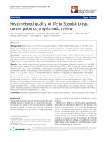
At the 10-month follow-up visit, 57% (34/60) of the patients exhibited disease progression. Using a cut-off of ≥5
CTCs, no significant difference was found in the median
PFS between the two groups (3.3 vs. 5.1 months, P =
0.4563, Figure 1A), consistent with a previous report [20].
Considering the lower detection rate of CTCs in HER2positive patients described above and previously [19,20],
we used a lower cut-off and found that patients with ≥1
CTC had a significantly shorter median PFS than those
with <1 CTC (2.5 vs. 7.5 months, P = 0.0125, Figure 1B).
We also analyzed the median PFS for groups divided
based on cut-offs of ≥2, ≥3 and ≥4 CTCs, but we found no
significant differences (Additional file 3: Figure S1).
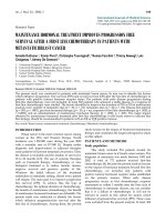
HER2 expression on CTCs
HER2 expression intensity in CTCs was given a score of
0, 1+, 2+, or 3+, according to the criteria described
previously [28,29], and representative images are shown
Page 4 of 8
in Figure 2. Additional file 2: Table S2 presents the
percentages of CTCs at given HER2 intensity scores in
both the HER2-positive and HER2–negative groups.
With the positive criterion defined as >30% of CTCs
over-expressing HER2 (3+), the positive and negative
coincidence rates of CTC HER2 were 48% (13/27) and
100% (9/9), respectively, compared with tumor tissue.
McNemar’s test demonstrated that the HER2 status of
CTCs was significantly different from that of tumor tissues (Table 2, χ2 = 12.07, P = 0.0005).
HER2 Expression in CTCs as a tool for predicting antiHER2 therapy efficacy
Twenty-seven patients with a CTC count ≥1 were divided
into 4 groups based on their CTC HER2 status and
whether they were receiving anti-HER2 therapy. Groups 1
and 2 consisted of patients with HER2 3+ CTC >30%, and
groups 3 and 4 consisted of patients with HER2 3+ CTC
≤30%. Although all patients were histologically positive for
HER2 and therefore should have received anti-HER2 therapy, patients in groups 2 and 4 did not receive the treatment for economic reasons. Kaplan-Meier plots of the
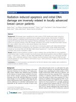
PFS values for all of the groups are shown in Figure 3.
Statistical analysis demonstrated that among the patients
who received anti-HER2 therapy (N = 18, groups 1 and 3),
only those with HER2-positive CTCs have benefited (8.8
vs. 2.5 months, P = 0.002). Among the patients with
HER2-positive CTCs (N = 13, groups 1 and 2), the median
PFS for those receiving anti-HER2 therapy was significantly longer than that for those without anti-HER2 therapy (8.8 vs. 1.5 months, P = 0.001). Notably, up to 52%
(14/27) of the patients who were histologically assessed as
HER2-positive had HER2-negative CTCs (N = 14, groups
3 and 4), and anti-HER2 therapy did not significantly improve the median PFS for these patients (2.5 vs. 0.9
months, P = 0.499). In addition, we also compared the PFS of
Figure 1 Kaplan-Meier PFS plots of HER2-positive MBC patients with a cut-off of ≥ 5 (A) and ≥1 (B) CTCs. PFS was calculated from the
time of the baseline blood draw. Coordinates of the dashed lines indicate median survival time.
Liu et al. BMC Cancer 2013, 13:202
/>
Page 5 of 8
Figure 2 Representative images for the 0, 1+, 2+, and 3+ intensities of HER2 expression on CTCs.
HER2 3+ >10% vs. < 10%, HER2 3+ vs. HER2 (2+ and 1+)
as well as HER2 (3+ and 2+) vs. HER2 (1+ and 0), but
found no significant difference (data not shown).
Discussion
In this study, we found that CTC enumeration with a
cut-off of ≥1 but not ≥5 CTCs could serve as a useful
prognostic factor for HER2-positive MBC patients. CTC
enumeration using CellSearch (with a cut-off of ≥5
CTCs) is widely accepted as a prognostic factor for
MBC patients [14-17]; however, its prognostic power for
the HER2-positive subgroup seems to be inadequate
Table 2 Comparison of HER2 status between tumor tissue
and CTCs
CTC
HER2
Tumor tissue HER2
Total
+
-
3+ >30%
13
0
13
3+ ≤30%
14
9
23
Total
27
9
36
χ = 12.07, P = 0.0005.
2
cut-off for all subgroups of MBC patients. We found
that HER2-positive patients had relatively lower CTC
counts than ER-positive/HER2-negative patients. Our results were consistent with the report of Giordano et al.,
in which a larger proportion of HR-positive/HER2-negative patients had ≥5 CTCs than those with other tumor
subtypes (P = 0.024) [20]. In line with these findings,
Punnoose et al. found that the CTCs in a population of
HR-positive/HER2-negative patients displayed higher
levels of EpCAM, a CTC enrichment marker used in the
CellSearch system [19]. It is possible that the ≥5-CTCs
cut-off is an unsuitable prognostic indicator for a subgroup of HER2-positive patients. We tried other possible
cut-offs and eventually found that the cut-off of ≥1 CTC
yielded significant differences in PFS. Our results indicated that the underlying molecular subtype and gene
expression patterns might not be same for different subtype of patients and that an adapted cut-off should be
considered to make prognosis judgments for various
subgroups of patients.
Based on clinical outcomes, our results indicated that a
CTC HER2-positive criterion defined as >30% of CTCs
over-expressing HER2 could improve the response
Liu et al. BMC Cancer 2013, 13:202
/>
Page 6 of 8
Figure 3 Kaplan-Meier PFS plots of patients who have >30% or ≤30% of their CTCs with an HER2 intensity score of 3+, with or
without anti-HER2 therapy. PFS was calculated from the time of the baseline blood draw. Coordinates of the dashed lines indicate median
survival time.
prediction in anti-HER2 therapy. In recent years, great
efforts have been undertaken to compare the HER2 status
of tumor tissue and CTCs and determine whether antiHER2 therapy would be beneficial. The studies summarized
in Additional file 4: Table S3 indicated that the HER2 status
of the CTCs was totally different from that of the tumor tissue. The overall discrepancy rate between the two sample
sources ranged from 15% to 61%. More importantly, the
HER2 detection methods used in these studies varied, and
there is no current consensus on how HER2 positivity
should be determined in CTCs.
For the IF-based HER2 staining method used in the
CellSearch system, two HER2 positivity criteria were proposed. According to Pestrin et al., CTCs can be defined as
HER2-positive if at least 50% of them were HER2-positive
by IF [27]. Riethdorf et al. [28] and Ignatiadis et al. [29]
noted that the intensity of HER2 staining using the
CellSearch system was variable, ranging from absent or
weak to intermediate and sometimes bright. They proposed a model in which HER2 expression in CTCs was
scored as 0, 1+, 2+, or 3+ according to the staining intensity of HER2 in 6 types of breast cancer cell lines with
known HER2 statuses [28,29]. CTCs were categorized as
HER2-positive if at least one CTC showed strong HER2
staining intensity; however, due to CTC heterogeneity, the
intensity of an individual CTC might not represent the
actual HER2 status of the patient.
We postulated that a reasonable CTC HER2-positive criterion should seek experience from IHC, which detect
HER2 protein expression with a similar technological
principle as IF. Most importantly, the criterion should be
validated by clinical evidence. The HER2 positive criterion
using IHC was defined as uniform and intense membrane
staining of >30% of invasive tumor cells membrane staining
(the original threshold was >10%) [30]. Accordingly, we
proposed and tested two criteria for HER2 positivity: >30%
or >10% of CTCs over-expressing HER2. Conceivably, such
criteria that combine qualitative and quantitative aspects
encompassed a comprehensive evaluation of the entire pool
of the isolated CTCs. Based on the patients’ clinical outcomes, we found that only the 30% threshold could give
more precise instruction for anti-HER2 therapy.
Using this threshold, we found that, surprisingly, only
patients who have both HER2-positive tumor tissue and
CTCs could substantially benefit from anti-HER2 therapy.
Conversely, up to 52% (14/27) of the histologically HER2positive patients had actually HER2-negative CTCs, and
these patients may not benefit from anti-HER2 therapy.
Our results are consistent with the recent work of Niikura
et al., who reported that patients with HER2-positive primary breast tumors could not benefit from trastuzumab
therapy due to loss of HER2 in the metastases [11].
Our data underscore the importance and urgency of
HER2 testing in CTCs, which is a real-time and dynamic
Liu et al. BMC Cancer 2013, 13:202
/>
procedure compared with HER2 testing on metastatic tumors. Through CTC characterization, patients with HER2positive tumors and CTCs are strongly recommended to
undergo anti-HER2 therapy. Furthermore, patients who
have HER2-positive tumors but HER2-negative CTCs could
avoid overtreatment with anti-HER2 agents.
Even though our study may help select patients for antiHER2 therapy, it was an exploratory single-center study, and
the number of the enrolled patients was not adequate for
powerful statistical analysis. To obtain more robust evidence,
large cohort, multi-center and prospective clinical trials
should be designed in the near future, in which therapeutic
decisions are based on HER2 analyses of both tumor tissue
and CTCs.
Conclusions
Our data demonstrate that CTC enumeration with a modified cut-off is a valuable prognostic tool for HER2-positive
MBC patients. The HER2 status of CTCs may be different
from that of tumor tissues and can predict responses to
anti-HER2 therapy. Our findings underscore the necessity
of a comprehensive CTC analysis (regarding both number
and HER2 status), which may be a valuable prognostic and
predictive tool for optimizing individually tailored therapies
for HER2-positive MBC patients.
Additional files
Additional file 1: Table S1. Pathological and Clinical Characteristics of
HER2-Negative Patients at Baseline.
Additional file 2: Table S2. The clinical data of patients who detected
CTC and the intensity and percentage of HER2 expression on CTCs.
Additional file 3: Figure S1. Kaplan-Meier PFS plots of HER2-positive
MBC patients with a cut-off of ≥ 2 (A) and ≥3 or 4 (B) CTCs. PFS was
calculated from the time of the baseline blood draw. Coordinates of
dashed lines indicate median survival time.
Additional file 4: Table S3. Previous literatures about HER2 status
comparison between tumor tissue and CTCs.
Competing interests
The authors declare that they have no competing interests.
Authors’ contributions
YL carried out the CTC analysis and wrote the manuscript. QL collected the
clinical data and carried out the statistical analysis. TW, LB, SHZ and SKW
collected blood and clinical data from the patients. HXH, SL and ZYH carried
out the CTC analysis. YL, BL and ZFJ participated in the design and
coordination of the study. All authors read and approved the final
manuscript.
Acknowledgements
We gratefully acknowledge Yaohua Huang for assistance with statistical
analyses.
This work was supported by the National High Technology Research and
Development Program of China [No.2006AA02246], the National Basic
Research Program of China [No.2010CB529404] and the Research Fund for
Capital Medical Development [No.2009-2044].
Page 7 of 8
Author details
1
Department of Breast Cancer, Affiliated Hospital of Academy of Military
Medical Sciences, No.8 Dongdajie, Beijing 100071, China. 2Translational
Medicine Center, Laboratory of Oncology, Affiliated Hospital of Academy of
Military Medical Sciences, No.8 Dongdajie, Beijing 100071, China. 3National
Center for Nanoscience and Technology, No.11 ZhongGuanCun BeiYiTiao,
Beijing 100190, China.
Received: 19 November 2012 Accepted: 18 April 2013
Published: 23 April 2013
References
1. Slamon DJ, Clark GM, Wong SG, Levin WJ, Ullrich A, McGuire WL: Human
breast cancer: correlation of relapse and survival with amplification of
the HER-2/neu oncogene. Science 1987, 235:177–182.
2. Slamon DJ, Godolphin W, Jones LA, Holt JA, Wong SG, Keith DE, Levin WJ,
Stuart SG, Udove J, Ullrich A, et al: Studies of the HER-2/neu protooncogene in human breast and ovarian cancer. Science 1989, 244:707–712.
3. Gusterson BA, Gelber RD, Goldhirsch A, Price KN, Säve-Söderborgh J,
Anbazhagan R, Styles J, Rudenstam CM, Golouh R, Reed R, et al: Prognostic
importance of c-erbB-2 expression in breast cancer. International
(Ludwig) Breast Cancer Study Group. J Clin Oncol 1992, 10:1049–1056.
4. Slamon DJ, Leyland-Jones B, Shak S, Fuchs H, Paton V, Bajamonde A,
Fleming T, Eiermann W, Wolter J, Pegram M, Baselga J, Norton L: Use of
chemotherapy plus a monoclonal antibody against HER2 for metastatic
breast cancer that overexpresses HER2. N Engl J Med 2001, 344:783–792.
5. Piccart-Gebhart MJ, Procter M, Leyland-Jones B, Goldhirsch A, Untch M, Smith
I, Gianni L, Baselga J, Bell R, Jackisch C, Cameron D, Dowsett M, Barrios CH,
Steger G, Huang CS, Andersson M, Inbar M, Lichinitser M, Láng I, Nitz U, Iwata
H, Thomssen C, Lohrisch C, Suter TM, Rüschoff J, Suto T, Greatorex V, Ward C,
Straehle C, McFadden E, Dolci MS, Gelber RD, Herceptin Adjuvant (HERA) Trial
Study Team: Trastuzumab after adjuvant chemotherapy in HER2-positive
breast cancer. N Engl J Med 2005, 353:1659–1672.
6. Joensuu H, Kellokumpu-Lehtinen PL, Bono P, Alanko T, Kataja V, Asola R,
Utriainen T, Kokko R, Hemminki A, Tarkkanen M, Turpeenniemi-Hujanen T,
Jyrkkiö S, Flander M, Helle L, Ingalsuo S, Johansson K, Jääskeläinen AS, Pajunen
M, Rauhala M, Kaleva-Kerola J, Salminen T, Leinonen M, Elomaa I, Isola J, FinHer
Study Investigators: Adjuvant docetaxel or vinorelbine with or without
trastuzumab for breast cancer. N Engl J Med 2006, 354:809–820.
7. Johnston SR, Leary A: Lapatinib: a novel EGFR/HER2 tyrosine kinase
inhibitor for cancer. Drugs Today (Barc) 2006, 42:441–453.
8. Perez EA: Breast cancer management: opportunities and barriers to an
individualized approach. Oncologist 2011, 16(Suppl 1):20–22.
9. Carney WP, Leitzel K, Ali S, Neumann R, Lipton A: HER-2/neu diagnostics in
breast cancer. Breast Cancer Res 2007, 9:207.
10. Pusztai L, Viale G, Kelly CM, Hudis CA: Estrogen and HER-2 receptor
discordance between primary breast cancer and metastasis.
Oncologist 2010, 15:1164–1168.
11. Niikura N, Liu J, Hayashi N, Mittendorf EA, Gong Y, Palla SL, Tokuda Y,
Gonzalez-Angulo AM, Hortobagyi GN, Ueno NT: Loss of Human Epidermal
Growth Factor Receptor 2 (HER2) Expression in Metastatic Sites of HER2Overexpressing Primary Breast Tumors. J Clin Oncol 2012, 30:593–599.
12. Pantel K, Alix-Panabières C: Circulating tumour cells in cancer patients:
challenges and perspectives. Trends Mol Med 2010, 16:398–406.
13. Alunni-Fabbroni M, Sandri MT: Circulating tumour cells in clinical practice:
methods of detection and possible characterization. Methods 2010, 50:289–297.
14. Cristofanilli M, Budd GT, Ellis MJ, Stopeck A, Matera J, Miller MC, Reuben JM,
Doyle GV, Allard WJ, Terstappen LW, Hayes DF: Circulating tumor cells,
disease progression, and survival in metastatic breast cancer. N Engl J
Med 2004, 351:781–791.
15. Hayes DF, Cristofanilli M, Budd GT, Ellis MJ, Stopeck A, Miller MC, Matera J,
Allard WJ, Doyle GV, Terstappen LW: Circulating tumor cells at each followup time point during therapy of metastatic breast cancer patients predict
progression-free and overall survival. Clin Cancer Res 2006, 12:4218–4224.
16. Budd GT, Cristofanilli M, Ellis MJ, Stopeck A, Borden E, Miller MC, Matera J,
Repollet M, Doyle GV, Terstappen LW, Hayes DF: Circulating tumor cells
versus imaging–predicting overall survival in metastatic breast cancer.
Clin Cancer Res 2006, 12:6403–6409.
17. De Giorgi U, Valero V, Rohren E, Dawood S, Ueno NT, Miller MC, Doyle GV, Jackson
S, Andreopoulou E, Handy BC, Reuben JM, Fritsche HA, Macapinlac HA, Hortobagyi
GN, Cristofanilli M: Circulating tumor cells and [18F]fluorodeoxyglucose positron
Liu et al. BMC Cancer 2013, 13:202
/>
18.
19.
20.
21.
22.
23.
24.
25.
26.
27.
28.
29.
30.
emission tomography/computed tomography for outcome prediction in
metastatic breast cancer. J Clin Oncol 2009, 27:3303–3311.
Flores LM, Kindelberger DW, Ligon AH, Capelletti M, Fiorentino M, Loda M,
Cibas ES, Jänne PA, Krop IE: Improving the yield of circulating tumour
cells facilitates molecular characterisation and recognition of discordant
HER2 amplification in breast cancer. Br J Cancer 2010, 102:1495–1502.
Punnoose EA, Atwal SK, Spoerke JM, Savage H, Pandita A, Yeh RF, Pirzkall A,
Fine BM, Amler LC, Chen DS, Lackner MR: Molecular biomarker analyses
using circulating tumor cells. PLoS One 2010, 5:e12517.
Giordano A, Giuliano M, De Laurentiis M, Arpino G, Jackson S, Handy BC,
Ueno NT, Andreopoulou E, Alvarez RH, Valero V, De Placido S, Hortobagyi
GN, Reuben JM, Cristofanilli M: Circulating tumor cells in
immunohistochemical subtypes of metastatic breast cancer: lack of
prediction in HER2-positive disease treated with targeted therapy.
Ann Oncol 2012, 23:1144–1150.
Gradilone A, Naso G, Raimondi C, Cortesi E, Gandini O, Vincenzi B, Saltarelli
R, Chiapparino E, Spremberg F, Cristofanilli M, Frati L, Aglianò AM, Gazzaniga
P: Circulating tumor cells (CTCs) in metastatic breast cancer (MBC):
prognosis, drug resistance and phenotypic characterization. Ann Oncol
2011, 22:86–92.
Tewes M, Aktas B, Welt A, Mueller S, Hauch S, Kimmig R, Kasimir-Bauer S:
Molecular profiling and predictive value of circulating tumor cells in
patients with metastatic breast cancer: an option for monitoring
response to breast cancer related therapies. Breast Cancer Res Treat 2009,
115:581–590.
Fehm T, Müller V, Aktas B, Janni W, Schneeweiss A, Stickeler E, Lattrich C,
Löhberg CR, Solomayer E, Rack B, Riethdorf S, Klein C, Schindlbeck C,
Brocker K, Kasimir-Bauer S, Wallwiener D, Pantel K: HER2 status of
circulating tumor cells in patients with metastatic breast cancer: a
prospective, multicenter trial. Breast Cancer Res Treat 2010, 124:403–412.
Fehm T, Becker S, Duerr-Stoerzer S, Sotlar K, Mueller V, Wallwiener D, Lane
N, Solomayer E, Uhr J: Determination of HER2 status using both serum
HER2 levels and circulating tumor cells in patients with recurrent breast
cancer whose primary tumor was HER2 negative or of unknown HER2
status. Breast Cancer Res 2007, 9:R74.
Wülfing P, Borchard J, Buerger H, Heidl S, Zänker KS, Kiesel L, Brandt B:
HER2-positive circulating tumor cells indicate poor clinical outcome in
stage I to III breast cancer patients. Clin Cancer Res 2006, 12:1715–1720.
Meng S, Tripathy D, Shete S, Ashfaq R, Haley B, Perkins S, Beitsch P, Khan A,
Euhus D, Osborne C, Frenkel E, Hoover S, Leitch M, Clifford E, Vitetta E,
Morrison L, Herlyn D, Terstappen LW, Fleming T, Fehm T, Tucker T, Lane N,
Wang J, Uhr J: HER-2 gene amplification can be acquired as breast
cancer progresses. Proc Natl Acad Sci USA 2004, 101:9393–9398.
Pestrin M, Bessi S, Galardi F, Truglia M, Biggeri A, Biagioni C, Cappadona S,
Biganzoli L, Giannini A, Di Leo A: Correlation of HER2 status between
primary tumors and corresponding circulating tumor cells in advanced
breast cancer patients. Breast Cancer Res Treat 2009, 118:523–530.
Riethdorf S, Müller V, Zhang L, Rau T, Loibl S, Komor M, Roller M, Huober J,
Fehm T, Schrader I, Hilfrich J, Holms F, Tesch H, Eidtmann H, Untch M, von
Minckwitz G, Pantel K: Detection and HER2 expression of circulating
tumor cells: prospective monitoring in breast cancer patients treated in
the neoadjuvant GeparQuattro trial. Clin Cancer Res 2010, 16:2634–2645.
Ignatiadis M, Rothé F, Chaboteaux C, Durbecq V, Rouas G, Criscitiello C, Metallo
J, Kheddoumi N, Singhal SK, Michiels S, Veys I, Rossari J, Larsimont D, Carly B,
Pestrin M, Bessi S, Buxant F, Liebens F, Piccart M, Sotiriou C: HER2-positive
circulating tumor cells in breast cancer. PLoS One 2011, 6:e15624.
Wolff AC, Hammond ME, Schwartz JN, Hagerty KL, Allred DC, Cote RJ,
Dowsett M, Fitzgibbons PL, Hanna WM, Langer A, McShane LM, Paik S,
Pegram MD, Perez EA, Press MF, Rhodes A, Sturgeon C, Taube SE, Tubbs R,
Vance GH, van de Vijver M, Wheeler TM, Hayes DF, American Society of
Clinical Oncology, College of American Pathologists: American Society of
Clinical Oncology/College of American Pathologists guideline
recommendations for human epidermal growth factor receptor 2 testing
in breast cancer. J Clin Oncol 2007, 25:118–145.
Page 8 of 8
Submit your next manuscript to BioMed Central
and take full advantage of:
• Convenient online submission
• Thorough peer review
• No space constraints or color figure charges
doi:10.1186/1471-2407-13-202
Cite this article as: Liu et al.: Circulating tumor cells in HER2-positive
metastatic breast cancer patients: a valuable prognostic and predictive
biomarker. BMC Cancer 2013 13:202.
• Immediate publication on acceptance
• Inclusion in PubMed, CAS, Scopus and Google Scholar
• Research which is freely available for redistribution
Submit your manuscript at
www.biomedcentral.com/submit