Evaluation of hypoxia in a feline model of head and neck cancer using 64Cu-ATSM positron emission tomography/computed tomography
Bạn đang xem bản rút gọn của tài liệu. Xem và tải ngay bản đầy đủ của tài liệu tại đây (2.37 MB, 11 trang )
Ballegeer et al. BMC Cancer 2013, 13:218
/>
RESEARCH ARTICLE
Open Access
Evaluation of hypoxia in a feline model of head
and neck cancer using 64Cu-ATSM positron
emission tomography/computed tomography
Elizabeth A Ballegeer1*, Nicole J Madrill1, Kevin L Berger3, Dalen W Agnew2 and Elizabeth A McNiel4
Abstract
Background: Human and feline head and neck squamous cell carcinoma (HNSCC) share histology, certain
molecular features, as well as locally aggressive and highly recurrent clinical behavior. In human HNSCC, the
presence of significant hypoxia within these tumors is considered an important factor in the development of a
more aggressive phenotype and poor response to therapy. We hypothesized that feline head and neck tumors,
particularly HNSCC, would exhibit hypoxia and that 64Cu-diacetyl-bis(N4-methylthiosemicarbazone) (Cu-ATSM)
positron emission tomography/computed tomography (PET/CT) would permit detection of intratumoral hypoxia.
Methods: 12 cats with measureable head and neck tumors were given 64Cu-ATSM and iodinated contrast for
PET/CT scan. The presence or absence of hypoxia was also assessed using an intratumoral fluorescent life-time
probe to quantitate pO2 and pimonidazole immunohistochemical staining in biopsy specimens. In two cats,
intratumoral O2 and 64Cu-ATSM uptake was measured before and after treatment with anti-angiogenic agents to
determine the effect of these agents on hypoxia.
Results: Eleven of twelve feline tumors demonstrated significant 64Cu-ATSM uptake, regardless of malignant or
benign etiology. The presence (and absence) of hypoxia was confirmed using the fluorescent O2 detection probe in
nine tumors, and using pimonidazole staining in three tumors. Squamous cell carcinomas (HNSCC) demonstrated
the highest degree of hypoxia, with Tmax/M ratios ranging from 4.3 to 21.8. Additional non-neoplastic tissues
exhibited 64Cu-ATSM uptake suggestive of hypoxia including reactive draining lymph nodes, non-malignant thyroid
pathology, a tooth root abscess, and otitis media. In two cats with HNSCC that received anti-vascular agents, the
pattern of 64Cu-ATSM uptake was altered after treatment, demonstrating the potential of the feline model to study
the modulation of tumor oxygenation.
Conclusion: Feline HNSCC serves as a clinically relevant model for the investigation of intratumoral hypoxia
including its measurement, modulation and targeting.
Keywords: Hypoxia, Head and neck cancer, Feline,
64
Cu-ATSM PET/CT, O2 probe, Pimonidazole
Background
Hypoxia occurs in tumors for a variety of reasons; these
include abnormal vessel growth [1,2], fluid accumulation
in the tumor extracellular matrix and rapid proliferation
of cancer cells causing high interstitial pressure [2,3], a
breakdown of the diffusion geometry within the tumor,
and paraneoplastic or therapy-related anemia leading to
* Correspondence:
1
Department of Small Animal Clinical Sciences, Michigan State University,
East Lansing, MI 48824, USA
Full list of author information is available at the end of the article
decreased oxygen delivery [4]. While tumor hypoxia was
initially recognized as a cause for cellular radiation
resistance, it is now known to contribute more generally to malignant progression and therapeutic failures
[5-7]. Lack of oxygen within tumors results in relative
resistance to ionizing radiation, since the presence of
oxygen permits irreversible peroxidation of DNA following ionizing radiation [5]. Furthermore, in acidic,
hypoxic conditions, an aggressive cellular phenotype,
with increased propensity for angiogenesis, invasion,
© 2013 Ballegeer et al.; licensee BioMed Central Ltd. This is an Open Access article distributed under the terms of the Creative
Commons Attribution License ( which permits unrestricted use, distribution, and
reproduction in any medium, provided the original work is properly cited.
Ballegeer et al. BMC Cancer 2013, 13:218
/>
and metastasis can emerge, an effect mediated by hypoxiainducible transcription factors [2,8-11].
Hypoxia and its contribution to malignant phenotype
and treatment failure are well-documented in head and
neck squamous cell carcinoma (HNSCC) [6,9,11-17].
Conversely, modulation of hypoxia may provide benefit
to patients with HNSCC [18], which underscores the
importance of understanding the impact of therapies on
tumor hypoxia and developing improved methods to
modulate tumor pO2 and the molecular response to
hypoxia. Unfortunately, animal models used to study
HNSCC may not completely recapitulate the larger,
invasive, and metastatic phenotype observed in human
clinical populations. Indeed for many cancers and agents,
there is a significant gap between preclinical rodent investigations and the clinical response of patients, suggesting a
need to understand the biology of therapeutic interventions
in models that more closely mimic human malignancies.
One potential model for HNSCC is head and neck
squamous cell carcinoma that occurs spontaneously in
pet cats. HNSCC is among the most common cancers
affecting cats [19,20]. Although its causation is not well
studied, it is thought that the fastidious grooming behavior
exhibited by cats may put the feline oropharynx at risk of
exposure to a variety of environmental carcinogens [21-23].
In addition to sharing histopathologic appearance, feline
HNSCC is characterized by invasive, highly recurrent, and
sometimes metastatic phenotype that is also observed in
people with this cancer [19]. Furthermore, feline and
human HNSCC may share their molecular underpinnings
including frequent expression of EGFR [24,25] and Cox-2
[26-28], as well as mutant p53 [23]. However, to our knowledge, the presence of hypoxia has not been previously
studied in feline HNSCC.
A great variety of techniques to detect hypoxia in
tumors have been developed. Traditionally, techniques for
evaluating tumor hypoxia have comprised tissue probes
and immunohistochemical evaluation of tissue [29]. However, these methods have limited clinical application given
that they are invasive and provide only focal assessment of
oxygenation. To provide a clinically applicable, global assessment of tumor hypoxia, imaging techniques have been
applied. In vivo imaging methods include both magnetic
resonance (MR) techniques such as dynamic contrast
enhanced-MR and nuclear-based imaging modalities,
including SPECT (Single Photon Emission Computed
Tomography) and PET (Positron Emission Tomography).
PET utilizes the detection of secondary, annihilation
photons produced by cyclotron-generated, positronemitting radionuclides, such as 18F, 13N, 15O, 11C, 62Cu,
and 64Cu. Suitable radionuclides are chemically coupled
with tracers targeted for detection of particular molecular
or physiologic parameters, such as hypoxia. Though
activity of the most commonly used PET agent, 2-deoxy-2-
Page 2 of 11
(18F)fluoro-D-glucose (FDG), has been correlated with gene
expression induced by hypoxia (HIF-1 α), FDG does not
directly detect hypoxia within the tissues [17]. A number
of PET tracers specifically designed for the detection
of hypoxia have been developed. These include either
misonidazole (MISO) or azomycinarabinofuranoside
(AZA) coupled to 18F, or ATSM coupled to a positronemitting isotope of Cu (62Cu of 64Cu) [13-16,30,31].
All such agents rely on the hypoxia-dependent trapping
of the tracer in cells that are hypoxic, yet viable. Cudiacetyl-bis(N4-methylthiosemicarbazone) (Cu-ATSM) has
been demonstrated to exhibit hypoxia associated cellular
uptake and is particularly advantageous due to its rapid
uptake and strong signal to noise ratio. However, there is
also evidence that some tumor subtypes may not demonstrate a direct relationship between Cu-ATSM signal and
hypoxia [16,32].
Our primary goal was to determine whether feline
head and neck tumors, particularly feline HNSCC,
exhibit biologically relevant hypoxia. For our purposes
we considered levels of hypoxia sufficient to confer
cellular radioresistance or to induce of HIF1α signaling
to be biologically relevant. Such consequences occur
below 1% O2 (7.5 mmHg). In addition, we planned to
evaluate the utility of 64Cu-ATSM PET to detect hypoxic
tumors in cats. To accomplish these aims, all cats were
imaged with 64Cu-ATSM PET/CT and were also evaluated
using at least one other technique to measure intratumoral
hypoxia including a fluorescent probe and/or immunohistochemical detection of pimonidazole. Herein, we demonstrate that most feline head and neck tumors concentrate
64
Cu-ATSM and that this signal is concomitant with low
intratumoral oxygen levels and pimonidazole uptake.
Feline HNSCC provides an opportunity to explore the
modulation of tumor oxygen and vascular physiology in a
clinically relevant system.
Methods
Animals
This study was conducted with approval from Michigan
State University’s Institutional Animal Care and Use
Committee and informed client consent. Twelve pet cats
with head and neck tumors were recruited for participation
in this study. Inclusion criteria were the presence of a
measureable and accessible tumor and lack of systemic
illness that would preclude anesthesia or would impact
oxygenation (e.g. severe anemia, respiratory disease). Initial
evaluation included a physical examination, complete blood
count, serum biochemical profile, and urinalysis.
Anesthesia
Cats were anesthetized for PET/CT and then the following
day for intratumoral oxygen probe measurements and
tumor biopsy. In order to allow cats to breathe room air
Ballegeer et al. BMC Cancer 2013, 13:218
/>
and not 100% oxygen, injectable rather than gas anesthesia
was used for PET and intratumoral O2 measurements.
Cats were switched to either Isoflurane (1–3% in oxygen)
or desflurane (5–9% in oxygen) anesthesia immediately
prior to biopsy. Cats were placed under general anesthesia
using either a combination of diazepam (0.5 mg/kg)/ketamine (10 mg/kg) or a continuous rate propofol infusion
(100 – 600 μg/kg/min to effect). Decisions regarding
anesthetic combination were made based on the physical
status and concurrent conditions of these older, in many
cases geriatric, cats. Diazepam/ketamine combinations
were augmented with either butorphanol (0.2 mg/kg),
buprenorphine and or dexmedetomidine (40 μg/kg) for improved immobilization. Cats were continuously monitored
visually and for heart rate, respiratory rate, and oxygenation
via a pulse-oximeter. Cats that received dexmedetomidine
were given atipamezole (250 μg/kg) intramuscularly for
reversal of sedation upon completion of the procedure.
Page 3 of 11
location. The value reached at the equilibration point was
recorded as the pO2 for that region. This process was
repeated to obtain three pO2 measurements at distinct
locations. In two instances, only two measurements were
obtained due to the small volume of accessible tumor.
Location of the probe was documented in the cases
treated with antiangiogenic agents and reevaluated, using
a diagrammatic representation of the feline oral cavity and
using digital photography to reproduce the area probed as
accurately as possible.
Pimonidazole immunohistochemistry
Cu-ATSM was produced with a commercially available
ligand kit (Proportional Technologies, Houston, TX) using
manufacturer instructions and 64-Cu obtained from the
Washington University Medical Center cyclotron. The
target dose was 74 MBq (2 mCi) of 64Cu- ATSM per cat
with actual dose ranging from 72.5 to 107 MBq (1.96 to
2.9 mCi) delivered intravenously through a catheter placed
in either the cephalic or saphenous vein. Scans were
performed following an uptake period of 20 minutes.
Following induction of general anesthesia, cats were positioned in sternal recumbency in a GE Discovery™ STE
PET/CT scanner (GE Healthcare). After a CT attenuation
correction scan was performed, PET imaging of the head
and thorax were performed in two, 15.7 cm bed positions,
with 3D acquisition parameters. Intravenous non-ionic
iodinated contrast (iohexol) was administered at a dosage
of 660 mg I/kg for a post-emmission CT scan.
There are no published feline doses for pimonidazole.
Therefore dose was based on that reported in the dog
[33,34]. Pimonidazole was administered intravenously at
the time of 64Cu-ATSM administration (24-hours before
biopsy) at a dose of 0.28 mg/m2 and 0.5 mg/m2 in four and
five cats, respectively. In three cats, pimonidazole was
administered at a dose of 0.5 mg/m2 IV between 20 and 60
minutes prior to biopsy. Biopsies were collected 24 hours
following the PET/CT imaging and immediately following
pO2 probe measurements. Following collection, biopsies
were fixed in 4% paraformaldehyde at 4°C for 24 hours.
Samples were then transferred to distilled water, 30% ethanol, 50% ethanol and 70% ethanol in series, each for 24
hours at 4°C. The fixed specimens were embedded in paraffin, sectioned onto slides, and stained using a commercially available monocolonal antibody against pimonidazole
tissue adducts ((Hypoxyprobe™- 1, Hypoxyprobe Inc,
Burlington, MA) according to manufacturer instructions.
Simultaneous examination of H&E stained sections was
performed using light microscopy by a board-certified
veterinary pathologist (DWA). Samples were scored to
determine the proportion of tumor cells exhibiting
pimonidazole binding, as previously described [35].
Intratumoral oxygen measurement
Vascular targeting
To quantify pO2 in particular locations within the tumor,
a fluorescent life-time probe (OxyLab pO2™, Oxford
Optronix, Oxford, England, UK) was used in conjunction
with a large area needle sensor to provide pO2 sampling
area of 0.8 – 1.0 mm2. PO2 was measured at three distinct
regions within each tumor. To perform the measurement,
a 22-gauge over- the-needle intravenous catheter was used
as a guide for the O2 sensor. The catheter was introduced
into the tumor and the catheter needle was retracted, leaving the polypropylene sheath in place. The 23-gauge
sensor was passed through the catheter to embed within
the tumor parenchyma beyond the catheter opening. The
probe was left in place until pO2 readout stabilized, with
less than 1–2 mmHg variation for a two minute period.
Several minutes were required to equilibrate at each
Two cats were treated with vascular targeting agents and
evaluated with 64Cu-ATSM PET/CT before and after
treatment. Pre- and Post- treatment imaging was
performed 7 days apart. The first agent evaluated was an
antivascular peptide, Anginex, that targets galectin-1 on
the surface of endothelial cells [36]. Anginex was administered subcutaneously at a dose of 5 mg/kg twice daily for a
total of 5 doses prior to the second scan. The second agent
used was a multiple tyrosine kinase inhibitor, toceranib
(Palladia®, Pfizer Animal Health, Kalamazoo, MI) that
targets vascular endothelial growth factor receptor 2
(VEGFR2) as well as platelet-derived growth factor 2 and
c-KIT. Toceranib was administered at a dose of 2.7 mg/kg
per os, every other day for a total of three treatments prior
to repeating the PET/CT.
PET/CT
64
Ballegeer et al. BMC Cancer 2013, 13:218
/>
Page 4 of 11
Imaging data analysis
PET/CT data was analyzed with MedImage Medview™ LE
version 11.7, by a board-certified veterinary radiologist
(EAB). Regions of interest were hand-drawn around each
tumor and within dorsal cervical muscles, to determine
maximum and average tumor uptake (Tmax and Tav), and
average muscle uptake (M). These are standardized uptake
values (SUVbw) normalized for body weight; SUV is the is
the ratio of the decay corrected activity per unit volume of
tissue (nCi/ml) to the administered activity per unit of
body weight (nCi/g) [37]. Ratios of uptake of tumor to
uptake of muscle were calculated (Tmax/M and Tav/M) as
relative measures of tumor hypoxia.
Statistical analysis
All numerical variables were tested for deviation from a
normal distribution using the D’Agostino Pearson Test.
Data were described using a median value or using mean
± standard deviation, if they failed or passed normality
testing, respectively. The Mann-Whitney test was used to
compare Tmax/M and Tav/M between HNSCC and other
tumor types. A Kruskal Wallis test was used to compare
Tmax/M and Tav/M in between HNSCC, sarcomas and
benign tumors.
Results
The twelve cats included in this study ranged in age from
7–16 years (mean = 12 ± 2.8 years), comprised 8 females
and 4 males, and were all of common domestic (rather
than purebred) origin. Of the twelve primary masses
examined in the cats, six were squamous cell carcinomas
(HNSCC), three were sarcomas, and three were benign
lesions, (Table 1). Size of the masses ranged from 1.4 cm
(benign) to 8.7 cm (malignant) maximum diameter with a
mean of 4.0 ± 2.0 cm.
With the exception of the bone cyst, all lesions demonstrated at least regional 64-Cu uptake (Table 1). Tmax/M
ratios were significantly higher (P < 0.005) than Tav/M
ratios, reflecting heterogeneity of uptake in tumors, which
in three tumors (both osteosarcomas and one HNSCC)
included signal voids. For the tumors exhibiting signal
voids, pre and post contrast CT images were compared.
Based on Hounsfield Unit (HU) analysis, the tumoral
regions exhibiting no 64-Cu uptake were also devoid of
CT contrast enhancement, which demonstrates lack of
perfusion and, likely, necrosis. Pre and post contrast
measurements in the HNSCC were 41 and 40 HU respectively, while in the osteosarcoma, in an area without
mineral attenuation, values were 40 and 42 HU pre and
post contrast; this compares to an area with contrast
enhancement and 64-Cu uptake in the same tumor, of 37
and 122 HU pre and post contrast. In the second osteosarcoma, histopathology of the entire tumor was performed
(Figure 1) and this demonstrated that the signal void
occurred within a necrotic cavity communicating with a
cutaneous ulcer.
64
Cu - ATSM uptake was highest for HNSCC (Median
Tmax/M = 11; Median Tav/M = 3.8) than for sarcomas
(Median Tmax/M = 7.3; Median Tav/M = 2.2) and the benign masses (Median Tmax/M = 6.0; Median Tav/M = 1.9).
However, given the small numbers and variability in the
data, there were no statistically significant differences in
comparing uptake parameters between HNSCC (P = 0.24
for Tmax/M; P = 0.09 for Tav/M) and other tumor types
Table 1 Measurement of tumor hypoxia in twelve feline head and neck tumors
Cat: Diagnosis
Location
Maximum dimension
(cm)
Tmax/
M
Tav/
M
%
PIM
pO2 1
(mmHg)
pO2 2
(mmHg)
pO2 3
(mmHg)
1
Polyp
Mandible
1.93
6.0
1.9
NE
32
5.5
0.6
2
Bone cyst
Maxilla
1.46
1.4
1.0
NE
61
68
NE
3
Eosinophilic
granuloma
Sublingual 1.37
6.4
3.0
NE
NE
NE
NE
4
SCC
Maxilla
4.16
14
4.7
NE
1.7
4.73
NE
5
SCC
Mandible
4.32
11
4.8
50%
NE
NE
NE
6
SCC
sublingual 3.37
4.8
2.2
60%
1.8
40
0.8
7
SCC
Maxilla
4.66
22
5.2
NE
50
0.3
3.3
8
SCC
Mandible
4.41
11
3
NE
2.2
26.3
2.6
9
SCC
Maxilla
4.18
4.3
1.8
NE
0.3
0
0.5
10
FSA
Maxilla
4.42
7.3
3.3
NE
0.4
0.8
0.38
11
OSA
Maxilla
8.73
7.5
1.5
NE
6.5
10.7
2.1
12
OSA
Maxilla
5.11
6.2
2.2
Fig 1
NE
NE
NE
Cats were assigned an arbitrary number from 1–12. The underlying etiology of the mass, location of the mass, maximum dimension of the mass, as well as values
for the three diagnostic tests are provided. Tmax/M is a ratio of maximum 64Cu-ATSM uptake over muscular uptake as a normalization for signal to background
uptake, Tav/M is the average uptake over the entire mass, %PIM is the percentage of pimonidazole uptake, and pO2 is the measured oxygen pressure with a
fluorescent life-time probe. HNSCC = squamous cell carcinoma, FSA = fibrosarcoma, OSA = osteosarcoma, NE = not evaluated, due to technical error.
Ballegeer et al. BMC Cancer 2013, 13:218
/>
Page 5 of 11
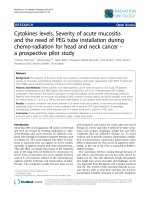
Figure 1 Spatial Correlation between 64Cu-ATSM and pimonidazole uptake in a cat with maxillary ostesarcoma. Formalin-fixation and
sectioning of the entire tumor from cat #12 was performed to compare spatial distribution of pimonidazole in relation to 64Cu-ATSM uptake on
PET. Panel A: Diagrammatic representation of a 5.1-cm osteosarcoma on the right lateral maxilla of a 7 year old spayed female domestic shorthair
cat. The position of two transverse sections are indicated by the letters B and C are shown in the diagram. The imaging and histologic sections at
these locations are provided in the panels below. Panels B and C: Top row: Transverse fused PET/CT image (left). H&E stained tissue section at 4×
magnification (middle). Pimonidazole at 4× magnification (reconstructed from tiled images) stained tissue section (right). Corresponding regions
in the PET/CT and histologic sections are marked by the numbers 1 and 2. Bottom Row (20× magnification of histologic sections): H&E stained
image from area marked “1” (Far left); Pimonidazole stained image from area marked “1” (Middle left). H&E stained image from area marked “2”
(Middle right); Pimonidazole stained image from area marked “2” (Far right). Note: The tumor tissue was friable and there were areas of necrotic
debris, such as the area marked by a star in panel B, that were lost during processing.
or between malignant and benign tumors (P = 0.15 for
Tmax/M; P = 0.21 to Tav/M).
Quantitative detection of tumor O2 using the intratumoral fluorescent probe confirmed, using a different technique, that tumors with 64Cu-ATSM uptake also exhibit
regions of very low oxygenation, ranging from 0.6 to
2.6 mmHg, which would be expected to have biologic
consequences including radioresistance and HIF1α induction (Table 1). Conversely, the tissues in the region of the
bone cyst that did not take up 64Cu-ATSM, appeared to be
normoxic (Table 1).
In addition to the fluorescent O2 detection probe,
pimonidazole immunohistochemistry was also used to
investigate tumor hypoxia. When pimonidazole was
Ballegeer et al. BMC Cancer 2013, 13:218
/>
administered 24 hours prior to biopsy, there was minimal
detectable immunostaining in samples, regardless of dose.
Whereas in three tumors, in which pimonidazole was
administered within an hour of biopsy, there was intense
immunohistochemical staining. The discrepancy in staining between samples collected 24 hours or 1 hour before
biopsy suggests that pimonidazole tissue adducts are
relatively short-lived in cats [33]. The patient with osteosarcoma was severely compromised by the primary tumor
and systemic metastasis and died following imaging. Thus
the entire tumor was available for examination and spatial
comparison of pimonidazole and 64Cu-ATSM uptake
(Figure 1). This comparison suggests a similar distribution
of pimonidazole and 64Cu-ATSM in this tumor.
Several additional tissues, distinct from the primary
tumor, demonstrated 64Cu-ATSM uptake, including
lymph nodes (medial and lateral retropharyngeal lymph
nodes, mandibular lymph nodes, and superficial cervical
lymph nodes) draining the primary tumor in six of the
cats with malignancies. In one of these six cats, there
was additional assessment of a mandibular lymph node
evaluated by fine needle aspiration cytology, which
demonstrated reactive change rather than metastatic
neoplasia.
Two of the cats had fluid within the tympanic bulla that
demonstrated 64Cu-ATSM uptake. One cat demonstrated
signal associated with a necrotic maxillary molar. Three of
the cats had 64Cu-ATSM uptake within the thyroid glands.
In one cat with bilateral thyroid uptake, clinical hyperthyroidism was confirmed by serum thyroid panel. In another
case, a large thyroid gland with increased 64Cu-ATSM
uptake on PET/CT was confirmed as a thyroid adenoma
at necropsy. In the third cat, there was PET signal in an
enlarged thyroid gland, but disease was not confirmed
with serum panel or histopathology. The cat with osteosarcoma that died immediately following PET/CT had a
diffuse increase in pulmonary signal and at necropsy there
were multiple 2–4 mm metastatic nodules in its lungs.
In two cats, intratumoral hypoxia was evaluated before
and after treatment with an antiangiogenic agent, either
a galectin-1 targeted peptide (Anginex) or a multiple
tyrosine kinase inhibitor that targets VEFGR2 (toceranib,
Palladia™, Pfizer Animal Health, Kalamazoo, MI). PET/
CT and intratumoral oxygen probe measurements were
performed one week apart with treatment administered
in the intervening interval. Similar location of the probe
was attempted as outlined in the materials and methods.
After one week, there was minimal change in tumor size
as measured by CT, with both tumors classifiable as
“stable” when applying the RECIST (Response Evaluation
Criteria in Solid Tumors) system used for human tumors
[38]. Nor was there appreciable change in CT appearance. However, both tumors exhibited a slight increase in
Tmax/M. While Tav/M increased slightly in the Anginex-
Page 6 of 11
treated cat, there was a slight decrease in Tav/M in the
toceranib-treated cat, with select regions of this second
tumor exhibiting less radiopharmaceutical uptake (see
Figure 2; Table 2). Intratumoral probe measurements
demonstrated variability in certain regions of both
tumors (Table 2). In the toceranib-treated tumor, pO2
values were consistently increased at each location. In
the Anginex- treated tumor the three regional measurements demonstrated decreased, increased, and stable
pO2 levels, respectively.
Discussion
The biologic effects and clinical consequences of intratumoral hypoxia have been the focus of decades of
research. It is well-established that hypoxic cells in vitro
and in animals are relatively radiation resistant [39].
Furthermore, it has been demonstrated that patients
with hypoxic tumors, including HNSCC, are more likely
to experience treatment failures both locally and systemically [12,18,39]. Therefore, a variety of methods to
increase tumor oxygenation or to target hypoxic cells
within tumors have been investigated. Traditionally, these
efforts have included measures such as hyperbaric oxygen
administration, inhalation of carbogen gas, and the use of
nitroimidazoles as hypoxic cell radiation sensitizers [18].
More recently, agents that specifically target hypoxic cell
populations have been developed [40]. Finally, it has also
been observed that anti-angiogenic and anti-vascular
therapies may also modulate tumor oxygenation [1,41].
However, despite these various efforts, clinical gains have
been modest. While a multitude of factors may contribute
to the gap between experimental and clinical results, two
issues are particularly problematic. First, of particular importance in the targeting of tumor hypoxia, the assessment
of relevant molecular and biologic surrogate endpoints is
challenging in humans [42]. Second, rodent models for
human cancer have significant limitations that do not
always permit direct clinical translation [43]. In this study,
we demonstrate the application of developing technology
to assess tumor oxygenation in a clinically relevant model,
spontaneous feline HNSCC.
There are a variety of methods for evaluating tumor
oxygenation and these have been thoroughly reviewed
elsewhere [29,42]. All of these techniques have strengths
and limitations, with no single technique offering complete
characterization of this dynamic, complex phenomenon
[42]. Imaging technology, by providing a noninvasive,
three-dimensional, real-time assessment of hypoxia, is particularly promising as a clinical tool. In this study, we investigated hypoxia using 64Cu-ATSM. Cu(II)-conjugated
ATSM enters cells by either passive diffusion or endocytosis
where is reduced and trapped, likely with the dissociation
of reduced Cu(I) from ATSM, within hypoxic, yet viable
cells [44,45]. Normoxic cells are able to oxidize the reduced
Ballegeer et al. BMC Cancer 2013, 13:218
/>
Page 7 of 11
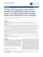
Figure 2 Uptake of 64Cu-ATSM within a maxillary squamous cell carcinoma. PET signalis presented in three planes of imaging; sagittal plane
image on the left, dorsal plane image in the middle, and transverse plane on the right. A similar area of transection through the head in each
plane was chosen between two time points, using anatomic landmarks of the orbit, mandibular rami, and medial canthus of the palpebrae. 2A
represents the mass before treatment with toceranib, 2B 7 days post treatment. In 2A, the mass is best seen as a large area of ATSM uptake on
dorsal plane PET image (white outline). Note the region of decreased uptake within the ventromedial portion of the mass, represented by the red
dot on dorsal plane PET image, yellow dot on sagittal plane PET image, and green dot on transverse plane PET image.
copper, which then is transported out of cells, either passively or, more likely, using a variety of chaperones or
transporters [42,45]. In preclinical studies, data demonstrate that tumor cells vary in their uptake of Cu-ATSM
even at constant pO2, implicating factors such as variable
transporter expression, microenvironmental pH, cellular
metabolism or the existence of alternative retention mechanisms [32,45]. Advantages of Cu-ATSM include, rapid uptake, strong signal to noise ratio, the availability of a variety
of Cu isotopes with variable half-lives and emission spectra,
and some potential for therapeutic as well as diagnostic
utility [46-48]. Cu-ATSM agents have subsequently been
Table 2 Evaluation of hypoxia in feline SCC before and after anti-angiogenic therapy
Column1
Diagnosis
Location
Maximum dimension (cm)
Tmax/M
Tav/M
pO2 #1 (mmHg)
pO2 #2 (mmHg)
PO2 #3 (mmHg)
Cat 8a
SCC
Mandible
4.41
11
3.05
2.2
26
2.6
Cat 8b
SCC
Mandible
4.41
11.8
3.16
24
2.8
2.6
Cat 9a
SCC
Maxilla
4.18
4.25
1.83
0.3
0.1
0.6
Cat 9b
SCC
Maxilla
4.06
5
1.73
14
19
20
Cat 8 was treated with Anginex, an anti-vascular peptide, while cat 9 was treated with toceranib, a VEGFR2 inhibitor. 64Cu-ATSM PET/CT and intratumoural
fluorescent O2 measurements were performed 7 days apart, with treatment occurring in the intervening interval. Lower case letter a and b indicates pre- and
post-treatment data, respectively. The location of the mass, maximum dimension of the mass, Tmax/M, Tav/M and pO2 in three tumor regions are provided.
HNSCC = head and neck squamous cell carcinoma.
Ballegeer et al. BMC Cancer 2013, 13:218
/>
used to image multiple tumors [16,32,44,46,49-53] and hypoxic tissues [54,55].
In this study, we demonstrate that most (11 of 12) feline
head and neck tumors take up 64Cu-ATSM with Tav/M
and Tmax/M greater than 1.5 and 4.3, respectively. In
studies that have investigated Cu-ATSM in human cancer
patients, Tav/M ratios ranging from 2.6 – 3.5 have been
used as arbitrary cutoff points for defining hypoxic and
normoxic tumors [56]. Indeed these levels of radionuclide
uptake have been associated with clinically relevant endpoints such as response to treatment and survival. However, these studies have not documented intratumoral
hypoxia using independent methods making it difficult to
determine whether these T/M ratios are best for determining actual hypoxic state. Furthermore, tumors with
significant radiopharmaceutical uptake also demonstrate
regions with quantitatively low pO2 (less than 7.5 mmHg)
or an affinity for pimonidazole, a hypoxia specific marker
that forms adducts when the pO2 is less than 10 mmHg.
Conversely, the bone cyst that failed to take up 64CuATSM, with T/M ratios was normoxic based on peritumoral pO2 measurements. These results support the
hypothesis that 64Cu-ATSM uptake occurs in hypoxic
rather than normoxic feline tumors. However, complete
spatial correlation between distribution of 64Cu-ATSM
was only possible in one case in which the animal died following imaging and the entire tumor, an osteosarcoma,
was available for sectioning and evaluation. Additionally,
no proof of 64Cu-ATSM uptake or lack thereof in these tumors’ normoxic cells was available. Subjectively, there
appeared to be concordance between pimonidazole and
64
Cu-ATSM uptake. Interestingly, in a xenograft study,
64
Cu-ATSM uptake failed to correlate with nitroimidazole
staining in a sarcoma, while demonstrating a strong correlation in both a carcinoma and a glioma [32].
While we were able to measure hypoxia using at least
one other technique in 11 of the 12 tumors, technical
problems precluded the use of all three techniques in
every case. The intratumoral probe was not operational at
the time of evaluation of the first three cats. We also limited our quantitation of tumor pO2 to a small number of
regions within the tumor. Studies of human tumors suggest that dozens of measurements may be needed to fully
map tumor oxygenation. However, our goal was simply to
verify the presence or absence of hypoxia in a few
intratumoral or peritumoral areas rather than to provide a
complete mapping of each tumor.
While the use of pimonidazole has been studied in the
dog [33,34], we were unable to find reports of the use of
this marker in cats. Therefore, doses were selected based
on those reported in dogs. Many drugs, including the
nitroimidazole, metronidazole, have similar or identical
doses in both cat and dog. We were unable to perform
additional procedures such as biopsy in the imaging
Page 8 of 11
facility, which necessitated a separate anesthetic episode.
Our initial plan had been to administer the pimonidazole
concomitant with the 64Cu-ATSM to permit side-by-side
comparison between the two. However, at the doses used,
we were not able to detect pimonidazole in cat biopsy
samples collected 24 hours after administration. In contrast, pimonidazole staining was strong and easily visualized when pimonidazole was administered shortly before
biopsy. These data suggest that the pimonidazole adducts
may turn over more quickly in feline tumors than in dogs
[33]. Factors that may have influenced pimonidazole staining intensity in the cat include species specific pharmacokinetic variables such as serum half life, which in humans is
about 5 hours and only 15 minutes in the mouse. Therefore recommended doses are several times higher in the
mouse than in humans. Unfortunately, these data are not
available for the cat. It is possible that with far larger doses
of pimonidazole we would have been able to visualize adducts in our biopsy specimens obtained 24 hours after
administration. Other factors that could have contributed
to poor retention of pimonidazole in tissues include rapidly
changing tissue perfusion or rapid turnover of cells in the
tumor. HNSCC in cats is considered a rapidly progressive
malignancy therefore it is possible that tumor growth kinetics may have also contributed. Pimonidazole dose
optimization should be performed in feline tumors to better utilize this technique.
It is not surprising to see heterogeneous distribution
of hypoxia within tumors, therefore significant differences between the Tmax/M and Tav/M in these PET studies is expected. However, signal voids were also observed
in areas with poor perfusion (based on CT contrast studies), which would presumably be hypoxic. In one cat
with osteosarcoma, the signal void corresponded to a
necrotic cavity identified at necropsy. It is possible that
poorly perfused regions contain necrotic rather than viable cells. Since uptake and retention of Cu-ATSM requires intact cell and likely lysosomal membranes, it is
unlikely that Cu-ATSM would accumulate in these necrotic regions [45]. A compounding factor in the specific
case of the osteosarcoma may be the high interstitial
pressures in bony areas of osteosarcomas leading to decreased perfusion [57-59].
In this study, while strongest 64Cu-ATSM uptake was
observed in HNSCC, sarcomas and benign tumors also
exhibited uptake and significant hypoxia. Thus, hypoxia is
not a characteristic of tumor type or malignancy. The increased uptake among feline HNSCC coupled with
intratumoral probe and pimonidazole data support that
these tumors are significantly hypoxic like their human
counterpart. However, we cannot rule out that some other
characteristic of HNSCC, in addition to hypoxia, has
influenced Cu-ATSM uptake and retention such as the
expression of specific transporters or metabolism. It has
Ballegeer et al. BMC Cancer 2013, 13:218
/>
been hypothesized that altered redox state associated with
glycolytic metabolism in some tumors might also promote
reduction and trapping of Cu-ATSM. It is likely that the
use of multiple methods to investigate tumor hypoxia may
yield the most accurate assessment.
Regardless of whether 64Cu-ATSM uptake is a direct reflection of tumor hypoxia, studies of human HNSCC indicate the clinical significance of this tracer. SUVmax [53]
and Tav/M ratio [56,60] cutoffs have been successfully
used to predict recurrence after radiation and prognosis,
respectively, in human cancer patients. It was not our objective to correlate these data with prognosis in cats nor
was it feasible given inconsistent follow-up therapy in
these cases. However, in using spontaneous HNSCC to investigate the biologic impact of therapeutic intervention,
these data may guide selection of appropriate thresholds.
Unexpectedly, certain other tissues in these cats exhibited
64
Cu-ATSM uptake. Uptake in lymph nodes draining the
primary tumor was seen in 8/12 cats. These lymph nodes
exhibited normal contrast enhancement on CT and only
mild to moderate enlargement. In one case, the lymph
nodes exhibited reactivity rather than metastasis. While
hypoxia is recognized in metastatic or primary tumors occurring in lymph nodes, its presence in reactive lymph
nodes has not been previously documented, to the authors’
knowledge [61,62]. It is interesting to consider how hypoxia
in draining lymph nodes might influence the development
of regional metastasis. Two cats also had 64Cu-ATSM uptake in association with presumptive otitis media. Hypoxia
has been demonstrated in rats with otitis media [63].
Hyperthyroidism is common in elderly felines and occurs secondary to adenomatous hyperplasia, thyroid adenomas, or least commonly functional thyroid carcinomas
[64]. Two of the three patients with 64Cu-ATSM uptake in
the thyroid had clinically proven functional hyperthyroidism prior to the scan. There are limited data concerning
hypoxia in non-malignant disorders of the thyroid, though
low level vascular endothelial growth factor (VEGF) expression, which is hypoxia inducible, has been observed in
follicular adenomas and adenomatous goiter of the thyroid
in humans [65]. This may be caused by the hypermetabolic
state and increased oxygen consumption [66] of the thyroid cells in these conditions. Human thyroid carcinoma
metastases, though not present in these patients, were also
demonstrated hypoxic when imaged with 99mTc-HL91, a
nitroimidazole, and SPECT [67]. Confirmation of hypoxia
in other tissues using another technique could not be easily
performed in these cases due to inaccessibility of lesions
and invasive nature of the other techniques used.
Two cats were evaluated before and after different
antivascular therapies. It has been proposed that modulation of tumor vasculature may affect intratumoral hypoxia
and preclinical studies have supported this notion [1,68].
In this study, treatment was accompanied by only slight
Page 9 of 11
changes in Cu-ATSM uptake. Since we do not have data
from untreated cats to demonstrate pattern on Cu-ATSM
uptake over time, it is not possible to determine whether
the changes observed were drug specific. However, in both
cats, there was a slight increase in Tmax/M possibly indicating regional vascular compromise. However, these
changes may be within range of error, as the inverse quartic relationship between partial pressure of oxygen and
Cu-ATSM uptake results in steep slope within the initial
decline of pO2, while at low pO2, slight changes may be
insufficient to alter uptake of 64Cu-ATSM [13]. At the
same time, in the cat treated with a tyrosine kinase inhibitor targeting VEGFR2, a slight decrease in Tav/M occurred
concomitantly with increased quantitative pO2 as measured with the intratumoral probe. Furthermore, focal
areas in the periphery of the tumors had decreased signal,
suggesting that further investigation into dose and time
frame of anti-angiogenic therapy administration as a hypoxia modulator might be useful.
Despite their experimental utility, rodent models fail to
completely recapitulate human cancer and to provide the
degree of heterogeneity that is characteristic of human
clinical populations. The gap between xenograft and
genetically-engineered mouse models and human clinical
studies are well recognized. Furthermore, as function of
animal size, the tumors seen are considerably smaller from
that expected in a human clinical population. Feline
HNSCC may provide a relevant alternative to rodent
models for this disease.
Conclusions
All of the feline HNSCC studied exhibited regional evidence of biologically relevant hypoxia, regardless of
measurement technique. Therefore, in addition to morphologic, clinical and molecular similarities, feline and
human HNSCC also share physiologic characteristics,
further demonstrating how closely the disease in cats
mimics its human counterpart. We also preliminarily illustrate, using anti-vascular agents, that feline tumors
can be used to study the biologic consequences of interventions and to develop and apply surrogate endpoints.
It is reasonable to assume that such studies could be
used to address specific issues of clinical translation and
inform the development of more effective human trials.
Abbreviations
HNSCC: Head and neck squamous cell carcinoma; ATSM: Diacetyl-bis(N4methylthiosemicarbazone); PET/CT: Positron emission tomography/computed
tomography; Tmax/M: Ratio of maximum tumor uptake to muscle uptake;
Tav/M: Ration of average tumor uptake to muscle uptake; EGFR: Epidermal
growth factor receptor; Cox-2: Cyclo-oxygenase isoform 2; MR: Magnetic
resonance; SPECT: Single photon emmision computed tomography;
FDG: Fluoro-D-Glucose; VEFGR2: Vascular endothelial growth factor receptor
2; SUVbw: Standardized uptake value adjusted for body weight;
HU: Hounsfield Unit; RECIST: Response evaluation criteria in solid tumors.
Ballegeer et al. BMC Cancer 2013, 13:218
/>
Competing interests
The authors declare that they have no competing interests.
Authors’ contributions
EAB was responsible for image interpretation and analysis and manuscript
preparation. NJM contributed to study design, case recruitment, O2
measurements, data management, table and figure preparation. KLB was
involved in study design, oversight of imaging, and manuscript editing. DWA
was involved in study design, histologic evaluation of biopsies and
pimonidazole staining and manuscript review. EAM was responsible for
study design, patient recruitment, clinical procedures, imaging, O2
measurement, data analysis, and manuscript preparation. All authors read
and approved the final manuscript.
Acknowledgements
This study was funded by a grant from the Michigan State University College
of Veterinary Medicine Companion Animal Fund.
The authors wish to gratefully acknowledge the assistance of Dr. Nathan
Nelson for project setup and Dr. Todd Erfourth for case management.
Performed at Michigan State University.
Author details
1
Department of Small Animal Clinical Sciences, Michigan State University,
East Lansing, MI 48824, USA. 2Department of Pathobiology and Diagnostic
Investigation, Michigan State University, East Lansing, MI 48824, USA.
3
Chesapeake Medical Imaging, Annapolis, MD 21401, USA. 4Tufts Cummings
School of Veterinary Medicine and Molecular Oncology Research Institute,
Boston, MA 02111, USA.
Received: 10 December 2012 Accepted: 25 April 2013
Published: 30 April 2013
References
1. Jain RK: Normalization of tumor vasculature: an emerging concept in
antiangiogenic therapy. Science 2005, 307(5706):58–62.
2. Vaupel P: Tumor microenvironmental physiology and its implications for
radiation oncology. Semin Radiat Oncol 2004, 14(3):198–206.
3. Padera TP, Stoll BR, Tooredman JB, Capen D, di Tomaso E, Jain RK:
Pathology: cancer cells compress intratumour vessels. Nature 2004,
427(6976):695.
4. Vaupel P, Mayer A: Hypoxia in cancer: significance and impact on clinical
outcome. Cancer Metastasis Rev 2007, 26(2):225–239.
5. Hall EJ, Giaccia AJ: Oxygen effec and re-oxygenation. In Radiobiology for
the Radiologist. 7th edition. Edited by Hall EJ, Giaccia AJ. Philadelphia:
Lipponcott, Williams & Wilkins; 2012.
6. Eckert AW, Lautner MH, Schutze A, Taubert H, Schubert J, Bilkenroth U:
Coexpression of hypoxia-inducible factor-1alpha and glucose
transporter-1 is associated with poor prognosis in oral squamous cell
carcinoma patients. Histopathology 2011, 58(7):1136–1147.
7. Hockel M, Knoop C, Schlenger K, Vorndran B, Baussmann E, Mitze M,
Knapstein PG, Vaupel P: Intratumoral pO2 predicts survival in advanced
cancer of the uterine cervix. Radiother Oncol 1993, 26(1):45–50.
8. Perez-Sayans M, Somoza-Martin JM, Barros-Angueira F, Diz PG, Rey JM,
Garcia-Garcia A: Multidrug resistance in oral squamous cell carcinoma:
the role of vacuolar ATPases. Cancer Lett 2010, 295(2):135–143.
9. Perez-Sayans M, Suarez-Penaranda JM, Pilar GD, Barros-Angueira F,
Gandara-Rey JM, Garcia-Garcia A: Hypoxia-inducible factors in OSCC.
Cancer Lett 2011, 313(1):1–8.
10. Unruh A, Ressel A, Mohamed HG, Johnson RS, Nadrowitz R, Richter E,
Katschinski DM, Wenger RH: The hypoxia-inducible factor-1 alpha is a
negative factor for tumor therapy. Oncogene 2003, 22(21):3213–3220.
11. Roh JL, Cho KJ, Kwon GY, Ryu CH, Chang HW, Choi SH, Nam SY, Kim SY:
The prognostic value of hypoxia markers in T2-staged oral tongue
cancer. Oral Oncol 2009, 45(1):63–68.
12. Brizel DM, Sibley GS, Prosnitz LR, Scher RL, Dewhirst MW: Tumor hypoxia
adversely affects the prognosis of carcinoma of the head and neck.
Int J Radiat Oncol Biol Phys 1997, 38(2):285–289.
13. Bowen SR, van der Kogel AJ, Nordsmark M, Bentzen SM, Jeraj R:
Characterization of positron emission tomography hypoxia tracer uptake
and tissue oxygenation via electrochemical modeling. Nucl Med Biol 2011,
38(6):771–780.
Page 10 of 11
14. Kikuchi M, Yamane T, Shinohara S, Fujiwara K, Hori SY, Tona Y, Yamazaki H,
Naito Y, Senda M: 18F-fluoromisonidazole positron emission tomography
before treatment is a predictor of radiotherapy outcome and survival
prognosis in patients with head and neck squamous cell carcinoma.
Ann Nucl Med 2011, 25(9):625–633.
15. Maftei CA, Shi K, Bayer C, Astner ST, Vaupel P: Comparison of (immuno-)
fluorescence data with serial [(1)(8)F]Fmiso PET/CT imaging for
assessment of chronic and acute hypoxia in head and neck cancers.
Radiother Oncol 2011, 99(3):412–417.
16. O'Donoghue JA, Zanzonico P, Pugachev A, Wen B, Smith-Jones P, Cai S,
Burnazi E, Finn RD, Burgman P, Ruan S, et al: Assessment of regional
tumor hypoxia using 18F-fluoromisonidazole and 64Cu(II)-diacetyl-bis
(N4-methylthiosemicarbazone) positron emission tomography:
comparative study featuring microPET imaging, pOo2 probe
measurement, autoradiography, and fluorescent microscopy in the
R3327-AT and FaDu rat tumor models. Int J Radiat Oncol Biol Phys
2005, 61(5):1493–1502.
17. Yamada T, Uchida M, Kwang-Lee K, Kitamura N, Yoshimura T, Sasabe E,
Yamamoto T: Correlation of metabolism/hypoxia markers and
fluorodeoxyglucose uptake in oral squamous cell carcinomas.
Oral Surg Oral Med Oral Pathol Oral Radiol Endod 2012, 113(4):464–471.
18. Overgaard J: Hypoxic modification of radiotherapy in squamous cell
carcinoma of the head and neck—a systematic review and
meta-analysis. Radiother Oncol 2011, 100(1):22–32.
19. Liptak JM, Withrow SJ: Oral tumors. In Withrow and MacEwen's Small
Animal Clinical Oncology. Volume 4th. Edited by Withrow SJ, Vail DM. St.
Louis: Saunders Elsevier Inc; 2007:455–510.
20. Dorn CR, Priester WA: Epidemiologic analysis of oral and pharyngeal
cancer in dogs, cats, horses, and cattle. J Am Vet Med Assoc 1976,
169(11):1202–1206.
21. Bertone ER, Snyder LA, Moore AS: Environmental and lifestyle risk factors
for oral squamous cell carcinoma in domestic cats. J Vet Intern Med 2003,
17(4):557–562.
22. McNiel EA, Carmella SG, Heath LA, Bliss RL, Le KA, Hecht SS: Urinary
biomarkers to assess exposure of cats to environmental tobacco smoke.
Am J Vet Res 2007, 68(4):349–353.
23. Snyder LA, Bertone ER, Jakowski RM, Dooner MS, Jennings-Ritchie J,
Moore AS: p53 expression and environmental tobacco smoke exposure
in feline oral squamous cell carcinoma. Vet Pathol 2004, 41(3):209–214.
24. Looper JS, Malarkey DE, Ruslander D, Proulx D, Thrall DE: Epidermal growth
factor receptor expression in feline oral squamous cell carcinomas.
Vet Comp Oncol 2006, 4(1):33–40.
25. Sabattini S, Marconato L, Zoff A, Morini M, Scarpa F, Capitani O, Bettini G:
Epidermal growth factor receptor expression is predictive of poor
prognosis in feline cutaneous squamous cell carcinoma. J Feline Med Surg
2010, 12(10):760–768.
26. Beam SL, Rassnick KM, Moore AS, McDonough SP: An
immunohistochemical study of cyclooxygenase-2 expression in various
feline neoplasms. Vet Pathol 2003, 40(5):496–500.
27. Hayes A, Scase T, Miller J, Murphy S, Sparkes A, Adams V: COX-1 and COX-2
expression in feline oral squamous cell carcinoma. J Comp Pathol 2006,
135(2–3):93–99.
28. DiBernardi L, Dore M, Davis JA, Owens JG, Mohammed SI, Guptill CF,
Knapp DW: Study of feline oral squamous cell carcinoma: potential
target for cyclooxygenase inhibitor treatment. Prostaglandins Leukot
Essent Fat Acids 2007, 76(4):245–250.
29. Dewhirst MW, Klitzman B, Braun RD, Brizel DM, Haroon ZA, Secomb TW:
Review of methods used to study oxygen transport at the
microcirculatory level. Int J Cancer 2000, 90(5):237–255.
30. Bruehlmeier M, Kaser-Hotz B, Achermann R, Bley CR, Wergin M,
Schubiger PA, Ametamey SM: Measurement of tumor hypoxia in
spontaneous canine sarcomas. Vet Radiol Ultrasound 2005, 46(4):348–354.
31. Foo SS, Abbott DF, Lawrentschuk N, Scott AM: Functional imaging of
intratumoral hypoxia. Mol Imaging Biol 2004, 6(5):291–305.
32. Yuan H, Schroeder T, Bowsher JE, Hedlund LW, Wong T, Dewhirst MW:
Intertumoral differences in hypoxia selectivity of the PET imaging agent
64Cu(II)-diacetyl-bis(N4-methylthiosemicarbazone). J Nucl Med 2006,
47(6):989–998.
33. Azuma C, Raleigh JA, Thrall DE: Longevity of pimonidazole adducts in
spontaneous canine tumors as an estimate of hypoxic cell lifetime.
Radiat Res 1997, 148(1):35–42.
Ballegeer et al. BMC Cancer 2013, 13:218
/>
34. Kleiter MM, Thrall DE, Malarkey DE, Ji X, Lee DY, Chou SC, Raleigh JA:
A comparison of oral and intravenous pimonidazole in canine tumors
using intravenous CCI-103F as a control hypoxia marker. Int J Radiat
Oncol Biol Phys 2006, 64(2):592–602.
35. Raleigh JA, Chou SC, Bono EL, Thrall DE, Varia MA: Semiquantitative
immunohistochemical analysis for hypoxia in human tumors. Int J Radiat
Oncol Biol Phys 2001, 49(2):569–574.
36. Griffioen AW, van der Schaft DW, Barendsz-Janson AF, Cox A, Struijker
Boudier HA, Hillen HF, Mayo KH: Anginex, a designed peptide that inhibits
angiogenesis. Biochem J 2001, 354(Pt 2):233–242.
37. Kubota K, Matsuzawa T, Ito M, Ito K, Fujiwara T, Abe Y, Yoshioka S, Fukuda H,
Hatazawa J, Iwata R, et al: Lung tumor imaging by positron emission
tomography using C-11 L-methionine. J Nucl Med 1985, 26(1):37–42.
38. Eisenhauer EA, Therasse P, Bogaerts J, Schwartz LH, Sargent D, Ford R,
Dancey J, Arbuck S, Gwyther S, Mooney M, et al: New response evaluation
criteria in solid tumours: revised RECIST guideline (version 1.1).
Eur J Cancer 2009, 45(2):228–247.
39. Hall EJ, Giaccia AJ: Oxygen effect and reoxygenation. In Radiobiology for
the Radiologist. Volume 7th. Edited by Hall EJ, Giacca AJ. Philadephia:
Lippincott, Williams & Wilkins; 2012:86–103.
40. Kawakami K, Hattori M, Inoue T, Maruyama Y, Ohkanda J, Kato N, Tongu M,
Yamada T, Akimoto M, Takenaga K, et al: A novel fusicoccin derivative
preferentially targets hypoxic tumor cells and inhibits growth in
xenografts. Anti Cancer Agents Med Chem 2012, 17(7):791–800.
41. Citrin D, Camphausen K: Advancement of antiangiogenic and vascular
disrupting agents combined with radiation. In Radiation Oncology
Advances. Edited by Bentzen SM, Harari PM, Tome WA, Mehta MP.
New York: Springer, LLC; 2008:153–172.
42. Tatum JL, Kelloff GJ, Gillies RJ, Arbeit JM, Brown JM, Chao KS, Chapman JD,
Eckelman WC, Fyles AW, Giaccia AJ, et al: Hypoxia: importance in tumor
biology, noninvasive measurement by imaging, and value of its
measurement in the management of cancer therapy. Int J Radiat Biol
2006, 82(10):699–757.
43. Smith LP, Thomas GR: Animal models for the study of squamous cell
carcinoma of the upper aerodigestive tract: a historical perspective with
review of their utility and limitations. Part A. Chemically-induced de
novo cancer, syngeneic animal models of HNSCC, animal models of
transplanted xenogeneic human tumors. Int J Cancer 2006,
118(9):2111–2122.
44. Padhani AR, Krohn KA, Lewis JS, Alber M: Imaging oxygenation of human
tumours. Eur Radiol 2007, 17(4):861–872.
45. Wood KA, Wong WL, Saunders MI: [(64)Cu]diacetyl-bis(N(4)-methylthiosemicarbazone)—a radiotracer for tumor hypoxia. Nucl Med Biol 2008,
35(4):393–400.
46. Yoshii Y, Furukawa T, Kiyono Y, Watanabe R, Mori T, Yoshii H, Asai T,
Okazawa H, Welch MJ, Fujibayashi Y: Internal radiotherapy with copper-64
-diacetyl-bis (N4-methylthiosemicarbazone) reduces CD133+ highly
tumorigenic cells and metastatic ability of mouse colon carcinoma.
Nucl Med Biol 2011, 38(2):151–157.
47. Weeks AJ, Paul RL, Marsden PK, Blower PJ, Lloyd DR: Radiobiological effects
of hypoxia-dependent uptake of 64Cu-ATSM: enhanced DNA damage
and cytotoxicity in hypoxic cells. Eur J Nucl Med Mol Imaging 2010,
37(2):330–338.
48. Vavere AL, Lewis JS: Cu-ATSM: a radiopharmaceutical for the PET imaging
of hypoxia. Dalton Trans 2007, 43:4893–4902.
49. de Lussanet QG, Beets-Tan RG, Backes WH, van der Schaft DW, van
Engelshoven JM, Mayo KH, Griffioen AW, Beets-Tan RGH, van der Schaft
DWJ, van Engelshoven JMA: Dynamic contrast-enhanced magnetic
resonance imaging at 1.5 Tesla with gadopentetate dimeglumine to
assess the angiostatic effects of anginex in mice. Eur J Cancer 2004,
40(8):1262–1268.
50. Black NF, McJames S, Rust TC, Kadrmas DJ: Evaluation of rapid dual-tracer
(62)Cu-PTSM + (62)Cu-ATSM PET in dogs with spontaneously occurring
tumors. Phys Med Biol 2008, 53(1):217–232.
51. Dietz DW, Dehdashti F, Grigsby PW, Malyapa RS, Myerson RJ, Picus J, Ritter J,
Lewis JS, Welch MJ, Siegel BA: Tumor hypoxia detected by positron
emission tomography with 60Cu-ATSM as a predictor of response and
survival in patients undergoing Neoadjuvant chemoradiotherapy for
rectal carcinoma: a pilot study. Dis Colon Rectum 2008, 51(11):1641–1648.
Page 11 of 11
52. Lewis JS, Sharp TL, Laforest R, Fujibayashi Y, Welch MJ: Tumor uptake of
copper-diacetyl-bis(N(4)-methylthiosemicarbazone): effect of changes in
tissue oxygenation. J Nucl Med 2001, 42(4):655–661.
53. Minagawa Y, Shizukuishi K, Koike I, Horiuchi C, Watanuki K, Hata M,
Omura M, Odagiri K, Tohnai I, Inoue T, et al: Assessment of tumor hypoxia
by 62Cu-ATSM PET/CT as a predictor of response in head and neck
cancer: a pilot study. Ann Nucl Med 2011, 25(5):339–345.
54. Ikawa M, Okazawa H, Kudo T, Kuriyama M, Fujibayashi Y, Yoneda M:
Evaluation of striatal oxidative stress in patients with Parkinson's disease
using [62Cu]ATSM PET. Nucl Med Biol 2011, 38(7):945–951.
55. Skovgaard D, Kjaer M, Madsen J, Kjaer A: Noninvasive 64Cu-ATSM and
PET/CT assessment of hypoxia in rat skeletal muscles and tendons
during muscle contractions. J Nucl Med 2009, 50(6):950–958.
56. Dehdashti F, Grigsby PW, Mintun MA, Lewis JS, Siegel BA, Welch MJ:
Assessing tumor hypoxia in cervical cancer by positron emission
tomography with 60Cu-ATSM: relationship to therapeutic response-a
preliminary report. Int J Radiat Oncol Biol Phys 2003, 55(5):1233–1238.
57. Nathan SS, DiResta GR, Casas-Ganem JE, Hoang BH, Sowers R, Yang R,
Huvos AG, Gorlick R, Healey JH: Elevated physiologic tumor pressure
promotes proliferation and chemosensitivity in human osteosarcoma.
Clin Cancer Res 2005, 11(6):2389–2397.
58. Nathan SS, Huvos AG, Casas-Ganem JE, Yang R, Linkov I, Sowers R,
DiResta GR, Gorlick R, Healey JH: Tumour interstitial fluid pressure may
regulate angiogenic factors in osteosarcoma. Ann Acad Med Singap 2009,
38(12):1041–1047.
59. Zachos TA, Aiken SW, DiResta GR, Healey JH: Interstitial fluid pressure and
blood flow in canine osteosarcoma and other tumors. Clin Orthop Relat
Res 2001, 385:230–236.
60. Dehdashti F, Mintun MA, Lewis JS, Bradley J, Govindan R, Laforest R, Welch
MJ, Siegel BA: In vivo assessment of tumor hypoxia in lung cancer with
60Cu-ATSM. Eur J Nucl Med Mol Imaging 2003, 30(6):844–850.
61. Gagel B, Reinartz P, Dimartino E, Zimny M, Pinkawa M, Maneschi P, Stanzel S,
Hamacher K, Coenen HH, Westhofen M, et al: pO(2) polarography versus
positron emission tomography ([(18)F] fluoromisonidazole, [(18)F]-2fluoro-2'-deoxyglucose). An appraisal of radiotherapeutically relevant
hypoxia. Strahlenther Onkol 2004, 180(10):616–622.
62. ibUnstructured>Postema EJ, McEwan AJ, Riauka TA, Kumar P, Richmond DA,
Abrams DN, Wiebe LI: Initial results of hypoxia imaging using 1-alpha-D: (5-deoxy-5-[18F]-fluoroarabinofuranosyl)-2-nitroimidazole (18F-FAZA).
Eur J Nucl Med Mol Imaging 2009, 36(10):1565–1573.
63. Cheeseman MT, Tyrer HE, Williams D, Hough TA, Pathak P, Romero MR,
Hilton H, Bali S, Parker A, Vizor L, et al: HIF-VEGF pathways are critical for
chronic otitis media in Junbo and Jeff mouse mutants. PLoS Genet 2011,
7(10):e1002336.
64. Peterson ME: Hyperthyroidism. In Textbook of Veterinary Internal Medicine.
Edited by Ettinger SJ, Feldman EC. Philadelphia: W.B. Saunders;
2000:1400–1419.
65. Itoh A, Iwase K, Jimbo S, Yamamoto H, Yamamoto N, Kokubo M, Senda T,
Nakai A, Nagagasaka A, Nagasaka T, et al: Expression of vascular
endothelial growth factor and presence of angiovascular cells in tissues
from different thyroid disorders. World J Surg 2010, 34(2):242–248.
66. Zhong Z, Li X, Yamashina S, von Frankenberg M, Enomoto N, Ikejima K,
Kolinsky M, Raleigh JA, Thurman RG: Cyclosporin A causes a
hypermetabolic state and hypoxia in the liver: prevention by dietary
glycine. J Pharmacol Exp Ther 2001, 299(3):858–865.
67. Cook GJ, Houston S, Barrington SF, Fogelman I: Technetium-99m-labeled
HL91 to identify tumor hypoxia: correlation with fluorine-18-FDG.
J Nucl Med 1998, 39(1):99–103.
68. Dings RP, Loren M, Heun H, McNiel E, Griffioen AW, Mayo KH, Griffin RJ,
Dings RPM, Loren M, Heun H, et al: Scheduling of radiation with
angiogenesis inhibitors anginex and Avastin improves therapeutic
outcome via vessel normalization. Clin Cancer Res 2007, 13(11):3395–3402.
doi:10.1186/1471-2407-13-218
Cite this article as: Ballegeer et al.: Evaluation of hypoxia in a feline
model of head and neck cancer using 64Cu-ATSM positron emission
tomography/computed tomography. BMC Cancer 2013 13:218.