Comparative performances of prognostic indexes for breast cancer patients presenting with brain metastases
Bạn đang xem bản rút gọn của tài liệu. Xem và tải ngay bản đầy đủ của tài liệu tại đây (579.25 KB, 8 trang )
Braccini et al. BMC Cancer 2013, 13:70
/>
RESEARCH ARTICLE
Open Access
Comparative performances of prognostic indexes
for breast cancer patients presenting with brain
metastases
Antoine-Laurent Braccini1*, David Azria1, Simon Thezenas2, Gilles Romieu3, Jean-Marc Ferrero4 and William Jacot3
Abstract
Background: Several prognostic indexes (PI) have been developed in the brain metastases (BM) setting to help
physicians tailor treatment options and stratify patients enrolled in clinical studies. The aim of our study was to
compare the clinical relevance of the major PI for breast cancer BM.
Methods: Clinical and biological data of 250 breast cancer patients diagnosed with BM at two institutions between
1995 and 2010 were retrospectively reviewed. The prognostic value and accuracy of recursive partitioning analysis
(RPA), graded prognostic assessment (GPA), basic score for BM (BS-BM), breast RPA, breast GPA, Le Scodan’s Score
and a clinico-biological score developed in a phase I study (P1PS) were assessed using Cox regression models. PI
comparison was performed using Harrell’s concordance index.
Results: After a median follow-up of 4.5 years, median overall survival (OS) from BM diagnosis was 8.9 months (CI
95%, 6.9–10.3 months). All PI were significantly associated with OS. Harrell’s concordance indexes C favored BS-BM
and RPA. In multivariate analysis, the RPA, Le Scodan’s score and GPA were found to be the best independent
predictors of OS. In multivariate analysis restricted to the 159 patients with known LDH and proteinemia, RPA 2 and
3, Le Scodan’s Score 3 and P1PS 2/3 were associated with worse survival. RPA was the most accurate score to
identify patients with long (superior to 12 months) and short (inferior to 3 months) life expectancy.
Conclusions: RPA seems to be the most useful score and performs better than new PI for breast cancer BM.
Keywords: Breast cancer, Brain metastases, Prognostic indexes, Biological subtype
Background
The Recursive Partitioning Analysis RPA [1] was the first
prognostic score developed in the brain metastases (BM)
setting. This classification was created in 1997 by the Radiation Therapy Oncology Group after analysis of the relative contributions of pretreatment variables to survival of
patients with BM. Since this date, several scores and prognostic indexes (PI), such as the Graded Prognosis Assessment (GPA) [2], the Basic Score for BM (BS-BM) [3], the
Phase 1 Prognostic Score (P1PS) [4], the Rotterdam score
[5], the Score Index for Radiosurgery (SIR) [6] and the
Rades’s score [7] have been developed both to help physicians tailor treatment options depending on patient
* Correspondence:
1
Department of Radiation Oncology, Val d’Aurelle Cancer Institute, 208 rue
des apothicaires, Montpellier 34298, France
Full list of author information is available at the end of the article
prognosis, and to stratify patients enrolled in clinical studies. However, it has been demonstrated that the prognostic
value of these scoring systems differs according to the primary tumor site [8], which raises the question of the usefulness of a breast-specific score.
Breast cancer is the second cause of BM, after lung cancer. Breast cancer is a heterogeneous disease with metastatic pattern and survival varying with the expression of
biological markers such as the hormonal receptor (HR)
status and human epidermal growth factor receptor-2
(HER2) overexpression. While the incidence of BM from
breast cancer has increased over the past decade, especially for the subgroup of HER2-overexpressing tumors,
several studies have shown that biological subtypes influence survival, even after BM diagnosis. In a series of 223
breast cancer patients irradiated for BM, Dawood et al.
showed that HER2 positive status was an independent
© 2013 Braccini et al.; licensee BioMed Central Ltd. This is an Open Access article distributed under the terms of the Creative
Commons Attribution License ( which permits unrestricted use, distribution, and
reproduction in any medium, provided the original work is properly cited.
Braccini et al. BMC Cancer 2013, 13:70
/>
favorable prognostic factor [9]. On the contrary, the triple
negative population seems to be associated with worse
prognosis [10,11]. These results have prompted the development of specific prognostic scores for BM from breast
cancer taking into account either tumor phenotypic characteristics [12,13] or not [14]. Given the number of scoring systems that have been devised for clinical use, the
aim of our study was to compare the clinical relevance of
the major existing prognostic scores in a cohort of breast
cancer patients with BM and known HER2 and HR status.
Page 2 of 8
Table 1 Prognostic indexes parameters
A: Clinical parameters used for 5 prognostic indexes (RPA, GPA, BSBM, Breast RPA, and Breast GPA).
RPA
Class 1
Age <65 y, KPS ≥ 70, controlled primary tumor, no
extracranial metastases
Class 2
All patients not in Class I or III
Class 3
KPS < 70
GPA
0
0,5
1
Age
> 60
50-59
<50
Study population
KPS
<70
70-80
90-100
Medical records of breast cancer patients with BM were
retrospectively extracted from the databases of two
French cancer centers. Patients were accrued over a 15year period, between 1995 and 2010. Inclusion criteria
were as follows: histologically proven breast carcinoma,
intradural BM detected by contrast-enhanced cerebral
computed tomography or magnetic resonance imaging,
and known HR and HER2 status. The tumor was considered HR positive when more than 10% of cells were labeled in immunohistochemistry (IHC) or when the
concentrations of estrogen and progesterone receptors
were above 10 ng/ml and 50 ng/ml using the radioligand
binding method, respectively. The tumor was considered
HER2 positive if the primary tumor was scored 3+ by
IHC or if the HER2 gene was amplified by fluorescence
in situ hybridization (FISH). If the tumor was scored 2+
by IHC, it was re-analyzed using FISH. Patients with history of other primitive carcinoma or leptomeningeal carcinomatosis were excluded. In addition, an additional
brain MRI was performed to all patient presenting with
1 to 3 BM at baseline CT-scan. Clinical data and, when
available, biological parameters were extracted in order
to score patients using the RPA [1], the GPA [2], the BSBM [3], the P1PS [4], the Breast-GPA [12], the BreastRPA [14] and Le Scodan’s score [13], whose constituting
parameters are detailed in Table 1. Ethical approval,
as well as permission to create, complete and access the
comprehensive database used in this study, was provided
by the local research ethics committee of the Val d’Aurelle
Cancer Institute. Due to the retrospective, non interventional nature of this study, no consent was requested by
the local research ethics committee.
Number of BM
>3
2-3
1
Yes
-
No
0
1
50-70
80-100
No
Yes
Yes
No
Methods
Statistical analyses
Categorical variables were reported by means of contingency tables. For continuous variables, median and range
values were computed. To investigate the association between study features, univariate statistical analyses were
performed using Pearson’s Chi-2 test or Fisher’s exact
test if applicable for categorical variables. The KruskalWallis test or Student T test were used for continuous
Extracranial metastases
BS-BM
KPS
Control of primary tumor
Extracranial metastases
Breast RPA
Class 1
1–2 brain metastases and extracranial disease absent
or controlled and KPS 100
Class 2
All patients not in Class I or III
Class 3
Multiple brain metastases and KPS ≤ 60
Breast GPA
0
0,5
Age
≥ 60
<60
KPS
≤ 50
60
Genetic subtype Basal
1
1.5
70-80
90-100
Luminal A
HER2
2
Luminal B
B: Clinico-biological parameters used for the P1PS and Le Scodan’s
prognostic indexes.
P1PS
Sites of metastases
Serum LDH
Albumin, g/L
0
1
0-2
>2
>ULN
≥35
<35
Le Scodan Score
Class I
HER2+ tumors treated with trastuzumab
Class II
All patients not in Class I or III
Class III
Tumors not treated with trastuzumab and:
lymphopenia at BM diagnosis or KPS < 70
and ≥ 50 years old at BM diagnosis or KPS ≥ 70
and triple negative tumors
RPA, Recursive Partitioning Analysis, GPA, Graded Partitioning Analysis, BS-BM,
Basic Score for Brain Metastases, BM, Brain Metastases, P1PS, phase 1
prognostic score, KPS, Karnofsky Performance Status, BM, Brain Metastases,
LDH, Lactate Dehydrogenase, ULN, Upper Limit of Normal.
Braccini et al. BMC Cancer 2013, 13:70
/>
Page 3 of 8
variables. Overall survival (OS) time was measured from
the date of BM diagnosis to the date of death from any
cause. Patients alive without event were censored at the
closing date of the study analysis (August 1st, 2011). OS
rates and median values were estimated according to the
Kaplan-Meier method [15], and presented with their
95% confidence intervals (95% CIs). The median length
of follow-up was estimated using a reverse Kaplan-Meier
method and presented with 95% CIs.
Pair wise comparisons of subgroups were performed
for each score. Survival curves were drawn and the logrank test was performed to assess differences between
groups. Harrell’s concordance Index (C index) was used
to assess the discriminating ability of the different PIs
[16]. To investigate prognostics factors, multivariate
analyses were carried out using the Cox’s proportional
hazards regression model with a stepwise selection procedure [17,18]. Hazard ratios (HR) with 95% CIs are
presented to display risk reductions. All p values
reported are two-sided, and the significance level was
set at 5% (p < 0.05). Statistical analysis was performed
using the STATA 11 software (Stata Corporation,
College Station, TX).
MOS times for the RPA classes I, II and III were 25.6
months (95% CI 18.4-32.9), 10.4 months (95% CI 8.912.6), and 2 months (95% CI 1.4-3.1), respectively. For
the GPA classes I, II and III, the MOS were 25.6 months
(95% CI 3.1-5.4), 12.3 months (95% CI 10.1-15.1), and
24.7 months (95% CI 12.7-27.1), respectively. In patients
stratified in the classes I, II, III and IV using the BS-BM
prognostic scores, the MOS were 2.2 months (95% CI
1.4-3.6), 8.7 months (95% CI 6.1-12.3), 12.7 months
(95% CI 12.7-27.1), and 21.6 months (95% CI 12.7-25.6),
respectively. With respect to the P1PS, ninety-one
patients could not be classified due to missing biological data. The MOS were 16.4 months (95% CI 11.9-
Results
Time between initial diagnosis and BM
diagnosis (months)
Patient characteristics
There were a total of two hundred and fifty patients
included in this analysis. Patient characteristics are
detailed in Table 2. At the time of BM diagnosis, the median age was 55 years (range 25–85), and 74% of patients
had good performance status (80–100). The brain was
the first metastatic site in about one third of patients
(34%), and the only site of metastatic disease in 12% of
patients. Of the 250 patients, 44% had a primary tumor
that over-expressed HER2, while 26% were diagnosed
with a triple negative breast cancer (negative HR and
HER2 status). A total of 47 patients (18.8%) underwent
targeted local treatment, namely stereotactic radiotherapy or surgery. Whole brain radiation therapy (WBRT),
used as primary treatment but also as adjuvant treatment after localized treatment, was given to 217 patients
(86.8%). Fifteen patients received best supportive care
only. After a median follow-up of 4.5 years, the median
OS (MOS) was 8.9 months (95% CI, 6.9-10.3 months).
The six-month, one-year and two-year overall survival
rates were 61% (95% CI, 54-67%), 40% (95% CI, 34-46%)
and 22% (95% CI, 17-27%), respectively.
Prognostic indexes analysis
Table 3 lists the study population distribution as well as
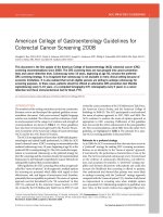
the MOS for each PI. Survival curves are depicted in
Figure 1. The results showed that all scores were able to
discriminate with statistical significance (p < 0.001)
patients for OS according to the prognostic category.
Table 2 Study population
Patient characteristics
Number of
patients
%
Age at breast cancer
diagnosis (years)
Median, range
50 (23–82)
Age at BM diagnosis (years)
Median, range
55 (25–85)
Median, range
39.4 (0–319.2)
Hormone receptor status
Positive
119
47.6
Negative
131
52.4
HER-2 status
Positive
109
43.6
Negative
141
56.4
66
26.4
Karnofsky Performance status
100
80–90
119
47.6
60–70
28
11.2
40–50
32
12.8
10-30
5
2
Extra-cerebral metastases
at BM diagnosis
Yes
219
87.6
No
31
12.4
Yes
177
70.8
No
73
29.2
Systemic treatment after BM diagnosis
Chemotherapy
Anti-HER2 treatment for HER2+ patients
Yes
20
18.3
No
89
81.7
BM, Brain Metastases, KPS, Karnofsky Performance Status.
Braccini et al. BMC Cancer 2013, 13:70
/>
Page 4 of 8
23.3) vs. 5.9 months (95% CI 3.4-8.4) in remaining
patients with P1PS scores of 0/1 vs. 2/3, respectively.
Based on the breast GPA scoring system, MOS were
found to be 2.3 months for a score of 0–1 (95% CI 1–
4.1), 5.7 months for a score of 1.5-2.5 (95% CI 4–8),
10.3 months for a score of 3 (95% CI 8.4-13.7), and 18.4
months for a score > 3 (95% CI 12.4-23.3). The MOS
were 21.3 months (95% CI 9.7-53.9) for class I, 9.8
months (95% CI 8.4-12.1) for class II, and 2.3 months
(95% CI 1.8-4.3) for class III according to the breast
RPA scoring system. Lastly, the MOS for Le Scodan’
scores I, II and III were 15.2 (95% CI 11.5-19.4), 9.7
(95% CI 7.5-12.4), and 4.2 (95% CI 3.3-6.1) months,
respectively.
Pairwise comparisons of each PI revealed statistically
significant differences in survival between prognostic classes except for the breast GPA classes I vs. II (p = 0.0609),
the BS-BM scores 1 vs. 2 (p = 0.27), and Le Scodan’s scores
I vs. II (p = 0.098).
Prognostic indexes comparison
There were no statistically significant differences between all PIs with regard to survival predicting ability (Table 3). Only minor differences were seen using
Harrell’s concordance index, with values of Harrell’s C
slightly higher for the BS-BM (0.6803) and RPA (0.6783)
scoring systems than for the breast RPA (0.6037), Le
Scodan’s score (0.6239), and P1PS (0.6251). In
Table 3 Distribution of the study population and median overall survival according to the class of prognostic scores;
Harrell’s concordance indexes (HCS)
Number of pts (%)
MOS (95% CI)
p
Hazard Ratio (95% CI)
HCS (95% CI)
1
26 (10.4)
25.6 M (18.4–32.9)
1
0.6783
2
166 (66.4)
10.4 M (8.9–12.6)
3
58 (23.2)
2 M (1.4–3.1)
11.38 (6.57–19.70)
2.16 (1.34–3.50)
(0.65–0.71)
≥3
32 (12.8)
24.7 M (12.7–27.1)
1
0.658
1.5-2.5
116 (46.4)
12.3 M (10.1–15.1)
1.70 (1.09–2.65)
(0.62–0.69)
0-1
102 (40.8)
4.2 M (3.1–5.4)
31 (12.4)
21.6 M (12.7–25.8)
RPA
<0.001
GPA
<0.001
4.15 (2.62–6.55)
BS-BM
3
<0.001
1
2
68 (27.2)
12.7 M (9.7–18.4)
1.30 (0.81–2.08)
0.6803
1
96 (38.4)
8.7 M (6.1–12.3)
2.20 (1.41–3.46)
(0.64–0.72)
0
55 (22.0)
2.2 M (1.4–3.6)
6.99 (4.25–11.47)
0–1
95 (38.0)
16.4 M (11.9–23.3)
2–3
64 (25.6)
5.9 M (3.4–8.4)
Not available
91 (36.4)
P1PS
1
0.6251
<0.001
2.89 (2.01–4.14)
(0.58–0.66)
1
0.6587
<0.001
1.37 (0.95–2.00)
(0.62–0.69)
Breast GPA
3.5-4
53 (21.2)
18.4 M (12.4–23.3)
3
90 (36.0)
10.3 M (8.4–13.7)
1.5-2.5
76 (30.4)
5.7 M (4–8)
2.09 (1.43–3.06)
0-1
31 (12.4)
2.3 M (1–4.1)
5.75 (3.56–9.27)
1
20 (8.0)
21.3 M (9.7–53.9)
1
0.6037
2
192 (76.8)
9.8 M (8.4–12.1)
2.05 (1.20–3.50)
(0.57–0.63)
3
38 (15.2)
2.3 M (1.8–4.3)
6.84 (3.69–12.72)
89 (35.6)
15.2 M (11.5–19.4)
1
0.6239
2
49 (19.6)
9.7 M (7.5–12.4)
1.32 (0.91–1.92)
(0.58–0.66)
3
112 (44.8)
4.2 M (3.3–6.1)
Breast RPA
<0.001
Le Scodan Score
1
<0.001
CI, Confidence Interval. MOS, Median Overall Survival (from brain metastases diagnosis). M, months. Pts, patients.
1.92 (1.43–2.59)
Braccini et al. BMC Cancer 2013, 13:70
/>
75
0
25
p< 0.001
50
p< 0.001
0
20
40
60
80
100
0
20
40
Months
80
100
75
Breast RPA 1 21.3 months (CI95%: 9.7 – 53.9)
Breast RPA 2 9.8 months (CI95%: 8.4 –12.1)
Breast RPA 3 2.3 months (CI95%: 1.8 – 4.3)
25
50
p< 0.001
0
25
50
p< 0.001
Proportion surviving (%)
75
GPA 0-1
4.2 months (CI95%: 3.1 – 5.4)
GPA 1.5-2.5 12.3 months (CI95%: 10.1-15.1)
GPA 3-4
24.7 months (CI95%: 12.7 – 27.1)
100
100
D
0
Proportion Surviving (%)
60
Months
C
0
20
40
60
80
0
100
Months
E
2.2 months (CI 95%: 1.4 – 3.6)
8.7 months (CI 95%: 6.1– 12.3)
12.7 months (CI 95%: 9.7 – 18.4)
21.6 months (CI 95%: 12.7 – 25.8)
25
Proportion Surviving (%)
75
RPA 1 25.6 months (CI95%: 18.4 – 32.9)
RPA 2 10.4 months (CI95%: 8.9 – 12.6)
RPA 3 2 months (CI95%: 1.4 – 3.1)
50
BS-BM 0
BS-BM 1
BS-BM 2
BS-BM 3
100
100
B
0
Proportion Surviving (%)
A
Page 5 of 8
20
40
60
80
100
Months
F
G
Figure 1 Overall survival according to (A) the RPA, (B) the BS-BM, (C) the GPA, (D) the Breast RPA, (E) the Breast GPA, (F) Le Scodan’s
score, (G) the P1PS score.
Braccini et al. BMC Cancer 2013, 13:70
/>
Page 6 of 8
Table 4 Multivariate Cox regression analyses (stepwise
procedure) on (a) general population and (b) population
with available biological parameters
a)
HR
95% CI
P
RPA 2
RPA 3
2.76
1.64–4.66
<0.001
8.42
4.36–16.26
<0.001
Le Scodan 2
1.49
1.01–2.19
0.041
Le Scodan 3
1.87
1.30–2.69
0.001
GPA 3
1.75
1.22–2.51
0.002
b)
HR
95% CI
P
RPA 2
2.42
1.36–4.29
0.003
RPA 3
13.26
6.94–25.30
<0.001
Le Scodan 3
1.57
1.03–2.39
0.035
P1PS 2/3
2.23
1.54–3.24
<0.001
RPA, recursive partitioning analysis, GPA, graded partitioning analysis, P1PS,
phase 1 prognostic score, HR, hazard ratio, CI, confidence interval.
multivariate analysis (excluding P1PS whose data were
only available for 159 patients), the RPA, Le Scodan’s
score, and GPA were found to be the best independent
predictors of overall survival. In a second multivariate
analysis restricted to the 159 patients with known serum
LDH level and proteinemia, the RPA 2 and 3, Le Scodan’s score 3 and P1PS 2/3 were associated with worse
survival (Table 4).
When evaluating the ability of the different scores to
correctly stratify patients with short or long life expectancy, the patients with a MOS longer than 12 months
accounted for 85%, 75%, 71%, 70%, 68%, 58% and 58% of
the “good prognosis” population defined as RPA 1,
GPA ≥ 3, BS-BM 3, Breast RPA 1, Breast GPA 3.5-4, Le
Scodan 1, BS 0–1, respectively. Patients with a MOS
shorter than 3 months accounted for 62%, 39%, 60%,
58%, 61%, 37.5%, and 34% of the “poor prognosis”:
population defined as RPA 3, GPA 0–1, BS-BM 0, Breast
RPA 3, Breast GPA 0–1, Le Scodan 3, BS 2–3, respectively. The misclassification rates in patients living more
than 12 months but classified as “poor prognosis” population were 3%, 17%, 5.5%, 8%, 6.5%, 26% and 25%, respectively. Conversely, the misclassification rates in
patients living less than 3 months but classified as “good
prognosis” population were 0%, 0%, 3%, 5%, 2%, 4.5%
and 7%, respectively.
Discussion
This comprehensive and simultaneous analysis of 7
prognostic scores was performed on a large, wellcharacterized and homogeneous population of 250
breast cancer patients with BM. This study examined
three common scores, namely the RPA, the GPA, and
the BS-BM, as well as four new scores incorporating biological or breast-specific parameters: the breast RPA, the
breast GPA, Le Scodan’s score, and the P1PS. With
respect to other scoring systems, the Rotterdam score
was not investigated since it uses, as a prognostic variable, the clinical response to steroid therapy prior to
panencephalic radiotherapy, which is a subjective information not necessarily collected in clinical observations
[5]. In the same way, neither the volume of the largest
BM, nor the time between BM diagnosis and the beginning of radiotherapy were available to calculate the SIR
[6] and Rades [7] scores, respectively.
Until recently, there have been few studies focusing on
BM prognostic scores in breast cancer. Yet, it has been
demonstrated that the reliability and clinical relevance of
these scores vary greatly depending on the type of primary tumor. Sperduto et al. found that, in a population
of 4,259 patients with 642 breast cancers, the GPA was
unfit not only for breast tumor, but also for gastrointestinal, melanoma, and renal cell cancer [8]. Similarly, the
widely used RPA index has some limitations in breast
disease as it does not consider specific tumor markers,
such as the status of HR and HER2. Moreover, the description of extra-cerebral disease is probably not the
best suited variable for this pathology, since the prognosis of women with bone metastases or locoregional
recurrences differs from that of patients with liver or
lung metastases. Recently, efforts have been made to improve accuracy of previous classifications by taking into
account breast cancer biomarkers. As such, the GPA
score has been replaced by a score specific to breast cancer integrating the status of both HER2 and HR [12].
Likewise, Le Scodan’s score, including the breast cancer
molecular subtype and treatment parameters, has been
proposed from a retrospective analysis of a selected
population of patients presenting with advanced disease
[12].
Overall, our results indicated that the different scores
were able to discriminate the prognosis of patients,
which is in keeping with the analysis of Nieder et al.
who compared a variety of prognostic classifications
from all published trials performed on more than 20
patients [19]. However, the new classifications failed to
improve patient selection, with the Breast GPA and
Breast RPA scores showing lower Harrell’s concordance
indexes than the original RPA score. The diversity of
populations between studies might explain discrepancies
in results and makes generalization difficult. Indeed, the
patients analyzed in the Breast GPA pivotal study did
not reflect daily clinical practice since 62% of patients
presented 1 to 3 BM, 35% had BM without extra-cranial
metastases, 37% were aged less than 50 years, 57% had
tumors overexpressing HER2 receptor, and 68% of
patients received targeted local treatments, which probably explains an impressively good survival (13.8
months). Regarding the results from the Breast RPA pivotal study, in comparison of our study population, the
Braccini et al. BMC Cancer 2013, 13:70
/>
irradiation of 98% of the population represents a selection bias related to the treatment received after BM
diagnosis compared to a general clinical practice situation [14]. Contrary to previous indexes, Le Scodan’s
score had an independent prognostic value in multiparametric analysis, emphasizing the importance of biological subtypes and blood parameters [13]. However,
the drawback is that the definition of biological subtype
varies depending on the author. Le Scodan et al. distinguished between HER2 positive population treated with
trastuzumab and triple negative breast cancer [13], while
Sperduto et al. [12] and Niwinska et al. [14] distinguished between luminal A, B, HER2, and basal tumors.
In these last two studies, 77% and 50% of the HER2+
population were treated using anti-HER2 agents, respectively. It would have been interesting to integrate, as
did Le Scodan, the anti-HER2 treatment in the biological
subtype since there is increasing evidence that antiHER2 treatments prolong survival of breast cancer
patients with BM [9-11,20]. Biological parameters,
such as lymphopenia for Le Scodan’s score and LDH
and proteinemia for the P1PS [4], have been shown to
have an independent prognostic value on multiparametric
analysis and thus warrant further evaluation. Evaluating
subclinical disease activity and the impact on nutritional
status may confer additional prognostic information.
One of the strengths of our study is to reflect routine
clinical practice population, without selection based on
performance status, number of metastases or treatment.
This is essential to provide physicians with a clinical tool
applicable to the whole patient population at the time of
BM diagnosis. According to our analysis, the RPA score
can still be considered as the reference score for several
reasons. Firstly, although Harrell’s concordance Indexes
were quite similar for all PIs, the hazard ratio of the
RPA was higher than those of other PIs in multivariate
analysis. Our results were consistent with those reported
by (i) Le Scodan et al. [21] and Mahmoud-Ahmed et al.
[22] who confirmed the prognostic value of the RPA
score in the setting of BM from breast cancer (ii) Viani
et al. who found a superiority of the RPA score over the
BS-BM one [23]. Secondly, one must keep in mind the
primary goal of these classifications which is to adapt
treatment options to the individual patient prognosis.
We need to mitigate the treatment burden for patients
with short life expectancy, and conversely to intensify
therapeutic interventions for patients for whom an improvement in overall survival is expected. Hence, it is
important to know how often the prognostic scores
wrongly categorize patients in inappropriate prognosis
groups. Nieder et al. studied their ability to correctly
classify patients with good prognosis (MOS longer than
6 months from the diagnosis of BM) and patients with
poor prognosis (MOS shorter than 2 months from the
Page 7 of 8
diagnosis of BM) [24]. In our study, the MOS was 8.9
months and 40% of the population was alive at 1 year, so
we decided to adapt the cut offs used by Nieder to our
study population, and we considered boundaries to be a
MOS of less than 3 months and a MOS of more than 12
months. In these circumstances, the RPA proved to be
more efficient than the other scores to predict median survival since 85% of patients classified as RPA 1 survived
more than 12 months, and 62% of patients classified as
RPA 3 survived less than 3 months. Furthermore, the RPA
misclassified a smaller proportion of patients than the
other scoring systems as no patients classified RPA 1 survived less than 3 months and only 3% of patients classified
as RPA 3 survived more than 12 months.
A particular weakness of some of the classification systems is the lack of homogeneous distribution of patients
between the different prognostic categories. Indeed, a
score that would identify a subgroup with excellent
prognosis in a very small number of patients, a situation
rarely seen in clinical practice, would have limited impact to aid therapeutic decision making in routine practice. This is one of the pitfalls of the GPA scoring since
the class 3.5-4 of better prognosis accounts only for
2.8% of our daily clinical practice population. Finally, an
ideal prognostic score should be simple and easily usable
in clinical practice. Our analysis at this stage differs from
that of Sperduto et al. [2] in so far that we believe that
the RPA score is more readily reproducible in practice
thanks to a limited number of variables to be collected
and fewer prognostic classes.
Nevertheless, due to its retrospective nature, our study
suffers some limitations. First, in retrospective analysis, it
could be difficult to assess controlled versus uncontrolled
distant metastases. As this information is required in
Breast RPA prognostic index, the retrospective analysis
of this factor could have misclassified some patients.
Similarly, a retrospective evaluation of KPS appears less
reliable than the evaluation of Performance Status using
ECOG classification, and could have led to some degrees
of misclassification.
Conclusion
The new PIs did not perform better than the original
scores. Although tumor subtypes, HER2 expression, and
blood parameters (LDH, proteinemia, lymphopenia) may
have an interesting additional prognostic value, the RPA
appears to be the most appropriate and simplest available tool to help clinicians select breast cancer patients
with BM.
Abbreviations
BM: Brain metastases; BS-BM: Basic score for brain metastases; GPA: Graded
prognostic assessment; HER2: Human epidermal growth factor receptor-2;
HR: Hormonal receptor; IHC: Immunohistochemistry; KPS: Karnofsky
performance status; LDH: Lactate dehydrogenase; MOS: Median overall
Braccini et al. BMC Cancer 2013, 13:70
/>
survival; OS: Overall survival; P1PS: Phase 1 prognostic score; PI: Prognostic
indexes; RPA: Recursive partitioning analysis; SIR: Score index for radiosurgery;
ULN: Upper limit of normal.
Page 8 of 8
12.
Competing interests
The authors declare that they have no conflict of interest.
13.
Authors’ contributions
Conception and design: ALB, WJ, D. Provision of study material or patients:
ALB, WJ, DA, J-MF, GR. Collection and assembly of data: ALB, WJ, ST. Data
analysis and interpretation: ALB, WJ, DA, ST. Manuscript writing: ALB, WJ, DA,
ST. Final approval of the manuscript: ALB, WJ, DA, J-MF, GR, ST.
Acknowledgment
The authors are grateful to Mrs Vanessa Guillaumon for her technical
assistance in the manuscript writing process.
Author details
1
Department of Radiation Oncology, Val d’Aurelle Cancer Institute, 208 rue
des apothicaires, Montpellier 34298, France. 2Department of Biostatistics, Val
d’Aurelle Cancer Institute, 208 rue des apothicaires, Montpellier 34298,
France. 3Department of Medical Oncology, Val d’Aurelle Cancer Institute, 208
rue des apothicaires, Montpellier 34298, France. 4Department of Medical
Oncology, Antoine Lacassagne Cancer Institute, 33, avenue de Valombrose,
Nice Cedex 02 06189, France.
Received: 31 July 2012 Accepted: 28 January 2013
Published: 8 February 2013
References
1. Gaspar L, Scott C, Rotman M, Asbell S, Phillips T, Wasserman T, McKenna
WG, Byhardt R: Recursive partitioning analysis (RPA) of prognostic factors
in three Radiation Therapy Oncology Group (RTOG) brain metastases
trials. Int J Radiat Oncol Biol Phys 1997, 37(4):745–751.
2. Sperduto PW, Berkey B, Gaspar LE, Mehta M, Curran W: A new prognostic
index and comparison to three other indices for patients with brain
metastases: an analysis of 1,960 patients in the RTOG database.
Int J Radiat Oncol Biol Phys 2008, 70(2):510–514.
3. Lorenzoni J, Devriendt D, Massager N, David P, Ruiz S, Vanderlinden B, Van
Houtte P, Brotchi J, Levivier M: Radiosurgery for treatment of brain
metastases: estimation of patient eligibility using three stratification
systems. Int J Radiat Oncol Biol Phys 2004, 60(1):218–224.
4. Nieder C, Dalhaug A: A new prognostic score derived from phase I study
participants with advanced solid tumours is also valid in patients with
brain metastasis. Anticancer Res 2010, 30(3):977–979.
5. Lagerwaard FJ, Levendag PC, Nowak PJ, Eijkenboom WM, Hanssens PE,
Schmitz PI: Identification of prognostic factors in patients with brain
metastases: a review of 1292 patients. Int J Radiat Oncol Biol Phys 1999,
43(4):795–803.
6. Weltman E, Salvajoli JV, Brandt RA, De Morais HR, Prisco FE, Cruz JC, De
Oliveira Borges SR, Wajsbrot DB: Radiosurgery for brain metastases: a
score index for predicting prognosis. Int J Radiat Oncol Biol Phys 2000,
46(5):1155–1161.
7. Rades D, Dziggel L, Haatanen T, Veninga T, Lohynska R, Dunst J, Schild SE:
Scoring Systems to Estimate Intracerebral Control and Survival Rates of
Patients Irradiated for Brain Metastases. Int J Radiat Oncol Biol Phys 2011,
80(4):1122–1127.
8. Sperduto PW, Chao ST, Sneed PK, Luo X, Suh J, Roberge D, Bhatt A, Jensen
AW, Brown PD, Shih H, et al: Diagnosis-specific prognostic factors,
indexes, and treatment outcomes for patients with newly diagnosed
brain metastases: a multi-institutional analysis of 4,259 patients.
Int J Radiat Oncol Biol Phys 2010, 77(3):655–661.
9. Dawood S, Broglio K, Esteva FJ, Ibrahim NK, Kau SW, Islam R, Aldape KD, Yu
TK, Hortobagyi GN, Gonzalez-Angulo AM: Defining prognosis for women
with breast cancer and CNS metastases by HER2 status. Ann Oncol 2008,
19(7):1242–1248.
10. Nam BH, Kim SY, Han HS, Kwon Y, Lee KS, Kim TH, Ro J: Breast cancer
subtypes and survival in patients with brain metastases. Breast Cancer Res
2008, 10(1):R20.
11. Niwinska A, Murawska M, Pogoda K: Breast cancer brain metastases:
differences in survival depending on biological subtype, RPA RTOG
14.
15.
16.
17.
18.
19.
20.
21.
22.
23.
24.
prognostic class and systemic treatment after whole-brain radiotherapy
(WBRT). Ann Oncol 2010, 21(5):942–948.
Sperduto PW, Kased N, Roberge D, Xu Z, Shanley R, Luo X, Sneed PK, Chao
ST, Weil RJ, Suh J, et al: Effect of Tumor Subtype on Survival and the
Graded Prognostic Assessment for Patients With Breast Cancer and Brain
Metastases. Int J Radiat Oncol Biol Phys 2012, 82(5):2111–2117.
Le Scodan R, Massard C, Jouanneau L, Coussy F, Gutierrez M, Kirova Y,
Lerebours F, Labib A, Mouret-Fourme E: Brain metastases from breast
cancer: proposition of new prognostic score including molecular
subtypes and treatment. J Neurooncol 2012, 106(1):169–176.
Niwinska A, Murawska M: New Breast Cancer Recursive Partitioning
Analysis Prognostic Index in Patients with Newly Diagnosed Brain
Metastases. Int J Radiat Oncol Biol Phys 2012, 82(5):2065–2071.
Kaplan EL, Meier P: Nonparametric estimation from incomplete
observations. J Am Stat Assoc 1958, 53:457–481.
Koziol JA, Jia Z: The concordance index C and the Mann–Whitney
parameter Pr(X > Y) with randomly censored data. Biom J 2009,
51(3):467–474.
Cox DR: The analysis of binary data. London: Methuen and Company; 1970.
Cox DR: Regression models and life tables. J R Stat Soc B 1972,
34:187–220.
Nieder C, Mehta MP: Prognostic indices for brain metastases–usefulness
and challenges. Radiat Oncol 2009, 4:10.
Bartsch R, Rottenfusser A, Wenzel C, Dieckmann K, Pluschnig U, Altorjai G,
Rudas M, Mader RM, Poetter R, Zielinski CC, et al: Trastuzumab prolongs
overall survival in patients with brain metastases from Her2 positive
breast cancer. J Neurooncol 2007, 85(3):311–317.
Le Scodan R, Massard C, Mouret-Fourme E, Guinebretierre JM, Cohen-Solal
C, De Lalande B, Moisson P, Breton-Callu C, Gardner M, Goupil A, et al: Brain
metastases from breast carcinoma: validation of the radiation therapy
oncology group recursive partitioning analysis classification and
proposition of a new prognostic score. Int J Radiat Oncol Biol Phys 2007,
69(3):839–845.
Mahmoud-Ahmed AS, Suh JH, Lee SY, Crownover RL, Barnett GH: Results of
whole brain radiotherapy in patients with brain metastases from breast
cancer: a retrospective study. Int J Radiat Oncol Biol Phys 2002,
54(3):810–817.
Viani GA, Castilho MS, Salvajoli JV, Pellizzon AC, Novaes PE, Guimaraes FS,
Conte MA, Fogaroli RC: Whole brain radiotherapy for brain metastases
from breast cancer: estimation of survival using two stratification
systems. BMC Cancer 2007, 7:53.
Nieder C, Molls M: Validation of graded prognostic assessment index for
patients with brain metastases: in regard to Sperduto et Al (Int J Radiat
Oncol Biol Phys 2008;70:510–514). Int J Radiat Oncol Biol Phys 2008,
72(5):1619. author reply 1619.
doi:10.1186/1471-2407-13-70
Cite this article as: Braccini et al.: Comparative performances of
prognostic indexes for breast cancer patients presenting with brain
metastases. BMC Cancer 2013 13:70.
Submit your next manuscript to BioMed Central
and take full advantage of:
• Convenient online submission
• Thorough peer review
• No space constraints or color figure charges
• Immediate publication on acceptance
• Inclusion in PubMed, CAS, Scopus and Google Scholar
• Research which is freely available for redistribution
Submit your manuscript at
www.biomedcentral.com/submit