The prospective application of a hypoxic radiosensitizer, doranidazole to rat intracranial glioblastoma with blood brain barrier disruption
Bạn đang xem bản rút gọn của tài liệu. Xem và tải ngay bản đầy đủ của tài liệu tại đây (2.61 MB, 9 trang )
Yasui et al. BMC Cancer 2013, 13:106
/>
RESEARCH ARTICLE
Open Access
The prospective application of a hypoxic
radiosensitizer, doranidazole to rat intracranial
glioblastoma with blood brain barrier disruption
Hironobu Yasui1, Taketoshi Asanuma2, Junichi Kino1, Tohru Yamamori1, Shunsuke Meike1, Masaki Nagane1,
Nobuo Kubota3, Mikinori Kuwabara1 and Osamu Inanami1*
Abstract
Background: Glioblastoma is one of the intractable cancers and is highly resistant to ionizing radiation. This
radioresistance is partly due to the presence of a hypoxic region which is widely found in advanced malignant
gliomas. In the present study, we evaluated the effectiveness of the hypoxic cell sensitizer doranidazole (PR-350)
using the C6 rat glioblastoma model, focusing on the status of blood brain barrier (BBB).
Methods: Reproductive cell death in the rat C6 glioma cell line was determined by means of clonogenic assay. An
intracranial C6 glioma model was established for the in vivo experiments. To investigate the status of the BBB in C6
glioma bearing brain, we performed the Evans blue extravasation test. Autoradiography with [14C]-doranidazole was
performed to examine the distribution of doranidazole in the glioma tumor. T2-weighted MRI was employed to
examine the effects of X-irradiation and/or doranidazole on tumor growth.
Results: Doranidazole significantly enhanced radiation-induced reproductive cell death in vitro under hypoxia, but
not under normoxia. The BBB in C6-bearing brain was completely disrupted and [14C]-doranidazole specifically
penetrated the tumor regions. Combined treatment with X-irradiation and doranidazole significantly inhibited the
growth of C6 gliomas.
Conclusions: Our results revealed that BBB disruption in glioma enables BBB-impermeable radiosensitizers to
penetrate and distribute in the target region. This study is the first to propose that in malignant glioma the
administration of hydrophilic hypoxic radiosensitizers could be a potent strategy for improving the clinical outcome
of radiotherapy without side effects.
Keywords: Doranidazole, Radiosensitizer, Glioblastoma, Hypoxia
Background
Glioblastoma, a highly malignant brain tumor, usually
has a poor prognosis despite surgical treatment, radiation therapy and/or chemotherapy [1,2]. Even when
recognizable tumor mass can be surgically removed and
adjuvant radiotherapy and chemotherapy are employed,
the mean survival of patients is only extended from 2–3 months to 1 year [3]. Several factors are considered to
be responsible for the radioresistance of glioblastomas
* Correspondence:
1
Laboratory of Radiation Biology, Department of Environmental Veterinary
Sciences, Graduate School of Veterinary Medicine, Hokkaido University, Kita
18 Nishi 9, Kita-ku, Sapporo, Hokkaido, Japan
Full list of author information is available at the end of the article
such as hypoxia [4], the up-regulation of the EGFR pathway [5] and the existence of glioma stem cells [6].
Tumor hypoxia, which is generally attributed to the imbalance between the demand and supply of oxygen and
poorly organized vasculature [7,8], is observed in many
tumor types especially glioblastoma. Hypoxia appears to
be the most important factor in the development of
radioresistance, invasiveness and more aggressive tumor
phenotypes [9]. Therefore, to develop therapies against
glioblastoma, an invariably fatal disease, enhancement of
the efficacy of radiotherapy by means of hypoxic
radiosensitizers is certainly a promising way to achieve
improved therapeutic outcome.
© 2013 Yasui et al.; licensee BioMed Central Ltd. This is an Open Access article distributed under the terms of the Creative
Commons Attribution License ( which permits unrestricted use, distribution, and
reproduction in any medium, provided the original work is properly cited.
Yasui et al. BMC Cancer 2013, 13:106
/>
Numerous radiosensitizers for hypoxic cells have been
developed and screened, both in preclinical studies and
clinical trials [10,11]. The nitroimidazole derivatives are
major compounds in this regard and have been tested
extensively. However, most clinical trials have failed to
demonstrate significant efficacy using these sensitizers,
mainly because of undesirable side effects such as neurotoxicity [12]. However, clinical trials in Denmark
reported that misonidazole and nimorazole were effective in chemoradiotherapy against carcinomas of the larynx and pharynx [13,14]. The efficacy of nitroimidazole
derivatives as hypoxic radiosensitizers remains controversial. It is currently difficult to determine which type
of tumor is susceptible to hypoxic radiosensitization and
which regimen is most efficient using nonproprietary
drugs, because of the lack of financial incentives for the
pharmaceutical industries to evaluate them [11].
Doranidazole (1-[1’,3’,4’-trihydroxy-2’-butoxy]-methyl2-nitroimidazole [PR-350]) is a hypoxic radiosensitizer,
and is a derivative of 2-nitroimidazole intended to reduce neurotoxicity due to its blood brain barrier (BBB)
impermeability [15,16]. Several studies have shown that
doranidazole has a radiosensitizing effect under hypoxia,
both in vitro [17-19] and in vivo [19-21]. Based on these
studies, a phase III trial of doranidazole against advanced
pancreatic cancer was performed; it was demonstrated
that treatment with doranidazole following radiation significantly improved the tumor mass reduction rate and
extended patient survival [22]. While various results
have suggested that doranidazole has promising potential in hypoxia-targeting chemoradiotherapy, to date
there have not been any reports on the use of this drug
for intracranial glioma.
It is known that the BBB restricts the transport of
hydrophilic or high-molecular-weight compounds into
the brain to maintain the brain internal milieu. Therefore, doranidazole, which has a hydrophilic residue, cannot cross the BBB and cause any toxicity to the intact
brain. However, in many advanced malignant gliomas,
disruption of the BBB has been reported [23-25]. These
facts led us to consider the possibility that doranidazole
might only reach the tumor regions and not the surrounding healthy brain.
In the present study, we examined the radiosensitizing
effect of doranidazole on C6 glioma both in vitro and
in vivo. We particularly focused on the extent of BBB
disruption in C6-bearing rat brain and also investigated
the uptake of doranidazole in the tumor region.
Methods
Page 2 of 9
from Hypoxyprobe Inc. (Burlington, MA, USA). A BD
Matrigel™ reagent was purchased from BD Biosciences
(Billerica, MA, USA). Ultrapure N2 gas (99.999%) was
obtained from Air Water Technical Supply (Ishikari,
Japan). Other chemicals were purchased from Wako
Pure Chemical Industries, Ltd. (Tokyo, Japan) unless
otherwise stated.
Cell culture
Rat glioma cell line C6 was obtained from the Health
Science Research Resources Bank (Osaka, Japan). The
cells were maintained in Dulbecco’s modified Eagle’s
medium (DMEM; Gibco-BRL/Invitrogen, Carlsbad, CA,
USA) supplemented with 10% fetal bovine serum (FBS:
Filtron, Brooklyn, Australia) at 37°C in 5% CO2/95% air.
Cell incubation, X-irradiation and drug treatment in vitro
Tumor cells attached to a 6-cm plastic dish were treated
with 10 mM doranidazole before hypoxic incubation.
The hypoxic condition (oxygen concentration ≤ 10 mmHg [1.3%]; unpublished data) for tumor cells in the
dish was achieved by placing it in a gas-exchangeable
chamber [18] and continuously passing ultrapure N2 gas
for 25 minutes on ice. The cells were then exposed to
20 Gy of X-rays while maintaining the gas flow. Xirradiation was performed with a Shimadzu PANTAK
HF-350 X-ray generator (1.0 mm Al filter; 200 kVp;
20 mA; Shimadzu, Kyoto, Japan).
Clonogenic survival assay
After X-irradiation under hypoxia or normoxia, C6 cells
were collected by trypsinization and washed with PBS.
The proper number (200–30000) of cells were seeded on
a 6-cm plastic dish containing fresh medium with 10%
fetal bovine serum, followed by incubation at 37°C for 8 days. The cells were then fixed with methanol, stained
with Giemsa solution and scored under a microscope.
Only colonies containing more than 50 cells were scored
as surviving cells. The surviving fraction at each dose
was calculated with respect to the plating efficiency of
the nonirradiated control.
Animals
WKAH/Hkm rats aged 9 weeks were purchased from
Japan SLC (Hamamatsu, Japan). All animal experiments
in this study were conducted according to the guidelines
of the Law for The Care and Welfare of Animals in Japan
and approved by the Animal Experiment Committee of
the Graduate School of Veterinary Medicine, Hokkaido
University.
Materials
Doranidazole and 2’-[14C]-labeled doranidazole ([14C]doranidazole) were supplied by POLA PHARMA INC.
(Tokyo, Japan). The Hypoxyprobe™-1 Kit was obtained
Intracranial tumor model
The C6 intracranial tumor model was established
according to the method detailed in our previous study
Yasui et al. BMC Cancer 2013, 13:106
/>
Page 3 of 9
[26]. Anesthetized rats were placed on a stereotaxic device (Narishige Scientific Instrument Lab., Tokyo, Japan).
A 1-mm hole was drilled through the skull 2 mm anterior and 2 mm lateral to the bregma on the right-hand
side of the head. One million of C6 cells in a mixture of
5 μL FBS(−) culture media and 5 μL Matrigel were
injected into the cortex at a 3-mm depth at a rate of
2 μL/min. A waiting time of 2 minutes was implemented
following injection and the hole was closed using bone
wax. The incision was sutured and covered with surgical glue.
Evaluation of the BBB disruption in C6-bearing rats
Vascular permeability in C6-bearing brain was evaluated by
perfusing it with Evans blue dye according to the method
described previously [27]. In brief, Evans blue dye solution
(2%) was intravenously administered to rats at a dose of
3 ml/kg and allowed to circulate for 60 minutes. To remove
intravascular dye, rats were transcardially perfused with saline for 20 minutes. Brains were removed and sectioned at
a thickness of 2 mm.
Treatment with doranidazole and X-irradiation
Doranidazole administration and X-irradiation were
performed when the tumor reached a size of 50–
100 mm3. Animals were randomized into four groups:
(1) no treatment; (2) X-irradiation (6 Gy) alone; (3)
doranidazole administration alone; and (4) doranidazole
administration at 30 minutes before X-irradiation (6 Gy).
Doranidazole at a dose of 200 mg/kg was intravenously
Surviving fraction
1
0.1
(i.v.) injected into rats. For irradiation of intracranial tumors, rats were shielded with lead panels, except for the
tumor-bearing cranium. X-irradiation was performed
with a Shimadzu PANTAK HF-350 X-ray generator at a
dose rate of 1.2 Gy/min.
MRI experiments
MRI was carried out using a 7.05 T superconducting magnet (Oxford Instruments, Oxford, UK) equipped with a
Unity/Inova 300/183 spectrometer (Varian, Palo Alto, CA,
USA). Rats were placed in the center of a 35 mm diameter
quadrature RF coil. After rapid assessment of the tumor
position using a multislice spin-echo (MSE) sequence, T2weighted images (T2WIs) were also obtained using a MSE
sequence with TR/TE = 2000 ms/60 ms, FOV = 80 × 80
and 60 × 60 mm (for sagittal and coronal images, respectively), image matrix = 128 × 128 and slice thickness =
1 mm. Using lengths of tumors measured in three orthogonal dimensions, tumor volume (V) was calculated as: V
(mm3) = π(a × b × c)/6, where a, b and c represent width,
height and thickness, respectively.
To measure leakage from the BBB, a gadolinium-chelate
(Gd-[DTPA]) contrast material (MagnevistW, gadopentetate
dimeglumine: Bayer Healthcare Pharmaceuticals, Montville,
NJ, USA) was i.v. injected at a concentration of 0.1 mmol/
kg body weight. Contrast-enhanced MRI (CE-MRI) images
were obtained using multislice T1-weighted images
(T1WIs) with spin-echo sequences. The parameters of the
CE-MRI were TR/TE = 500 ms/16 ms, slice thickness =
1 mm, FOV = 51.2 × 51.2 mm, and image matrix = 256 ×
256. The quantification of the signal enhancement due to
Gd-[DTPA] uptake to glioma was performed using Image J
software (National Institutes of Health, Bethesda, MD,
USA) by calculating the ratio of signal intensity in tumor
region to that in normal brain region.
Hypoxia
Autoradiography
0.01
Hypoxia
+ doranidazole
0.001
Normoxia
Normoxia
+ doranidazole
0.0001
0
5
10
15
20
25
30
Dose (Gy)
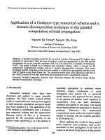
Figure 1 Sensitization of C6 cells to radiation under hypoxia
using doranidazole. Dose–response curves of X-irradiated C6 cells.
Tumor cells were X-irradiated under normoxia (red closed circles),
under normoxia with doranidazole (red open circles), under hypoxia
(blue closed squares) and under hypoxia with doranidazole (blue
open squares). The surviving fraction at each dose was calculated
and corrected according to the plating efficiency of the
nonirradiated control. Data are expressed as the mean ± S.E. for
three experiments.
To examine the distribution of doranidazole in the rat
brain, we performed autoradiographic analysis using
[14C]-doranidazole. Tumor-bearing rats were i.v. injected
with 500 μL of [14C]-doranidazole (4.9 MBq/head). At
90 minutes after drug administration, rats were decapitated without prior perfusion with saline. Their brains
were immediately removed and frozen. Frozen sections
that were 20-μm thick were exposed to a radiosensitive
imaging plate (BAS-SR2040: Fuji Film Co. Ltd., Tokyo,
Japan) for 4 days with a radioactive standard slide (ARC146: American Radiolabeled Chemicals Inc., St Louis,
MO, USA). The image acquisition was performed using
a BAS-2500 Bioimage Analyzer system (Fuji Film Co.
Ltd. Tokyo, Japan). After the acquisition of autoradiographic images, parts of sections were fixed with 4%
buffered formaldehyde and stained with hematoxylin/
eosin (H/E).
Yasui et al. BMC Cancer 2013, 13:106
/>
Page 4 of 9
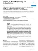
Figure 2 Disruption of the BBB in the brain of a C6-bearing rat. (A) Representative photographs of the dorsal surface (I), ventral surface
(II), coronal slice (III) and sagittal slice (IV) of control brain (a) and C6-bearing brain (b) after perfusion with Evans blue dye. (B) Representative
T1-weighted MR images obtained before and after Gd-[DTPA] injection. White lines show the region with high signal intensity, indicating the
BBB-disrupted region. (C) Quantitative data for Gd-[DTPA]-based CE-MRI. Relative MRI signal intensities are expressed as ratios relative to the
normal brain region.
Immunohistochemistry
At 1 day after treatment with doranidazole and/or Xirradiation tumor-bearing rats were i.v. injected with
pimonidazole (Hypoxyprobe™-1 Kit; 60 mg/kg). At 90 minutes after drug administration, rats were perfused
with saline and subsequently 4% buffered formaldehyde.
Removed brain tissues were fixed, embedded in paraffin
and sectioned at 5-μm thickness. The immunostaining
procedure for pimonidazole was carried out in accordance with the manufacturer’s instructions. Serial sections
Yasui et al. BMC Cancer 2013, 13:106
/>
Page 5 of 9
were also stained with H/E. The stained images of each
section were acquired using a fluorescence microscope
(BZ-9000: Keyence, Osaka, Japan).
Statistical analysis
All results were expressed as the mean ± S.E. The variance ratio was estimated using the F-test and differences
in means of groups were determined using Student’s ttest or Welch’s t-test. The minimum level of significance
was set at P < 0.05.
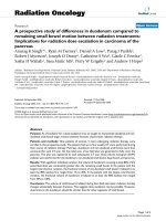
Figure 3 The distribution of [14C]-doranidazole in C6 intracranial
glioma. (A) A 20-μm thick tissue section of rat brain that was used for
autoradiography (a) and subsequent H/E staining (b). Black lines show
the C6 glioma. The annotated words “T” and “N” represent tumor and
normal brain regions, respectively. (B) Using these images, quantitative
data for the accumulation of [14C]-doranidazole in normal cortex and
C6 glioma was acquired. Data are expressed as the mean ± S.E. for four
different tumors. *: P < 0.05 vs. normal cerebrum.
Results
The clonogenic survival curves for C6 glioma cells irradiated in vitro under normoxic and hypoxic conditions,
with or without doranidazole, are shown in Figure 1.
Under conditions without doranidazole, X-irradiation
under hypoxia reduced the radiosensitivity of C6 cells,
and the oxygen enhancement ratio (OER) was approximately 1.9. The hypoxic condition set in this experiment
was ≤ 10 mmHg for pO2, and this OER value coincided
with that reported in a previous study [28]. Under
normoxic conditions without irradiation, the survival
fractions with or without doranidazole were 0.703 ±
0.019 and 0.677 ± 0.031, respectively. Hypoxic conditions
decreased the plating efficiency of C6 cells to 0.675 ±
0.006 and the addition of doranidazole resulted in a further decline to 0.667 ± 0.032, although no significant differences were observed among the groups. Under both
normoxia and hypoxia without irradiation, the toxicity
of 10 mM doranidazole against C6 cells was less than
30%. While doranidazole had no sensitizing effect when
combined with aerobic irradiation, it had significant sensitizing activity when combined with irradiation under
hypoxic conditions. The dose that reduces cell survival
to 10% (D10) obtained from the hypoxic cell survival
curve was 20.2 Gy, and it decreased to 13.3 Gy when
cells were irradiated in the presence of 10 mM
doranidazole. The sensitizing enhancement ratio (SER)
for doranidazole after irradiation under hypoxic conditions was ~1.5, whereas the SER after irradiation under
normoxic conditions was ~1.0.
To examine the disruption of the BBB in the C6tumor-bearing rat brain, we employed the Evans blue extravasation method. Evans blue dye is known to bind to
albumin producing a 68 kDa compound that does not
cross the BBB [29]. In fact, normal control brain after
intra-arterial infusion of Evans blue showed no staining
in the cerebral hemisphere (Figure 2A [a-I, II]). Using
this Evans blue extravasation test, we evaluated the permeability of the BBB in C6-bearing brain. Figure 2A (b-I,
II) shows a clearly stained region in the frontal cortex of
right hemisphere, in which the C6 tumor was located.
The photographs in Figure 2A (III, IV) are views of sectioned slices from control and C6-bearing brains. They
Yasui et al. BMC Cancer 2013, 13:106
/>
also demonstrated the apparent correspondence of the
stained region with the tumor region in C6-bearing
brain, while no staining was observed in the control
brain. To confirm this disruption of the BBB in the
tumor region, we performed CE-MRI analysis using a
BBB-impermeable reagent, Gd-[DTPA]. Figure 2B displays representative pre- and post-contrast T1WIs of
brains in C6-glioma-bearing rats, with the region of
interest (ROI) placed on the glioma. After Gd-[DTPA]
Figure 4 Effects of the combination of doranidazole and
X-irradiation on tumor growth in C6 glioma. When the tumor
reached a size of 50–100 mm3, rats were treated with doranidazole
(200 mg/kg) and/or X-irradiation (6 Gy). (A) Typical T2-weighted MR
images of a C6-bearing brain before and after each treatment.
(B) The quantitative data for suppression of tumor growth by
doranidazole administration and/or X-irradiation. The sizes of tumors
were estimated using T2-weighted MRI before treatment and at
7 days after treatment. Data are expressed as the mean ± S.E. for 5–8
different tumors. *: P < 0.05, **: P < 0.01.
Page 6 of 9
injection, MRI signal enhancement due to the accumulation of Gd-[DTPA] was clearly observed around the
tumor region. The quantitative data showed that the
relative signal intensities in glioma before and after Gd[DTPA] injection were 0.933 ± 0.008 and 1.597 ± 0.042,
respectively (Figure 2C).
We next investigated the distribution of doranidazole
in the brains of C6-bearing rats. Ninety minutes after
the i.v. administration of [14C]-doranidazole, rats were
decapitated. Brain tissue sections were analyzed using
autoradiography and subsequent H/E staining. In the
autoradiographic image shown in Figure 3A(a), [14C]doranidazole is clearly distributed in the tumor region
but not in the normal brain cortex. We then quantified
the accumulation of [14C]-doranidazole in each region
of the normal cortex and tumor region defined by H/E
staining (Figure 3A[b]). Tumor regions showed significantly higher [14C] radioactivity levels (1926.5 ±
523.3 Bq/mm2) than the normal cortex region (138.7 ±
14.6 Bq/mm2) (Figure 3B). These results suggested that
doranidazole could penetrate into the tumor region due
to the breakdown of the BBB in the C6-bearing brain.
We also examined the radiosensitizing effect of doranidazole on the growth of transplanted C6 glioma. Rats
with 50–100 mm3 of glioma tumor were treated with
200 mg/kg doranidazole and/or 6 Gy of X-rays. We estimated the tumor volumes before and after each treatment
using T2WIs to indicate the definite tumor area (Figure 4A).
As shown in Figure 4B, without any treatment tumor size
increased ~2.5-fold in 7 days and reached 165.3 ±
35.5 mm3. X-irradiation or doranidazole alone induced no
statistically significant inhibition of tumor growth. The
tumor volumes at 7 days after treatment were 121.0 ±
24.9 mm3 after X-irradiation alone and 152.0 ± 30.3 mm3
after doranidazole alone. X-irradiation at 30 minutes after
doranidazole treatment induced a significant retardation in
tumor growth (56.0 ± 22.7 mm3). To examine the suppressive effect of doranidazole on the hypoxic region
in the C6 glioma, histological analysis with pimonidazole staining and H/E staining was performed.
Immunohistological images for pimonidazole revealed a
characteristic cord-like structure of hypoxia in viable
tumor, within specimens resected from tumors receiving radiation or doranidazole alone. However, the great
majority of the tumor containing hypoxic region was
necrotic after combined treatment (Figure 5).
Discussion
In the present study, we investigated the radiosensitizing
effect of a hypoxic cell radiosensitizer, doranidazole, on C6
intracranial glioma. Doranidazole has a 2-nitroimidazole
-based chemical structure with a side chain having low lipophilicity. It is designed to be less neurotoxic due to its
BBB-impermeability [15,16]. In common with other
Yasui et al. BMC Cancer 2013, 13:106
/>
Page 7 of 9
Figure 5 Effects of the combination of doranidazole and X-irradiation on tumor hypoxia in C6 glioma. Histological evaluation of C6
tumors at 1 day after treatment. (A) Immunohistochemical images for pimonidazole. Animals received vehicle (a), doranidazole (200 mg/kg)
(b), 6 Gy of X-rays (c), or a combination (d) as described in Figure 4. A representative field for each condition is shown. Bar = 500 μm.
(B) Representative images of pimonidazole staining and H/E staining taken at high magnification in C6 tumors resected from the control group
(a) and the combination group (b). Bar = 100 μm.
2-nitroimidazole derivatives such as misonidazole and
etanidazole, doranidazole is reduced under hypoxic conditions and imported into the cell nucleus, leading to
fixation of radiation damage in a manner similar to
oxygen [30]. In the present study, it was clearly demonstrated in vitro that doranidazole radiosensitized hypoxic cells as determined by clonogenic survival assay
(Figure 1). This radiosensitizing effect was consistent
with previous reports [15,21].
Because the delivery of hydrophilic doranidazole into the
tumor region is crucial for its radiosensitizing effect, we
investigated the extent of the BBB disruption using Evans
blue dye extravasation. Figure 2A clearly shows the penetration of this dye into the tumor region, but not normal brain
tissue. The disrupted BBB allows MR-based detection of
glioblastoma by extravasation and accumulation of contrast
agents such as Gd-DTPA in the interstitial spaces [31]. By
using this method, the breakdown of the BBB in C6 glioma
was confirmed by CE-MRI with Gd-[DTPA] (Figure 2B
and C). Due to its trihydroxyl structure, doranidazole is less
lipophilic than misonidazole and etanidazole, with reduced
neurotoxicity. The disruption of the BBB as shown in
Yasui et al. BMC Cancer 2013, 13:106
/>
Figure 2 may indicate the feasibility of using doranidazole
to treat some intracranial tumors. In fact in the current
study, autographic analysis in vivo indicated the obvious accumulation of [14C]-doranidazole in the tumor region. To
our knowledge, our results have clarified for the first time
that disruption of the BBB, which has been observed in
some types of glioblastoma such as C6 glioma, enabled a
lipophobic nitroimidazole analog, doranidazole to be incorporated into the tumor region. To reveal the variability in
tumor response to doranidazole based on levels of hypoxia,
further investigation using other glioma models will be
required.
As mentioned, a number of clinical trials involving a
few 2-nitroimidazole-derivatives in combination with
radiotherapy have been performed with the objective of
improving therapeutic benefit. However, most of them
have provided disappointing results with poor enhancement of the efficacy of radiotherapy and severe side
effects such as neurotoxicity. To develop an effective
therapy with few side effects and sufficient radiosensitizing effects, it is necessary to identify the appropriate tumor type using optimal parameters such as
oxygenation status and vascular permeability. Currently,
several noninvasive tools are being established for the
monitoring of tumor oxygenation and blood perfusion
[32,33]. To confirm the rationale for using hypoxic cell
sensitizers, microenvironmental information on the target tumor should be obtained in preclinical and clinical
studies.
Conclusions
In conclusion, we demonstrated that doranidazole had a
radiosensitizing effect on C6 glioma, a tumor model that
shows a wide range of hypoxia and disruption of the
BBB. The observation of synergistic tumor growth inhibition by combined treatment with X-irradiation and
doranidazole, as shown in Figure 4, clearly indicates the
possibility of clinical administration of this drug in the
treatment of intracranial glioma. Our study also demonstrated that this radio-sensitization effect was induced
through the selective accumulation of doranidazole in a
BBB-disrupted tumor. Thus, doranidazole may be a candidate radiosensitizer for use against malignant glioma.
Abbreviations
BBB: Blood brain barrier; DMEM: Dulbecco’s modified Eagle’s medium;
FBS: Fetal bovine serum; i.v.: intravenous; MSE: Multislice spin-echo; T2WI: T2weighted image; Gd-DTPA: Gadopentetate dimeglumine; CE-MRI: Contrastenhanced MRI; T1WI: T1-weighted image; H/E: Hematoxylin/eosin;
SER: Sensitizing enhancement ratio; ROI: Region of interest.
Competing interests
NK is an employee of POLA PHARMA INC.; all of the other authors have no
competing interests to declare.
Page 8 of 9
Authors’ contributions
HY, TA and JK performed the in vitro and in vivo experiments, analyzed the
data and prepared the manuscript. TY and SM also participated in the
performance of the in vitro experiments. MN prepared the gliomatransplanted animal model. NK synthesized doranidazole and [14C]doranidazole. MK and OI designed the research and interpreted the data. All
authors approved the final version of the manuscript.
Acknowledgements
This work was supported, in part, by Grants-in-Aid for Basic Scientific
Research from the Ministry of Education, Culture, Sports, Science and
Technology, Japan (No. 21658106 and No. 21380185 [O.I.], No. 21780267
[T.Y.] and No. 23791375 [H.Y.]), and by the Akiyama Life Science Foundation
[H.Y. and T.Y.].
Author details
Laboratory of Radiation Biology, Department of Environmental Veterinary
Sciences, Graduate School of Veterinary Medicine, Hokkaido University, Kita
18 Nishi 9, Kita-ku, Sapporo, Hokkaido, Japan. 2Laboratory of Veterinary
Radiology, Department of Veterinary Sciences, University of Miyazaki, 1-1,
Gakuen Kibanadai-nishi, Miyazaki, Miyazaki, Japan. 3POLA PHARMA INC, 8-9-5,
Nishigotanda, Shinagawa-ku, Tokyo, Japan.
1
Received: 21 June 2012 Accepted: 3 March 2013
Published: 8 March 2013
References
1. Burton EC, Prados MD: Malignant gliomas. Curr Treat Options Oncol 2000,
1(5):459–468.
2. Forsyth PA, Cairncross JG: Treatment of malignant glioma in adults.
Curr Opin Neurol 1995, 8(6):414–418.
3. Robins HI, Chang S, Butowski N, Mehta M: Therapeutic advances for
glioblastoma multiforme: current status and future prospects. Curr Oncol
Rep 2007, 9(1):66–70.
4. Jensen RL: Brain tumor hypoxia: tumorigenesis, angiogenesis, imaging,
pseudoprogression, and as a therapeutic target. J Neurooncol 2009,
92(3):317–335.
5. Chakravarti A, Dicker A, Mehta M: The contribution of epidermal growth
factor receptor (EGFR) signaling pathway to radioresistance in human
gliomas: a review of preclinical and correlative clinical data. Int J Radiat
Oncol Biol Phys 2004, 58(3):927–931.
6. Baumann M, Krause M, Hill R: Exploring the role of cancer stem cells in
radioresistance. Nat Rev Cancer 2008, 8(7):545–554.
7. Brown JM, Wilson WR: Exploiting tumour hypoxia in cancer treatment.
Nat Rev Cancer 2004, 4(6):437–447.
8. Dewhirst MW: Relationships between cycling hypoxia, HIF-1,
angiogenesis and oxidative stress. Radiat Res 2009, 172(6):653–665.
9. Brat DJ, Mapstone TB: Malignant glioma physiology: cellular response to
hypoxia and its role in tumor progression. Ann Intern Med 2003,
138(8):659–668.
10. Nagasawa H, Uto Y, Kirk KL, Hori H: Design of hypoxia-targeting drugs as
new cancer chemotherapeutics. Biol Pharm Bull 2006, 29(12):2335–2342.
11. Overgaard J: Hypoxic radiosensitization: adored and ignored. J Clin Oncol
2007, 25(26):4066–4074.
12. Kaanders JH, Bussink J, van der Kogel AJ: Clinical studies of hypoxia
modification in radiotherapy. Semin Radiat Oncol 2004, 14(3):233–240.
13. Overgaard J, Hansen HS, Andersen AP, Hjelm-Hansen M, Jorgensen K,
Sandberg E, Berthelsen A, Hammer R, Pedersen M: Misonidazole combined
with split-course radiotherapy in the treatment of invasive carcinoma of
larynx and pharynx: report from the DAHANCA 2 study. Int J Radiat Oncol
Biol Phys 1989, 16(4):1065–1068.
14. Overgaard J, Hansen HS, Overgaard M, Bastholt L, Berthelsen A, Specht L,
Lindelov B, Jorgensen K: A randomized double-blind phase III study of
nimorazole as a hypoxic radiosensitizer of primary radiotherapy in
supraglottic larynx and pharynx carcinoma. Results of the Danish Head
and Neck Cancer Study (DAHANCA) Protocol 5–85. Radiother Oncol 1998,
46(2):135–146.
15. Oya N, Shibamoto Y, Sasai K, Shibata T, Murata R, Takagi T, Iwai H, Suzuki T,
Abe M: Optical isomers of a new 2-nitroimidazole nucleoside analog
(PR-350 series): radiosensitization efficiency and toxicity. Int J Radiat
Oncol Biol Phys 1995, 33(1):119–127.
Yasui et al. BMC Cancer 2013, 13:106
/>
16. Kuwabara M, Iida Y, Inanami O, Sawamura S, Yokoyama K, Tsujitani M:
Radiation-chemical properties of the hypoxic cell radiosensitizer
doranidazole (PR-350). J Radiat Res (Tokyo) 2002, 43(1):77–88.
17. Aoki M, Furusawa Y, Shibamoto Y, Kobayashi A, Tsujitani M: Effect of a
hypoxic cell sensitizer doranidazole on the radiation-induced apoptosis
of mouse L5178Y lymphoma cells. J Radiat Res (Tokyo) 2002,
43(2):161–166.
18. Hamasu T, Inanami O, Tsujitani M, Yokoyama K, Takahashi E, Kashiwakura I,
Kuwabara M: Post-irradiation hypoxic incubation of X-irradiated MOLT-4
cells reduces apoptotic cell death by changing the intracellular redox
state and modulating SAPK/JNK pathways. Apoptosis 2005, 10(3):557–567.
19. Shibamoto Y, Kubota T, Kishii K, Tsujitani M: Radiosensitivity of human
pancreatic cancer cells in vitro and in vivo, and the effect of a new
hypoxic cell sensitizer, doranidazole. Radiother Oncol 2000, 56(2):265–270.
20. Murata R, Tsujitani M, Horsman MR: Enhanced local tumour control after
single or fractionated radiation treatment using the hypoxic cell
radiosensitizer doranidazole. Radiother Oncol 2008, 87(3):331–338.
21. Yahiro T, Masui S, Kubota N, Yamada K, Kobayashi A, Kishii K: Effects of
hypoxic cell radiosensitizer doranidazole (PR-350) on the radioresponse
of murine and human tumor cells in vitro and in vivo. J Radiat Res
(Tokyo) 2005, 46(3):363–372.
22. Sunamura M, Karasawa K, Okamoto A, Ogata Y, Nemoto K, Hosotani R,
Nishimura Y, Matsui K, Matsuno S: Phase III trial of radiosensitizer PR-350
combined with intraoperative radiotherapy for the treatment of locally
advanced pancreatic cancer. Pancreas 2004, 28(3):330–334.
23. Deeken JF, Loscher W: The blood–brain barrier and cancer: transporters,
treatment, and Trojan horses. Clin Cancer Res 2007, 13(6):1663–1674.
24. Schneider SW, Ludwig T, Tatenhorst L, Braune S, Oberleithner H, Senner V,
Paulus W: Glioblastoma cells release factors that disrupt blood–brain
barrier features. Acta Neuropathol 2004, 107(3):272–276.
25. Towner RA, Smith N, Doblas S, Garteiser P, Watanabe Y, He T, Saunders D,
Herlea O, Silasi-Mansat R, Lupu F: In vivo detection of inducible nitric
oxide synthase in rodent gliomas. Free Radic Biol Med 2010, 48(5):691–703.
26. Asanuma T, Doblas S, Tesiram YA, Saunders D, Cranford R, Yasui H, Inanami
O, Smith N, Floyd RA, Kotake Y, et al: Visualization of the protective ability
of a free radical trapping compound against rat C6 and F98 gliomas
with diffusion tensor fiber tractography. J Magn Reson Imaging 2008,
28(3):574–587.
27. Liu R, Wen Y, Perez E, Wang X, Day AL, Simpkins JW, Yang SH: 17betaEstradiol attenuates blood–brain barrier disruption induced by cerebral
ischemia-reperfusion injury in female rats. Brain Res 2005,
1060(1–2):55–61.
28. Schwartz DL, Powis G, Thitai-Kumar A, He Y, Bankson J, Williams R, Lemos R,
Oh J, Volgin A, Soghomonyan S, et al: The selective hypoxia inducible
factor-1 inhibitor PX-478 provides in vivo radiosensitization through
tumor stromal effects. Mol Cancer Ther 2009, 8(4):947–958.
29. Rapoport SI: Osmotic opening of the blood–brain barrier: principles,
mechanism, and therapeutic applications. Cell Mol Neurobiol 2000,
20(2):217–230.
30. Weinmann M, Welz S, Bamberg M: Hypoxic radiosensitizers and hypoxic
cytotoxins in radiation oncology. Curr Med Chem Anticancer Agents 2003,
3(5):364–374.
31. Ludemann L, Grieger W, Wurm R, Wust P, Zimmer C: Quantitative
measurement of leakage volume and permeability in gliomas,
meningiomas and brain metastases with dynamic contrast-enhanced
MRI. Magn Reson Imaging 2005, 23(8):833–841.
32. Matsumoto S, Yasui H, Batra S, Kinoshita Y, Bernardo M, Munasinghe JP,
Utsumi H, Choudhuri R, Devasahayam N, Subramanian S, et al:
Simultaneous imaging of tumor oxygenation and microvascular
permeability using Overhauser enhanced MRI. Proc Natl Acad Sci USA
2009, 106(42):17898–17903.
33. Yasui H, Matsumoto S, Devasahayam N, Munasinghe JP, Choudhuri R, Saito
K, Subramanian S, Mitchell JB, Krishna MC: Low-field magnetic resonance
imaging to visualize chronic and cycling hypoxia in tumor-bearing mice.
Cancer Res 2010, 70(16):6427–6436.
Page 9 of 9
Submit your next manuscript to BioMed Central
and take full advantage of:
• Convenient online submission
• Thorough peer review
• No space constraints or color figure charges
• Immediate publication on acceptance
doi:10.1186/1471-2407-13-106
Cite this article as: Yasui et al.: The prospective application of a hypoxic
radiosensitizer, doranidazole to rat intracranial glioblastoma with blood
brain barrier disruption. BMC Cancer 2013 13:106.
• Inclusion in PubMed, CAS, Scopus and Google Scholar
• Research which is freely available for redistribution
Submit your manuscript at
www.biomedcentral.com/submit