Detection and characterization of classical and “uncommon” exon 19 Epidermal Growth Factor Receptor mutations in lung cancer by pyrosequencing
Bạn đang xem bản rút gọn của tài liệu. Xem và tải ngay bản đầy đủ của tài liệu tại đây (936.77 KB, 9 trang )
Righi et al. BMC Cancer 2013, 13:114
/>
TECHNICAL ADVANCE
Open Access
Detection and characterization of classical and
“uncommon” exon 19 Epidermal Growth Factor
Receptor mutations in lung cancer by
pyrosequencing
Luisella Righi1*, Alessandra Cuccurullo3, Simona Vatrano1, Susanna Cappia1, Daniela Giachino2, Paolo De Giuli3,
Mara Ardine4, Silvia Novello5, Marco Volante1, Giorgio V Scagliotti5 and Mauro Papotti1
Abstract
Background: The management of advanced stage non-small cell lung cancer is increasingly based on diagnostic
and predictive analyses performed mostly on limited amounts of tumor tissue. The evaluation of Epidermal Growth
Factor Receptor (EGFR) mutations have emerged as the strongest predictor of response to EGFR-tyrosine kinase
inhibitors mainly in patients with adenocarcinoma. Several EGFR mutation detection techniques are available,
having both sensitivity and specificity issues, being the Sanger sequencing technique the reference standard, with
the limitation of a relatively high amount of mutated cells needed for the analysis.
Methods: A novel nucleotide dispensation order for pyrosequencing was established allowing the identification
and characterization of EGFR mutation not definable with commercially and clinically approved kits, and validated in
a consecutive series of 321 lung cancer patients (246 biopsies or cytology samples and 75 surgical specimens).
Results: 61/321 (19%) mutated cases were detected, 17 (27.9%) in exon 21 and 44 (72.1%) in exon 19, these latter
corresponding to 32/44 (72.7%) classical and 12/44 (27.3%) uncommon mutations. Furthermore, a novel, never
reported, point mutation, was found, which determined a premature stop codon in the aminoacidic sequence that
resulted in a truncated protein in the tyrosine kinase domain, thus impairing the inhibitory effect of specific
therapy.
Conclusions: The novel dispensation order allows to detect and characterize both classical and uncommon EGFR
mutations. Although several phase III studies in genotypically defined groups of patients are already available,
further prospective studies assessing the role of uncommon EGFR mutations are warranted.
Keywords: Lung cancer, EGFR mutation, Exon 19, Pyrosequencing, Adenocarcinoma
Background
The old dichotomic distinction between small cell and
non-small cell lung cancer (NSCLC) in the last 10 years
has been replaced by a more accurate morphological
and immunohistochemical subtyping associated to the
identification of specific molecular profiles [1,2]. For the
management of lung cancer, a crucial issue is the availability of adequate and sufficient tumor tissue not only
* Correspondence:
1
Divisions of Pathology, University of Torino, Regione Gonzole 10, Torino,
Orbassano 10043, Italy
Full list of author information is available at the end of the article
for pathological diagnosis, but also to allow additional
immunohistochemical and molecular studies [3]. Since
at diagnosis up to 70% of patients with NSCLC present
with inoperable, advanced-stage disease, the histological
definition and molecular characterization, including the
assessment of the epidermal growth factor receptor
(EGFR) sensitizing and resistant mutations, is often
based on lung biopsies (endoscopic or transthoracic)
or cytological specimens, only. These tumor samples
are often characterized by poor cellularity and/or
inflammatory or necrotic background containing large
amounts of tumor-associated normal cells, which may
© 2013 Righi et al.; licensee BioMed Central Ltd. This is an Open Access article distributed under the terms of the Creative
Commons Attribution License ( which permits unrestricted use, distribution, and
reproduction in any medium, provided the original work is properly cited.
Righi et al. BMC Cancer 2013, 13:114
/>
potentially impair the accuracy of tumor subtyping and
molecular characterization [4]. This issue can be
improved by the enrichment of tumor cells using tissue
microdissection prior to mutational analysis [5].
The detection of activating EGFR mutations is nowadays
the best predictive marker to treat NSCLC with EGFRTyrosine Kinase Inhibitors (TKI) [6], but most trials
conducted so far are based on a limited number of known
EGFR mutations, including the point mutation at codon
858 of exon 21 (NM_005228.3 p.Leu858Arg) and the
numerous in-frame deletions in exon 19, which account for
more than 90% of mutations [7]. Furthermore, a single,
standardized method to perform the mutational analysis is
not yet available [8], making rigorous quality control tests
mandatory for each laboratory. Numerous methodological
approaches are currently available although affected by
great inter- and intra-laboratory variability in terms of
performance and lack of adequate quality controls. The use
of commercial kits certified by the FDA and/or EMEA
is therefore recommended in the clinical practice [9].
The most commonly used mutation detection techniques
(i.e. Sanger sequencing, Pyrosequencing, HRMA - High
Resolution Melting Analysis and ARMS – Amplification
Refractory Mutation System analysis) were established to
offer sensitive molecular analysis, all including DNA
extraction, PCR amplification and subsequent genetic test.
Thus far, the Sanger DNA sequencing method is the
reference method used for the detection and identification
of EGFR mutations in tumor cells, because it provides the
exact nucleotide sequence of the segment amplified,
despite its sensitivity is lower than others, especially in the
case of small tumor samples, since it requires at least 50%
of mutated tumor cells [10], corresponding to 20–25% of
mutated DNA in an heterozygous case [8]. More recently,
several assays have been developed to improve mutation
detection in terms of sensitivity (to better perform
molecular analyses even in very small specimens) and also
of specificity (to recognize and characterize multiple
mutations at the same time). Indeed recently, beyond the
classical therapy-responsive mutations, some “uncommon”
mutations were described whose clinical significance is still
poorly understood [11].
Pyrosequencing is a DNA sequencing technology “by
synthesis” with luminometric detection. Due to its
modalities of analysis and nucleotide dispensation [12],
any change from normal in the target sequence is
detected as a pyrogram alteration, but not characterized
unless corresponding to an expected genetic alteration [13].
Furthermore, the commercially available pyrosequencing
kit properly identifies commonest EGFR point mutations
(i.e. exons 18 NM_005228.3 p.Gly719Ser, p.Gly719Cys,
p.Gly719Ala, p.Gly719Asp and exon 21 NM_005228.3
p.Leu858Arg, p.Leu861Gln), but is certified to detect
just a positive mutational status in the presence of
Page 2 of 9
the two most frequent (classical) deletions in exon 19
(NM_005228.3 c.2235_2249del15 and c.2236_2250del15 p.Glu746_Ala750del). On the contrary, all the other uncommon exon 19 mutations are detected as an altered
signal, although not further identifiable.
The aim of the present study was to improve the
performance of pyrosequencing assay for EGFR mutation
detection by setting up a novel dispensation order (NDO)
capable not only to detect but also to characterize the type
of mutations of exon 19 associated to the responsiveness
to TKI therapy [14], and to validate its efficacy in the
clinical setting in a consecutive prospectively collected
series of lung cancer specimens.
Methods
Cell lines and plasmids
The human lung cancer HCC827 and H522 cell lines were
obtained from the American Type Culture Collection and
were cultured in RPMI 1640 supplemented with 10% fetal
bovine serum at 37°C in air containing 5% CO2. The
HCC827 cell line harboured in homozygosis one of the two
classical EGFR deletions in the tyrosine kinase (TK) domain
(NM_005228.3 p.Glu746_Ala750del, c.2236_2250del15),
while the control H522 cell line was wild type (wt) for exon
19 mutations. DNA from H522 was used to dilute the
mutated HCC827 DNA cell line at 50% and 25% of
mutated allele in order to test the NDO accuracy to detect
the presence of the classical EGFR deletions.
Furthermore, to test the sensitivity of NDO detection
(set as the minimum percentage of mutation detection),
the DNA plasmid pUC57 (Eurogentec, Belgium, kindly
purchased by Diatech Company - Jesi, Italy), containing
an insertion of 400 bp harbouring three different in-frame
deletions of exon 19 EGFR in homozygosis (the other classical NM_005228.3 p.Glu746_Ala750del, c.2236_2250del15
(M1882), and the uncommon c.2240_2257del18 (M1883)
in-frame deletion and c.2239_2248delinsC (M1884)
complex mutation) was mixed with the wt plasmid
M1880 at serial descending dilutions, obtaining 50%,
25%, 12.5% and 6.25% of exon 19 mutated DNA.
Tissue samples
From January 2010 to December 2011, 334 consecutive
NSCLC samples (including 75 surgical resections, 139
transthoracic or endoscopic biopsies and 96 cytological specimens) were considered for EGFR (GenBank
NM_005228.3) exons 18, 19, 21 mutational analysis in the
Pathology Division of the University of Turin at San Luigi
Hospital, and subsequently prospectively collected for
NDO pyrosequencing analysis. A pathologist (LR) evaluated
the adequacy of all cases selecting the tissue specimens
having the highest tumor cell content. Adequacy was set at
a minimum of 200 tumor cells and a percentage of tumor
cells in the DNA sample of at least 30%. Such adequacy
Righi et al. BMC Cancer 2013, 13:114
/>
assessment is in line to current national and international
recommendations/guidelines [9,15]. Enrichment of tumor
cells was obtained by manual microdissection under light
microscopy from one to ten sections for each case. Briefly,
5 μm thick sections of tumor samples were collected on
glass slides and processed with a fast haematoxylin-eosin
staining. Tumoral cells were scraped with a 1 mm-gauge
needle in 70% ethanol and collected in vials. After centrifugation at 14000 rpm for 20 minutes, the microdissected
pellet obtained for each sample was dehydrated with
absolute ethanol, followed by another centrifugation at
14000 rpm for 20 minutes. The dried pellets were
processed for DNA extraction.
All histological material was de-identified and cases
were anonymized by a pathology staff member not
involved in the study. Clinical data were compared and
analysed through coded data, only. The study was
approved by the institutional review board of San Luigi
Hospital, Turin, Italy.
DNA extraction and PCR amplification
Genomic DNA from formalin-fixed paraffin-embedded
(FFPE) cell lines and tissues was extracted and purified
using QIAmp DNA FFPE Tissue kit (Qiagen, Hilden,
Germany) specific for purification from FFPE samples,
according to the manufacturer’s instructions. The amount
of DNA obtained was quantified by spectrophotometry
(Eppendorf, Hamburg, Germany). Genomic DNA from cell
lines, tissues and plasmid was amplified by real-time
end-point PCR using EGFR TKI response (sensitivity) kit
(CE-IVD, Diatech, Jesi, Italy) according to the manufacturer’s instructions, using Rotor-Gene Q (Qiagen, Hilden,
Germany). After amplification, the presence of PCR products was detected by melting-analysis with a denaturation
step from 65°C up to 95°C. The specific melting temperatures of exon 18, 19 and 21 amplicons were 84.5°C, 81.3°C
and 83°C, respectively. Wild type and no template samples
were added in each assay as positive and negative controls.
Mutational analysis by pyrosequencing and novel
nucleotide dispensation order
The mutational analysis was performed by pyrosequencing
with PyroMark Q96MA apparatus (Biotage, Uppsala,
Sweden) using EGFR TKI response (sensitivity) kit. PCR and
mutational analysis were conducted in duplicate for each
sample, as requested by the guidelines. Only the genomic
regions frequently harbouring mutations relevant for TKI
therapy [14] were analyzed (i.e. for ex 18: codons from
2149 to 2157; for ex 21: codons from 2572 to 2585; for ex
19: codons from 2234 to 2250). The pyrograms obtained
were analysed following the manufacturer’s instructions.
To better characterize both common and uncommon
mutations affecting EGFR exon 19 in the studied genomic
region, PCR primers generating an amplimer of 113 bp
Page 3 of 9
(sense- 50-TCCCAGAAGGTGAGAAAGTTAAA-30 and
antisense BIO-50-CCACACAGCAAAGCAGAAAC-30) and
a sequencing primer (50-TTCCCGTCGCTATCA-30) were
designed using the PSQ Assay Design software (Biotage)
and a novel NDO (50-TACGCAGTCATGAGAGTCGAGC
AGTCTCG-30) for pyrosequencing was developed analyzing the codons from 2234 to 2259. The NDO consisted
in a sequence of 29 dispensed nucleotides, longer than
commercial kit dispensation order, in which additional
bases are introduced in strategic positions, according to
possible expected mutated sequences (Figure 1A). The
obtained pyrograms allowed to characterize the sequences
different from wt resulting from either classical or uncommon mutations (Figure 1B, C, D).
Mutational analysis by ARMS
To validate the results obtained by pyrosequancing, as
indicated by guidelines [9], all tumor samples were also
tested for the presence of the two most frequent EGFR
exon 19 deletions (NM_005228.3 c.2235_2249del15 and
c.2236_2250del15, p.Glu746_Ala750del) by ARMS using the
following primers: sense 50-TGCATCGCTGGTAACAT-30
and antisense 50-CGGAGATGTTTTGATAGCG-30 [16].
Briefly 25 μl of PCR reaction mix contained 2X Master mix
(Promega, Madison - USA), 20X EVA Green dye (Biotium,
Hayward, CA - USA), 0.6 μM of each primer and 50 ng
DNA template. PCR conditions on Rotor-Gene Q (Qiagen
Hilden, Germany) were as follows: 95°C for 5 minutes, then
40 cycles of 95°C for 30 seconds, 56.6°C for 30 seconds and
72°C for 30 seconds, followed by a melting profile with a
ramping range of 65° to 95°C and rising steps of 1 degree.
Positive and no template controls were added to each tests.
Sanger sequencing analysis
In selected tissue cases, EGFR exon 19 uncommon mutation were also confirmed by di-deoxy Sanger direct sequencing analysis, supported by Eurofins MWG Operon service,
(Ebersberg, Germany), after DNA amplification. Briefly, the
target region was amplified with sense 50-ACAATTGCCA
GTTAACGTCTTCCT-30 and antisense 50-ATGAGAAAA
GGTGGGCCTGA-30 primers under the following conditions: 95°C for 5 minutes, then 40 cycles of 95°C for 30 seconds, 55°C for 40 seconds and 72°C for 40 seconds,
followed by 72°C for 4 minutes. Then, the PCR products
were resolved by 2% agarose gel electrophoresis to confirm
successful amplification and purified using MicroSpin™
S-400 HR Columns kit (GE Healthcare, Buckinghamshire,
UK), according to the manufacturer’s instructions.
Statistical analysis
Distribution of EGFR mutations as compared to patients’
variables were analyzed with One-way ANOVA test and
Fisher’s test using GraphPad Prism software, version 5.0.
The level of significance was set at p < 0.05.
Righi et al. BMC Cancer 2013, 13:114
/>
Page 4 of 9
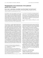
Figure 1 Nucleotide sequences and representative pyrograms of the commercial kit and novel dispensation order for EGFR exon 19
mutation analysis. (A) In the home-made dispensation order a number of nucleotides major than commercial is present to characterize a wider
group of deleted sequences (classical and uncommon) than commercial kit. Circles exemplify anomalous nucleotides identified by NDO only.
Example of pyrograms obtained using the commercial KDO (left panels) and NDO (right panels) for pyrosequencing in samples with EGFR exon 19
(B) wild type, (C) classical deletion (c.2235-2249del15), (D) uncommon deletion (c.2240-2257del18). Arrows indicate peak modifications corresponding
to anomalous nucleotides identified by NDO only (Abbreviations: KDO: kit dispensation order; NDO: novel dispensation order; WT: wild type).
Righi et al. BMC Cancer 2013, 13:114
/>
Results
Validation of the novel dispensation order on cell lines
and plasmids
On cell lines, the EGFR TKI response (sensitivity) kit and
the NDO procedure were both able to detect the classical
deletion in the HCC827 DNA cell line at the higher
(50%) and lower (25%) dilutions in terms of peak profile
alteration, whereas EGFR wt DNA cell line resulted in
the normal profile.
In the analysis of DNA plasmid with the EGFR TKI
response (sensitivity), the classical mutation was detected
up to 25% of dilution, while uncommon mutations were
reported as indeterminate or wt when the dilution
reached the lowest percentages of mutated alleles. On
the contrary, the NDO showed specific peak alterations
corresponding to the three different deletions on plasmid
allowing to characterize all mutations up to 6.25% of
mutant allele dilution.
Mutational analysis on tissue samples
Thirteen cases (3.8%) were excluded because not reaching
the adequacy standards set for molecular analysis, based on
national and international recommendations [9,15] even
after tumor cell enrichment by manual microdissection.
The study samples derived from 186 men (57.9%) and
135 women (42.1%), with a median age of 65 years
(range 16 to 89 years); specimens were represented by
75 (23.4%) primary lung tumor resections and 246
(76.6%) non-surgical (150 endoscopic transthoracic or
lymph node biopsies and 96 cytological) samples; 226
(70.4%) tissues were from pulmonary tumor location
and 95 (29.6%) were metastases. The final diagnoses
included: 269 (83.8%) adenocarcinomas (ADC), 37 (11.6%)
NSCLC, favor ADC, four (1.2%) NSCLC not otherwise
specified and 11 (3.4%) non-ADC. In particular, surgical
case series included: 69 ADC and 6 non-ADC (2 squamous
cell carcinoma; 2 large cell carcinoma, 1 mucoepidermoid
low grade carcinoma and 1 sarcomatoid carcinoma having 80% of ADC component). Non-surgical case series
corresponded to 200 cases with a morphology-only based
ADC diagnosis, 37 NSCLC favouring ADC after immunohistochemistry (IHC), four NSCLC not otherwise specified
for an ambiguous immunophenotype and five non-ADC
cases (4 squamous and 1 undifferentiated carcinoma).
Patients’ characteristics are summarised in Table 1.
EGFR mutational status was distributed as follows:
total mutated cases were 61/321 (19.0%), including 17/
61 (27.9%) in exon 21 and 44/61 (72.1%) in exon 19.
Among surgical samples, 17/75 (22.6%) mutated cases
were found, including 10/17 (58.8%) mutations in exon 19
and 7/17 (41.2%) mutations in exon 21 (all p.L858R type).
In non-surgical samples, 44/246 (17.8%) mutated cases
were found, of which 34/44 (77.2%) mutations were in
EGFR exon 19 and 10/44 (22.7%) point mutations in exon
Page 5 of 9
21 (9 p.L858R and 1 p.L861Q). As to concern primary
pulmonary versus metastatic tumor samples, mutations were detected in 45/226 (19.9%) and 16/95
(16.8%) cases, respectively. Moreover, distribution of
mutated cases according to diagnosis was 53/269
(19.7%) ADCs, 7/37 (18.9%) NSCLC favour ADC and
1/11 (9.1%) non-ADC (sarcomatoid carcinoma). No
mutations in exon 18 were found.
The distribution of EGFR mutations among male and
female patients was significantly different: the percentage
of mutations in women was nearly double than that of
male patients (Fisher test, p < 0.0001). On the contrary, no
significant differences were recorded as compared to other
characteristics, including type of sample (surgical, biopsy
or cytology) (Table 1).
The EGFR TKI response (sensitivity) commercial kit
was able to detect all mutated cases as an altered signal
with respect to wild-type sequence either in exon 21 and
exon 19; of these latter 32/44 (72.7%) were attributable
to one of the two classical more common in-frame
deletions, while 12/44 (27.2%) were referred as altered
signals by the kit with no possibility of further
characterization. On the other hand, ARMS analysis was
able to confirm all samples (32/44 cases) harboring the
classical exon 19 deletions with a specific amplification.
Nevertheless the remaining samples resulted negative
because did not show amplification, included not only the
true wt cases but also those cases harboring uncommon
mutations in exon 19. No amplification was detected in
no-template controls. On the contrary, using the above
described newly constructed NDO, it was possible not only
to detect all the 61/321 alterations (with a 100% concordance with EGFR TKI response commercial kit), but also to
exactly characterize both the 44/61 classical and the 12/61
uncommon mutations of exon 19, with a similar performance on cytological and histological material (Figure 2).
The uncommon mutations identified by NDO were subsequently sequenced by Sanger method for confirmation
only, although this latter procedure would not be necessary
to determine the type of mutation and could be avoided in
the clinical practice.
Moreover, using this approach, a new mutation, which
had never been described in the literature nor reported in
any database of genetic variants ( />genetics/CGP/cosmic/; /> http://www.
ensembl.org/Homo_sapiens/Search/Details?db=core;end=
498;idx=Somatic_mutation;q=egfr; species = Homo_sapiens),
was identified in exon 19. This occurred in a lung ADC case
that showed no alteration at ARMS analysis (Figure 3A), an
altered pyrogram with the EGFR TKI response (sensitivity)
kit (Figure 3B) but not attributable neither to classical or
uncommon mutations affecting EGFR exon 19 so far described. On the contrary, with the NDO mutational analysis
Righi et al. BMC Cancer 2013, 13:114
/>
Page 6 of 9
Table 1 Case series characteristics and EGFR mutations distribution
N
EGFR mutations
Total mutated
Total
321
61 (19.0%)
Male
186 (57.9%)
21 (11.3%)
Female
135 (42.1%)
40 (29.6%)
75 (23.4%)
17 (22.6%)
p value
EXON 19
EXON 21
44 (72.1%)
17 (27.9%)
14 (66.7%)
7 (33.3%)
Sex
Surgical samples
Non surgical samples
<0.0001
30 (75.0%)
10 (25.0%)
0.40
10 (58.8%)
7 (41.2%)
34 (77.2%)
10 (22.7%)
0.86
20 (76.9%)
6 (23.1%)
246 (76.6%)
44 (17.8%)
Biopsies
150 (46.7%)
26 (17.3%)
Cytology (cell blocks)
87 (27.1%)
16 (18.3%)
14 (87.5%)
2 (12.5%)
9 (2.8%)
2 (22.2%)
0
2 (100%)
Primary tumors
226 (70.4%)
45 (19.9%)
33 (73.3%)
12 (26.7%)
Metastases
95 (29.6%)
16 (16.8%)
11 (68.8%)
5 (31.2%)
ADC
269 (83.8%)
53 (19.7%)
37 (69.8%)
16 (30.2%)
NSCLC (favor ADC)
37 (11.6%)
7 (18.9%)
7 (100%)
0 (0%)
Cytology (smears)
Location
0.64
Diagnosis
0.83
NSCLC NOS
4 (1.2%)
0
-
-
Non-ADC
11 (3.4%)
1 (9.1%)
0 (0%)
1 (100%)
Legend: ADC: adenocarcinoma; NSCLC: non small cell lung cancer; NOS: non otherwise specified.
a nucleotide substitution was hypothesized (Figure 3C).
Subsequently, we developed a further dispensation
order (50-CAGTGATAG-30) based on the hypothesized
nucleotidic change capable to better characterize the
alteration identified. The new, more specific pyrogram
obtained showed the presence of a point mutation,
c.2236 G > T (ENST275493; NM_005228.3) (p.Glu749* ENSP00000275493; NP_005219.2; P00533) (Figure 3D), further confirmed by dideoxy Sanger sequencing (Figure 3E).
This mutation occurred in the coding region of the
Tyrosine Kinase protein domain (from aa 712 to aa 968) of
the EGFR receptor (P00533_ and
generates a premature stop-codon possibly leading to a
shorter transcript and subsequently to the formation of a
truncated protein, possibly causing the lack of a part of the
Tyrosine Kinase domain and of the C-terminal portion
(from aa 749 to aa 1210). In silico studies using bioinformatic simulations ( />html; http://
www.cbs.dtu.dk/services/NetGene2/; />sun/webgene/) performed to evaluate the possible effects
of this mutation on primary transcript splicing didn’t show
any possible significant modification.
Figure 2 EGFR exon 19 mutational analysis on 321 prospectively
collected samples. The diagram illustrates the results obtained using
ARMS technique, EGFR TKI response (sensitivity) kit for pyrosequencing
and the home-made dispensation order for EGFR exon 19 mutational
analysis. As compared to EGFR TKI response (sensitivity) kit, the NDO
allowed to characterize the specific nucleotidic change in the
presence of uncommon mutations in a single step, avoiding further
need of Sanger sequencing. (Abbreviations: Pyro-kit: commercial
pyrosequencing analysis; Pyro-NDO: novel dispensation order for
pyrosequencing; wt: wild type; ARMS: Amplification Refractory
Mutation System; mut: mutations).
Discussion
In the present study, we performed EGFR mutational
analysis in a prospectively collected NSCLC sample series
with pyrosequencing technique and we further constructed
an home-made dispensation order for pyrosequencing that
allowed not only to recognize in a single step but also to
characterize both the classical and the uncommon
mutations affecting exon 19 in the region from codon
2234 to 2259, that is the region most frequently affected
by mutations relevant for TKI therapy [14], The analysis
Righi et al. BMC Cancer 2013, 13:114
/>
Figure 3 Sequence analysis of the tumor sample harbouring the
novel mutation in EGFR exon 19. (A) ARMS analysis: the amplification
of EGFR exon 19 is positive in the cases harbouring the two classical
deletions (c.2235-2249del15 and c.2236-2250del15) in comparison to
negative wt sample, no template control and the test tumor sample
harbouring the new mutation. (B). EGFR TKI response (sensitivity) kit
sequencing using commercial dispensation order: the pyrogram is
altered but not referable to any classical or uncommon mutation.
(C). EGFR TKI response (sensitivity) kit sequencing using NDO: the
pyrogram allows to determine a suspected substitution (arrow) of a new
nucleotide. (D) Pyrogram trace of the sample obtained using a specific
nucleotide dispensation order allows to characterize the new mutation
as an insertion of T nucleotide (arrow) in the place of a G nucleotide.
(E) Sanger confirmation of the new mutation identified (arrow).
Page 7 of 9
was informative of EGFR mutational status in more than
95% cases of NSCLC, while non-eligible specimens were
mainly cytological material with a very low amount of
neoplastic cells. For the remaining samples, when the
tumor tissue was present only in a small fraction of the
biopsy and/or dense inflammatory infiltrates were detected,
the preliminary procedure of microscope-assisted manual
microdissection and sample enrichment for neoplastic cells
allowed to obtain at least 100 tumor cells and all samples
could successfully be analysed for EGFR mutations [8,17].
The interest in the mutational analysis on small cytological samples increased in recent years [4] and several
studies are ongoing with different techniques to develop
new guidelines [2]. In our study, no difference in term of
mutation detection was found between surgical and
non-surgical samples; furthermore, among non-surgical,
none of different fixation methods (formalin for biopsies
and alcohol for cytology) impaired the mutation detection.
A similar detection rate was observed in tumor samples
from primary and metastatic locations thus confirming the
same distribution of EGFR mutations within the primary
tumor and between primary tumor and metastases that are
still matters of debate [18,19]. Recent studies found high
disease control rate even though small biopsy or cytology
specimens were a source for EGFR test [20,21]. Thus, testing of small cytological or biopsy samples from the primary
site or metastases may be representative of the whole
carcinoma genotype [8].
Furthermore, no difference in mutation distribution was
found between morphologically determined ADC and
NSCLC favouring ADC after IHC [6], thus confirming
that the histotyping of NSCLC non otherwise specified is
important to select patients for mutational analysis.
In this study, in agreement with data from others [22],
the majority of EGFR mutations were in-frame deletions
in exon 19. Furthermore, a slightly higher amount of
mutated cases compared to the literature data in the
Western population [23] was observed, and this could
be attributed to the high sensitivity of pyrosequencing.
Such results were confirmed either with ARMS assay,
that demonstrated a good concordance though providing
information only in terms of presence/absence of
classical mutations (with no information about any other
alteration), and with Sanger sequencing that provides
the exact nucleotide sequence (though with a lower
sensitivity [10]). Recent technological advances enabled
the development of several more sensitive and rapid
methods than direct sequencing for the detection of EGFR
mutations in multiple biological samples. These methods
(including LNA/PNA clamp, TheraScreen, SNAPshot PCR
procedures) are able to detect mutant alleles occurring at
frequencies as low as 0.1%, but the results obtained are
restricted to a screening of mutant versus wild type tumors,
in the lack of any further characterization, as currently
Righi et al. BMC Cancer 2013, 13:114
/>
required [9,24]. Pyrosequencing performs well on shorter
DNA sequences than Sanger sequencing (for this reason it
represents one of the most sensitive methods in those cases
associated to poor cellularity because of small sample
size and/or to potentially damaged DNA). Nevertheless, the commercial EGFR TKI response (sensitivity)
kit for pyrosequencing was able to detect all abnormal
cases, but not to characterize EGFR mutations other
than the two most common in-frame deletions
(NM_005228.3 c.2235_2249del15 and c.2236_2250del15 p.Glu746_Ala750del). Thus, in the presence of an uncommon mutation occurring in the studied sequence for TKI
therapy, it is difficult, and somehow arbitrary, to associate
the “atypical” pyrogram profile with the corresponding
mutation. Furthermore, in case of an unknown, never
described, mutation (deletion, insertion or any other type)
that occurred in the same analyzed sequence, the pyrogram
will be altered with respect to the wild type, but with no
way to describe the exact mutation. To overcome this limitation, we re-sequenced all cases with the above described
home-made NDO for pyrosequencing, allowing to better
define the 12/44 (27.2%) uncommon mutations in exon 19,
occurring in the region from codon 2234 to 2259.
Moreover, we further identified a new uncommon
mutation, a point mutation occurred in the Exon 19
(c.2236 G > T ENST275493; NM_005228.3) not possible
to be characterized by the conventional EGFR kit,
resulting by means of NDO in a transversion possibly
leading to an early stop codon and to the synthesis of a
truncated EGFR protein lacking of a part of the Tyrosine
Kinase domain and also of the near C-terminal portion.
Such mutation was also confirmed by Sanger sequencing.
The lost portion of the protein is probably the most
essential for its intracellular function, therefore its
almost total absence might lead to a signal transduction
interruption and to an inactive EGFR protein, thus rendering useless the TKI drug administration. As a matter of
fact, the patient failed to respond to the TKI therapy,
although functional studies are necessary to better
clarify the patho-physiological implications of this
particular mutation. The significance of uncommon
mutations is uncertain. In fact, on the one side it is well
known that care must be taken when working with
small amounts of DNA (e.g. from FFPE biopsies) to
avoid artifactual mutation detection [25,26]. In this
respect, large amounts of template DNA were used and
multiple amplifications examined, as recommended
[17]. On the other side, the clinical relevance of rare
mutations is necessarily to be linked with response to
specific treatment. Some lung cancer patients harbouring
never described mutations and experiencing an unexpected
response to gefitinib have already been reported [11]. By
contrast, other rare mutations such as the insertion in exon
19 recently described by Otto and co-workers [27], which is
Page 8 of 9
not recognizable by our actual NDO protocol, needs to be
further validated in the clinical practice as markers of
responsiveness to TKI therapy. For this reason, new information are expected from clinical and outcome data on
patient bearing uncommon EGFR mutations [7], as well as
the results of trials with EGFR inhibitors designed introducing common versus uncommon mutations as stratification
factor in the randomization schema [28].
In our series, clinical outcome data on 26/44 exon 19
mutated patients who underwent second- or third-line
therapy with gefitinib were available, including 20/26
classical and 6/26 uncommon deletions. Among the six
(of 26) responsive patients, 3/20 (15%) bore classical and
3/6 (50%) had uncommon deletions. These preliminary
data may indicate that uncommon mutations in general
are more probably associated to sensitivity rather than
resistance to therapy.
Conclusions
Our results overall strengthen the overwhelming necessity
of cost-effective and practical methods for EGFR mutation
detailed characterization [7] for NSCLC patient management, even when dealing with small amount of tumoral
tissue. Correlative studies comparing each type of EGFR
mutation with specific clinical response to EGFR inhibitors
are necessary, with special attention to poorly responding
patients.
Abbreviations
NSCLC: Non small cell lung cancer; EGFR: Epidermal Growth Factor Receptor;
HRMA: High resolution melting analysis; ARMS: Amplification refractory
mutation system; NDO: Novel dispensation order; TK: Tyrosine Kinase;
FFPE: Formalin-fixed paraffin-embedded; TKI: Tyrosine kinase inhibitor;
ADC: Adenocarcinoma; IHC: Immunohistochemistry.
Competing interests
GVS: honoraria from Eli-Lilly, Astra-Zeneca, Roche, Pfizer. All the other authors
have no competing interest to declare with regard to the topic covered in
this study.
Authors’ contributions
LR, SC: have made substantial contributions to conception and design, to
analysis and interpretation of data and have been involved in drafting the
manuscript; AC, VS, DG: have made substantial contributions to conception
and design and to acquisition and analysis of data; PDG, MA, SN: have made
contributions to acquisition of data, MV, MP, GVS: have been involved in
revising it critically for important intellectual content and have given final
approval of the version to be published. All authors read and approved the
final manuscript.
Authors’ information
CA is recipient of a scholarship supported by Fondazione Elena e Gabriella
Miroglio, ONLUS, Alba (Cuneo). VS is a graduate student of PhD program,
University of Turin.
Author details
1
Divisions of Pathology, University of Torino, Regione Gonzole 10, Torino,
Orbassano 10043, Italy. 2Department of Clinical and Biological Sciences,
Medical Genetics, University of Torino, Regione Gonzole 10, Torino
10043Orbassano, Italy. 3Pathology Unit, ASLCN2, Cuneo, Alba, Italy.
4
Oncology Unit, ASLTO5, Torino, Carmagnola, Italy. 5Department of
Oncology, Medical Oncology, University of Torino, Regione Gonzole 10,
Torino, Orbassano 10043, Italy.
Righi et al. BMC Cancer 2013, 13:114
/>
Received: 27 August 2012 Accepted: 6 March 2013
Published: 13 March 2013
References
1. Travis WD, Rekhtman N, Riley GJ, Geisinger KR, Asamura H, Brambilla E, Garg
K, Hirsch FR, Noguchi M, Powell CA, et al: Pathologic diagnosis of
advanced lung cancer based on small biopsies and cytology: a
paradigm shift. J Thorac Oncol 2010, 5(4):411–414.
2. Travis WD, Brambilla E, Noguchi M, Nicholson AG, Geisinger KR, Yatabe Y,
Beer DG, Powell CA, Riely GJ, Van Schil PE, et al: International association
for the study of lung cancer/american thoracic society/european
respiratory society international multidisciplinary classification of lung
adenocarcinoma. J Thorac Oncol 2011, 6(2):244–285.
3. Moreira AL, Thornton RH: Personalized medicine for non-small-cell lung
cancer: implications of recent advances in tissue acquisition for
molecular and histologic testing. Clin Lung Canc 2012, 13(5):334–339.
4. Rekhtman N, Brandt SM, Sigel CS, Friedlander MA, Riely GJ, Travis WD,
Zakowski MF, Moreira AL: Suitability of thoracic cytology for new
therapeutic paradigms in non-small cell lung carcinoma: high accuracy
of tumor subtyping and feasibility of EGFR and KRAS molecular testing.
J Thorac Oncol 2011, 6(3):451–458.
5. Querings S, Altmuller J, Ansen S, Zander T, Seidel D, Gabler F, Peifer M,
Markert E, Stemshorn K, Timmermann B, et al: Benchmarking of mutation
diagnostics in clinical lung cancer specimens. PLoS One 2011, 6(5):e19601.
6. Ladanyi M, Pao W: Lung adenocarcinoma: guiding EGFR-targeted therapy
and beyond. Mod Pathol 2008, 21(Suppl 2):S16–S22.
7. Yatabe Y, Pao W, Jett J: Encouragement to submit data of clinical
response to EGFR-TKIs in patients with uncommon EGFR mutations.
J Thorac Oncol 2012, 7(5):775–776.
8. Thunnissen E, Kerr KM, Herth FJ, Lantuejoul S, Papotti M, Rintoul RC, Rossi G,
Skov BG, Weynand B, Bubendorf L, et al: The challenge of NSCLC diagnosis
and predictive analysis on small samples. Practical approach of a
working group. Lung Cancer 2011, 76(1):1–18.
9. Marchetti A, Normanno N, Pinto C, Taddei GL, Adamo V, Ardizzoni A, Botti
G, Bardelli A, Comin C, Crino L, et al: Recommendations for mutational
analysis of EGFR in lung carcinoma. Pathologica 2010, 102(3):119–126.
10. Pao W, Ladanyi M: Epidermal growth factor receptor mutation testing in
lung cancer: searching for the ideal method. Clin Cancer Res 2007,
13(17):4954–4955.
11. Sharma A, Tan TH, Cheetham G, Scott HS, Brown MP: Rare and novel
epidermal growth factor receptor mutations in non-small cell lung
cancer and lack of clinical response to gefitinib in two cases.
J Thorac Oncol 2012, 7(5):941–942.
12. Dufort S, Richard MJ, Lantuejoul S, de Fraipont F: Pyrosequencing, a
method approved to detect the two major EGFR mutations for anti
EGFR therapy in NSCLC. J Exp Clin Cancer Res 2011, 30:57.
13. Guo DC, Qi Y, He R, Gupta P, Milewicz DM: High throughput detection of
small genomic insertions or deletions by Pyrosequencing. Biotechnol Lett
2003, 25(20):1703–1707.
14. Gazdar AF: Activating and resistance mutations of EGFR in non-small-cell
lung cancer: role in clinical response to EGFR tyrosine kinase inhibitors.
Oncogene 2009, 28(Suppl 1):S24–S31.
15. Pirker R, Herth FJ, Kerr KM, Filipits M, Taron M, Gandara D, Hirsch FR,
Grunenwald D, Popper H, Smit E, et al: Consensus for EGFR mutation
testing in non-small cell lung cancer: results from a European workshop.
J Thorac Oncol 2010, 5(10):1706–1713.
16. Ohnishi H, Ohtsuka K, Ooide A, Matsushima S, Goya T, Watanabe T: A
simple and sensitive method for detecting major mutations within the
tyrosine kinase domain of the epidermal growth factor receptor gene in
non-small-cell lung carcinoma. Diagn Mol Pathol 2006, 15(2):101–108.
17. Marchetti A, Felicioni L, Buttitta F: Assessing EGFR mutations. N Engl J Med
2006, 354(5):526–528. author reply 526-528.
18. Nakano H, Soda H, Takasu M, Tomonaga N, Yamaguchi H, Nakatomi K,
Fujino S, Hayashi T, Nakamura Y, Tsukamoto K, et al: Heterogeneity of
epidermal growth factor receptor mutations within a mixed
adenocarcinoma lung nodule. Lung Cancer 2008, 60(1):136–140.
19. Gow CH, Chang YL, Hsu YC, Tsai MF, Wu CT, Yu CJ, Yang CH, Lee YC, Yang
PC, Shih JY: Comparison of epidermal growth factor receptor mutations
between primary and corresponding metastatic tumors in tyrosine
kinase inhibitor-naive non-small-cell lung cancer. Ann Oncol 2009,
20(4):696–702.
Page 9 of 9
20. Yoshida K, Yatabe Y, Park JY, Shimizu J, Horio Y, Matsuo K, Kosaka T,
Mitsudomi T, Hida T: Prospective validation for prediction of gefitinib
sensitivity by epidermal growth factor receptor gene mutation in
patients with non-small cell lung cancer. J Thorac Oncol 2007, 2(1):22–28.
21. Mitsudomi T, Morita S, Yatabe Y, Negoro S, Okamoto I, Tsurutani J, Seto T,
Satouchi M, Tada H, Hirashima T, et al: Gefitinib versus cisplatin plus
docetaxel in patients with non-small-cell lung cancer harbouring
mutations of the epidermal growth factor receptor (WJTOG3405): an
open label, randomised phase 3 trial. Lancet Oncol 2010, 11(2):121–128.
22. Paez JG, Janne PA, Lee JC, Tracy S, Greulich H, Gabriel S, Herman P, Kaye FJ,
Lindeman N, Boggon TJ, et al: EGFR mutations in lung cancer: correlation with
clinical response to gefitinib therapy. Science 2004, 304(5676):1497–1500.
23. Martinez-Navarro EM, Rebollo J, Gonzalez-Manzano R, Sureda M, Evgenyeva
E, Valenzuela B, Fernandez FJ, Forteza J, Brugarolas A: Epidermal growth
factor receptor (EGFR) mutations in a series of non-small-cell lung
cancer (NSCLC) patients and response rate to EGFR-specific tyrosine
kinase inhibitors (TKIs). Clin Transl Oncol 2011, 13(11):812–818.
24. Han HS, Lim SN, An JY, Lee KM, Choe KH, Lee KH, Kim ST, Son SM, Choi SY,
Lee HC, et al: Detection of EGFR mutation status in lung adenocarcinoma
specimens with different proportions of tumor cells using two methods
of differential sensitivity. J Thorac Oncol 2012, 7(2):355–364.
25. Williams C, Ponten F, Moberg C, Soderkvist P, Uhlen M, Ponten J, Sitbon G,
Lundeberg J: A high frequency of sequence alterations is due to formalin
fixation of archival specimens. Am J Pathol 1999, 155(5):1467–1471.
26. Akbari M, Hansen MD, Halgunset J, Skorpen F, Krokan HE: Low copy
number DNA template can render polymerase chain reaction error
prone in a sequence-dependent manner. J Mol Diagn 2005, 7(1):36–39.
27. Otto C, Csanadi A, Fisch P, Werner M, Kayser G: Molecular modeling and
description of a newly characterized activating mutation of the EGFR
gene in non-small cell lung cancer. Diagn Pathol 2012, 7:146.
28. Yang JC, Schuler MH, Yamamoto N, O’Byrne KJ, Hirsh V, Mok T, Geater SL,
Orlov SV, Tsai C, Boyer MJ, et al: LUX-Lung 3: A randomized, open-label,
phase III study of afatinib versus pemetrexed and cisplatin as first-line
treatment for patients with advanced adenocarcinoma of the lung
harboring EGFR-activating mutations. J Clin Oncol 2012,
30(18 Supplement):LBA7500.
doi:10.1186/1471-2407-13-114
Cite this article as: Righi et al.: Detection and characterization of
classical and “uncommon” exon 19 Epidermal Growth Factor Receptor
mutations in lung cancer by pyrosequencing. BMC Cancer 2013 13:114.
Submit your next manuscript to BioMed Central
and take full advantage of:
• Convenient online submission
• Thorough peer review
• No space constraints or color figure charges
• Immediate publication on acceptance
• Inclusion in PubMed, CAS, Scopus and Google Scholar
• Research which is freely available for redistribution
Submit your manuscript at
www.biomedcentral.com/submit