Pyloric gland adenoma of the cystic duct with malignant transformation: Report of a case with a review of the literature
Bạn đang xem bản rút gọn của tài liệu. Xem và tải ngay bản đầy đủ của tài liệu tại đây (3.78 MB, 10 trang )
Schaefer et al. BMC Cancer 2012, 12:570
/>
CASE REPORT
Open Access
Pyloric gland adenoma of the cystic duct with
malignant transformation: report of a case with a
review of the literature
Inga-Marie Schaefer1*, Silke Cameron4, Peter Middel4, Kia Homayounfar3, Harald Schwörer2, Michael Vieth4
and Lothar Veits4
Abstract
Background: Pyloric gland adenoma consists of closely packed pyloric-type glands lined by mucus-secreting cells.
To date, approximately 230 cases have been reported, mostly of gastric localization with a tumour size up to 3.5 cm
and a mean age of occurrence around 70 years. Adenocarcinoma develops in about 40% of cases and may be
difficult to detect due to relatively mild nuclear atypia.
Case presentation: We present the first case of a pyloric gland adenoma of the cystic duct in a 62-year-old male
patient and demonstrate the clinicopathologic characteristics, including radiographic, molecular, and cytogenetic
findings. The 2 cm-tumour developed in the cystic duct and protruded into the hepatic and common bile duct. On
microscopic examination, it displayed closely packed pyloric-type glands, and focal architectural distortion with mild
nuclear atypia. Immunohistochemically, it expressed MUC1, MUC5AC, MUC6 and p53, but not MUC2 and CD10. The
Ki67-proliferation index was 25%. Furthermore, high-grade intraepithelial neoplasia was observed in the surrounding
bile duct. We detected chromosomal gains at 7p, 7q11q21, 15q, 16p, 20, losses at 6p23pter, 6q, 18, and
amplifications at 1q and 6p21p22 in the pyloric gland adenoma by comparative genomic hybridization. A KRAS
codon 12 mutation (c.35G>T; p.G12V) was detected in the pyloric gland adenoma and in the adjacent dysplasia by
sequencing analysis. The diagnosis of pyloric gland adenoma was established with transition into well-differentiated
adenocarcinoma and high-grade biliary intraepithelial neoplasia.
Conclusion: Pyloric gland adenoma evolving in the cystic duct is a rare differential diagnosis of obstructive bile
duct tumours. Other premalignant bile duct lesions may be associated. Due to the risk of developing
adenocarcinoma, surgical resection should be performed.
Keywords: Pyloric gland adenoma, Adenocarcinoma, Cystic duct, Comparative genomic hybridization (CGH),
KRAS mutation
Background
Pyloric gland adenoma was first described in 1976 by
Kurt Elster. At that time, a neoplasm was not recognized, but since 1990 pyloric gland adenoma has been
categorized as a distinct neoplastic entity in the WHO
classification of gastric tumours [1-3]. In the approximately 230 previously reported cases, the lesion was
mostly localized in the stomach (69%), followed by
* Correspondence:
1
Department of Pathology, University Medical Center Göttingen,
Robert-Koch-Straße 40, Göttingen D-37075, Germany
Full list of author information is available at the end of the article
gallbladder (14%), duodenum (12%), esophagus, gastroesophageal junction, bile duct, pancreatic duct, and rectum (together <5%) [2-15]. In the stomach, the pyloric
gland adenoma accounts for <3% of gastric polyps [3].
Extra-gastric cases are even rarer and their incidence is
not known [3]. However, pyloric gland adenoma is
reported to be the most common type of benign epithelial neoplasm of the gallbladder, although it rarely occurs
in the extrahepatic bile ducts [16]. The lesion occurs in
patients with a mean age of approximately 70 years, with
a reported mean tumour size of 0.6-3.5 cm, and a slight
female predominance [2-15]. It harbors the risk of
© 2012 Schaefer et al.; licensee BioMed Central Ltd. This is an Open Access article distributed under the terms of the Creative
Commons Attribution License ( which permits unrestricted use, distribution, and
reproduction in any medium, provided the original work is properly cited.
Schaefer et al. BMC Cancer 2012, 12:570
/>
malignant transition into adenocarcinoma, occurring in
up to 47% of cases of all locations [3]. The diagnosis of
pyloric gland adenoma can be established according to
the histological criteria proposed by Watanabe et al.:
closely packed pyloric-type glands, lined by cuboidal or
columnar mucus-secreting cells with round or oval, relatively small, hyperchromatic nuclei with a parabasal location; so-called lateral expansion or fusion of neighboring
foveolae indicate adenocarcinoma [3].
Three cases of pyloric gland adenoma of the common
bile duct have up to now been reported [7]. Here, we
present the first reported case of pyloric gland adenoma
evolving in the cystic duct, with transition into welldifferentiated adenocarcinoma, and associated high-grade
intraepithelial neoplasia of the adjacent bile duct. The
clinico-pathologic characteristics, including radiologic as
well as molecular and cytogenetic findings, will be demonstrated with a review of the literature.
Case presentation
A 62-year-old male patient was admitted with a threeweek history of colic-like pain in the upper abdomen
and jaundice. He had a metabolic syndrome (body mass
index 45 kg/m2) including a fatty liver disease with beginning fibrosis, and a history of smoking (25 pack
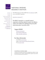
years). Abdominal computed tomography (CT) revealed
an approximately 3 × 2 cm polypoid mass lesion apparently located in the common bile duct and along the
bifurcation into the cystic duct with consecutive dilation of the central intra- and extrahepatic bile ducts
(Figure 1A, B), gallbladder hydrops and cholecystolithiasis. Laboratory tests detected an increase in total bilirubin (1.4 mg/dL; normal ≤1.2 mg/dL), aspartate amino
transferase (76 U/L; normal ≤35 U/L), alanine amino
Page 2 of 10
transferase (79 U/L; normal ≤45 U/L), and γ-glutamyl
transferase (223 U/L; normal ≤55 U/L). Levels of alphafetoprotein (AFP) and carbohydrate antigen 19–9 (CA19-9)
were within normal range (AFP 3 μg/L; normal <7 μg/L,
and CA19-9 31 kU/L; normal <37 kU/L). As abdominal
ultrasound showed a dilated common hepatic duct of up to
2 cm, endoscopic retrograde cholangiopancreatography
(ERCP) was performed (Figure 1C). It revealed a mass
in the common hepatic duct and hematobilia. Via
passage with a blocked balloon, material was obtained
for histoxpathology. A stent was inserted into the bile
duct, producing immediate bile drainage. After that
intervention, the jaundice steadily declined and the cholestatic parameters normalized. The initial pathological
diagnosis of the obtained tissue was a tubulo-villous
adenoma of the bile duct.
After a four-week interval for weight reduction, ERCP
was re-performed and the stent was removed. Fluoroscopic guidance with contrast application revealed the
tumour in the middle part of the common bile duct. After
obtaining biopsies from the tumour, a stent was inserted
again for drainage until the operation. Endosonography
located the tumour in the common bile duct (Figure 1D)
and the cystic duct, protruding into the infundibulum of
the gallbladder. A suspect lymph node was detected
between bile duct and cystic duct.
Histopathological examination revealed tubulo-papillary
neoplastic proliferations and closely packed glandular structures with eosinophilic cytoplasm, round to oval nuclei and
inconspicuous nucleoli. At the surface, small papillary proliferations were observed. Focally, marked architectural
distortion with nuclear atypia, hyperchromatic nuclei with
prominent nucleoli, and a back-to-back formation of
stellar glands were present. Squamous morules were not
Figure 1 Radiographic findings of the pyloric gland adenoma of the cystic duct. Abdominal computed tomography revealed markedly
dilated intrahepatic bile ducts (A, arrow), and a polypoid tumour in the common hepatic duct just below the bifurcation (B, arrow). Endoscopic
retrograde cholangiopancreatography confirmed the polypoid mass lesion (C, arrow) and demonstrated consecutive dilation of the central
intrahepatic bile ducts. Endosonography verified the polypoid intraluminal tumour of 2.1 x 1.1 cm in the common hepatic duct (D, arrow) next to
the portal vein.
Schaefer et al. BMC Cancer 2012, 12:570
/>
Page 3 of 10
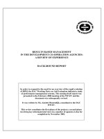
Figure 2 Gross findings of the resected pyloric gland adenoma. The resected specimen comprised the common bile duct (star), the bifurcation
into common hepatic duct (arrow) and cystic duct with attached gallbladder (A). Cross sections revealed an intraluminal tumour of 2.5 cm length
and 2 cm in diameter developing in the cystic duct (B), and protruding into the common bile duct and common hepatic duct (C, arrow).
Schaefer et al. BMC Cancer 2012, 12:570
/>
observed. The diagnosis of a pyloric gland adenoma
with possible transition into well-differentiated adenocarcinoma was established and confirmed by reference
pathology.
In the meantime the patient developed a thrombosis
of the right cephalic vein after catheter infection and a
non-ST-elevation myocardial infarction, which were treated non-interventionally with antibiotics and heparin for
prolongation of the prothrombin time. Two weeks
later, resection of the extrahepatic bile ducts including
gallbladder with biliodigestive anastomosis was performed.
The resected specimen of the common bile duct, cystic
duct with attached gallbladder, and common hepatic duct
presented a 2.5 × 2 cm tumour with a gray-brown cut surface, developing in the cystic duct, and protruding
through the bifurcation into both the common bile duct
and common hepatic duct, with partial obstruction of the
lumen (Figure 2). The surrounding cystic duct showed
epithelial cell proliferations in the mucosa with moderate
and focal high-grade cellular atypia, outstretching into
small branching bile duct (Figure 3). Immunohistochemical staining with MUC1 (clone MRQ-17, 1:300, Cell
marque/Medac, Wedel, Germany), MUC2 (clone MRQ-18,
1:100, Cell marque/Medac), MUC5AC (clone MRQ-19,
1:300, Cell marque/Medac), MUC6 (clone MRQ-20, 1:300,
Cell marque/Medac), vascular endothelial growth factor
Page 4 of 10
receptor (VEGF; clone SP28, prediluted, Abcam, Cambridge,
MA, USA), CD10 (clone 56C6, 1:25, Zytomed Systems,
Berlin, Germany), CDX2 (clone EPR2764Y, 1:100, Cell
Marque, Rocklin, CA, USA), p53 (clone DO-7, 1:50, Dako,
Glostrup, Denmark), p21 (clone DCS-60.2, 1:100, Thermo
Scientific, Fremont, CA, USA), p16 (clone JC8, 1:100,
Santa Cruz Biotechnology, Heidelberg, Germany), and
Ki67 (clone K-2, 1:200 Zytomed Systems) was performed.
The pyloric gland adenoma showed focal positive staining
for MUC1, and negative staining for MUC2, as well as
superficial staining for MUC5AC, and positive expression
of MUC6 and VEGF (Figure 4). CD10 and CDX2 were
not expressed. Nuclear expression of p53, focal p16, and
p21 was observed. Proliferative activity was assessed by
Ki67 and estimated at 25%. The high-grade intraepithelial
neoplasia of the bile duct, in contrast, did not express
MUC1, MUC2, and MUC5AC, but MUC6 and VEGF.
Staining with CD10 and CDX2, p53, and p16 was negative, whereas p21 was only focally expressed. Proliferative activity was assessed by Ki67, and estimated at
10%. The diagnosis of a pyloric gland adenoma with
focal high-grade intraepithelial neoplasia and transition into well-differentiated gastric-type adenocarcinoma associated with BilIN-3 of the cystic duct
resembling gastric-type intraductal papillary neoplasm
in areas with low-grade intraepithelial neoplasia was
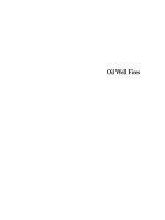
Figure 3 Microscopic findings of the pyloric gland adenoma and adjacent bile duct lesions. On microscopic view, the pyloric gland
adenoma arose in the cystic duct (right) and displayed a papillary, intraluminal growth pattern with protrusion into the common bile duct (left)
(A, H&E, x40). Closely packed pyloric type glands were lined by cuboidal to columnar mucus-secreting cells (B, x100) with focal architectural
distortion (C, x100) and high-grade dysplasia with nuclear atypia, indicating transition into well-differentiated adenocarcinoma (D, x200). Focal
high-grade intraepithelial neoplasia (BilIN-3) of the cystic duct was detected (E, x200), focally resembling gastric-type intraductal papillary
neoplasm (IPN) with direct transition into the pyloric gland adenoma (F, x200).
Schaefer et al. BMC Cancer 2012, 12:570
/>
Page 5 of 10
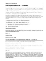
Figure 4 Immunohistochemical findings of the pyloric gland adenoma and adjacent bile duct lesions. The pyloric gland adenoma (A, HE)
immunohistochemically expressed focal MUC1 (B), but no MUC2 (C). MUC5AC (D) was positive predominantly in the superficial luminal cell layers,
whereas MUC6 (E) was expressed throughout. The tumour cell also expressed vascular endothelial growth factor receptor (VEGF) (F), but no CD10 (G)
and CDX2 (H). Nuclear p53 (I) was positive, p16 (J) was focally observed, and p21 (K) was positive. Ki67 (L) was observed in approximately 25% (x100).
thus confirmed histopathologically, and the tumour
was finally staged at pT1, pNX, pMX, G1, L0, V0, R0.
Metastases had been excluded by CT scan.
CGH analysis was performed from the pyloric gland adenoma as described previously [17] and revealed chromosomal gains at 7p, 7q11q21, 15q, 16p, 20, losses at
6p23pter, 6q, 18, and amplifications at 1q and 6p21p22 in
the pyloric gland adenoma (Figure 5). Sequencing analysis
of KRAS exon 1 and 2 showed a point mutation at exon 1,
codon 12 (c.35G>T; p.G12V) and wildtype sequence at
exon 2 in the pyloric gland adenoma and the same mutation in the adjacent BilIN-3. The patient recovered well
and was discharged 12 days after surgery. He presently
shows no signs of tumour relapse 12 months after
tumour resection.
The clinico-pathologic findings of previously reported
cases of pyloric gland adenoma [2-15] as well as the present
case are summarized in Table 1. The developmental etiology
of pyloric gland adenoma is still unclear, particularly when
observed in localizations other than gastric. However, the sequence of metaplasia-dysplasia and carcinoma is well
accepted as a histogenetic pathway of the gallbladder and
bile duct carcinogenesis [16,18]. For the development of pyloric gland adenoma/adenocarcinoma, it is believed that the
first step may be initiated by the presence of gastric metaplasia or gastric heterotopias [6,7]. An association of pyloric
gland adenomas with heterotopic gastric mucosa or gastric metaplasia in the gallbladder [14], pancreas [4], duodenum [15], and rectum [6] has previously been reported.
Above the designation as hyperplasia or hamartoma, some
authors suggest an unstable and precancerous nature by
proposing a “pyloric gland adenoma-adenocarcinoma sequence” by CGH analyses, and underline its high potential
for invasive malignancy [3,18]. Immunohistochemically, as
in the present case, the tumour typically expresses MUC6
and variably MUC5AC [2,3,6,8,9,11,12,14,15]. MUC2 and
Schaefer et al. BMC Cancer 2012, 12:570
/>
Page 6 of 10
Figure 5 Results of comparative genomic hybridization (CGH). CGH of the pyloric gland adenoma revealed ish cgh amp(1)(q),amp(6)
(p21p22),dim(6)(p23pter),dim(6)(q),enh(7)(p),enh(7)(q11q21),enh(15)(q),enh(16)(p),dim(18),enh(20) as indicated by green (gains) and red (losses)
bars. The number of chromosomes included in the CGH analysis is indicated at the bottom of each individual profile.
CD10 are generally negative, but may indicate transition
from gastric to intestinal differentiation [2,3,8,12]. Additionally, Ki67 expression and p53 mutations can be used
to detect malignant transformation [3]. The presence of
adenocarcinoma may be difficult to recognize since the
cytology of adenocarcinoma developing in pyloric gland
adenoma is known to show rather mild abnormalities
without the classical signs of intraepithelial neoplasia/dysplasia [3]. However, transition from round or oval nuclei
to elongated or pleomorphic nuclei with loss of polarity in
conjunction with so-called lateral expansion or branching
of glands indicates the presence of adenocarcinoma, as
found focally also in the present case (Figure 3) [3]. In
another study describing 3 pyloric gland adenocarcinomas
of the extrahepatic bile ducts the authors observed pyloric
gland metaplasia adjacent to the adenocarcinoma in 2 of 3
cases and suggested them as probable precursor lesion
[16]. These tumours did not arise from pyloric gland adenomas, but displayed a similar immunophenotype with
positive expression of MUC5AC and MUC6 and negativity
for MUC2 and CDX2 [16]. Squamous morules (or spindle
cell metaplasias) are reported to occur in 23% of cases [9],
but were not observed in the present case.
Histopathological differential diagnoses of polypous
intraductal bile duct lesions include adenomas of the
gallbladder and extrahepatic bile ducts. They can be
divided into a tubular, papillary, and tubulopapillary type
based on their growth pattern, and cytologically into a
pyloric-gland, intestinal, foveolar, and biliary type [1].
IPN of gastric, pancreatobiliary, intestinal or oncocytic
phenotype, and mucinous cystic neoplasms should also
be considered [1]. Furthermore, concomitant intraductal
papillary mucinous neoplasms (IPMN) [5,13] have been
observed in cases of pancreatic pyloric gland adenomas.
In the pancreas, gastric-type IPMN are usually located
as small cystic lesions in branching ducts, harboring
only mild/low-grade atypia and immunohistochemically
expressing MUC5AC, but not MUC1, MUC2 or CDX2.
They are associated with a rather favorable clinical prognosis compared to the other subtypes of IPMN [19];
adenocarcinoma occurs in 10-15% [19]. Invasive adenocarcinoma may also develop in IPN of the bile duct [1].
BilIN of the gallbladder and extrahepatic bile ducts are
associated with lithiasis in up to 3%, familial adenomatous
polyposis coli, sclerosing cholangitis, and pancreatobiliary
reflux [1]. Additionally, BilIN-3 usually arises in a association with pyloric and intestinal metaplasia, as observed in
the present case, with an abrupt transition between normal
and atypical columnar cells [1]. As reported, BilIN
immunohistochemically also expresses p53 [1].
KRAS codon 12 mutations have been previously
reported in two cases of pyloric gland adenoma of the
main pancreatic duct, and probably support the neoplastic nature of this tumour [4,5]. KRAS (13-100%)
and TP53 (50%) mutations have been described for
IPMN of the pancreas before [19]. Also in the present
case, the pyloric gland adenoma and the BilIN-3 harbored a KRAS codon 12 mutation, indicating a possible metaplasia-dysplasia-carcinoma sequence with a
common tumourigenesis.
Previous CGH results of a pyloric gland adenoma of
the esophagus revealed chromosomal aberrations which
overlapped with findings in Barrett’s dysplasia and
adenocarcinoma as well as gastric cardia adenocarcinoma
Author (year)
Number of
cases
Age/Sex
Site
Associated lesion
Malignancy
developing
in PGA
Size (cm)
Immunoreactivity
Genetics
Kushima (1996)
1
61/F
Gallbladder
Gastric metaplasia
–
1.5
MUC6 (= M2)
n.k.
Bakotic (1999)
1
69/F
Pancreas (main duct)
Heterotopic gastric corpus
mucosa
–
0.9
PAS, negative: Alcian blue,
chromogranin, serotonin,
somatostatin, gastrin
KRAS exon 1 (p.G12R; c.34G>C)
Kushima (1999)
1
67/F
Duodenum
Heterotopic gastric corpus
mucosa
–
2.5
MUC5AC (= M1), MUC6
(= M2)
n.k.
Kato (2002)
1
70/M
Pancreas (main duct)
IPMN
–
0.6
PCS, HIK1083, negative:
neuroendocrine markers,
hormones
KRAS exon 1, codon 12
Amaris (2002)
1
73/M
Pancreas (branch duct)
IPMN
–
n.k.
PAS, negative: Alcian blue
n.k.
Vieth (2003)
90
73/F:M = 3:1
Stomach (n =77 ),
duodenal bulb (n = 7),
duodenum (n = 1),
common bile duct
(n = 3), gallbladder
(n = 2)
Gastritis (A-, B-, and C-type)
(20-34%), tubular adenoma
(n = 1), carcinoid tumour
(n = 1), adenocarcinoma
(n = 1)
Adenocarcinoma
(30%)
1.6
n.k.
n.k.
Vieth (2005)
1
46/M
Rectum
Heterotopic gastric corpus
mucosa
–
3
MUC6, MUC5AC
n.k.
Kushima (2005)
1
62/M
Esophagus
Barrett's mucosa
–
3
MUC6, MUC5AC, negative:
MUC2, CD10
CGH: losses at 2p24p25.2,
2q14.1pter, 5q31.3q32,
6q23q24, 8q23q24.2,
11q22.3q24, 18q21.1q22
Chen (2009)
41 tumours,
36 patients
73/F:M = 25:11
Stomach (19), duodenum
(19), gastroesophageal
junction (2), pancreas (1)
Gastritis (A-type) (40%),
intestinal metaplasia (60%)
Adenocarcinoma
(12.2%)
n.k.
MUC6, MUC5AC, negative:
CDX2, MUC2
n.k.
Wani (2008)
29
n.k.
Gallbladder
Intestinal metaplasia (34.4%),
squamous morules (24.1%)
–
0.82
MUC6, MUC5AC, M-GGMC-1,
morules: CDX2, beta-catenin
n.k.
Golger (2008)
1
79/F
Stomach
Helicobacter-negative gastritis
–
2
n.k.
n.k.
Oh (2010)
1
n.k.
Stomach
–
Adenocarcinoma
3.5
MUC6
n.k.
Schaefer et al. BMC Cancer 2012, 12:570
/>
Table 1 The clinico-pathologic characteristics of previously reported cases of pyloric gland adenoma [2-15] and the case reported here
Page 7 of 10
Vieth (2010)
60
70-71/F=M
Stomach
–
Adenocarcinoma
(46.7%)
0.9-1.5
MUC6, MUC5AC (MUC2,
CD10), p53 and Ki67 in
malignant transition
n.k.
GutierrezGrobe (2010)
1
49/F
Stomach
–
–
n.k.
MUC6, MUC5AC, negative:
MUC2
n.k.
Present case
1
62/M
Cystic duct
IPN, BilIN-3
Adenocarcinoma
2
MUC5AC, MUC6, VEGF, p53,
p21, Ki67, (MUC1, p16),
negative: MUC2, CD10, CDX2
KRAS exon 1 (p.G12V; c.35G>T);
CGH: gains at 1q, 6p11p22, 7p,
15q, 20p, and losses at 6p23pter,
6q14qter, 11q12q13, 18
PGA: Pyloric gland adenoma.
PAS: Periodic Schiff's acid.
PCS: paradoxical concanavalin A.
IPMN: intraductal papillary mucinous neoplasm.
IPN: intraductal papillary neoplasm.
BilIN: biliary intraepithelial neoplasia.
CGH: comparative genomic hybridization.
Schaefer et al. BMC Cancer 2012, 12:570
/>
Table 1 The clinico-pathologic characteristics of previously reported cases of pyloric gland adenoma [2-15] and the case reported here (Continued)
Page 8 of 10
Schaefer et al. BMC Cancer 2012, 12:570
/>
[8]. In pyloric gland adenomas of the stomach, previous
CGH analyses revealed chromosomal abnormalities common to invasive gastric adenocarcinoma, including -5q
(50%), -6 (40%), -4q, +17pq and +20 [18,20]. Additional
gains were observed at 1, 3q, 5q, 7, 9q, 11q, 12q, 13q, 15q,
17 and 22q, and losses at 1p, 2q, 4, 9p, 10, 12q 13q, 14q,
16, 18q, 20q, and 21 [20]. Of these aberrations, gains at 7p
and 15q, and losses at 6q, and 18q were also detected in
the present case. Interestingly, losses at 6q and 18q have
been demonstrated in pancreatic IPMN, before [21].
Furthermore, the amplicon at 6p21p22 harbors the
VEGF (VEGF-A) gene at 6p21.1 (MIM ID *192240)
which was shown to be expressed by the pyloric
gland adenoma by immunohistochemical staining,
suggesting VEGF upregulation. VEGF plays a crucial
role in angiogenesis of normal tissues and several
types of tumours [22]. Altogether, the relatively high
number of chromosomal imbalances in the present
case of pyloric gland adenoma of the cystic duct suggests
an instable karyotype and underlines the risk of malignant
transformation.
Conclusions
In conclusion, a pyloric gland adenoma evolving in the
cystic duct is very rare, but may sometimes be overlooked and therefore should be considered as a differential diagnosis for obstructive bile duct tumours. An
association with other premalignant bile duct lesions
such as BilIN may be observed. ERCP-guided biopsy
with histopathological examination is necessary to establish the diagnosis. Due to the high risk of evolving
adenocarcinoma, surgical resection should be performed
whenever possible.
Consent
Written informed consent was obtained from the patient
for publication of this Case report and any accompanying images. A copy of the written consent is available for
review by the Series Editor of this journal.
Competing interests
The authors declare that they have no competing interests.
Authors' contributions
IMS, PM, LV, and MV performed the histopathological, immunohistochemical
and genetic examinations and established the diagnosis. SC, KH, and HS
examined, treated and observed the patient, including follow-up. IMS, SC,
PM, KH, HS, LV, and MV participated in writing the manuscript. SC and HS
provided the radiographic, and IMS the histological and CGH images. All
authors read and approved of the final manuscript.
Acknowledgements
The authors thank Sabine Schäfer, Radiology group practice Göttingen,
Germany, for providing radiographic images.
Author details
1
Department of Pathology, University Medical Center Göttingen,
Robert-Koch-Straße 40, Göttingen D-37075, Germany. 2Gastroenterology and
Endocrinology, University Medical Center Göttingen, Göttingen, Germany.
Page 9 of 10
3
General and Visceral Surgery, University Medical Center Göttingen,
Göttingen, Germany. 4Institute of Pathology, Klinikum Bayreuth, Germany.
Received: 22 September 2012 Accepted: 29 November 2012
Published: 4 December 2012
References
1. Bosman FT, Carneiro F, Hruban RH, Theise ND: WHO classification of tumours of
the digestive system. Lyon: International Agency for Research on Cancer; 2010.
2. Chen ZM, Scudiere JR, Abraham SC, Montgomery E: Pyloric gland
adenoma: an entity distinct from gastric foveolar type adenoma.
Am J Surg Pathol 2009, 33:186–193.
3. Vieth M, Kushima R, Mukaisho K, Sakai R, Kasami T, Hattori T:
Immunohistochemical analysis of pyloric gland adenomas using a series
of Mucin 2, Mucin 5AC, Mucin 6, CD10, Ki67 and p53. Virchows Arch 2010,
457:529–536.
4. Bakotic BW, Robinson MJ, Sturm PD, Hruban RH, Offerhaus GJ, AlboresSaavedra J: Pyloric gland adenoma of the main pancreatic duct.
Am J Surg Pathol 1999, 23:227–231.
5. Kato N, Akiyama S, Motoyama T: Pyloric gland-type tubular adenoma
superimposed on intraductal papillary mucinous tumor of the
pancreas. Pyloric gland adenoma of the pancreas. Virchows Arch 2002,
440:205–208.
6. Vieth M, Kushima R, de Jonge JP, Borchard F, Oellig F, Stolte M: Adenoma
with gastric differentiation (so-called pyloric gland adenoma) in a
heterotopic gastric corpus mucosa in the rectum. Virchows Arch 2005,
446:542–545.
7. Vieth M, Kushima R, Borchard F, Stolte M: Pyloric gland adenoma: a
clinico-pathological analysis of 90 cases. Virchows Arch 2003, 442:317–321.
8. Kushima R, Vieth M, Mukaisho K, Sakai R, Okabe H, Hattori T, Neuhaus H,
Borchard F, Stolte M: Pyloric gland adenoma arising in Barrett's
esophagus with mucin immunohistochemical and molecular cytogenetic
evaluation. Virchows Arch 2005, 446:537–541.
9. Wani Y, Notohara K, Fujisawa M: Aberrant expression of an "intestinal
marker" Cdx2 in pyloric gland adenoma of the gallbladder. Virchows Arch
2008, 453:521–527.
10. Golger D, Probst A, Wagner T, Messmann H: Pyloric-gland adenoma of the
stomach: case report of a rare tumor successfully treated with endoscopic
submucosal dissection. Endoscopy 2008, 40(Suppl 2):E110–E111.
11. Oh MG, Cho SJ, Lee JH, Kook MC, Park SY: A spongiform mass in the
stomach: pyloric gland adenoma with a transition to adenocarcinoma.
Korean J Gastroenterol 2010, 56:1–5.
12. Gutierrez-Grobe Y, Gavilanes-Espinar J, Uribe M, Kobashi-Margain R, MendezSanchez N: Pyloric Gland Adenoma: Case Report. Rev Gastroenterol Mex
2010, 75:360–362.
13. Amaris J: Intraductal mucinous papillary tumor and pyloric gland
adenoma of the pancreas. Gastrointest Endosc 2002, 56:441–444.
14. Kushima R, Remmele W, Stolte M, Borchard F: Pyloric gland type adenoma
of the gallbladder with squamoid spindle cell metaplasia. Pathol Res Pract
1996, 192:963–969.
15. Kushima R, Ruthlein HJ, Stolte M, Bamba M, Hattori T, Borchard F: 'Pyloric
gland-type adenoma' arising in heterotopic gastric mucosa of the
duodenum, with dysplastic progression of the gastric type. Virchows Arch
1999, 435:452–457.
16. Albores-Saavedra J, Chable-Montero F, Mendez-Sanchez N, Mercado MA,
Vilatoba-Chapa M, Henson DE: Adenocarcinoma with pyloric gland
phenotype of the extrahepatic bile ducts: a previously unrecognized and
distinctive morphologic variant of extrahepatic bile duct carcinoma. Hum
Pathol 2012, 43:2292–8.
17. Schaefer IM, Martinez R, Enders C, Loertzer H, Bruck W, Rohde V, Fuzesi L,
Gutenberg A: Molecular cytogenetics of malignant pheochromocytoma
with cerebral metastasis. Cancer Genet Cytogenet 2010, 200:194–197.
18. Kushima R, Vieth M, Borchard F, Stolte M, Mukaisho K, Hattori T: Gastrictype well-differentiated adenocarcinoma and pyloric gland adenoma of
the stomach. Gastric Cancer 2006, 9:177–184.
19. Sipos B, Henopp T: Precursor lesions of pancreatobiliary cancer. Pathologe
2011, 32:224–31.
20. Buffart TE, Carvalho B, Mons T, Reis RM, Moutinho C, Silva P, van Grieken NC,
Vieth M, Stolte M, van de Velde CJ, et al: DNA copy number profiles of
gastric cancer precursor lesions. BMC Genomics 2007, 8:345.
Schaefer et al. BMC Cancer 2012, 12:570
/>
Page 10 of 10
21. Fritz S, Fernandez-del CC, Mino-Kenudson M, Crippa S, Deshpande V,
Lauwers GY, Warshaw AL, Thayer SP, Iafrate AJ: Global genomic analysis of
intraductal papillary mucinous neoplasms of the pancreas reveals
significant molecular differences compared to ductal adenocarcinoma.
Ann Surg 2009, 249:440–447.
22. Ferrara N: Vascular endothelial growth factor: basic science and clinical
progress. Endocr Rev 2004, 25:581–611.
doi:10.1186/1471-2407-12-570
Cite this article as: Schaefer et al.: Pyloric gland adenoma of the cystic
duct with malignant transformation: report of a case with a review of
the literature. BMC Cancer 2012 12:570.
Submit your next manuscript to BioMed Central
and take full advantage of:
• Convenient online submission
• Thorough peer review
• No space constraints or color figure charges
• Immediate publication on acceptance
• Inclusion in PubMed, CAS, Scopus and Google Scholar
• Research which is freely available for redistribution
Submit your manuscript at
www.biomedcentral.com/submit