Blood vessel hyperpermeability and pathophysiology in human tumour xenograft models of breast cancer: A comparison of ectopic and orthotopic tumours
Bạn đang xem bản rút gọn của tài liệu. Xem và tải ngay bản đầy đủ của tài liệu tại đây (1.01 MB, 10 trang )
Ho et al. BMC Cancer 2012, 12:579
/>
RESEARCH ARTICLE
Open Access
Blood vessel hyperpermeability and
pathophysiology in human tumour xenograft
models of breast cancer: a comparison of ectopic
and orthotopic tumours
Karyn S Ho1,2, Peter C Poon1, Shawn C Owen1,2 and Molly S Shoichet1,2,3*
Abstract
Background: Human tumour xenografts in immune compromised mice are widely used as cancer models because
they are easy to reproduce and simple to use in a variety of pre-clinical assessments. Developments in
nanomedicine have led to the use of tumour xenografts in testing nanoscale delivery devices, such as nanoparticles
and polymer-drug conjugates, for targeting and efficacy via the enhanced permeability and retention (EPR) effect.
For these results to be meaningful, the hyperpermeable vasculature and reduced lymphatic drainage associated
with tumour pathophysiology must be replicated in the model. In pre-clinical breast cancer xenograft models, cells
are commonly introduced via injection either orthotopically (mammary fat pad, MFP) or ectopically (subcutaneous,
SC), and the organ environment experienced by the tumour cells has been shown to influence their behaviour.
Methods: To evaluate xenograft models of breast cancer in the context of EPR, both orthotopic MFP and ectopic
SC injections of MDA-MB-231-H2N cells were given to NOD scid gamma (NSG) mice. Animals with matched
tumours in two size categories were tested by injection of a high molecular weight dextran as a model nanocarrier.
Tumours were collected and sectioned to assess dextran accumulation compared to liver tissue as a positive
control. To understand the cellular basis of these observations, tumour sections were also immunostained for
endothelial cells, basement membranes, pericytes, and lymphatic vessels.
Results: SC tumours required longer development times to become size matched to MFP tumours, and also
presented wide size variability and ulcerated skin lesions 6 weeks after cell injection. The 3 week MFP tumour
model demonstrated greater dextran accumulation than the size matched 5 week SC tumour model (for P < 0.10).
Immunostaining revealed greater vascular density and thinner basement membranes in the MFP tumour model
3 weeks after cell injection. Both the MFP and SC tumours showed evidence of insufficient lymphatic drainage, as
many fluid-filled and collagen IV-lined spaces were observed, which likely contain excess interstitial fluid.
Conclusions: Dextran accumulation and immunostaining results suggest that small MFP tumours best replicate the
vascular permeability required to observe the EPR effect in vivo. A more predictable growth profile and the absence
of ulcerated skin lesions further point to the MFP model as a strong choice for long term treatment studies that
initiate after a target tumour size has been reached.
Keywords: Tumour xenograft models, Orthotopic transplantation, Ectopic transplantation, Enhanced permeability
and retention, Breast cancer, Blood vessel hyperpermeability, Nanomedicine, Targeting
* Correspondence:
1
Department of Chemical Engineering & Applied Chemistry, 200 College
Street, Toronto, ON M5S 3E5, Canada
2
Institute of Biomaterials & Biomedical Engineering, Terrence Donnelly Centre
for Cellular and Biomolecular Research, University of Toronto, Room 514 –
160 College Street, Toronto, ON M5S 3E1, Canada
Full list of author information is available at the end of the article
© 2012 Ho et al.; licensee BioMed Central Ltd. This is an Open Access article distributed under the terms of the Creative
Commons Attribution License ( which permits unrestricted use, distribution, and
reproduction in any medium, provided the original work is properly cited.
Ho et al. BMC Cancer 2012, 12:579
/>
Background
Pre-clinical development of anti-cancer therapeutics relies on availability of relevant and reproducible in vivo
tumour models. Human tumour xenograft models in
immunodeficient mice are widely used to assess pharmacokinetics, biodistribution, and treatment efficacy because they are inexpensive and easy to replicate [1].
However, their utility in evaluating potential treatment
strategies depends on their capacity to recapitulate
human disease conditions.
Progress in nanomedicine seeks to shift distribution of
therapeutic compounds to tumour tissue by targeting
hyperpermeable tumour vasculature [2,3]. Tumours are
restricted in size until they can trigger greater blood vessel density through angiogenesis and blood vessel remodeling [4,5]. Compared to normal tissue, tumour
tissue has been demonstrated to be more permissive to
extravasation of macromolecules as a result of abnormal
blood vessel structure [3]. Moreover, tumour tissue is
subject to poor lymphatic drainage, leading to greater retention of material in the extravascular space. These
combined phenomena are called enhanced permeability
and retention (EPR) and form the basis for improved selectivity of nanoscale drug delivery for solid tumour targeting [2,4,6].
Several pathological features of tumour vasculature
lead to its utility in targeting applications. Pathological
tumour vessels are dynamic, and can result both from
angiogenesis and remodeling of existing vessels [5,7].
Endothelial cells that comprise tumour blood vessels
have poor organization, leading to gaps between cells,
multiple endothelial cell layers, and unusual tortuosity
and branching [8,9]. These openings allow unregulated
movement of macromolecules and nanoscale carriers
across tumour vessel walls and into the surrounding tissue [10]. In response, the associated basement membrane is also often thickened or absent [9,11]. This
apparent dichotomy stems from a dynamic interaction
between increased and multilayered collagen deposition
in the basement membrane [10,12-14] and increased expression of matrix metalloproteinases (MMPs) that can
result in collagen degradation [15]. Further enhancing
the aberrant permeability of tumour blood vessels, the
pericytes that normally cover and stabilize the outer vessel wall can also be missing or detached, leading to a
more immature vessel structure [5]. The absence of
these contractile support cells may lead to further
increased vessel permeability and weakened control over
blood flow [7]. Lymphatic vessels are closely associated
with and derived from the blood vessel network. They
are responsible for transporting waste out of tissues, but
tumours are often deficient in lymphatic drainage, leading to increased accumulation of macromolecular material in tumour tissue [4]. Each of these features
Page 2 of 10
contributes to the pathophysiology that enables the EPR
effect. Vascular permeability factors, such as bradykinin
and nitric oxide, also mediate and enhance vascular
hyperpermeability [16]; however, our analysis will be
limited to physical vascular defects and their contributions to EPR.
To validate the use of human tumour xenografts in
mouse models of breast cancer to investigate tumour
targeting via EPR, we studied MDA-MB-231-H2N cells
transplanted in NOD scid gamma (NSG) mice and compared two common cell injection sites in the context of
EPR permissive pathology. Owing to its simplicity,
tumour cells are often introduced ectopically as subcutaneous (SC) injections, regardless of their native tissue
type [17-19]. Cells injected orthotopically (eg. breast
cancer cells into mammary tissue) are subject to biological cues present in the relevant organ environment
[18]. Allowing tumour cells to grow in their orthotopic
environment influences growth rate, blood and lymphatic vessel development, metastatic potential, interstitial
pressure, and response to therapy [5,18-21]. We
hypothesized that the orthotopic environment may also
influence the permeability of the resulting tumour vasculature. Notably, to promote successful tumour engraftment in both locations, our chosen cell line is known to
be tumourigenic in the absence of external factors, such
as estrogen [22]. Groups of animals were compared as
cohorts of matching tumour size becaus����������������������������������������������������������������������������������������������������������������������������������������������������������������������������������������������������������������������������������������������������������������������������������������������������������������������������������������������������������������������������������������������������������������������������������������������������������������������������������������������������������������������������������������������������������������������������������������������������������������������������������������������������������������������������������������������������������������������������������������������������������������������������������������������������������������������������������������������������������������������������������������������������������������������������������������������������������������������������������������������������������������������������������������������������������������������������������������������������������������������������������������������������������������������������������������������������������������������������������������������������������������������������������������������������������������������������������������������������������������������������������������������������������������������������������������������������������������������������������������������������������������������������������������������������������������������������������������������������������������������������������������������������������������������������������������������������������������������������������������������������������������������������������������������������������������������������������������������������������������������������������������������������������������������������������������������������������������������������������������������������������������������������������������������������������������������������������������������������������������������������������������������������������������������������������������������������������������������������������������������������������������������������������������������������������������������������������������������������������������������������������������������������������������������������������������������������������������������������������������������������������������������������������������������������������������������������������������������������������������������������������������������������������������������������������������������������������������������������������������������������������������������������������������������������������������������������������������������������������������������������������������������������������������������������������������������������������������������������������������������������������������������������������������������������������������������������������������������������������������������������������������������������������������������������������������������������������������������������������������������������������������������������������������������������������������������������������������������������������������������������������������������������������������������������������������������������������������������������������������������������������������������������������������������������������������������������������������������������������������������������������������������������������������������������������������������������������������������������������������������������������������������������������������������������������������������������������������������������������������������������������������������������������������������������������������������������������������������������������������������������������������������������������������������������������������������������������������������������������������������������������������������������������������������������������������������������������������������������������������������������������������������������������������������������������������������������������������������������������������������������������������������������������������������������������������������������������������������������������������������������������������������������������������������������������������������������������������������������������������������������������������������������������������������������������������������������������������������������������������������������������������������������������������������������������������������������������������������������������������������������������������������������������������������������������������������������������������������������������������������������������������������������������������������������������������������������������������������������������������������������������������������������������������������������������������������������������������������������������������������������������������������������������������������������������������������������������������������������������������������������������������������������������������������������������������������������������������������������������������������������������������������������������������������������������������������������������������������������������������������������������������������������������������������������������������������������������������������������������������������������������������������������������������������������������������������������������������������������������������������������������������������������������������������������������������������������������������������������������������������������������������������������������������������������������������������������������������������������������������������������������������������������������������������������������������������������������������������������������������������������������������������������������������������������������������������������������������������������������������������������������������������������������������������������������������������������������������������������������������������������������������������������������������������������������������������������������������������������������������������������������������������������������������������������������������������������������������������������������������������������������������������������������������������������������������������������������������������������������������������������������������������������������������������������������������������������������������������������������������������������������������������������������������������������������������������������������������������������������������������������������������������������������������������������������������������������������������������������������������������������������������������������������������������������������������������������������������������������������������������������������������������������������������������������������������������������������������������������������������������������������������������������������������������������������������������������������������������������������������������������������������������������������������������������������������������������������������������������������������������������������������������������������������������������������������������������������������������������������������������������������������������������������������������������������������������������������������������������������������������������������������������������������������������������������������������������������������������������������������������������������������������������������������������������������������������������������������������������������������������������������������������������������������������������������������������������������������������������������������������������������������������������������������������������������������������������������������������������������������������������������������������������������������������������������������������������������������������������������������������������������������������������������������������������������������������������������������������������������������������������������������������������������������������������������������������������������������������������������������������������������������������������������������������������������������������������������������������������������������������������������������������������������������������������������������������������������������������������������������������������������������������������������������������������������������������������������������������������������������������������������������������������������������������������������������������������������������������������������������������������������������������������������������������������������������������������������������������������������������������������������������������������������������������������������������������������������������������������������������������������������������������������������������������������������������������������������������������������������������������������������������������������������������������������������������������������������������������������������������������������������������������������������������������������������������������������������������������������������������������������������������������������������������������������������������������������������������������������������������������������������������������������������������������������������������������������������������������������������������������������������������������������������������������������������������������������������������������������������������������������������������������������������������������������������������������������������������������������������������������������������������������������������������������������������������������������������������������������������������������������������������������������������������������������������������������������������������������������������������������������������������������������������������������������������������������������������������������������������������������������������������������������������������������������������������������������������������������������������������������������������������������������������������������������������������������������������������������������������������������������������������������������������������������������������������������������������������������������������������������������������������������������������������������������������������������������������������������������������������������������������������������������������������������������������������������������������������������������������������������������������������������������������������������������������������������������������������������������������������������������������������������������������������������������������������������������������������������������������������������������������������������������������������������������������������������������������������������������������������������������������������������������������������������������������������������������������������������������������������������������������������������������������������������������������������������������������������������������������������������������������������������������������������������������������������������������������������������������������������������������������������������������������������������������������������������������������������������������������������������������������������������������������������������������������������������������������������������������������������������������������������������������������������������������������������������������������������������������������������������������������������������������������������������������������������������������������������������������������������������������������������������������������������������������������������������������������������������������������������������������������������������������������������������������������������������������������������������������������������������������������������������������������������������������������������������������������������������������������������������������������������������������������������������������������������������������������������������������������������������������������������������������������������������������������������������������������������������������������������������������������������������������������������������������������������������������������������������������������������������������������������������������������������������������������������������������������������������������������������������������������������������������������������������������������������������������������������������������������������������������������������������������������������������������������������������������������������������������������������������������������������������������������������������������������������������������������������������������������������������������������������������������������������������������������������������������������������������������������������������������������������������������������������������������������������������������������������������������������������������������������������������������������������������������������������������������������������������������������������������������������������������������������������������������������������������������������������������������������������������������������������������������������������������������������������������������������������������������������������������������������������������������������������������������������������������������������������������������������������������������������������������������������������������������������������������������������������������������������������������������������������������������������������������������������������������������������������������������������������������������������������������������������������������������������������������������������������������������������������������������������������������������������������������������������������������������������������������������������������������������������������������������������������������������������������������������������������������������������������������������������������������������������������������������������������������������������������������������������������������������������������������������������������������������������������������������������������������������������������������������������������������������������������������������������������������������������������������������������������������������������������������������������������������������������������������������������������������������������������������������������������������������������������������������������������������������������������������������������������������������������������������������������������������������������������������������������������������������������������������������������������������������������������������������������������������������������������������������������������������������������������������������������������������������������������������������������������������������������������������������������������������������������������������������������������������������������������������������������������������������������������������������������������������������������������������������������������������������������������������������������������������������������������������������������������������������������������������������������������������������������������������������������������������������������������������������������������������������������������������������������������������������������������������������������������������������������������������������������������������������������������������������������������������������������������MFP 4 wks
SC 5 wks
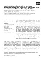
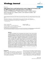
Figure 3 CD31 and collagen IV immunostaining. Mean blood vessel wall thickness visualized through A CD31 (endothelial cells) and B
collagen IV (basement membrane). Both are abnormally thick as compared to healthy liver control tissue, which is denoted by the dashed line. C
shows that mean blood vessel density assayed using CD31 staining is greatest in 3 week old MFP tumours. D indicates mean vascular area as a
measure of blood vessel size and capacity. Their small size categorizes them as microvasculature. All data are shown as the mean of n = 4
animals ± SD. Starred lines connecting bars denote statistical significance, P < 0.05.
in other studies utilizing models such as albumin
(~7 nm) [30,31].
Separate sections were also co-stained for collagen IV
to visualize the thickness of the associated basement
membrane. The basement membrane forms a physical
barrier that inhibits transport of high molecular weight
materials across blood vessel walls [15,32]. In tumour
pathophysiology, opposing phenomena have been
observed: the basement membrane can thicken, thin, or
even be absent. In the MFP and SC tumour models, the
basement membrane was thickened compared to healthy
liver blood vessels (Figure 3B). This observation is consistent with the xenografted MDA-MB-231-H2N cell
line being poorly invasive like its parental line, MDAMB-231 [33]. Conversely, a more metastatic cell line is
often capable of using MMPs to degrade the basement
membrane to enable cell migration through neighbouring blood vessels [33]. The 5 week old SC tumours were
observed to have the highest basement membrane thickness, indicating the greatest mass transport barrier
against nanocarrier delivery.
CD31 staining also revealed differences in vascular
density, with the 3 week old MFP tumours having a
significantly greater vessel density than the other groups
(Figure 3C). The decrease in vascular density from
3 weeks to 4 weeks in the MFP model suggests that
the tumour cell growth may be too rapid for the corresponding new blood vessels to form. The thick basement membranes observed in the tumour tissue may
also contribute to this deficiency as the basement membrane must be degraded before vascular branching can
occur [15]. Although the 3 week old MFP and 5 week
old SC tumours were size matched, the MFP model
had greater blood vessel density, which may be attributed to greater vascular density in the MFP. Together
these observations suggest that remodeling blood vessels
already present in the transplantation site are important in establishing relevant tumour vasculature. The
relatively poor vascular density in SC tumours may
also explain the poor engraftment after 6 weeks, as a
lack of blood flow may inhibit further growth and lead
to necrosis.
Ho et al. BMC Cancer 2012, 12:579
/>
The mean vascular area was also quantified, giving an
indication of the size, and therefore the capacity of the
blood vessels present in each tumour type. The vascular
area in 3 week old MFP tumours was significantly higher
than the 4 week old MFP tumours (Figure 3D), indicating that in addition to decreasing vessel density with increasing tumour size, there is on average a lower
capacity for blood in the vessels present. Having a
greater density and capacity for blood perfusion
enhances the likelihood for delivery of materials to the
3 week old MFP tumours through systemic circulation.
At the same time, all of the evaluated models are likely
underperfused as their small size categorizes them as
microvasculature [34]. This low overall capacity for
blood flow impacts their utility in assessing nanocarrier
accumulation via EPR, and likely results in regions of
hypoxia and heterogeneous drug distribution.
CD31 was also co-stained with αSMA to visualize differences in pericyte association with blood vessels. Pericytes are important blood vessel support cells that help
to regulate blood flow and vessel permeability, but are
often detached in tumour pathophysiology. The
observed staining patterns suggest that this was the case
across all tumour models (Figure 4A-C). Pericytes (violet) were distributed throughout tumour tissue instead
of associating exclusively with blood vessels (brown) and
forming uniform layers around the endothelial cell layer,
as observed in healthy liver tissue (Figure 4D).
LYVE-1 staining was used to detect lymphatic vessels
in tumour tissue. Lymphatic vessels provide a network
Page 7 of 10
to drain protein rich interstitial fluid back into circulation. By the nature of their function, these vessels are
porous to allow macromolecules to be transported [35],
and therefore nanocarrier accumulation in tumour tissue
may increase when their expression is impaired. Mouse
models of lymphatic impairment can be generated by
surgically ablating lymphatic vessels in the tail, resulting
in lymphedema. In these models, the surrounding tissue
attempts to restore homeostasis by generating new
lymphatic vessels and dilating the remaining lymphatic
vessels, suggesting that both density and diameter impact drainage capacity [36]. LYVE-1 stained sections
were used to quantify lymphatic vessel size and density
(Figure 5A-B). Both of these measures gave different variances between groups (P < 0.05 by Bartlett’s test of equality of variances) meaning that the groups tested were not
equivalent. While the mean lymphatic vessel density was
highest in the 3 week old MFP tumours, the 5 week old
SC tumours demonstrated the highest mean lymphatic
vessel area. These factors counterbalance one another, as
density and capacity each contribute to overall drainage.
There is evidence that both the MFP and SC tumour
models yielded poor lymphatic drainage compared to
healthy tissue. Accumulation of interstitial fluid in cases
of lymphedema has been shown to lead to the deposition
of collagen [37]. Visual examination of the tumour slices
revealed a high density of collagen IV-lined spaces
that were CD31 negative, which likely represent fluidfilled cavities in the tumour tissue (Figure 5C-D). These
likely contain excess interstitial fluid resulting from a
A
B
C
D
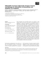
Figure 4 CD31 and αSMA co-staining. Representative images of pericytes (αSMA, violet) that are not associated with blood vessels (CD31,
brown) in: A 3 week MFP, B 4 week MFP, and C 5 week SC tumours. Several blood vessels are highlighted with black arrows; blue staining
represents cell nuclei. D shows that pericytes are exclusively associated with blood vessels in healthy liver control tissue. Scale bars represent
200 μm.
Ho et al. BMC Cancer 2012, 12:579
/>
Page 8 of 10
A
B
250
LYVE-1
LYVE-1
Mean vessel area (µm2)
Mean vessel density (#/mm2)
25
20
15
10
5
200
150
100
50
0
0
MFP 3 wks
MFP 4 wks
MFP 3 wks MFP 4 wks
SC 5 wks
C
SC 5 wks
D
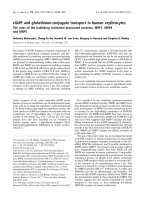
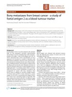
Figure 5 LYVE-1 immunostaining. A shows mean lymphatic vessel density, and B shows mean vessel area, both of which are indicators of
lymphovascular capacity. Both measures were found to have unequal variance between groups, and therefore although the groups were not
equivalent, ANOVA could not be used to verify their differences. While 3 week old MFP tumours had the highest mean lymphatic vessel density,
5 week old SC tumours had greater mean vessel size, both of which contribute to overall lymphatic drainage capacity. All data are shown as the
mean of n = 4 animals ± SD. Representative images of fluid-filled spaces lined with collagen (violet) but not with endothelial cells (negative for
CD31, brown)are shown in: C 3 week MFP and D 5 week SC tumours. Several of these spaces, which indicate lymphedema, are highlighted with
black arrows; blue staining represents cell nuclei. Scale bars represent 200 μm.
combination of increased vascular permeability and deficient lymphatic drainage.
Taken together, the data gathered through CD31 and
collagen IV immunostaining suggest that, of the models
tested, the 3 week MFP tumour best replicates the vascular permeability required to observe the EPR effect
in vivo. However, the blood vessels visualized are sparse
and small, contributing to low accumulation of the
model nanocarrier used in this study. Both MFP and SC
tumours showed evidence of excess interstitial fluid accumulation, suggesting poor lymphatic drainage in both
models. While MFP tumours demonstrated greater
lymphatic vessel density, SC tumours had greater lymphatic vessel size, both of which contribute to drainage,
making it difficult to easily differentiate the two models
in terms of drainage capacity. MFP tumours demonstrated greater utility for long-term treatment studies, as
their growth is more consistent at large tumour sizes,
and no skin ulcerations were observed.
Conclusions
This study provides insight into the vascular properties
of human tumour xenograft models of breast cancer in
both MFP (orthotopic) and SC (ectopic) environments,
two common pre-clinical models. When both animal
models were challenged with a high molecular weight
dextran as a model nanocarrier, there was higher accumulation in MFP tumours 3 weeks after cell injection. Further adding to the evidence that MFP tumour vasculature
has greater permeability to macromolecules – a pathological feature relevant to nanocarrier accumulation via
EPR – CD31 and collagen IV immunostaining revealed
greater vascular density and size, as well as thinner basement membranes, in MFP tumours collected 3 weeks
after cell injection. Both models demonstrated poor dextran accumulation compared to the liver as a positive
control, suggesting that although several pathological features were observed, low vascular density and small blood
vessel size led to relatively poor tumour perfusion. Both
the MFP and SC tumour models showed evidence of poor
lymphatic drainage, as several CD31 negative and collagen
IV-lined fluid-filled cavities were observed. The MFP
environment offered several practical benefits, including
shorter development times to reach a target tumour size,
more consistent growth profiles, and the absence of ulcerated skin lesions observed in SC tumour animals.
Ho et al. BMC Cancer 2012, 12:579
/>
Abbreviations
α-SMA: Alpha smooth muscle actin; DAB: 3,3’-diaminobenzidine;
EPR: Enhanced permeability and retention; FBS: Fetal bovine serum; LYVE1: Lymphatic vessel endothelial hyaluronan receptor; MFP: Mammary fat pad;
MMP: Matrix metalloproteinase; NGS: Normal goat serum; NSG mice: NOD
scid gamma mice; PBS: Phosphate buffered saline, pH 7.4; SC: Subcutaneous.
Page 9 of 10
12.
13.
Competing interests
The authors declare that they have no competing interests.
14.
Authors’ contributions
KSH designed the study and protocols, performed animal experiments,
immunostained tissue, collected images, maintained and prepared cells for
transplantation, executed the data analysis, and prepared the manuscript. PP
was responsible for the breeding the mouse colony, performing cell
injections, monitoring tumour growth, and assisted in designing protocols,
performing the animal experiments, immunostaining tissue, and collecting
images. SCO participated in designing the study and protocols, and assisted
in performing SC cell injections. MSS participated in study design and was
involved in writing the manuscript. All authors read and approved the final
manuscript.
15.
Acknowledgements
We thank: Drs. Robert Kerbel (Sunnybrook Health Science Centre), Armand
Keating and Yoko Kosaka (Princess Margaret Hospital) for their help and
advice in establishing the mouse tumour model. We are grateful to the
Canadian Institutes of Health Research (CIHR to MSS) for funding of this
research.
19.
Author details
1
Department of Chemical Engineering & Applied Chemistry, 200 College
Street, Toronto, ON M5S 3E5, Canada. 2Institute of Biomaterials & Biomedical
Engineering, Terrence Donnelly Centre for Cellular and Biomolecular
Research, University of Toronto, Room 514 – 160 College Street, Toronto, ON
M5S 3E1, Canada. 3Department of Chemistry, University of Toronto, 80 St.
George Street, Toronto, ON M5S 3H6, Canada.
21.
Received: 21 June 2012 Accepted: 12 November 2012
Published: 5 December 2012
16.
17.
18.
20.
22.
23.
24.
25.
References
1. Teicher BA: Human tumor xenografts and mouse models of human
tumors: re-discovering the models. Expert Opin Drug Dis 2009,
4(12):1295–1305.
2. Torchilin V: Tumor delivery of macromolecular drugs based on the EPR
effect. Adv Drug Deliver Rev 2011, 63(3):131–135.
3. Fang J, Nakamura H, Maeda H: The EPR effect: unique features of tumor
blood vessels for drug delivery, factors involved, and limitations and
augmentation of the effect. Adv Drug Deliver Rev 2011, 63(3):136–151.
4. Carmeliet P, Jain RK: Angiogenesis in cancer and other diseases.
Nature 2000, 407(6801):249–257.
5. Kerbel RS: Tumor angiogenesis: past, present and the near future.
Carcinogenesis 2000, 21(3):505–515.
6. Matsumura Y, Maeda H: A new concept for macromolecular therapeutics
in cancer-chemotherapy - mechanism of tumoritropic accumulation
of proteins and the antitumor agent smancs. Cancer Res 1986,
46(12):6387–6392.
7. Morikawa S, Baluk P, Kaidoh T, Haskell A, Jain RK, McDonald DM:
Abnormalities in pericytes on blood vessels and endothelial sprouts in
tumors. Am J Pathol 2002, 160(3):985–1000.
8. Dreher MR, Liu WG, Michelich CR, Dewhirst MW, Yuan F, Chilkoti A: Tumor
vascular permeability, accumulation, and penetration of macromolecular
drug carriers. J Natl Cancer I 2006, 98(5):335–344.
9. Jain RK: Normalization of tumor vasculature: an emerging concept in
antiangiogenic therapy. Science 2005, 307(5706):58–62.
10. Hashizume H, Baluk P, Morikawa S, McLean JW, Thurston G, Roberge S,
Jain RK, McDonald DM: Openings between defective endothelial cells
explain tumor vessel leakiness. Am J Pathol 2000, 156(4):1363–1380.
11. Sorensen AG, Batchelor TT, Zhang WT, Chen PJ, Yeo P, Wang MY,
Jennings D, Wen PY, Lahdenranta J, Ancukiewicz M, et al: A "Vascular
Normalization Index" as potential mechanistic biomarker to predict
26.
27.
28.
29.
30.
31.
32.
33.
survival after a single dose of cediranib in recurrent glioblastoma
patients. Cancer Res 2009, 69(13):5296–5300.
Hida K, Hida Y, Shindoh M: Understanding tumor endothelial cell
abnormalities to develop ideal anti-angiogenic therapies. Cancer Sci 2008,
99(3):459–466.
McDonald DM, Baluk P: Significance of blood vessel leakiness in cancer.
Cancer Res 2002, 62(18):5381–5385.
Baluk P, Morikawa S, Haskell A, Mancuso M, McDonald DM: Abnormalities
of basement membrane on blood vessels and endothelial sprouts in
tumors. Am J Pathol 2003, 163(5):1801–1815.
Lu P, Weaver VM, Werb Z: The extracellular matrix: a dynamic niche in
cancer progression. J Cell Biol 2012, 196(4):395–406.
Wu J, Akaike T, Maeda H: Modulation of enhanced vascular permeability
in tumors by a bradykinin antagonist, a cyclooxygenase inhibitor, and a
nitric oxide scavenger. Cancer Res 1998, 58(1):159–165.
Kerbel RS, Cornil I, Theodorescu D: Importance of orthotopic
transplantation procedures in assessing the effects of transfected genes
on human tumor-growth and metastasis. Cancer Metast Rev 1991,
10(3):201–215.
Killion JJ, Radinsky R, Fidler IJ: Orthotopic models are necessary to predict
therapy of transplantable tumors in mice. Cancer Metast Rev 1998,
17(3):279–284.
Lunt SJ, Kalliomaki TMK, Brown A, Yang VX, Milosevic M, Hill RP: Interstitial
fluid pressure, vascularity and metastasis in ectopic, orthotopic and
spontaneous tumours. BMC Cancer 2008, 8:2.
Wilmanns C, Fan D, Obrian CA, Bucana CD, Fidler IJ: Orthotopic and
ectopic organ environments differentially influence the sensitivity of
murine colon-carcinoma cells to doxorubicin and 5-fluorouracil. Int J
Cancer 1992, 52(1):98–104.
Francia G, Cruz-Munoz W, Man S, Xu P, Kerbel RS: Mouse models of
advanced spontaneous metastasis for experimental therapeutics. Nat Rev
Cancer 2011, 11(2):135–141.
Holliday DL, Speirs V: Choosing the right cell line for breast cancer
research. Breast Cancer Res 2011, 13(4):215.
Edge SB, Compton CC: The American Joint Committee on Cancer: the
7th edition of the AJCC cancer staging manual and the future of TNM.
Ann Surg Oncol 2010, 17(6):1471–1474.
Tomayko MM, Reynolds CP: Determination of subcutaneous tumor size in
athymic (Nude) mice. Cancer Chemoth Pharm 1989, 24(3):148–154.
Schiffelers RM, Metselaar JM, Fens MHAM, Janssen APCA, Molema G, Storm
G: Liposome-encapsulated prednisolone phosphate inhibits growth of
established tumors in mice. Neoplasia 2005, 7(2):118–127.
Ito M, Hiramatsu H, Kobayashi K, Suzue K, Kawahata M, Hioki K, Ueyama Y,
Koyanagi Y, Sugamura K, Tsuji K, et al: NOD/SCID/gamma(null)(c) mouse:
an excellent recipient mouse model for engraftment of human cells.
Blood 2002, 100(9):3175–3182.
Cogger VC, McNerney GP, Nyunt T, DeLeve LD, McCourt P, Smedsrod B,
Le Couteur DG, Huser TR: Three-dimensional structured illumination
microscopy of liver sinusoidal endothelial cell fenestrations. J Struct Biol
2010, 171(3):382–388.
Davies B, Morris T: Physiological parameters in laboratory-animals and
humans. Pharmaceut Res 1993, 10(7):1093–1095.
Hori K, Saito S, Takahashi H, Sato H, Maeda H, Sato Y: Tumor-selective
blood flow decrease induced by an angiotensin converting enzyme
inhibitor, temocapril hydrochloride. Jpn J Cancer Res 2000, 91(2):261–269.
Chen B, Pogue BW, Zhou XD, O'Hara JA, Solban N, Demidenko E,
Hoopes PJ, Hasan T: Effect of tumor host microenvironment on
photodynamic therapy in a rat prostate tumor model. Clin Cancer Res
2005, 11(2):720–727.
Tong RT, Boucher Y, Kozin SV, Winkler F, Hicklin DJ, Jain RK: Vascular
normalization by vascular endothelial growth factor receptor 2 blockade
induces a pressure gradient across the vasculature and improves drug
penetration in tumors. Cancer Res 2004, 64(11):3731–3736.
Kong G, Braun RD, Dewhirst MW: Hyperthermia enables tumor-specific
nanoparticle delivery: effect of particle size. Cancer Res 2000,
60(16):4440–4445.
Abdelkarim M, Vintonenko N, Starzec A, Robles A, Aubert J, Martin M-L,
Mourah S, Podgorniak M-P, Rodrigues-Ferreira S, Nahmias C, et al: Invading
basement membrane matrix is sufficient for MDA-MB-231 breast cancer
cells to develop a stable in vivo metastatic phenotype. PLoS One 2011,
6(8):e23334.
Ho et al. BMC Cancer 2012, 12:579
/>
Page 10 of 10
34. Perles-Barbacaru AT, van der Sanden BPJ, Farion R, Lahrech H: How
stereological analysis of vascular morphology can quantify the blood
volume fraction as a marker for tumor vasculature: comparison with
magnetic resonance imaging. J Cerebr Blood F Met 2012, 32(3):489–501.
35. Rockson SG: Diagnosis and management of lymphatic vascular disease.
J Am Coll Cardiol 2008, 52(10):799–806.
36. Schneider M, Ny A, Ruiz De Almodovar C, Carmeliet P: A new mouse
model to study acquired lymphedema. PLoS Med 2006, 3(7):e264.
37. Szuba A, Rockson SG: Lymphedema: anatomy, physiology and
pathogenesis. Vasc Med 1997, 2(4):321–326.
doi:10.1186/1471-2407-12-579
Cite this article as: Ho et al.: Blood vessel hyperpermeability and
pathophysiology in human tumour xenograft models of breast cancer: a
comparison of ectopic and orthotopic tumours. BMC Cancer 2012 12:579.
Submit your next manuscript to BioMed Central
and take full advantage of:
• Convenient online submission
• Thorough peer review
• No space constraints or color figure charges
• Immediate publication on acceptance
• Inclusion in PubMed, CAS, Scopus and Google Scholar
• Research which is freely available for redistribution
Submit your manuscript at
www.biomedcentral.com/submit