HOME: A histogram based machine learning approach for effective identification of differentially methylated regions
Bạn đang xem bản rút gọn của tài liệu. Xem và tải ngay bản đầy đủ của tài liệu tại đây (5.67 MB, 15 trang )
Srivastava et al. BMC Bioinformatics
(2019) 20:253
/>
METHODOLOGY ARTICLE
Open Access
HOME: a histogram based machine
learning approach for effective
identification of differentially methylated
regions
Akanksha Srivastava1, Yuliya V. Karpievitch1,2, Steven R. Eichten3, Justin O. Borevitz3 and Ryan Lister1,2*
Abstract
Background: The development of whole genome bisulfite sequencing has made it possible to identify methylation
differences at single base resolution throughout an entire genome. However, a persistent challenge in DNA
methylome analysis is the accurate identification of differentially methylated regions (DMRs) between samples.
Sensitive and specific identification of DMRs among different conditions requires accurate and efficient algorithms,
and while various tools have been developed to tackle this problem, they frequently suffer from inaccurate DMR
boundary identification and high false positive rate.
Results: We present a novel Histogram Of MEthylation (HOME) based method that takes into account the inherent
difference in the distribution of methylation levels between DMRs and non-DMRs to discriminate between the two
using a Support Vector Machine. We show that generated features used by HOME are dataset-independent such
that a classifier trained on, for example, a mouse methylome training set of regions of differentially accessible
chromatin, can be applied to any other organism’s dataset and identify accurate DMRs. We demonstrate that DMRs
identified by HOME exhibit higher association with biologically relevant genes, processes, and regulatory events
compared to the existing methods. Moreover, HOME provides additional functionalities lacking in most of the
current DMR finders such as DMR identification in non-CG context and time series analysis. HOME is freely available
at />Conclusion: HOME produces more accurate DMRs than the current state-of-the-art methods on both simulated
and biological datasets. The broad applicability of HOME to identify accurate DMRs in genomic data from any
organism will have a significant impact upon expanding our knowledge of how DNA methylation dynamics affect
cell development and differentiation.
Keywords: Whole genome bisulfite sequencing, DNA methylation, Epigenetics, DMR identification, SVM
Background
DNA methylation plays an important role in the regulation of various cell functions including genomic imprinting,
X-chromosome
inactivation
and
cellular
differentiation [1–3]. However, analysis of DNA methylation presents various challenges as the modification is
highly dynamic in space and time [4, 5]. DNA
* Correspondence:
1
ARC Centre of Excellence in Plant Energy Biology, The University of Western
Australia, Perth, Australia
2
Harry Perkins Institute of Medical Research, Perth, Australia
Full list of author information is available at the end of the article
methylation levels vary between distinct genomic features such as promoters, enhancers, gene bodies, transposable elements, and repeat elements [6–12].
Furthermore, widespread variation in the distribution of
DNA methylation has been observed between different
cell types, cell lines, tissues, individuals and species [13–
18]. Moreover, the distribution of DNA methylation is
not uniform across all cytosines in the genome. In mammals, DNA methylation predominantly occurs in the CG
dinucleotide context, however multiple studies have uncovered the presence of non-CG (CH, where H = A, T,
© The Author(s). 2019 Open Access This article is distributed under the terms of the Creative Commons Attribution 4.0
International License ( which permits unrestricted use, distribution, and
reproduction in any medium, provided you give appropriate credit to the original author(s) and the source, provide a link to
the Creative Commons license, and indicate if changes were made. The Creative Commons Public Domain Dedication waiver
( applies to the data made available in this article, unless otherwise stated.
Srivastava et al. BMC Bioinformatics
(2019) 20:253
or C) methylation in certain cell types including embryonic stem cells and brain cells [5, 9, 19, 20]. In contrast,
DNA methylation in plants occurs in all sequence context, namely CG, CHG, and CHH [10]. Furthermore,
CH methylation is often found at much lower levels
compared to CG methylation, as measured by the proportion of reads displaying methylation, making the accurate analysis of CH DNA methylation more
challenging given the typical sequencing depth of experiments to date.
High-throughput sequencing methods such as whole
genome bisulfite sequencing (WGBS) have been developed to provide detection and quantitative measurement
of DNA methylation at single base resolution throughout whole genomes [9, 21]. Sodium bisulfite treatment
of genomic DNA converts cytosines, but not methylcytosines, into uracils, and during subsequent PCR amplification of the bisulfite treated DNA the uracils are
replaced by thymines. High-throughput sequencing of
bisulfite converted DNA and alignment to a reference
genome enables the methylation level of any covered
cytosine to be computed by counting the number of
methylated and unmethylated bases in reads that cover
that cytosine position. Sensitive and accurate DMR detection from such data is important in characterization
of the differences and dynamics of DNA methylation
state, exploration of potential roles in genome regulation, and as disease biomarkers [22]. However, accurate
DMR detection remains a significant challenge. Most of
the existing DMR identification methods such as bsseq
[23], RADMeth [24], MACAU [25] and BiSeq [26] are
more appropriate to identify DMRs when two or more
replicates are available for each of the treatment groups
[27]. Other methods such as Comet [28] and swDMR
[29] have been developed to identify DMRs for single
replicate treatment groups. Two of the recently developed methods, DSS and DSS-single [30, 31] (referred to
as DSS hereafter), and Metilene [32] can be used for single or multiple replicate treatment groups and have been
shown to outperform the aforementioned methods.
However, both of these methods are limited to DMR
identification between two treatment groups and cannot be directly used for more complex experimental
designs with multiple groups and/or time points.
Moreover, multiple characteristics need to be considered for accurate prediction of DMRs, including
spatial correlation present between neighboring cytosine sites, sequencing depth that takes into account
sampling variability that occurs during sequencing,
and biological variation among replicates of treatment
groups [23, 27, 33, 34]. Most of the DMR identification tools described above do not consider either all
or some of the characteristics required for accurate
prediction of DMRs.
Page 2 of 15
To overcome these limitations we have developed
HOME, a novel DMR finder that takes into account important characteristics such as cytosine spatial correlation, sequencing depth, and biological variation
between replicates for predicting accurate DMRs for
both single and multiple replicate treatment groups.
HOME utilizes high quality orthogonal datasets such as
differential ATAC-seq peaks or differentially expressed
genes that are available for samples, for which accompanying DNA methylome data is utilized to generate the
training data. Moreover, HOME is computationally very
efficient for predicting DMRs in the CH context, where
the number of potential sites of methylation in the genome are significantly greater than in the CG context.
Furthermore, HOME has the functionality to identify
DMRs in time-series data to accurately identify temporal
changes in DNA methylation state. A detailed comparison of HOME with the most commonly used method,
DSS, and a recently developed method, Metilene, demonstrates that HOME achieves high performance on
both simulated and biological data. HOME outperforms
both DSS and Metilene by predicting more accurate
DMR boundaries and having lower false positive and
false negative rates.
Methods
The method developed here approaches the problem of
DMR identification from the perspective of binary classification in machine learning, classifying a region as
DMR or non-DMR using a Support Vector Machine
(SVM) classifier [35]. Features that distinguish the
DMRs from non-DMRs are used to train the classifier
for automated prediction of DMRs in unseen datasets.
Successful
employment
of
a
supervised
or
semi-supervised learning algorithm requires access to a
high quality training dataset. Due to the lack of a biological dataset with known DMRs and non-DMRs, we
generated a training dataset using publicly available
DNA methylomes and associated complementary datasets from the same biological samples such as differential Assay for Transposase Accessible Chromatin
sequencing (ATAC-seq) peaks or RNA-seq data that has
been shown to have strong correlation with DNA
methylation [36]. ATAC-seq peaks mark the regions of
open chromatin which are strongly associated with low
methylation levels [36]. Therefore, differential ATAC-seq
peak locations between treatment groups can be used to
determine locations of potential DMRs, allowing selection of a training set based on orthogonal data. Similarly,
highly expressed genes are often associated with low
methylation levels and silenced genes are often associated with high methylation levels. Consequently, differentially expressed gene locations between treatment
groups can be used as potential locations of DMRs. The
Srivastava et al. BMC Bioinformatics
(2019) 20:253
Page 3 of 15
regions excluding the differential ATAC-seq peaks or
differentially expressed genes can be used as potential
non-DMRs.
for the difference in methylation level, are combined to
generate histogram features for each cytosine site in generated DMRs and non-DMRs.
Training data generation
Histogram computation
We used publicly available WGBS DNA methylation
data along with available complementary ATAC-seq or
RNA-seq data generated from the same biological samples to construct the training data [36]. We produced
two training datasets, for CG and CH methylation contexts, as they exhibit different methylation characteristics. For the CG context, we used differential ATAC-seq
peaks between excitatory pyramidal neurons (EX) and
vasoactive intestinal peptide-expressing interneurons
(VIP) as potential DMRs (Additional file 1: Section 1.1).
To select robust and accurate training data, we used the
differential ATAC-seq peaks that exhibit high average
methylation difference (> 0.3) and that were within the
size range of 500–2500 bp. For non-DMRs, we used regions excluding the differential ATAC-seq peaks and
that exhibit low average methylation difference (< 0.1).
Furthermore, we only selected the non-DMR regions
that lie within the size range of 500–2500 bp. Note that
while we perform the filtering of the ATAC-seq peaks to
obtain more confident DMRs and non-DMRs for the
training set, it does not bias the training set in recognizing the DMRs and non-DMRs of any particular size or
pattern. This is because the cutoffs are applied at the region level on the entire differential and non-differential
ATAC-seq peaks and we use all the individual cytosines
within the selected regions as independent training samples for training the classifier (details in Section:
“Histogram computation”). The distribution of methylation level difference for all the cytosines in the training
dataset before and after filtering is shown in Additional
file 1: Figure S1.
For CH training data, differential ATAC-seq peaks did
not exhibit a clear methylation difference between
DMRs and non-DMRs. Therefore, we used RNA-seq
data showing differentially expressed genes between EX
and VIP neurons as DMRs. We selected differentially
expressed genes with the size range of 500–5000 bp and
average methylation difference > 0.05 as DMRs. For
non-DMRs, we selected regions not containing differentially expressed genes and size between 500 and 5000 bp
with an average methylation difference < 0.02. The details on the number of training DMRs and non-DMRs
used for CG and CH context are provided in Additional
file 1: Tables S1 and S2, respectively. It is critical to note
that for classifier training, each individual cytosine site
(in the selected DMR and non-DMR) is an independent
training sample.
Thereafter, the important information, including the
methylation difference and the measure of significance
HOME uses novel histogram based features for identification of DMRs. The method starts by combining the
Watson and Crick strand counts for mc and t for the
CG context. For the CH context, no strand combination
is performed. Next, HOME computes the methylation
level difference between the two samples and estimates
the p-value for the difference at each cytosine. For training data, which has biological replicates within each
treatment group, p-values are computed using weighted
logistic regression to model methylation levels in relation to the treatment groups and variation between replicates. More specifically, at a given cytosine site, we
model methylation level through weighted logistic regression model. Logistic regression is used to estimate
the p-values for the methylation difference with a continuous predictor (methylation level) and a binary outcome representing each treatment group.
Weighted logistic regression estimates the p-value for
the discriminatory ability of each cytosine to distinguish
between treatment groups with the chi-square test. The
test compares how well our model with intercept and
methylation level as a predictor fits the data as compared to the null model that includes only the intercept.
We use z-test for p-value estimation in case of treatment
groups without replicate data. The underlying null hypothesis for modeling p-values for both tests described
above is that methylation levels are the same among
treatment groups for a given cytosine. The alternative
hypothesis is that there is a difference in methylation
levels among the treatment groups. To account for uneven read coverage, HOME uses a logistic function to
compute the weights for all cytosines for weighted logistic regression. The weights are computed from t, such
that the range of weight is between 0 and 1 when calculating the p-value. More specifically, if the coverage is
low for a particular cytosine, its weight will be lower
compared to a cytosine with high coverage. Thereafter,
the absolute difference in methylation level at each cytosine is weighted by its p-value (p) to compute a bin value
(b) as shown in Eq. 1 below.
b ¼ jm1 −m2 j:eð1−pÞ
ð1Þ
Where, m1 and m2 are the methylation levels of treatment groups under comparison and exponentiation of
the 1-p allows smaller p-values to contribute more to
the produced bin value than larger (insignificant)
p-values. To account for the spatial correlation between
the neighboring cytosines, moving average smoothing
Srivastava et al. BMC Bioinformatics
(2019) 20:253
Page 4 of 15
(default: 3 cytosines) is performed for each chromosome
separately, on values of b to compute final bin value bs.
Thereafter, bs is scaled to range [0,1], for each chromosome, as shown in Eq. 2 below.
bs ¼
ðbs −bs min Þ
ðbs max −bs min Þ
ð2Þ
The final bin values (bs) are then binned into a histogram of 10 bins with the width of 0.1, to generate the
proposed novel histogram based features for each cytosine, in generated DMRs and non-DMRs. We tested different bin sizes of 5, 10 and 20, and selected a bin size of
10 based on ROC curve for DMRs and non-DMR training data (Additional file 1: Figure S2 A & B). We used
DMRs and non-DMRs from chromosome 2, 4, 6, 8, and
10 for testing and remaining chromosomes for training.
The histogram feature is computed for every individual cytosine present in each DMR and non-DMR training data. For a given cytosine, to compute the histogram
feature, a fixed window of size w centered around it is
used where w is the number of cytosines in a window (w
is set to 11 for CG and 51 for CH context). We tested
different window sizes of 5, 11, 21 and 51 and selected a
window size of 11 for CG context as the ROC curve was
very similar for window sizes of 11, 21 and 51 (Additional file 1: Figure S2 C and D). Similarly, we selected a
window size of 51 for the CH context. To capture the
spatial correlation between neighboring cytosine sites,
for each window, the bin values bs are binned using a
weighted voting approach such that for a given cytosine,
its contribution v to the bin is computed as a weighted
distance from the center cytosine which is normalized
by the maximum allowed distance as shown in Eq. 3
below.
(
)
jl−lc j
v ¼ 1− d ; if jl−lc j < d
ð3Þ
0;
otherwise
where, l is the location of the cytosine being binned, lc
is the location of the center cytosine of w, and d (default:
250 bp) is the normalization constant signifying the
maximum allowed distance from the center cytosine.
Consequently, the cytosines close to the center cytosine
will have higher weights and will contribute more to the
histogram feature. On the other hand, if the distance between the cytosine being binned and the center cytosine
of the window is larger than d, then that cytosine will
have zero contribution.
Next, for a given cytosine, a histogram feature is computed by using bs and v for each cytosine in the window.
More specifically, bs defines the bin of the histogram in
which the contribution will be placed and v defines the
value of that contribution. Subsequently, the histogram
feature vector is normalized such that the feature vector
sums to unity.
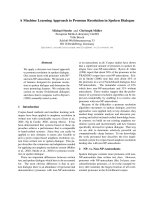
The schematic of the method described above is illustrated with an example DMR and non-DMR selected
from the training dataset in Fig. 1 (a-h). The proposed
histogram based features (Fig. 1d and h) show a clear
demarcation between DMRs and non-DMRs. In particular, the distributions of non-DMRs show low mean
values for the bins representing the higher difference in
methylation level (> 0.3), indicating low number of votes
falling in the bins that correspond to higher methylation
differences (Fig. 1i). In contrast, DMRs exhibit higher
differences in methylation level and have consistently
higher mean for bins that correspond to higher methylation differences (Fig. 1i). This indicates that the histogram based features are highly discriminative between
treatment states, which makes the problem of DMR detection suitable for machine learning analysis.
Training via SVM
The algorithm then uses the normalized histogram feature vectors described above to train a classifier based
on the label (DMR or non-DMR) provided for each individual cytosine. We tested various classifiers such as
Random forest, SVM with linear kernel and SVM with
RBF kernel [37–39] and selected a linear classifier based
on the ROC curve (Additional file 1: Figure S2 E and F).
Moreover, linear SVM is computationally very efficient
and showed comparable performance to the more computationally expensive non-linear RBF kernel and random forest classifier (Additional file 1: Figure S2 E and
F). However, note that the most crucial aspect of the
training is the use of the novel highly discriminative normalized histogram based feature vectors that robustly
discriminate between DMRs and non-DMRs. Hence, any
other classifier of choice can be used instead of linear
SVM without any significant changes to the proposed
method.
Testing and DMR prediction on new datasets
HOME requires input files containing basic information
of methylation, including chromosome numbers, genomic coordinates, type of cytosine (CG, CHG, CHH),
and mc and t for cytosines.
Pairwise
HOME can be used to predict DMRs from methylomes
of two treatment groups with single or multiple replicates. To predict the DMRs, the normalized histogram
features are computed for each cytosine on a particular
chromosome, which are then provided to the trained
SVM model to obtain the prediction scores that are normalized using the logistic function from the generalized
linear model (GLM) to lie in the range [0,1]. Individual
Srivastava et al. BMC Bioinformatics
(2019) 20:253
Page 5 of 15
a
b
c
d
e
f
g
h
i
j
Fig. 1 Feature generation overview. (a) Methylation level of sample 1 (S1) and sample 2 (S2) for a DMR from the training set. The overlapping
fixed size window is used around individual cytosine (C) in the DMR for feature extraction. (b) Extracted features: p-value and difference in
methylation level for each CG site. (c) Histogram of scores computed from the extracted features and (d) histogram of normalized scores. (e)
Methylation level of S1 and S2 for a non-DMR from the training dataset. The overlapping fixed size window is used around individual C in the
DMR for feature extraction. (f) Extracted features: p-value and difference in methylation level for each CG. (g) Histogram of scores computed from
the extracted features and (H) histogram of normalized scores. (i) Mean and standard deviation of histogram features for complete training data
for DMRs (blue) and non-DMRs (pink). (j) Testing and DMR prediction on new dataset
cytosines are grouped together into preliminary DMRs
based on the prediction scores (default: > 0.1) and the
distance between neighboring cytosines (default: < 500
bp). A low prediction score (< 0.1) from the classifier for
a cytosine site indicates low confidence in the site being
differentially methylated and a high prediction score (>
0.1) indicates high confidence in a site being methylated.
To produce the final DMRs, our method performs a
boundary refinement of the preliminary DMRs such that
boundaries are trimmed until k consecutive cytosines
(default: 3) have the value of the b (Eq.1) greater than or
equal to the defined threshold (default: 0.1).
Srivastava et al. BMC Bioinformatics
(2019) 20:253
Time-series and multi-group comparisons
HOME can be used to predict DMRs from time-series
and multi-group studies. Given a number of time points
or treatments, n, a total of nC2 pairwise combinations
are possible. HOME computes SVM prediction scores
for each of these pairwise combinations in the same
manner as for pairwise method described above. The
prediction scores are then normalized to lie in the range
[0,1] using the logistic function from the generalized linear model (GLM) to allow further analysis among all
pairwise comparisons. The scores are summed for each
cytosine to get a final score. The cytosines are then
grouped into DMRs.
In summary, once the SVM has been trained, the
histogram based features for new methylomes can be
computed, and HOME scans the entire methylome to
provide a prediction score (between 0 and 1) from the
SVM classifier for each cytosine site. Then, the individual cytosine sites are grouped together into DMRs based
on the user defined prediction score cutoff and the distance between neighboring cytosines. The testing and
DMR prediction on new dataset is shown in Fig. 1j.
Here, we independently applied HOME to both CG and
CH contexts, and compared its performance to two
other commonly used DMR finders, Metilene and DSS,
using both simulated and biological data. Furthermore,
we also show that HOME can be used for time-series
DMR analysis on biological data.
Results and discussion
Analysis of simulated DNA methylation data
The DMRs were simulated using the approach reported
by Dolzhenko and Smith [24]. For generation of simulated data, we utilised the read coverage and CG site distribution from WGBS datasets of neuronal and
non-neuronal cell types [5]. Only the methylated reads
in the actual DNA methylation data were replaced by
the simulated reads, generated using the beta-binomial
distribution from the work of Rakyan et al. [40], as
followed by Dolzhenko and Smith. Equal numbers of
DMRs and non-DMRs (2142) were simulated with two
distinct beta binomial settings, each increasing in their
difficulty for identification. The number of Cs in simulated DMRs were 49,442 and the number of Cs in simulated non-DMRs were 1,395,693. The length distribution
of the simulated DMRs and non-DMRs is shown in
Additional file 1: Figure S3. For each setting two treatment groups were simulated, each with three replicates
and 5 random simulations were performed to get more
accurate results. The read coverage for replicates was
taken from WGBS datasets of neuronal and
non-neuronal cell types [5]. For both settings, the
methylation level of cytosine sites in DMRs were generated from a beta distribution of (6,1.5) and a beta
Page 6 of 15
distribution of (1.5,6) for the two treatment groups, respectively. For the first setting (class 1), the non-DMR
portion of the genome displayed a fixed methylation
level (0.7), with only DMRs showing variation from this
value. In the second setting (class 2), non-DMR cytosine
methylation level was simulated from beta parameters
(2,2), such that the methylation level was not fixed for
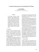
either DMRs or non-DMRs. As shown in Fig. 2a, class 1
showed no variation in methylation level for non-DMRs
between groups, and therefore DMRs are easier to identify for this class compared to class 2, which show variation both within and outside the DMR boundaries. To
demonstrate the difference between simulated class 1
and class 2 further, we plot the absolute methylation difference for both classes at the level of individual Cs
(Additional file 1: Figure S4A & B). For class 1, there is a
marginal overlap in absolute methylation difference between Cs in the DMRs and non-DMRs (Additional file 1:
Figure S4A). Whereas, for class 2 there is a significant
overlap in the absolute methylation difference between Cs
in the DMRs and non-DMRs (Additional file 1: Figure
S4B) and it is therefore much harder to detect DMRs of
class 2 than class 1.
We performed an extensive parameter search to identify settings for each DMR finder that resulted in the
best performance (Additional file 1: Section 1.2). Simulated DMRs are predicted by all the DMR finders for
Class 1, however, HOME is more accurate in predicting
the boundaries, compared to DSS and Metilene. Predicted DMRs by DSS and Metilene, for class 2 DMRs,
are either fragmented or have inaccurate boundaries,
whereas, HOME identifies all DMRs with more accurate
boundaries (Fig. 2a).
Precise definition of DMR boundaries is essential
for accurate downstream analysis and biological interpretation of differential methylation, for example
when associating DMRs to regulatory regions such as
promoters or enhancers to explore potential connections between methylation changes and local chromatin state and transcription. Imprecise DMR boundary
identification will result in the inappropriate inclusion
in the DMR of cytosine sites that are not differentially methylated, or exclusion of bona fide differentially methylated cytosines. Consequently, mean
methylation levels calculated for all cytosines within a
DMR would be inaccurate, and analysis of genomic or
chromatin features at the DMR boundaries would be imprecise. We evaluated the DMR boundary accuracy for all
three DMR finders by calculating the true positive rate
(TPR) and positive predictive value (PPV) for the range of
overlap (50–100%) between simulated and predicted
DMRs (Additional file 1: Section 1.3). TPR and PPV have
previously been used as performance metrics for comparing DMR finders [32, 41]. PPV has been used instead of
Srivastava et al. BMC Bioinformatics
(2019) 20:253
a
Page 7 of 15
b
Fig. 2 Comparison of DMR detection methods (HOME, DSS and Metilene) on simulated data. (a) Browser representation showing the quality and
boundary accuracy of predicted DMRs by HOME, DSS and Metilene on two simulated classes. The horizontal bars indicate the DMRs. Simulated
DMRs (black) and the scale are the same for both classes. (b) The performance of HOME, DSS, and Metilene was assessed in terms of true positive
rate (TPR) and positive predictive value (PPV) for both classes. The plots show mean and standard deviation of TPR and PPV for 5 random
simulations. The evaluation was performed in terms of percent reciprocal overlap ranging from 50 to 100% between simulated and predicted
DMRs by HOME, DSS and Metilene for two classes
specificity as a measure of false positive rate, as methods
with high specificity may still return a large number of
false positive findings [41].
TPR is defined as the number of overlaps between
simulated and predicted DMRs out of total simulated
DMRs. PPV is defined as the number of overlaps between simulated and predicted DMRs out of all predicted DMRs. Both HOME and Metilene showed
higher TPR and PPV compared to DSS for both classes (Fig. 2b). For class 1, HOME and Metilene
showed comparable performance for both TPR and
PPV for 50–90% overlap between simulated and predicted DMRs (Fig. 2b). However, HOME outperformed Metilene in both TPR and PPV for 90–100%
overlap between simulated and predicted DMRs. This
demonstrates that HOME predicts more accurate
boundaries compared to DSS and Metilene. For Class
2, HOME showed higher TPR compared to DSS and
Metilene for 50–95% overlap between simulated and
predicted DMRs (Fig. 2b). HOME showed higher PPV
for all overlap ranges between simulated and predicted DMRs. Overall, HOME predicted the DMR
boundaries with very small margin of error compared
to both DSS and Metilene.
Analysis of performance on biological datasets
On biological datasets we tested the performance of different DMR finders for pairwise comparisons (Section “Pairwise differential methylation analysis”) on boundary
accuracy and agreement with known biological knowledge.
For this, we used both plant and animal datasets as they
display different characteristics such as level of methylation, context, and length of DMRs. Importantly, the use of
biological knowledge to assess the performance of DMR
finders uses information from orthogonal experimental approaches that probe biological events that are highly associated with differential DNA methylation state. We consider
this to be a valuable additional approach to assess whether
DMRs identified by different algorithms are associated with
changes in other genomic regulatory layers, in particular
for the DMRs identified uniquely by each approach.
Pairwise differential methylation analysis
Accuracy: We compared the performance of HOME
with DSS and Metilene for the CG context on published
WGBS data [36] for two neuronal cell types: excitatory
pyramidal neurons (EX) and parvalbumin-expressing
fast-spiking interneurons (PV), each with two replicates.
Srivastava et al. BMC Bioinformatics
(2019) 20:253
Among the DMR finders that were compared, DMRs
identified by HOME were less fragmented and exhibited
more accurate boundary detection compared to the
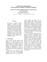
DMRs produced by the other finders (Fig. 3a), despite
equivalent DMR merging parameters being used for
each finder (Additional file 1: Section 1.4). DMRs predicted by HOME showed consistently higher methylation level differences inside the DMR boundaries when
compared to the other finders (Fig. 3b). For closer inspection of the above observation, the mean and standard deviation of the absolute methylation level difference
between analyzed samples for the 5 CG sites immediately inside and outside the DMR boundary is shown
(Fig. 3c). The mean methylation level difference for the
Page 8 of 15
boundary CG site and CG sites inside the DMRs is
higher for HOME than DSS and Metilene (Fig. 3c), while
the mean methylation level difference for the CG sites
immediately outside the DMR boundaries is lower for
HOME as compared to DSS and Metilene. This demonstrates the higher DMR border detection precision of
HOME on real biological datasets, as also observed for
the simulated WGBS data (Fig. 2b).
A more detailed comparison of the DMRs uniquely
predicted by each finder showed that DMRs uniquely
identified by HOME consistently had a higher methylation level difference for the CG sites located within the
DMRs (Fig. 3d). In contrast, DMRs uniquely predicted
by DSS and Metilene consistently had a lower
Fig. 3 Quality assessment of CG-DMRs predicted in mammalian WGBS data by HOME, DSS and Metilene. (a) Browser representation showing the
quality and boundary accuracy of predicted CG context DMRs for the PV specific gene Syt2. (b) Heatmap of methylation level difference for all
predicted DMRs by HOME, DSS and Metilene. The DMRs are sorted by length. The bin size is 200 bp for all heatmaps. (c) Mean and standard
deviation of absolute methylation difference for all predicted CG DMRs for 5 CGs upstream and downstream of the DMR start (left) and stop
(right) marked as 0, respectively. (d) Heatmap of methylation level difference for uniquely predicted DMRs by HOME, DSS and Metilene
Srivastava et al. BMC Bioinformatics
(2019) 20:253
methylation difference for the CG sites within the
DMRs. Furthermore, the boundaries of the DMRs identified by HOME are more precise compared to DSS and
Metilene (Fig. 3d). To investigate the biological significance of the uniquely predicted DMRs, we tested
whether the uniquely predicted DMRs identified by each
finder are located in the genomic regions that are relevant
to neuronal development and function. For this analysis,
phenotype and gene expression annotations provided by
the Genomic Regions Enrichment of Annotations Tool
(GREAT) were used [42]. Briefly, among the top 20 terms
produced by GREAT for gene expression and phenotype,
we counted the enrichment terms related to neuronal development and function for uniquely predicted DMRs by
each finder. The significance of enrichment terms was
ranked according to the binomial distribution-based
P-values obtained from GREAT. A similar approach for
exploring biological functions of DMRs has been performed previously [41]. The parameter details used for the
analysis are provided in Table 1. Among the top 20 terms
for phenotype annotation, 85% of terms for DMRs
uniquely predicted by HOME were directly related to
neural system functions. In contrast, terms related to
neural systems for DMRs uniquely predicted by DSS and
Metilene were 60 and 10% respectively. For associated
gene expression annotations provided by GREAT, we
found that uniquely predicted HOME DMRs were located
on or near to genes related to neuronal development and
function. Among the top 20 terms for gene expression annotation, 70% of terms for unique HOME DMRs were directly related to neural system function. Terms related to
neural systems for unique DMRs by DSS and Metilene
DMRs were 35 and 15%, respectively (Table 1). The details
of phenotype and gene expression annotations are summarized in Additional file 1: Figure S5.
To investigate the incidence of false positive DMRs
predicted by each finder, we permuted the labels among
the EX and PV WGBS samples to generate two artificial
datasets: (1) EX replicate 1 and PV replicate 1 (comprising treatment group 1) versus EX replicate 2 and PV
replicate 2 (comprising treatment group 2), and (2) EX
replicate 1 and PV replicate 2 versus EX replicate 2 and
PV replicate 1 (comprising treatment groups 1 and 2 respectively). Due to randomness in the shuffled data, it is
Page 9 of 15
expected that there will be shorter regions with contiguous methylation level differences occurring by chance,
and these short regions will be identified by all methods
as DMRs. However, it is also expected that there will be
significantly fewer long DMRs in the shuffled data, because as the length of the DMRs increases the likelihood
of obtaining such DMRs due to random chance decreases rapidly. These shuffled datasets were analyzed
with HOME, DSS and Metilene, and as expected the
number of DMRs predicted by each method in comparison to the unshuffled data was significantly reduced
(Additional file 1: Figure S6 A & B). In addition, DMRs
identified by HOME in the shuffled data were smaller
and contained low number of CGs. In contrast, the
DMRs identified by DSS and Metilene were longer and
had a higher number of CGs per DMR and therefore are
more likely to be false positive DMRs (Additional file 1:
Figure S6 A & B).
To test the performance of HOME for detecting differential methylation in the CH sequence context, we used
WGBS datasets of neuronal and non-neuronal cell types
isolated from the frontal cortex of 7 week old male
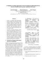
mouse prefrontal cortex [5]. A genome browser view of
a representative genomic region showed that the CH
DMRs predicted by HOME between neurons (NeuN+)
and glia (NeuN-) were regions of contiguous hyper- or
hypo-methylation (Fig. 4a). The directionality of the
DMRs (hyper/hypo) are defined with respect to NeuN+
cells. We observed a high number of hyper-methylated
DMRs (429,421) in NeuN+ cells as compared to a very
low number of hypo-methylated DMRs (21,829) (Fig.
4b). These findings are consistent with previously published results, where CH methylation accumulates to a
high level in neurons compared to glia [5, 36]. CH
DMRs predicted by HOME have accurate boundaries
with higher inter-sample methylation level difference at
and within DMR boundaries, and low methylation level
difference immediately outside the DMR boundaries
(Fig. 4b). Although the observed methylation difference
in hypo-methylated NeuN+ DMRs was very low (< 0.02),
gene ontology analysis using GREAT showed that these
DMRs were located in genomic regions related to neuronal development and function (Fig. 4c), suggesting that
the HOME CH DMRs are biologically relevant.
Table 1 Biological annotations of unique DMRs predicted by HOME, DSS and Metiene on the PV and EX methylome data. The top
20 terms were counted for neural system function related terms using the mouse phenotype annotation and MGI gene expression
annotation. No. of Cs refers to the minimum number of cytosines required in a DMR and delta refers to the absolute change in
methylation level
DMR
finders
Best results were obtained for below
threshold
No. of DMRs after
thresholding
Phenotype (%) (terms counted
out of 20)
Expression (%) (terms counted
out of 20)
HOME
No. of Cs > 5 and delta > 0.2
4721
85
70
DSS
No. of Cs > 5 and delta > 0.25
3066
60
35
Metilene
delta > 0.15
2495
10
15
Srivastava et al. BMC Bioinformatics
(2019) 20:253
a
Page 10 of 15
b
c
Fig. 4 Quality assessment of CH-DMRs predicted in mammalian WGBS data. (a) Genome browser representation of the quality and boundary
accuracy of HOME predicted CH context DMRs for neurons (NeuN+) and glia (NeuN-) methylation data. Top panel represents hyper-methylated
DMRs in NeuN+ and bottom panel represents hypo-methylated DMRs in NeuN+. (b) Heatmap of methylation level difference for hypermethylated HOME DMRs and hypo-methylated HOME DMRs. (c) Biological annotations of hypo-methylated HOME DMRs in NeuN+ cells
displaying the top 20 terms using the mouse phenotype annotation and the MGI gene expression annotation (neuron, or glia, and brain tissue
related terms are highlighted in orange)
To assess the generalizability of HOME performance
for species that have very distinct methylation patterns
and distributions compared to mammals, we next tested
the performance of HOME on Arabidopsis WGBS data,
comparing DNA methylation in wild-type (WT) and
CHROMOMETHYLASE 2 mutant (cmt2), a well
characterised mutant that exhibits differences in CH
methylation [43]. CMT2 is a functional non-CG methyltransferase, known to mediate DNA methylation at both
CHG and CHH context in vitro and in vivo [43, 44]. We
compared the performance of HOME with DSS, which
has previously been used for DMR prediction in plant
WGBS datasets [16, 45]. Note that we used the same
model that was trained on the mammalian WGBS dataset for predicting the DMRs between cmt2 and WT, to
assess whether the trained model is generalizable. The
heatmap for all predicted DMRs by HOME and DSS
showed that HOME DMRs are more accurate in boundary prediction than DSS, predominantly for the CG and
CHG contexts (Fig. 5a). Moreover, for the CHG context,
HOME detected a large number of DMRs (n = 13,402)
of a large median length DMRs (593 bp), whereas DSS
Srivastava et al. BMC Bioinformatics
(2019) 20:253
Page 11 of 15
Fig. 5 Qualitative analysis of predicted DMRs by HOME and DSS in plant WGBS data. (a) Heatmap of methylation level difference for all predicted
HOME and DSS DMRs, between cmt2 and WT, for CG, CHG and CHH contexts. (b) Mean and standard deviation of absolute methylation difference for
all predicted CHH DMRs by HOME and DSS for 5 CGs upstream and downstream of the DMR start (left) and stop (right) marked as 0, respectively. (c)
Heatmap of methylation level difference for uniquely predicted HOME and DSS DMRs, between cmt2 and WT, for CG, CHG and CHH contexts
Srivastava et al. BMC Bioinformatics
(2019) 20:253
Page 12 of 15
only predicted small number of DMRs (n = 3083) with a
short median length (133 bp), indicating that HOME is
more sensitive for DMR detection over a greater range
of sizes. For the CHH context, the mean and standard
deviation of 5 cytosines upstream and downstream of
the predicted DMRs start and stop sites, respectively,
showed that the methylation level difference is high
within the DMR and low just outside the DMRs for
HOME (Fig. 5b). In contrast, DMRs predicted by DSS
showed similar mean and standard deviation of methylation level difference both inside and outside the DMR
boundaries (Fig. 5b). These results indicate more accurate boundary prediction by HOME for all contexts.
Similarly, genome browser screenshots exemplify how
HOME DMRs are more precise than DSS DMRs for all
contexts (Additional file 1: Figure S7).
Furthermore, the heatmaps of DMRs predicted
uniquely by HOME exhibit more accurate boundaries
than DSS for all sequence contexts (Fig. 5c). We further
examined the genomic distribution of the DMRs
uniquely predicted by HOME and DSS. Over 60% of
HOME CG DMRs were present in gene bodies, while
only a small fraction (< 30%) of CHG and CHH DMRs
overlapped with gene bodies (Table 2). These results are
consistent with the distribution of DNA methylation in
plant genes, where gene bodies mainly exhibit CG
methylation [21, 43, 46]. Compared to HOME CG
DMRs, a smaller fraction (58%) of CG DMRs predicted
by DSS overlapped with gene bodies (Table 2). Non-CG
methylation plays an important role in silencing transposable elements (TEs) [43]. Thus, it is expected that
most of the non-CG DMRs detected between WT and
cmt2 will overlap with TEs, given the role of CMT2 in
mediating methylating of TEs, particularly long TEs, in
the non-CG context. We found that > 70% of CHG
DMRs and 50% of CHH DMRs predicted by HOME
overlapped with TEs, and only 21% of the CG DMRs
intersected with TEs (Table 2). On the other hand,
DMRs predicted by DSS showed a similar percentage
overlap with TEs to HOME for CG and CHG. However,
DSS showed significantly less overlap with TEs for the
CHG context (Table 2). Previous studies have shown
that cmt2 exhibits loss of non-CG methylation predominantly at long TEs (> 1 kb) [43]. Hence, we further
inspected the location of uniquely predicted DMRs in
TEs. The overall genomic distribution of long TEs (> 1
Table 2 Percentage of uniquely predicted DMRs by HOME and
DSS in gene bodies and TEs for CG, CHG and CHH contexts
Gene body
kb) and short TEs (< 1 kb) in the genome is 20 and 80%,
respectively. A higher percentage of CHG and CHH
DMRs predicted by HOME overlapped with long TEs
than with short TEs (Table 3). The fraction of TEs (long
and short) that overlapped with HOME CG DMRs was
very small (< 7%). For CHH DMRs both HOME and
DSS exhibited a similar overlap with TEs. Most CHG
DMRs predicted by DSS did not overlap either long or
short TEs, while a larger fraction of long TEs overlapped
CG DMRs predicted by DSS, compared to short TEs
(Table 3). Our results suggest that DMRs predicted by
HOME fit better with the known biological function of
cmt2, than DMRs predicted by DSS.
Runtime: The run time for HOME, DSS and Metilene
for all the analysis in Section: Accuracy is summarized
below. For DMR identification between neuronal cell types
(EX and PV) in the CG context, both DSS and HOME
showed very similar run time (~ 2 h), while Metilene completed in ~ 4 min. Because of a high computational runtime
requirement for both DSS and Metilene for the CH context
analysis, the run did not complete after 12 days of execution and had to be terminated, being deemed an unfeasible
analysis to undertake with compared methods given reasonable timeframes for analysis. Therefore, the results for
DMR identification in the CH context (Section: Accuracy)
are only shown for HOME (runtime: 4 days). For the plant
dataset, HOME took 12 min to predict CG DMRs compared to 27 min for DSS. For the CHG context DMRs, both
HOME and DSS showed similar execution times of 27 min,
while HOME was > 3 times faster compared to DSS for
DMR prediction in the CHH sequence context, taking 2
and 7 h, respectively. Overall, HOME showed similar or
better execution times compared to DSS and Metilene, particularly for non-CG context.
Time-series differential methylation analysis
An additional feature of HOME is the ability to predict
DMRs in time-series data. The HOME time-series analysis algorithm can be successfully used for identification
of DMRs in datasets where DNA methylation varies over
time or between development stages, for example, during seed germination [47], cell reprogramming [48, 49],
and mammalian brain development [5]. Current
Table 3 Percentage of short (< 1 kb) and long (> 1 kb) TEs that
overlap with uniquely predicted DMRs by HOME and DSS, for
CG, CHG and CHH contexts
TE
Long TE (> 1 kb)
Short TE (< 1 kb)
DMR finders
CG
CHG
CHH
CG
CHG
CHH
DMR finders
CG
CHG
CHH
CG
CHG
CHH
HOME
67%
16.7%
29%
21%
72%
50%
HOME
6%
69%
13%
DSS
58%
31.7%
15%
27%
48%
52%
DSS
13%
3%
12%
0.9%
34%
8%
2%
0.3%
4%
Srivastava et al. BMC Bioinformatics
(2019) 20:253
methods can be used to call pairwise DMRs for each
combination of groups in time-series data, and thereafter
the DMRs could be merged to obtain final predicted
time-series DMRs. However, taking this approach, the
complexity of all possible combinations increases and
becomes tedious for users, as the number of groups increases in time-series data. With HOME, we provide an
easy and convenient way to compare many time points
with a single command. Moreover, the output of the
HOME time-series module contains many useful metrics
that allow users to trace methylation changes through
time and determine the stability or stochasticity of the
methylation state in the DMRs. For example, the output
summarizes the mean methylation level difference and
directionality of methylation level change, for each pair
combination in the time-series data.
We tested the performance of HOME on another
time-series dataset of mouse embryonic fibroblast (MEF)
reprogramming to induced pluripotent stem cells
(iPSCs) [50]. The dataset contains 6 time points: MEF,
day 3, day 6, day 9, day 12 and iPSCs. The browser representations in Fig. 6, show that HOME is able to identify DMRs with gradual methylation changes effectively.
Conclusions
Here we present a novel histogram of methylation based
machine learning method to detect DMRs from single
nucleotide resolution DNA methylation data. Our
method treats the problem of DMR detection as a binary
classification problem and requires a high quality training dataset. Due to the lack of a biological dataset with
known DMRs and non-DMRs, we generated our own
training dataset using publicly available DNA methylomes and complementary datasets such as differential
ATAC-seq peaks or differentially expressed genes.
Page 13 of 15
HOME showed more accurate DMR prediction and precise DMR boundary identification compared to both
DSS and Metilene. The key features of HOME are: (i)
novel histogram based features which combines important information such as methylation level difference,
measure of significance for the difference in methylation,
and distance between neighbouring cytosines; (ii) a robustly trained model that is effective for a wide variety
of species; (iii) a flexible method that can be used for
prediction of DMRs in both CG and CH contexts with
high border accuracy; and (iv) a tool that can identify
DMRs in time-series data.
The most important qualities of any DMR finder are
accurate prediction of DMR boundaries and low number
of spurious DMRs (false positives). HOME outperforms
both DSS and Metilene in both of these measures. One
of the reasons underlying the low false positive rate of
HOME is the use of biological training data for DMRs
and non-DMRs to train the classifier. In addition, the
histogram based features can robustly discriminate between DMRs and non-DMRs, thereby reducing the
probability of detecting spurious DMRs. Histogram
based features are also able to capture the information
present around each cytosine site with the use of
weighted voting, thereby, enabling accurate identification
of the DMR boundaries.
HOME accounts for biological variation present between the replicates and uneven read coverage through
weighted logistic regression while computing the
p-value. The spatial correlation present among neighboring cytosine sites is captured by moving average smoothing and the use of weighted voting for histogram based
features. We demonstrate that HOME can be used to
predict accurate DMRs in both CG and non-CG (CHG
and CHH) sequence contexts for both mammalian and
Fig. 6 Browser representations of CG DMRs predicted by HOME in time-series WGBS data. Six time points: mouse embryonic fibroblast (MEF), day
3, day 6, day 9, day 12 and induced pluripotent stem cell (iPSC)
Srivastava et al. BMC Bioinformatics
(2019) 20:253
plant WGBS methylome data by using the same training
data. Although the classifier was trained on mammalian
WGBS data for CG and CH contexts, HOME can accurately predict DMRs in plants and for specific non-CG
contexts (CHG and CHH), demonstrating its versatility.
However, if users wish to retrain the HOME model on
their own data, it can easily be done from the approach
mentioned above (see Methods section).
Finally, another standout feature of HOME is the prediction of DMRs in time-series data. Time-series DNA
methylation experiments are commonly used to study a
wide range of biological processes such as development
[5] and stress responses [51]. HOME is an efficient
method to directly predict accurate DMRs in studies
with multiple timepoints. This added functionality of
HOME will greatly facilitate and expand the study of
epigenome dynamics in numerous biological systems
and disease models. Taken together, HOME is a highly
effective and robust DMR finder that accounts for uneven cytosine coverage in WGBS data, accounts for biological variation present between the samples in the
same treatment group, predicts DMRs in various genomic contexts, and accurately identifies DMRs among
any number of treatment groups in experiments with or
without replicates.
Additional file
Additional file 1: Contains supplementary information that includes
parameters tested for different DMR finders, Table S1 and S2, and
Figure S1 to S7. (PDF 382 kb)
Abbreviations
ATAC-seq: Assay for Transposase Accessible Chromatin sequencing;
CMT2: CHROMOMETHYLASE 2; DMRs: Differentially methylated regions;
EX: Excitatory pyramidal neurons; GREAT: Genomic Regions Enrichment of
Annotations Tool; iPSCs: Induced pluripotent stem cells; MEF: Mouse
embryonic fibroblast; PPV: Positive predictive value; PV: Parvalbuminexpressing fast-spiking interneurons; SVM: Support vector machine;
TEs: Transposable elements; TPR: True positive rate; VIP: Vasoactive intestinal
peptide-expressing interneurons; WGBS: Whole genome bisulfite sequencing;
WT: Wild-type
Acknowledgements
We thank Dr. Egor Dolzhenko for his helpful discussions regarding the
generation of simulated data. We thank members of the Lister Lab and the
Borevitz Lab for their suggestions and comments.
Funding
This work was supported by the Australian Research Council (ARC) Centre of
Excellence program in Plant Energy Biology (CE140100008). RL was
supported by a Sylvia and Charles Viertel Senior Medical Research
Fellowship, ARC Future Fellowship (FT120100862), and Howard Hughes
Medical Institute International Research Scholarship (RL).
Availability of data and materials
WGBS data from 7 weeks old male mouse brain for neuron and glia cell
types were obtained from GSM1173786 and GSM1173787, respectively.
Datasets for EX, PV and VIP neuronal cell types were downloaded from NCBI
GEO (GSE63137). Arabidopsis WT and cmt2 datasets were obtained from
NCBI GEO repositories GSM1242401 and GSM1242405, respectively. Time-
Page 14 of 15
series dataset for MEF, day 3, day 6, day 9, day 12 and iPSCs were obtained
from GEO accessions GSM2718419, GSM2718420, GSM2718421, GSM2718422,
GSM2718423 and GSM2718424, respectively.
Authors’ contributions
AS and RL devised the project. AS conceptualized, designed and
implemented HOME with statistical guidance from YVK. RL, SRE and JOB
supervised experiments. AS and YVK processed the biological data. AS tested
HOME on simulated and biological data. AS and YVK drafted the manuscript,
all authors contributed to writing the manuscript.
Ethics approval and consent to participate
Not applicable.
Consent for publication
Not applicable.
Competing interests
The authors declare no conflicts of interest.
Publisher’s Note
Springer Nature remains neutral with regard to jurisdictional claims in
published maps and institutional affiliations.
Author details
1
ARC Centre of Excellence in Plant Energy Biology, The University of Western
Australia, Perth, Australia. 2Harry Perkins Institute of Medical Research, Perth,
Australia. 3ARC Centre of Excellence in Plant Energy Biology, The Australian
National University, Canberra, Australia.
Received: 14 October 2018 Accepted: 24 April 2019
References
1. Richardson BC. Role of DNA methylation in the regulation of cell function:
autoimmunity, aging and cancer. J Nutr. 2002;132(8 Suppl):2401S–5S.
2. Khavari DA, Sen GL, Rinn JL. DNA methylation and epigenetic control of
cellular differentiation. Cell Cycle. 2010;9(19):3880–3.
3. Messerschmidt DM, Knowles BB, Solter D. DNA methylation dynamics
during epigenetic reprogramming in the germline and preimplantation
embryos. Genes Dev. 2014;28(8):812–28.
4. Jones PA. Functions of DNA methylation: islands, start sites, gene bodies
and beyond. Nat Rev Genet. 2012;13(7):484–92.
5. Lister R, Mukamel EA, Nery JR, Urich M, Puddifoot CA, Johnson ND, Lucero J,
Huang Y, Dwork AJ, Schultz MD, et al. Global epigenomic reconfiguration
during mammalian brain development. Science. 2013;341(6146):1237905.
6. Kass SU, Landsberger N, Wolffe AP. DNA methylation directs a timedependent repression of transcription initiation. Current biology : CB. 1997;
7(3):157–65.
7. Jones PA. The DNA methylation paradox. Trends in genetics : TIG. 1999;
15(1):34–7.
8. Meissner A, Mikkelsen TS, Gu H, Wernig M, Hanna J, Sivachenko A, Zhang X,
Bernstein BE, Nusbaum C, Jaffe DB, et al. Genome-scale DNA methylation
maps of pluripotent and differentiated cells. Nature. 2008;454(7205):766–70.
9. Lister R, Pelizzola M, Dowen RH, Hawkins RD, Hon G, Tonti-Filippini J, Nery
JR, Lee L, Ye Z, Ngo QM, et al. Human DNA methylomes at base resolution
show widespread epigenomic differences. Nature. 2009;462(7271):315–22.
10. Law JA, Jacobsen SE. Establishing, maintaining and modifying DNA
methylation patterns in plants and animals. Nat Rev Genet. 2010;11(3):204–20.
11. Stadler MB, Murr R, Burger L, Ivanek R, Lienert F, Scholer A, van Nimwegen E,
Wirbelauer C, Oakeley EJ, Gaidatzis D, et al. DNA-binding factors shape the
mouse methylome at distal regulatory regions. Nature. 2011;480(7378):490–5.
12. Bogdanovic O, Smits AH, de la Calle Mustienes E, Tena JJ, Ford E, Williams R,
Senanayake U, Schultz MD, Hontelez S, van Kruijsbergen I et al: Active DNA
demethylation at enhancers during the vertebrate phylotypic period. Nat
Genet 2016, 48(4):417–426.
13. Heyn H, Moran S, Hernando-Herraez I, Sayols S, Gomez A, Sandoval J, Monk
D, Hata K, Marques-Bonet T, Wang L, et al. DNA methylation contributes to
natural human variation. Genome Res. 2013;23(9):1363–72.
Srivastava et al. BMC Bioinformatics
(2019) 20:253
14. Kundaje A, Meuleman W, Ernst J, Bilenky M, Yen A, Heravi-Moussavi A,
Kheradpour P, Zhang Z, Wang J, Ziller MJ, et al. Integrative analysis of 111
reference human epigenomes. Nature. 2015;518(7539):317–30.
15. Schultz MD, He Y, Whitaker JW, Hariharan M, Mukamel EA, Leung D,
Rajagopal N, Nery JR, Urich MA, Chen H, et al. Human body epigenome
maps reveal noncanonical DNA methylation variation. Nature. 2015;
523(7559):212–6.
16. Eichten SR, Stuart T, Srivastava A, Lister R, Borevitz JO. DNA methylation
profiles of diverse Brachypodium distachyon align with underlying genetic
diversity. Genome Res. 2016;26(11):1520–31.
17. Kawakatsu T, Stuart T, Valdes M, Breakfield N, Schmitz RJ, Nery JR, Urich MA,
Han X, Lister R, Benfey PN, et al. Unique cell-type-specific patterns of DNA
methylation in the root meristem. Nature plants. 2016;2(5):16058.
18. Niederhuth CE, Bewick AJ, Ji L, Alabady MS, Kim KD, Li Q, Rohr NA, Rambani
A, Burke JM, Udall JA, et al. Widespread natural variation of DNA
methylation within angiosperms. Genome Biol. 2016;17(1):194.
19. Xie W, Barr CL, Kim A, Yue F, Lee AY, Eubanks J, Dempster EL, Ren B. Baseresolution analyses of sequence and parent-of-origin dependent DNA
methylation in the mouse genome. Cell. 2012;148(4):816–31.
20. Varley KE, Gertz J, Bowling KM, Parker SL, Reddy TE, Pauli-Behn F, Cross MK,
Williams BA, Stamatoyannopoulos JA, Crawford GE, et al. Dynamic DNA
methylation across diverse human cell lines and tissues. Genome Res. 2013;
23(3):555–67.
21. Cokus SJ, Feng S, Zhang X, Chen Z, Merriman B, Haudenschild CD, Pradhan
S, Nelson SF, Pellegrini M, Jacobsen SE. Shotgun bisulphite sequencing of
the Arabidopsis genome reveals DNA methylation patterning. Nature. 2008;
452(7184):215–9.
22. Guo S, Diep D, Plongthongkum N, Fung HL, Zhang K, Zhang K.
Identification of methylation haplotype blocks aids in deconvolution of
heterogeneous tissue samples and tumor tissue-of-origin mapping from
plasma DNA. Nat Genet. 2017;49(4):635–42.
23. Hansen KD, Langmead B, Irizarry RA. BSmooth: from whole genome bisulfite
sequencing reads to differentially methylated regions. Genome Biol. 2012;
13(10):R83.
24. Dolzhenko E, Smith AD. Using beta-binomial regression for high-precision
differential methylation analysis in multifactor whole-genome bisulfite
sequencing experiments. BMC bioinformatics. 2014;15:215.
25. Lea AJ, Tung J, Zhou X. A flexible, efficient binomial mixed model for
identifying differential DNA methylation in bisulfite sequencing data. PLoS
Genet. 2015;11(11):e1005650.
26. Hebestreit K, Dugas M, Klein HU. Detection of significantly differentially
methylated regions in targeted bisulfite sequencing data. Bioinformatics.
2013;29(13):1647–53.
27. Shafi A, Mitrea C, Nguyen T, Draghici S. A survey of the approaches for
identifying differential methylation using bisulfite sequencing data. Brief
Bioinform. 2018;19(5):737–53.
28. Saito Y, Tsuji J, Mituyama T. Bisulfighter: accurate detection of methylated
cytosines and differentially methylated regions. Nucleic Acids Res. 2014;
42(6):e45.
29. Wang Z, Li X, Jiang Y, Shao Q, Liu Q, Chen B, Huang D. swDMR: a sliding
window approach to identify differentially methylated regions based on
whole genome bisulfite sequencing. PLoS One. 2015;10(7):e0132866.
30. Feng H, Conneely KN, Wu H. A Bayesian hierarchical model to detect
differentially methylated loci from single nucleotide resolution sequencing
data. Nucleic Acids Res. 2014;42(8):e69.
31. Wu H, Xu T, Feng H, Chen L, Li B, Yao B, Qin Z, Jin P, Conneely KN.
Detection of differentially methylated regions from whole-genome bisulfite
sequencing data without replicates. Nucleic Acids Res. 2015;43(21):e141.
32. Juhling F, Kretzmer H, Bernhart SH, Otto C, Stadler PF, Hoffmann S. Metilene:
fast and sensitive calling of differentially methylated regions from bisulfite
sequencing data. Genome Res. 2016;26(2):256–62.
33. Eckhardt F, Lewin J, Cortese R, Rakyan VK, Attwood J, Burger M, Burton J,
Cox TV, Davies R, Down TA, et al. DNA methylation profiling of human
chromosomes 6, 20 and 22. Nat Genet. 2006;38(12):1378–85.
34. Jaffe AE, Feinberg AP, Irizarry RA, Leek JT. Significance analysis and statistical
dissection of variably methylated regions. Biostatistics. 2012;13(1):166–78.
35. Cortes C, Vapnik V. Support-vector networks. Mach Learn. 1995;20(3):273.
36. Mo A, Mukamel EA, Davis FP, Luo C, Henry GL, Picard S, Urich MA, Nery JR,
Sejnowski TJ, Lister R, et al. Epigenomic signatures of neuronal diversity in
the mammalian brain. Neuron. 2015;86(6):1369–84.
37. Breiman L. Random forests. Mach Learn. 2001;45(1):5–32.
Page 15 of 15
38. Karpievitch YV, Hill EG, Leclerc AP, Dabney AR, Almeida JS. An introspective
comparison of random forest-based classifiers for the analysis of clustercorrelated data by way of RF++. PLoS One. 2009;4(9):e7087.
39. Pedregosa F, Varoquaux G, Gramfort A, Michel V, Thirion B, Grisel O, Blondel
M, Prettenhofer P, Weiss R, Dubourg V, et al. Scikit-learn: machine learning
in python. J Mach Learn Res. 2011;12:2825–30.
40. Rakyan VK, Down TA, Balding DJ, Beck S. Epigenome-wide association
studies for common human diseases. Nat Rev Genet. 2011;12(8):529–41.
41. Wen Y, Chen F, Zhang Q, Zhuang Y, Li Z. Detection of differentially
methylated regions in whole genome bisulfite sequencing data using local
Getis-Ord statistics. Bioinformatics. 2016;32(22):3396–404.
42. McLean CY, Bristor D, Hiller M, Clarke SL, Schaar BT, Lowe CB, Wenger AM,
Bejerano G. GREAT improves functional interpretation of cis-regulatory
regions. Nat Biotechnol. 2010;28(5):495–501.
43. Stroud H, Do T, Du J, Zhong X, Feng S, Johnson L, Patel DJ, Jacobsen SE.
Non-CG methylation patterns shape the epigenetic landscape in
Arabidopsis. Nat Struct Mol Biol. 2014;21(1):64–72.
44. Zemach A, Kim MY, Hsieh PH, Coleman-Derr D, Eshed-Williams L, Thao K,
Harmer SL, Zilberman D. The Arabidopsis nucleosome remodeler DDM1
allows DNA methyltransferases to access H1-containing heterochromatin.
Cell. 2013;153(1):193–205.
45. Crisp PA, Ganguly DR, Smith AB, Murray KD, Estavillo GM, Searle I, Ford E,
Bogdanovic O, Lister R, Borevitz JO, et al. Rapid recovery gene
downregulation during excess-light stress and recovery in Arabidopsis. Plant
Cell. 2017;29(8):1836–63.
46. Lister R, O'Malley RC, Tonti-Filippini J, Gregory BD, Berry CC, Millar AH, Ecker
JR. Highly integrated single-base resolution maps of the epigenome in
Arabidopsis. Cell. 2008;133(3):523–36.
47. Narsai R, Secco D, Schultz MD, Ecker JR, Lister R, Whelan J. Dynamic and
rapid changes in the transcriptome and epigenome during germination
and in developing rice (Oryza sativa) coleoptiles under anoxia and reoxygenation. The Plant journal : for cell and molecular biology. 2017;89(4):
805–24.
48. Lister R, Pelizzola M, Kida YS, Hawkins RD, Nery JR, Hon G, AntosiewiczBourget J, O’Malley R, Castanon R, Klugman S, et al. Hotspots of aberrant
epigenomic reprogramming in human induced pluripotent stem cells.
Nature. 2011;471(7336):68–73.
49. Lee DS, Shin JY, Tonge PD, Puri MC, Lee S, Park H, Lee WC, Hussein SM,
Bleazard T, Yun JY, et al. An epigenomic roadmap to induced pluripotency
reveals DNA methylation as a reprogramming modulator. Nat Commun.
2014;5:5619.
50. Knaupp AS, Buckberry S, Pflueger J, Lim SM, Ford E, Larcombe MR, Rossello
FJ, de Mendoza A, Alaei S, Firas J, et al. Transient and permanent
reconfiguration of chromatin and transcription factor occupancy drive
reprogramming. Cell Stem Cell. 2017;21(6):834–845 e836.
51. Dowen RH, Pelizzola M, Schmitz RJ, Lister R, Dowen JM, Nery JR, Dixon JE,
Ecker JR. Widespread dynamic DNA methylation in response to biotic stress.
Proc Natl Acad Sci U S A. 2012;109(32):E2183–91.