An integrated strategy for identifying new targets and inferring the mechanism of action: Taking rhein as an example
Bạn đang xem bản rút gọn của tài liệu. Xem và tải ngay bản đầy đủ của tài liệu tại đây (2.17 MB, 11 trang )
Sun et al. BMC Bioinformatics (2018) 19:315
/>
RESEARCH ARTICLE
Open Access
An integrated strategy for identifying new
targets and inferring the mechanism of
action: taking rhein as an example
Hao Sun1,2, Yiting Shen1, Guangwen Luo1, Yuepiao Cai1* and Zheng Xiang1*
Abstract
Background: Target identification is necessary for the comprehensive inference of the mechanism of action of a
compound. The application of computational methods to predict the targets of bioactive compounds saves cost
and time in drug research and development. Therefore, we designed an integrated strategy consisting of ligandprotein docking, network analysis, enrichment analysis, and an experimental surface plasmon resonance (SPR)
method to identify and validate new targets, and then used enriched pathways to elucidate the underlying
pharmacological mechanisms. Here, we used rhein, a compound with various pharmacological activities, as an
example to find some of its previously unknown targets and to determine its pharmacological activity.
Results: A total of nine candidate targets were discovered, including LCK, HSP90AA1, RAB5A, EGFR, CDK2, CDK6,
GSK3B, p38, and JNK. LCK was confirmed through SPR experiments, and HSP90AA1, EGFR, CDK6, p38, and JNK were
validated through previous reports. Rhein network regulations are complex and interconnected. The therapeutic
effect of rhein is the synergistic and comprehensive result of this vast and complex network, and the perturbation
of multiple targets gives rhein its various pharmacological activities.
Conclusions: This study provided a new integrated strategy to identify new targets of bioactive compounds and
reveal their molecular mechanisms of action.
Keywords: Target identification, Rhein, Ligand-protein docking, Network analysis, Enrichment analysis, SPR
Background
In real biological systems, bioactive compounds generally bind to more than one target proteins to exert their
biological activities [1]. Target identification is therefore
necessary for the comprehensive inference of the action
mechanisms of a compound. Although wet lab experiments are more convincing, the application of in silico
computational methods to predict targets of bioactive
compounds has become more important in recent years
[2]. Current computational methods for drug target discovery fall into three categories: structure-based,
ligand-based, and phenotype-based virtual screening [3].
The structure-based methods involve the molecular
docking between a ligand and a target, and the scoring
function is used to assess the likelihood of the ligand
* Correspondence: ;
1
School of Pharmaceutical Sciences, Wenzhou Medical University, Wenzhou
325035, China
Full list of author information is available at the end of the article
binding to a protein. The disadvantages of this method include high false positives and weak accuracies [4]. The
ligand-based methods are based on using similarities between known ligands to speculate on unknown structures
of receptor sites; thus, such methods are not appropriate
for the analysis of proteins without known ligands [5].
The phenotype-based methods aim at analysing phenotypic responses, such as gene expression profiles in cell
lines or proteomic information, but may neglect valuable
information from other types of data sources [6]. Perhaps,
any method used alone will have its own short board, so
the combination of multiple methods is a train of thought.
Actually, an effective drug often regulate several biological processes by acting on multiple targets, which
can form a complex interaction network [2]. The complex network can provide a lot of target topological
information through network analysis. Therefore, the
network analysis can be used to study the complex interactions between targets and may be a good method for
© The Author(s). 2018 Open Access This article is distributed under the terms of the Creative Commons Attribution 4.0
International License ( which permits unrestricted use, distribution, and
reproduction in any medium, provided you give appropriate credit to the original author(s) and the source, provide a link to
the Creative Commons license, and indicate if changes were made. The Creative Commons Public Domain Dedication waiver
( applies to the data made available in this article, unless otherwise stated.
Sun et al. BMC Bioinformatics (2018) 19:315
new target identification. However, it cannot reflect the
whole biological processes since how targets influence
the biological processes are lacked. The enrichment analysis can link interactions between proteins and biological processes. Therefore, the enrichment analysis can
supplement the deficiency of network analysis for identifying targets and inferring their regulation on biological
processes [7]. Nowadays, network visualisation and bioinformatics enrichment tools have promoted the understanding of complex drug-target and target-target
interactions, accelerated the drug discovery through the
identification of topological structures in biological networks, developed a systematic understanding of drug action and disease complexity, and improved the efficiency
and safety of drug design [8–10].
Rhein is an active alipophilic anthraquinone that is
mainly extracted from several traditional plant rhizomes,
including Rheum palmatum L., Aloe barbadensis Miller,
Cassia angustifolia Vahl., and Polygonum multiflorum
Thunb. [11]. Rhein has various pharmacological effects,
such as anti-inflammatory, anti-tumour, antioxidant,
antifibrotic, hepatoprotective, and nephroprotective activities [12, 13]. According to our research, more than
1000 articles about rhein have been published in
PubMed; over 100 of these have discussed its pharmacological mechanism of action [13]. Many targets of rhein
have been identified in recent years. Rhein could suppress
all the tested RXRA-involved homo-or-heterodimeric
transcription activities, decrease the expression of VEGFA,
EGF, HIF1A, ERBB2, and PTGS2 proteins, decrease the
activity of NFKB1 and RELA proteins [14, 15], and increase the levels of apoptosis-related proteins including
BAX, CASP3, and CASP8 [16]. Moreover, the regulation
of multiple pathways by rhein, such as the MAPK,
PI3K-AKT, NF-κB, and TGF-β signalling pathways, cell
cycle, and cell apoptosis, has been a particular focus of research [17–19]. Since rhein affects so many different targets and regulates multiple pathways in the body, we
believe that rhein can be repurposed to treat even more
diseases, and its new targets can still be discovered.
In this study, an integrated strategy consisting of
ligand-protein docking, network analysis, enrichment
analysis, and experimental validation was developed and
applied to identify new rhein targets and infer the mechanisms underlying the pharmacological effects of rhein.
Using this approach, we could easily identify the targets
of one drug or one bioactive compound and infer their
molecular mechanisms.
Methods
The integrated strategy for target identification involved
four main steps: (1) Preliminary screening by
ligand-protein docking; (2) Further screening by network
analysis; (3) Final screening by enrichment analysis; (4)
Page 2 of 11
Validating candidate targets through the surface plasmon
resonance (SPR) interaction experiment. The strategy of
target identification is shown in Fig. 1.
Ligand-protein docking for potential targets
Here, two steps were designed for the preliminary
screening of targets. First, the inverse molecular docking,
one of the ligand-based virtual screening, was used to
quickly narrow the screening range of potential targets
by the fit scores. Then, the accurate molecular docking,
one of the structure-based virtual screening, was used to
further screen potential targets.
For the inverse molecular docking analysis, the 3D
molecular structure of a compound of interest (downloaded from the ZINC database [20]) was uploaded to
the PharmMapper Server. The PharmMapper was a
freely accessible web server designed to discover potential targets for given molecules using the pharmacophore
mapping approach. It was backed up by a large pharmacophore database that includes 2241 human protein targets extracted from TargetBank, DrugBank, BindingDB,
and PDTD [21]. Here, the “select targets set” parameter
was set as “human protein targets only”, and all other
parameters were set to their default values. Based on the
fit score, the top 300 proteins (default values) were obtained and referred to as the potential targets; their 3D
molecular structures were downloaded from the Protein
Data Bank [22].
Due to the low screening threshold for the inverse molecular docking, accurate molecular docking is used for further screening. All the potential targets were pre-processed
with PyMOL [23]. Water molecules, metal ions, and other
small molecules were removed from the model. Hydrogen
atoms were then added, and all non-hydrogen atoms were
not allowed to move. The search space for each target was
determined according to the coordinate and size of the
experimental-bound ligand structure. Subsequently, all
structure files of the pre-processed targets and their experimental ligands were saved. To obtain the most stable conformations, all experimental ligands and rhein were
optimised using the CHARMM force field. Next, a docking
protocol was performed to determine the interactions between the ligands and the proteins. This study was conducted using the free software AutoDock Vina, which
calculates the mode of combination and affinity [24]. The
scoring function was used to evaluate the binding intensity,
with a smaller score representing stronger binding. Therefore, if the docking score was less than that of the experimental ligands, the corresponding potential target was
selected for further studies.
Network analysis for potential targets
The network construction is a key step in the network
analysis. Before building the network, known targets of a
Sun et al. BMC Bioinformatics (2018) 19:315
Page 3 of 11
Fig. 1 The strategy of the target identification
given compound were collected from the STITCH (a
database of known interactions between chemicals and
proteins) [25]. Next, the known targets and the potential
ones were integrated. They were mapped to several protein–protein interaction (PPI) databases, including BIOGRID, INTACT, MINT, DIP, BIND, and HPRD, by
BisoGenet [26] to construct a target PPI network. Subsequently, an extended PPI (EPPI) network was further
constructed by adding the nearest PPI neighbours. In
these networks, each node is a protein, and two proteins
are connected if there are interactions between them.
The network visualisation was performed using Cytoscape (version 2.8) [27].
To reduce the false-positive rate in the molecular
docking, a network analysis was then performed, and
the topological parameters of the network were obtained. The network topological parameters, including
the node degree, betweenness centrality, clustering coefficient, closeness centrality, and topological coefficient,
reflect the structural relationship between each node in
a network. These five topological parameters were calculated by the NetworkAnalyzer [28]. Next, the resulting
receiver operating characteristic (ROC) curves of five
topological parameters were plotted using GraphPad
Prism (Version 6.01). The ROC curve, which could be
used to evaluate the ability of topological parameters to
identify targets, was a graphical plot with the false positive rate (FPR, i.e. 1-Specificity) as the horizontal axis
and true positive rate (TPR, i.e. Sensitivity) as the vertical axis. Here, the FPR was the rate of potential targets
considered as true targets, and the TPR was the rate of
known targets considered as true targets. Subsequently,
the network parameter with the largest area under the
ROC curve (AUC) was selected to be the key parameter,
and the best cut-off value of this parameter was determined to be the value with the largest Youden index
(Youden index = Sensitivity + Specificity - 1). Finally, all
of the potential targets with key parameter values greater
than the cut-off value were selected.
Enrichment analysis for potential targets
The enrichment analysis made it easy to associate proteins with biological processes. In this method, we assumed that potential target proteins would be selected
as candidate targets if the enrichment analysis indicated
that they were in the same biological process with
known ones. Therefore, the enrichment analysis of the
known and potential targets was performed using the
DAVID tool [10]. The pathways with significant enrichment derived from the KEGG pathway database were selected if p-value < 0.05 [29]. Next, all potential targets in
enriched pathways were eventually screened. These potential targets for final screening were defined as candidate targets, which meant that these targets were highly
likely to be the true targets if experimentally proven.
Experimental validation of the candidate targets
SPR is an important tool to determine the interactions
between drugs and targets [30], and is widely used for
detecting binding events, such as antibody–antigen, protein–protein, and receptor–ligand interactions [31, 32].
Binding experiments and kinetic analyses were performed using the PlexArray® HT system (Plexera®, LLC),
based on SPR imaging (SPRi) at 25 °C with an injection
Sun et al. BMC Bioinformatics (2018) 19:315
rate of 2 μL·s− 1. The sample (object compound), positive
control (rapamycin), and negative control (dimethyl
sulphoxide) were printed on a 3D photo-crosslinking
chip via a photo-crosslinking instrument (Amersham)
[33]. The candidate protein solution in the running buffer (10 mM HEPES (pH 7.4), 150 mM NaCl, 0.005%
Tween-20, and 3.4 mM EDTA) was used as the analyte
at 375, 750, 1500, and 3000 nM by serial dilution. The
sample injection cycle consisted of a 300 s association
phase with an analyte solution and a 300 s dissociation
phase with a running buffer. For the sensor chip regeneration, 10 mM glycine-HCl (pH 2.0, 3 μL·s− 1, 300 s)
was then injected. All data were collected and monitored
by the Plexera SPRi system and analysed using PlexeraDE software.
Results
Virtual screening based on ligand-protein docking
Ligand-protein docking was the first step in this study.
Taking rhein as an example, 300 potential targets were
quickly obtained from 2241 human protein targets by inverse molecular docking (Additional file 1: Table S1).
However, many false positives could have existed in
these 300 potential targets because of the low threshold
present in the inverse docking. To decrease the
false-positive rate, accurate molecular docking was used
for further screening, reducing the number of potential
targets to 67 (Additional file 1: Table S2).
Virtual screening based on network analysis
Network analysis was the second step. The PPI and
EPPI networks was constructed after integrating potential and known targets of Rhein. Fig. 2a represents the
Page 4 of 11
integrated results of the 10 known targets (RXRA,
CASP3, CASP8, BAX, LOX, RELA, NFKB1, VEGFA,
RARA, and SRD5A2) and 67 potential targets. This
network consisted of 77 nodes; more than half of the
nodes were linked by 60 edges to form a cluster. As
shown in Fig. 2b, the EPPI network included 3349
nodes and 66,348 edges; only three isolated nodes
existed. Clearly, most of the known targets and potential targets had a close relationship with each other.
In a complex network, the topology of the network
carried a lot of important information that would help
the target identification. Therefore, the degree, betweenness centrality, clustering coefficient, closeness centrality, and topological coefficient were chosen to further
analyse the EPPI network to reduce the FPR. In the network analysis results (Additional file 1: Table S3), the
ROC curves of betweenness centrality, degree, and
closeness centrality were above the reference line,
whereas the clustering coefficient and topological coefficient were under the reference line (Fig. 3). In this study,
only the parameters above the reference line made sense.
The betweenness centrality describes the capacity of carrying traffic; the degree reflects the importance of a node
in the network; the closeness centrality represents the
degree of closeness between a node and other nodes in
the network [34]. The AUCs of all the network parameters were displayed in Table 1. Typically, the larger AUC
value was corresponding to the better target identification ability for the parameter. Although all three parameters are critical, betweenness centrality was selected as
the key parameter because it had the largest AUC
(0.710). Finally, 21 nodes were screened because they
were above the cut-off of betweenness centrality
Fig. 2 Network construction of rhein targets. a Rhein target protein–protein interaction network (PPI). b Extended rhein target PPI network (EPPI).
In these networks, each node is a protein, and an edge indicates that two proteins interact with each other. Purple nodes represent known rhein
targets; green nodes represent potential rhein targets; light blue nodes represent extended adjacent proteins of rhein targets
Sun et al. BMC Bioinformatics (2018) 19:315
Page 5 of 11
Fig. 3 The receiver-operator characteristic (ROC) curves of five topological parameters in the extended protein–protein interaction (EPPI) network
(0.0016) in the EPPI network. These 21 nodes included
7 known targets and 14 potential targets, and they were
displayed in Table 2.
Virtual screening based on enrichment analysis
Enrichment analysis was the third step to supplement the
deficiency of network analysis for identifying targets. As a
result, 15 out of 21 proteins were enriched including 6
known targets (RELA, NFKB1, CASP3, CASP8, RXRA,
and VEGFA) and 9 potential ones (LCK, HSP90AA1,
RAB5A, EGFR, CDK2, CDK6, GSK3B, MAPK8, and
MAPK14). Thus, these 9 potential targets were regarded
as rhein candidate targets. In addition, all 15 proteins were
respectively present in 11 items in KEGG pathways (see
Additional file 1: Table S4).
SPR experimental validation for rhein candidate targets
According to the literature search results, 5 of the 9 candidate targets, including EGFR [35, 36], MAPK8 [17],
MAPK14 [37], CDK6 [38], and HSP90AA1 [15], had
Table 1 Area under the ROC curve
Test Result Variable(s)
Area
Std. Errora
Asymptotic Sig.b
Lower Bound
Upper Bound
Betweenness Centrality
.710
.090
.033
.533
.886
Degree
.690
.093
.054
.508
.871
Closeness Centrality
.627
.109
.198
.413
.841
Clustering Coefficient
.383
.068
.234
.250
.515
Topological Coefficient
.248
.059
.010
.133
.363
a
Under the nonparametric assumption
b
Null hypothesis: true area = 0.5
Asymptotic 95% Confidence Interval
Sun et al. BMC Bioinformatics (2018) 19:315
Page 6 of 11
Table 2 21 selected targets based on network analysis
Target Name
Gene
Symbol
Target
Type
Be Enriched or
Not
Betweenness
Centrality
Heat shock protein 90 kDa alpha (cytosolic), class A member 1
HSP90AA1
Candidate
Yes
0.04743
Epidermal growth factor receptor
EGFR
Candidate
Yes
0.02710
Cyclin-dependent kinase 2
CDK2
Candidate
Yes
0.01959
Albumin
ALB
Candidate
No
0.01653
Glycogen synthase kinase 3 beta
GSK3B
Candidate
Yes
0.01317
V-rel reticuloendotheliosis viral oncogene homolog A (avian)
RELA
Known
Yes
0.00777
Mitogen-activated protein kinase 14
MAPK14
Candidate
Yes
0.00765
Nuclear factor of kappa light polypeptide gene enhancer in B-cells 1
NFKB1
Known
Yes
0.00364
Dipeptidyl-peptidase 4
DPP4
Candidate
No
0.00348
Mitogen-activated protein kinase 8
MAPK8
Candidate
Yes
0.00321
Lymphocyte-specific protein tyrosine kinase
LCK
Candidate
Yes
0.00279
Cyclin-dependent kinase 6
CDK6
Candidate
Yes
0.00277
RAB5A, member RAS oncogene family
RAB5A
Candidate
No
0.00275
Serpin peptidase inhibitor, clade A (alpha-1 antiproteinase, antitrypsin),
member 1
SERPINA1
Candidate
No
0.00244
Cathepsin B
CTSB
Candidate
No
0.00241
Caspase 3, apoptosis-related cysteine peptidase
CASP3
Known
Yes
0.00238
K(lysine) acetyltransferase 2B
KAT2B
Candidate
No
0.00237
Retinoic acid receptor, alpha
RARA
Known
Yes
0.00198
Vascular endothelial growth factor A
VEGFA
Known
Yes
0.00178
Caspase 8, apoptosis-related cysteine peptidase
CASP8
Known
Yes
0.00165
Retinoid X receptor, alpha
RXRA
Known
Yes
0.00163
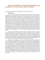
Fig. 4 The surface plasmon resonance (SPR) results of the interaction between LCK and rhein. Increased concentration of LCK protein showed a
trend of increased binding with rhein; the equilibrium dissociation constant (KD) was 1.060 × 10− 6 M
Sun et al. BMC Bioinformatics (2018) 19:315
been previously reported, in spite of not being included
in the STITCH database. Therefore, the remaining four
candidate targets (LCK, RAB5A, CDK2, and GSK3B)
were selected for further research using SPR. The positive and negative control signals were shown in supplementary materials (Additional file 1: Figure S1), which
indicated that the sensor chip quality was normal. In the
experimental results, for LCK, the binding tendency to
rhein increased with increasing the concentration of the
protein, whereas for RAB5A, CDK2, and GSK3B, the
tendency was not obvious. The binding curves of rhein
with LCK were shown in Fig. 4. The kinetic parameters
were fitted and obtained using the LCK signals bound
with rhein. The association rate constant (ka), dissociation rate constant (kd), and equilibrium dissociation
constant (KD) were 186 (M·s)− 1, 1.97 × 10− 4 s− 1, and
1.060 × 10− 6 M, respectively. Therefore, after the experimental verification of SPR, we had reason to believe that
LCK was a new target of rhein.
Discussion
At present, there had been many successful cases of
ligand-protein docking for target identification [39, 40].
The use of ligand-protein docking provided the conditions
for the rapid screening of potential targets, rather than the
aimless trial of luck. In this study, virtual screening based
on ligand-protein docking was divided into two steps. The
first step was inverse rhein molecular docking analysis. In
this step, 300 potential targets were selected from 2241
human protein targets. The second step was the accurate
rhein molecular docking analysis. In this step, the 300 targets were further reduced to 67 potential targets. These
two steps were designed to reduce the rate of false positives and obtain more accurate targets. Although the
ligand-protein docking was popular for drug target identification, challenges remained for this method due to its
limitations that included insufficiencies of the database resources, imperfections of the scoring functions, and inaccurate selection of binding sites and docking poses [41].
Due to these limitations, there may still be a few false positives among the 67 potential targets. In addition, the direct verification of 67 potential targets by experiments was
time-consuming and costly. Therefore, a further method
was needed to screen potential targets and reduce false
positive targets.
The network analysis was a new strategy to comprehensively screen drug targets [8]. In biological networks,
the targets of one bioactive compound always gathered
in a cluster. For instance, there were close interactions
between the targets of nearly any bioactive compound in
the STITCH database [42], which meant that the adjacent nodes of a known target were likely to be a target
as well. To clearly illustrate the principle of network
analysis, the diagrammatic sketch of the idea was
Page 7 of 11
constructed as shown in Fig. 5. In this diagrammatic
sketch, plane a represented the target PPI of one bioactive compound, targets of which were mapped to a
biological network (plane b). All the known targets of
this bioactive compound clustered together, and the target EPPI of this compound was the network with broken
circle in plane b. Then, the plane c was selected from
the EPPI according to the importance of nodes in the
EPPI network. Thus, the potential targets in plane c
were used for further screen. In this study, the PPI network of rhein targets was consisted of a big cluster with
40 nodes linked by 60 interactions along with 37 isolated
nodes. Further research should consider whether these
37 isolated nodes were connected to other known targets via neighbouring nodes such that one whole cluster
forms. Certainly, each node in the cluster had a high
probability of being a target. Therefore, the EPPI network was further constructed to filter targets. Topological characteristics offered significant insight into
biologically relevant connectivity patterns, and pinpoint
likely key targets in the network [43]. The node degree
represented the number of other nodes connected to a
node. A high degree node was generally considered to
be important because of its extensive connectivity [44,
45]. Similarly, the closeness centrality represented the
degree of closeness between a node and other nodes in
the network. The node with a large closeness centrality
was also a protein of great importance. The betweenness
centrality was another basic property of a network. The
node with a large betweenness centrality was always a
key transmit point for biological information flow; if this
node was lost or blocked in a network, it resulted in the
emergence of many modules [34, 46]. Here, betweenness
centrality was determined as a key parameter because it
had the largest AUC (0.710), which implied the best predictive rate. Then, the 21 nodes were screened according
to the highest cut-off (0.0016) of betweenness centrality.
These 21 nodes included 7 known targets and 14 potential targets. Examples used in this study demonstrated
that our network analysis method was very efficient, reducing 67 potential targets to 14 ones. However, our
network analysis needed two prerequisite conditions: 1.
There must be a certain number of known targets; 2.
There should be direct or indirect links between known
and potential targets. In other words, if the number of
known targets was insufficient enough or the known targets were not closely related to potential targets, the
false positives or false negatives might increase in the results. In addition, the network analysis could not reflect
the flow of biological information because the network
used in network analysis was usually undirected. Therefore, the enrichment analysis was another required
method in order to further reduce the false positives and
to consider the flow of bio-information closer to reality.
Sun et al. BMC Bioinformatics (2018) 19:315
Page 8 of 11
Fig. 5 Diagrammatic sketch of the idea for network analysis and enrichment analysis. In this diagrammatic sketch, plane a represents the target
protein–protein interaction (PPI) of one bioactive compound, targets of which were mapped to a biological network (plane b). In fact, the target
extended PPI (EPPI) of this bioactive compound is the network with broken circle in plane b. According to the importance of nodes in the
network, plane c was selected from the EPPI via network analysis. The plane d represents the enriched pathway of proteins in plane c. Thus, the
potential targets of this bioactive compound in plane d could be considered to be candidate targets
The pathway enrichment analysis was usually used to
assess the distribution of given proteins in the KEGG
pathway and determine their contribution to biological
processes. This method would calculate the hypergeometric distributions between given proteins and pathways
and return a P-value for each pathway in which the given
proteins existed. Based on the P-value, it was assessed
whether the given proteins were enriched in that pathway
[10]. Obviously, the enrichment analysis had significant
implications for establishing the relationships between
proteins and pathways. Here, the enrichment analysis was
innovatively used for the target identification since the
enriched proteins often played similar and important biological roles in the biological process, and were likely to be
the targets of the bioactive molecule. For example, the
activation of JAK2 and STAT3 induced the expression of
TNF-α and IL-6 in acute renal injury, while curcumin protected against the acute renal injury by distinctly inhibiting
the activation of JAK2 and STAT3 in the JAK2/STAT3
pathway [47]. As shown in Fig. 5, the proteins with the
flow of biological information in plane d were enriched
from plane c, and thus the range of potential targets
Sun et al. BMC Bioinformatics (2018) 19:315
would be more accurate after the enrichment analysis. In
this study, 21 proteins from the network analysis screening were subjected to the enrichment analysis. The results
showed that 15 proteins were enriched and 9 of the 15
proteins were potential targets and determined to be candidate targets. Interestingly, 5 of the 9 candidate targets
had been previously reported, in spite of not being included in the STITCH database. This situation further
verified the accuracy and reliability of the integration strategy used in this study. Moreover, 11 KEGG pathways that
were significantly enriched interacted closely through the
15 enriched proteins, as shown in Fig. 6. All 11 KEGG
pathways were associated with inflammation, proliferation
and apoptosis, which were consistent with the pharmacological activities of rhein, again suggesting that each
enriched protein was likely to be a target.
LCK, a member of the Src family of protein tyrosine kinases [48], was a new rhein target identified by our strategy.
Our SPR experiment revealed that LCK could interact with
rhein, and the binding tendency was proportional to the
protein concentration. In biological systems, LCK played an
important role in the T-cell antigen receptor (TCR)-linked
signal transduction pathway as a non-receptor tyrosine
Page 9 of 11
kinase [49]. LCK constitutively associated with the cytoplasmic portions of the CD4 and CD8 surface receptors, and
then initiated the TCR-linked signaling pathway [50]. Upon
TCR stimulation, LCK phosphorylated the TCR, thus leading to the recruitment, phosphorylation, and activation of
ZAP70 [51]. Activated ZAP70 then directly or indirectly
regulated the MAPK and the NFKB signalling pathways,
subsequently affecting cell proliferation and inflammatory
processes [52, 53]. As a new target of rhein, LCK might play
an important role in the treatment of cancer or inflammation. Of course, the therapeutic effect of rhein was not only
due to regulating the LCK target, but also was the result of
synergistic and comprehensive regulation of multiple targets in different pathways [13]. Rhein could inhibit the
phosphorylation of EGFR, p38 and JNK in the classical
MAPK cascade [17, 35–37], repress the activity of RELA
and NFKB1 in the NF-κB signalling pathway [17, 54–56],
promote apoptosis through the activation of CASP3 and
CASP8 in the apoptotic pathway [57], induce G0/G1 arrest
through CDK6 inhibition in the cell cycle [38], decrease the
expression of VEGFA and the activity of HSP90AA1 and
RXRA in other pathways [14, 15, 58]. Apparently, the
rhein-mediated biological network was vast and complex.
Fig. 6 The integrated network of enrichment pathways of rhein targets. This pathway was constructed via manually extracting the biological
process which is related to enriched targets of rhein from the KEGG pathway. The main body of a biological process was extracted if a rhein
target was in this biological process. The protein marked by star is the rhein target. Purple and green stars represent known and candidate
targets, respectively
Sun et al. BMC Bioinformatics (2018) 19:315
The therapeutic effect of rhein was the synergistic and
comprehensive result of this vast and complex network
[13], and the perturbation of multiple targets gave rhein a
variable and effective pharmacological activity.
Conclusion
In this study, ligand-protein docking, network analysis,
and enrichment analysis were integrated to identify new
targets of rhein, followed by the validation of these targets using SPR experiments. Although any one of these
methods had been applied to the target identification before, the rational combination of them for the target
identification was novel. The integrated network of
enriched pathways was used to elucidate the comprehensive pharmacological mechanisms of rhein. This
study provided a new strategy to effectively identify candidate targets and infer the molecular mechanisms of
bioactive compounds.
Additional file
Additional file 1: Table S1. Inverse Docking Result. Table S2. Potential
Targets of Rhein after Accurate Molecular Docking. Table S3. Sorting
results of topological parameters. Table S4. 15 Enriched Proteins in 11
KEGG Pathways. Figure S1. The positive and negative control signal for
SPR. (PDF 637 kb)
Abbreviations
AUC: Area under the curve; EPPI: Extended PPI; FPR: False positive rate;
PPI: Protein–protein interaction; ROC: Receiver operating characteristic;
SPR: Surface plasmon resonance; TPR: True positive rate
Acknowledgements
The authors acknowledge financial support from Wenzhou Science and
Technology Major Project, China (ZS2017018), the National Natural Science
Foundation of China (No. 81773691 and 81703815), and granted by the
Opening Project of Zhejiang Provincial Top Key Discipline of Pharmaceutical
Sciences.
Funding
This work was supported by Wenzhou Science and Technology Major
Project, China (ZS2017018), the National Natural Science Foundation of China
(No. 81773691 and 81703815), and granted by the Opening Project of
Zhejiang Provincial Top Key Discipline of Pharmaceutical Sciences.
Availability of data and materials
The 3D molecular structure of rhein is downloaded from the ZINC database
( The 3D molecular structures of potential targets
are downloaded from the Protein Data Bank ( The
known targets of rhein are collected from the STITCH database (http://
stitch.embl.de/). The other datasets used and analysed during the current
study are available from the supplementary materials (Additional file 1).
Authors’ contributions
ZX and YC engaged in study design and coordination, material support for
supervised study. ZX and HS designed the experimental validation and
drafted the manuscript. GL and YS performed SPR experiment. All authors
read and approved the final manuscript.
Ethics approval and consent to participate
Not applicable
Consent for publication
Not applicable
Page 10 of 11
Competing interests
The authors declare that they have no competing interests.
Publisher’s Note
Springer Nature remains neutral with regard to jurisdictional claims in
published maps and institutional affiliations.
Author details
1
School of Pharmaceutical Sciences, Wenzhou Medical University, Wenzhou
325035, China. 2Pharmacy Department, Women’s Hospital, Zhejiang
University School of Medicine, Hangzhou 310006, Zhejiang, China.
Received: 24 April 2018 Accepted: 29 August 2018
References
1. Chen S, Jiang H, Cao Y, Wang Y, Hu Z, Zhu Z, Chai Y. Drug target
identification using network analysis: taking active components in Sini
decoction as an example. Sci Rep. 2016;6:24245.
2. Zhou W, Wang Y, Lu A, Zhang G. Systems pharmacology in small molecular
drug discovery. Int J Mol Sci. 2016;17(2):246.
3. Xie L, Xie L, Kinnings SL, Bourne PE. Novel computational approaches to
polypharmacology as a means to define responses to individual drugs.
Annu Rev Pharmacol Toxicol. 2012;52:361–79.
4. Cerqueira NM, Gesto D, Oliveira EF, Santos-Martins D, Bras NF, Sousa SF,
Fernandes PA, Ramos MJ. Receptor-based virtual screening protocol for
drug discovery. Arch Biochem Biophys. 2015;582:56–67.
5. Yan X, Liao C, Liu Z, Hagler AT, Gu Q, Xu J. Chemical structure similarity
search for ligand-based virtual screening: methods and computational
resources. Curr Drug Targets. 2016;17(14):1580–5.
6. Fang Y. Combining label-free cell phenotypic profiling with
computational approaches for novel drug discovery. Expert Opin Drug
Discov. 2015;10(4):331–43.
7. Huang da W, Sherman BT, Lempicki RA. Systematic and integrative analysis
of large gene lists using DAVID bioinformatics resources. Nat Protoc. 2009;
4(1):44–57.
8. Westerhoff HV. Network-based pharmacology through systems biology.
Drug Discov Today Technol. 2015;15:15–6.
9. Engin HB, Gursoy A, Nussinov R, Keskin O. Network-based strategies can
help mono- and poly-pharmacology drug discovery: a systems biology
view. Curr Pharm Des. 2014;20(8):1201–7.
10. Huang da W, Sherman BT, Lempicki RA. Bioinformatics enrichment tools:
paths toward the comprehensive functional analysis of large gene lists.
Nucleic Acids Res. 2009;37(1):1–13.
11. Ge JH, Liu XH, Xu H, Xu DY, Bai FP. Identification of different varieties of
Rhei Radix et Rhizoma based on chemical analysis. Zhongguo Zhong Yao
Za Zhi. 2015;40(12):2309–13.
12. Zhou YX, Xia W, Yue W, Peng C, Rahman K, Zhang H. Rhein: a review of
pharmacological activities. Evid Based Complement Alternat Med. 2015;
2015:578107.
13. Sun H, Luo G, Chen D, Xiang Z. A comprehensive and system review
for the pharmacological mechanism of action of Rhein, an active
Anthraquinone ingredient. Front Pharmacol. 2016;7:247.
14. Zhang H, Chen L, Chen J, Jiang H, Shen X. Structural basis for retinoic X
receptor repression on the tetramer. J Biol Chem. 2011;286(28):24593–8.
15. Fernand VE, Losso JN, Truax RE, Villar EE, Bwambok DK, Fakayode SO,
Lowry M, Warner IM. Rhein inhibits angiogenesis and the viability of
hormone-dependent and -independent cancer cells under normoxic or
hypoxic conditions in vitro. Chem Biol Interact. 2011;192(3):220–32.
16. Heo SK, Yun HJ, Park WH, Park SD. Rhein inhibits TNF-alpha-induced human
aortic smooth muscle cell proliferation via mitochondrial-dependent
apoptosis. J Vasc Res. 2009;46(4):375–86.
17. Legendre F, Bogdanowicz P, Martin G, Domagala F, Leclercq S, Pujol JP,
Ficheux H. Rhein, a diacerhein-derived metabolite, modulates the
expression of matrix degrading enzymes and the cell proliferation of
articular chondrocytes by inhibiting ERK and JNK-AP-1 dependent
pathways. Clin Exp Rheumatol. 2007;25(4):546–55.
18. Zhang K, Jiao XF, Li JX, Wang XW. Rhein inhibits lipopolysaccharide-induced
intestinal injury during sepsis by blocking the toll-like receptor 4 nuclear
factor-kappaB pathway. Mol Med Rep. 2015;12(3):4415–21.
Sun et al. BMC Bioinformatics (2018) 19:315
19. Li Y, Xu Y, Lei B, Wang W, Ge X, Li J. Rhein induces apoptosis of
human gastric cancer SGC-7901 cells via an intrinsic mitochondrial
pathway. Braz J Med Biol Res. 2012;45(11):1052–9.
20. Irwin JJ, Sterling T, Mysinger MM, Bolstad ES, Coleman RG. ZINC: a free tool
to discover chemistry for biology. J Chem Inf Model. 2012;52(7):1757–68.
21. Wang X, Shen Y, Wang S, Li S, Zhang W, Liu X, Lai L, Pei J, Li H.
PharmMapper 2017 update: a web server for potential drug target
identification with a comprehensive target pharmacophore database.
Nucleic Acids Res. 2017;45(W1):W356–60.
22. Berman HM, Westbrook J, Feng Z, Gilliland G, Bhat TN, Weissig H,
Shindyalov IN, Bourne PE. The Protein Data Bank. Nucleic Acids Res.
2000;28(1):235–42.
23. Lill MA, Danielson ML. Computer-aided drug design platform using
PyMOL. J Comput Aid Mol Des. 2010;25(1):13–9.
24. Trott O, Olson AJ. AutoDock Vina: improving the speed and accuracy of
docking with a new scoring function, efficient optimization, and
multithreading. J Comput Chem. 2010;31(2):455–61.
25. Szklarczyk D, Santos A, von Mering C, Jensen LJ, Bork P, Kuhn M.
STITCH 5: augmenting protein-chemical interaction networks with tissue
and affinity data. Nucleic Acids Res. 2016;44(D1):D380–4.
26. Martin A, Ochagavia ME, Rabasa LC, Miranda J, Fernandez-de-Cossio J,
Bringas R. BisoGenet: a new tool for gene network building,
visualization and analysis. BMC Bioinform. 2010;11:91.
27. Smoot ME, Ono K, Ruscheinski J, Wang PL, Ideker T. Cytoscape 2.8: new
features for data integration and network visualization. Bioinformatics. 2011;
27(3):431–2.
28. Assenov Y, Ramirez F, Schelhorn SE, Lengauer T, Albrecht M. Computing
topological parameters of biological networks. Bioinformatics. 2008;24(2):282–4.
29. Kanehisa M, Furumichi M, Tanabe M, Sato Y, Morishima K. KEGG: new
perspectives on genomes, pathways, diseases and drugs. Nucleic Acids Res.
2017;45(D1):D353–61.
30. Puiu M, Bala C. SPR and SPR imaging: recent trends in developing
Nanodevices for detection and real-time monitoring of biomolecular events.
Sensors. 2016;16(6)
31. D'Orazio P. Biosensors in clinical chemistry - 2011 update. Clin Chim Acta.
2011;412(19–20):1749–61.
32. Olaru A, Bala C, Jaffrezic-Renault N, Aboul-Enein HY. Surface plasmon
resonance (SPR) biosensors in pharmaceutical analysis. Crit Rev Anal Chem.
2015;45(2):97–105.
33. Li SP, Yang M, Zhou WF, Johnston TG, Wang R, Zhu JS. Dextran hydrogel
coated surface plasmon resonance imaging (SPRi) sensor for sensitive and
label-free detection of small molecule drugs. Appl Surf Sci. 2015;355:570–6.
34. Joy MP, Brock A, Ingber DE, Huang S. High-betweenness proteins in
the yeast protein interaction network. J Biomed Biotechnol. 2005;
2005(2):96–103.
35. Lin YJ, Zhen YZ, Shang BY, Zhen YS. Rhein lysinate suppresses the growth
of tumor cells and increases the anti-tumor activity of Taxol in mice. Am J
Chin Med. 2009;37(5):923–31.
36. Lin YJ, Zhen YS. Rhein lysinate suppresses the growth of breast cancer cells
and potentiates the inhibitory effect of Taxol in athymic mice. Anti-Cancer
Drugs. 2009;20(1):65–72.
37. Heo SK, Yun HJ, Noh EK, Park SD. Emodin and rhein inhibit LIGHTinduced monocytes migration by blocking of ROS production. Vasc
Pharmacol. 2010;53(1–2):28–37.
38. Hsia TC, Yang JS, Chen GW, Chiu TH, Lu HF, Yang MD, Yu FS, Liu KC,
La KC, Lin CC, et al. The roles of endoplasmic reticulum stress and ca(2
+) on Rhein-induced apoptosis in A-549 human lung Cancer cells.
Anticancer Res. 2009;29(1):309–18.
39. Luo H, Mattes W, Mendrick DL, Hong H. Molecular docking for
identification of potential targets for drug repurposing. Curr Top Med
Chem. 2016;16(30):3636–45.
40. Xu X, Huang M, Zou X. Docking-based inverse virtual screening:
methods, applications, and challenges. Biophys Rep. 2018;4(1):1–16.
41. Chen YC. Beware of docking! Trends Pharmacol Sci. 2015;36(2):78–95.
42. Kuhn M, von Mering C, Campillos M, Jensen LJ, Bork P. STITCH:
interaction networks of chemicals and proteins. Nucleic Acids Res. 2008;
36(Database issue):D684–8.
43. Shi SH, Cai YP, Cai XJ, Zheng XY, Cao DS, Ye FQ, Xiang Z. A network
pharmacology approach to understanding the mechanisms of action of
traditional medicine: Bushenhuoxue formula for treatment of chronic
kidney disease. PLoS One. 2014;9(3):e89123.
Page 11 of 11
44. Barabasi AL, Oltvai ZN. Network biology: understanding the cell's functional
organization. Nat Rev Genet. 2004;5(2):101–13.
45. Azuaje FJ, Zhang L, Devaux Y, Wagner DR. Drug-target network in
myocardial infarction reveals multiple side effects of unrelated drugs. Sci
Rep. 2011;1:52.
46. Hwang W, Cho YR, Zhang A, Ramanathan M. A novel functional module
detection algorithm for protein-protein interaction networks. Algorithms
Mol Biol. 2006;1:24.
47. Zhu S, Zhang C, Weng Q, Ye B. Curcumin protects against acute renal injury
by suppressing JAK2/STAT3 pathway in severe acute pancreatitis in rats. Exp
Ther Med. 2017;14(2):1669–74.
48. Chiang YJ, Hodes RJ. T-cell development is regulated by the coordinated
function of proximal and distal Lck promoters active at different
developmental stages. Eur J Immunol. 2016;46(10):2401–8.
49. Gelkop S, Gish GD, Babichev Y, Pawson T, Isakov N. T cell activation-induced
CrkII binding to the Zap70 protein tyrosine kinase is mediated by Lckdependent phosphorylation of Zap70 tyrosine 315. J Immunol. 2005;175(12):
8123–32.
50. Parrish HL, Deshpande NR, Vasic J, Kuhns MS. Functional evidence for TCRintrinsic specificity for MHCII. Proc Natl Acad Sci U S A. 2016;113(11):3000–5.
51. Lo WL, Shah NH, Ahsan N, Horkova V, Stepanek O, Salomon AR, Kuriyan J,
Weiss A. Lck promotes Zap70-dependent LAT phosphorylation by bridging
Zap70 to LAT. Nat Immunol. 2018;
52. Round JL, Humphries LA, Tomassian T, Mittelstadt P, Zhang M, Miceli MC.
Scaffold protein Dlgh1 coordinates alternative p38 kinase activation,
directing T cell receptor signals toward NFAT but not NF-kappaB
transcription factors. Nat Immunol. 2007;8(2):154–61.
53. Jiang Y, Cheng H. Evidence of LAT as a dual substrate for Lck and Syk in T
lymphocytes. Leuk Res. 2007;31(4):541–5.
54. Oeckinghaus A, Hayden MS, Ghosh S. Crosstalk in NF-kappaB signaling
pathways. Nat Immunol. 2011;12(8):695–708.
55. Martin G, Bogdanowicz P, Domagala F, Ficheux H, Pujol JP. Rhein inhibits
interleukin-1 beta-induced activation of MEK/ERK pathway and DNA
binding of NF-kappa B and AP-1 in chondrocytes cultured in hypoxia: a
potential mechanism for its disease-modifying effect in osteoarthritis.
Inflammation. 2003;27(4):233–46.
56. Martin G, Bogdanowicz P, Domagala F, Ficheux H, Pujol JP. Articular
chondrocytes cultured in hypoxia: their response to interleukin-1 beta and
rhein, the active metabolite of diacerhein. Biorheology. 2004;41(3–4):549–61.
57. Lin ML, Chen SS, Lu YC, Liang RY, Ho YT, Yang CY, Chung JG. Rhein induces
apoptosis through induction of endoplasmic reticulum stress and Ca2+
−dependent mitochondrial death pathway in human nasopharyngeal
carcinoma cells. Anticancer Res. 2007;27(5a):3313–22.
58. Ersahin T, Tuncbag N, Cetin-Atalay R. The PI3K/AKT/mTOR interactive
pathway. Mol BioSyst. 2015;11(7):1946–54.