The present and future role of photodynamic
Bạn đang xem bản rút gọn của tài liệu. Xem và tải ngay bản đầy đủ của tài liệu tại đây (1.21 MB, 12 trang )
Review
Photodynamic therapy
The present and future role of photodynamic
therapy in cancer treatment
Stanley B Brown, Elizabeth A Brown, and Ian Walker
It is more than 25 years since photodynamic therapy (PDT)
was proposed as a useful tool in oncology, but the
approach is only now being used more widely in the clinic.
The understanding of the biology of PDT has advanced,
and efficient, convenient, and inexpensive systems of light
delivery are now available. Results from well-controlled,
randomised phase III trials are also becoming available,
especially for treatment of non-melanoma skin cancer and
Barrett’s oesophagus, and improved photosensitising
drugs are in development. PDT has several potential
advantages over surgery and radiotherapy: it is
comparatively non-invasive, it can be targeted accurately,
repeated doses can be given without the total-dose
limitations associated with radiotherapy, and the healing
process results in little or no scarring. PDT can usually be
done in an outpatient or day-case setting, is convenient
for the patient, and has no side-effects. Two
photosensitising drugs, porfirmer sodium and temoporfin,
have now been approved for systemic administration, and
aminolevulinic acid and methyl aminolevulinate have been
approved for topical use. Here, we review current use of
PDT in oncology and look at its future potential as more
selective photosensitising drugs become available.
Lancet Oncol 2004 5: 497–508
© David Parker/Science Photo Library
Figure 1. Red light from a non-coherent lamp activates a topically
applied drug, killing cancer cells.
Photodynamic therapy (PDT) uses the combination of a
photosensitising drug and light (figure 1) to cause selective
damage to the target tissue. An adequate concentration of
molecular oxygen is also needed for tissue damage. If any
one of these components is absent, there is no effect, and the
overall effectiveness therefore requires careful planning of
both drug and light dosimetry. The drugs are generally given
systemically, but because the targeting process is mainly
achieved through precise application of the light—usually
from a laser source—the effect is local rather than systemic.
The local nature of the effect of PDT should be recognised
from the outset because it contributes to both the
limitations and the opportunities for PDT as a successful
treatment in cancer.
A limitation of PDT is that it cannot cure advanced
disseminated disease because irradiation of the whole body
with appropriate doses is not possible (at least with current
technologies). Nevertheless, for advanced disease, PDT can
improve quality of life and lengthen survival. For early or
localised disease, PDT can be a selective and curative therapy
with many potential advantages over available alternatives.
A single treatment can eradicate disease and can have an
Oncology Vol 5 August 2004
Rights were not
granted to
include this
image in
electronic media.
Please refer to
the printed
journal.
excellent cosmetic result (figure 2). Although the clinical
potential of PDT has been recognised for more than
25 years,1 it is only now starting to be used in the clinic.
PDT harnesses the energy of light to damage or destroy
target tissue (see panel). A sensitiser absorbs energy directly
from a light source, which it then transfers to molecular
oxygen to create an activated form of oxygen called singlet
oxygen. It is this singlet oxygen that is the true cytotoxic agent
and that reacts rapidly with cellular components2 to cause the
damage that ultimately leads to cell death and tumour
destruction. During this process, the sensitiser is regenerated
so that it acts catalytically, and many cycles of singlet-oxygen
production can occur for each molecule of sensitiser.
SBB is Yorkshire Cancer Research Professor of Biochemistry, EAB
is Clinical Database Co-ordinator, and IW is a research fellow; all at
the Centre for Photobiology and Photodynamic Therapy, School of
Biochemistry and Microbiology, University of Leeds, UK.
Correspondence: Prof Stanley Brown, Centre for Photobiology and
Photodynamic Therapy, School of Biochemistry and Microbiology,
University of Leeds, Leeds, LS2 9JT, UK. Tel: +44 (0)113 233 3166.
Fax: +44 (0)113 233 3017. Email:
497
Review
Photodynamic therapy
result after 2–3 months is usually
excellent—ie, the effect of healing is
itself a type of selectivity.
In the past 10 years, substantial
advances have been made in the
understanding of the behaviour of
light in human tissues7,8 and in the
development of equipment for light
delivery for PDT. Light of adequate
dose can now be delivered precisely to
most tumour sites (both internal and
external), and PDT is now rarely
rejected because of difficulties in
delivery of light. Generally, a laser
source is needed for internal treatment
by use of endoscopy or for interstitial
treamtent because lasers are the most
efficient way of channelling light into
one or more optical fibre. For
cutaneous or subcutaneous lesions, a
non-laser source is usually effective.
The power of the source is important
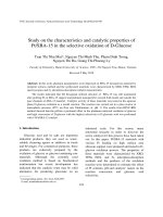
Figure 2. Patient with Bowen’s disease before treatment with aminolevulinic acid PDT (A), and
2 months after treatment (B). The single treatment was shown by histological analysis to have
because it will determine treatment
irradicated the lesion, which did not recur. The excellent healing after treatment is apparent.
times. However, achievement of
sufficient power is rarely difficult with
PDT uses several different mechanisms to destroy modern laser or non-laser sources, which have typical
tumours. A photosensitiser can target tumour cells directly, treatment times of 5–20 min.
inducing necrosis or apoptosis.3 Alternatively, by the
One of the main advances has been the availability of
targeting of tumour vasculature (or indeed of healthy diode lasers, which are small, portable, very reliable, and
surrounding vasculature), the tumour can be starved of inexpensive (about £20 000 or less) compared with earlier
oxygen-carrying blood. Thus, together with inflammatory lasers for PDT. Diode lasers are ideal for routine use as
and immune responses, damage to the tumour can be clinical tools and need little technical expertise for use.
maximised by use of PDT.4
However, because their wavelength is fixed and must be
specified for use with a particular photosensitiser, diode
Practical considerations
lasers are less useful for research in which different
At a predetermined time after administration of the drug photosensitising drugs are being assessed. Many non-laser
(called the drug-to-light interval), light is directed into the light sources have also been developed, especially for
tumour and surrounding healthy tissue. The tumour is treatment of skin lesions. These non-laser sources can be
destroyed rapidly, and any damage to healthy tissue heals either various types of filtered lamps or, more recently, lightover the following 6–8 weeks.
emitting diodes.9 In all cases, the light field produced needs
The targeting and selectivity of PDT is aided by several to be uniform so that the dose delivered can be calculated
factors, the first of which is the delivery of light. By use of precisely. As with most fixed-cost medical equipment, the
modern fibre-optic systems and various types of endoscopy, real cost of lasers for PDT depends on the intensity of their
light can be targeted accurately to almost any part of the use. A PDT laser that treats only one patient a week is
body. Singlet oxygen generated by the activated photosen- expensive, whereas the same laser that treats 20–30 patients a
sitiser has a very short life, and is deactivated before it can week gives a low cost per treatment.
escape from the cell in which it was produced, further
Photosensitisers
assisting targeting.
Some photosensitising drugs can reach higher Systemic sensitisers
concentrations in tumour tissue than in surrounding healthy Early preparations of photosensitisers for PDT were based
tissue. Although the exact mechanisms that drive this on a complex mixture of porphyrins called haematoprocess are not understood fully, the abnormal physiology of porphyrin derivative.10,11 Porfimer sodium was the first drug
tumours (eg, poor lymphatic drainage, leaky vasculature,
Mechanisms by which photodynamic therapy harnesses
decreased pH,5 increased number of receptors for lowthe energy of light to damage or destroy target tissue
density lipoprotein, and abnormal stromal composition)
might contribute to the selectivity of photosensitisers.6
Sensitiser + light
→ Activated sensitiser
Furthermore, the healing of healthy tissue after PDT is very
Activated sensitiser + oxygen → Sensitiser + activated (singlet) oxygen
efficient, usually without scarring (figure 2). Even if healthy
Activated oxygen + target
→ Oxidised (damaged) target
tissue is damaged at the time of treatment, the cosmetic
498
Oncology Vol 5 August 2004
Review
Photodynamic therapy
to receive approval for PDT and is based on
haematoporphyrin derivative, with some of the non-active
components removed. Although porfimer sodium is a
complex mixture, it is now used widely and remains
the most common photosensitiser for treatment of nondermatological tumours. The drug has been approved
for use in advanced and early-stage lung cancers, superficial
gastric cancer, oesophageal adenocarcinoma, cervical
cancer, and bladder cancer (and has been used on a
trial basis for many other indications). The advantages
of porfimer sodium are that it: destroys tumours
effectively, is non-toxic in the absence of light, and can
be easily formulated in a water-soluble preparation for
intravenous administration. As the first drug product
to be approved, porfimer sodium has also highlighted
the fundamental safety and advantages of PDT as a
treatment option for cancer: the drug has been used
in thousands of patients for more than 20 years. No
long-term safety issues have emerged, and it seems that
PDT can be used repeatedly without limit (ie, there are
no lifetime dose limitations, as there can be with
radiotherapy).
Despite the continuing effectiveness of porfimer
sodium, it has several disadvantages that could potentially
be overcome in subsequent candidate compounds. The drug
induces protracted skin photosensitivity,12 and the intial
selectivity between tumour tissue and healthy tissue can be
low.13 Although reasonable selectivity is seen after
2–3 months, this selectivity might be mainly the result of
selective healing of healthy tissue, rather than selective initial
damage by porfimer sodium. Furthermore, the time
between administration of porfimer sodium and light is
typically 48–72 h, during which the patient must be
protected from light.
Much chemical and biological research has been done
over the past 20 years to identify new photosensitisers with
improved properties over porfimer sodium. However, most
of this work has been aimed at development of
photosensitising drugs that are pure chemically and that
absorb more strongly at longer wavelengths, rather than
placing a high priority on development of improved
biological properties. Table 1 shows the typical wavelength
of maximum absorption and the molar-absorption
coefficients for various photosensitising drugs.
With the exception of porfimer sodium, the only other
PDT drug currently approved for systemic use in cancer
treatment is temoporfin (table 1). A mixture of aluminiumsulphonated phthalocyanine has been used widely in
Russia, but not in any other country. Temoporfin is
effective for the palliative treatment of head and neck
cancer and was approved in Europe for this indication in
2001. It is a very active photosensitiser and thus requires a
much lower dose of both the drug and light than does
porfimer sodium.10 Furthermore, temoporfin is a pure
compound with a very strong absorption at 652 nm.
However, like porfimer sodium, the drug is also associated
with a pronounced and lengthy generalised skin
photosensitivity and can show little initial selectivity, with
the selective benefits arising later from selective healing of
healthy tissue. Temoporfin also needs to be administered
up to 96 h before light is applied.
Verteporfin (benzoporphyrin derivative) has been
developed for the treatment of macular degeneration
(table 1).14,15 Although not indicated for cancer, this drug is
one of the most useful ophthalmology drugs ever developed
and thus might have lessons for the development of PDT
drugs for cancer. Verteporfin is cleared rapidly and does not
induce a generalised skin photosensitivity that lasts longer
than 24 h. Moreover, treatment with this drug is convenient
for patient and clinician: 10 min intravenous infusion,
followed 15 min later by 83 s of laser light (690 nm) at
600 mW/cm2.
Sensitisers for topical application
None of the systemically administered sensitisers shown in
table 1 have been developed for topical application to treat
skin lesions, despite many attempts. Furthermore,
achievement of effective PDT through the injection of
photosensitisers directly into the lesion has been
unsuccessful. In both cases, delivery of photosensitisers into
sensitive subcellular sites, through binding to serum
proteins, seems necessary for effective PDT.
All nucleated cells in the body contain the biochemical
apparatus needed to make haem for cytochromes and
other haemoproteins (figure 3). The immediate precursor
of haem (which is not a photosensitiser), is
protoporphyrin IX, which is a powerful photosensitiser.
The concentration of porphyrin that will support PDT can
Table 1. Types of photosensitising drugs
Class
Approved drugs for photodynamic therapy
Porphyrins
Porfimer sodium
Protoporphyrin IX (eg, from methyl aminolevulinate
and aminolevulinic acid)
Chlorins
Verteporfin (benzoporphyrin derivative)
Temoporfin (meta-tetrahydroxyphenylchlorin)
Bacteriochlorins
None yet approved
Phthalocyanines
Sulphonated aluminium phthalocyanine mixture
(approved in Russia)
Phenothiazinium compounds None yet approved
Texafrins
None yet approved
Typical maximum
absorption (nm)
630
Typical absorption
coefficient*
10 000
633
690
652
740
10 000
35 000
30 000
32 000
680
670
734
110 000
60 000
42 000
*Absorption of light cm–1 mol–1 L–1.
Oncology Vol 5 August 2004
499
Review
Photodynamic therapy
Treatment of skin cancer
O
H2 N
OH
NH
O
N
Aminolevulinic acid
N
HN
Light
HO
Porphobilinogen
O
HO
O
Protoporphyrin IX
Intermediate
products
Haem
Figure 3. A simplified scheme of the haem biosynthetic pathway. After
the accumulation of porphyrin, light of an appropriate wavelength
(633 nm) can be administered to obtain a therapeutic response.
be achieved by topical application of either aminolevulinic
acid or methyl aminolevulinate to the site of a skin cancer
or precancerous lesion. This finding has led to approval of
aminolevulinic acid in the USA, and of methyl aminolevulinate in Europe.
PDT in clinical practice
Thousands of patients have been given PDT over the past
20 years but most trials have involved only a few patients,
commonly have provided anecdotal data, and have not been
sufficiently convincing to persuade medical practitioners
and health-service providers of the benefits of PDT as
standard treatment. This situation has partly been caused by
difficulties in establishing the optimum treatment
conditions for an approach that requires the setting of
several variables (ie, drug and light dose, and drug-to-light
interval), as well as difficulties in skin photosensitivity and
low selectivity. However, greatly improved understanding of
the tissue and cellular factors that control PDT4,16 and
increased experience in the clinic has led to much larger,
better-controlled clinical trials and the approval of four
PDT drugs for cancer (table 2).
Hopper1 presented a comprehensive account of clinical
trials on PDT up to 2000; several of which have contributed
to the approval of the drugs outlined in table 2. Table 3
shows the scope of these trials. Here, we discuss subsequent
and continuing clinical trials, and assesses the future of PDT
in clinical practice.
The very high incidence of skin cancer and the striking
rates of increase in white populations (up to 5% per year)17
place an increasing burden on both patients and health
services. PDT already plays a substantial part in treatment
of non-melanoma skin cancer and will expand with new
trials and with approval of aminolevulinic acid for
treatment of actinic keratinosis in the USA, and of methyl
aminolevulinate for actinic keratinosis and basal-cell
carcinoma in Europe. Use of PDT for melanoma has not
yet been pursued substantially in any study partly because
of the difficulty in achieving good penetration of light
through pigmented lesions, and partly because of ethical
considerations about the aggressive nature of the disease.
Non-melanoma skin cancer is very common and
includes both superficial and nodular basal-cell carcinoma,
superficial squamous-cell carcinoma, squamous-cell
carcinoma, and Bowen’s disease (squamous-cell carcinoma
in situ). Actinic (solar) keratoses are potentially
precancerous lesions that can progress to squamous-cell
carcinoma. Non-melanoma skin cancer is not usually lifethreatening because it rarely metastasises and is treated
readily. However, the treatment options have been
associated with morbidity effects (eg, scarring), and the
drugs can be expensive (especially in view of the demands
on the time of dermatologists and plastic surgeons). PDT
has the potential to substantially decrease morbidity effects
and improve health economics.18
Intravenous administration of porfimer sodium19 or
temoporfin20,21 is effective in treatment of cutaneous
lesions. However, systemic administration of these drugs is
unlikely to be justified for large-scale treatment of local
disease (with the corresponding long periods of
photosensitivity).
By contrast, use of PDT with topical applications of
either aminolevulinic acid or methyl aminolevulinate is
simple and convenient, without substantial systemic toxic
effects. A cream or solution that contains either drug is
applied to the lesion and secured under a dressing.
Aminolevulinic acid is licensed in the USA for application
as a solution for 14–18 h, but in Europe (where the drug is
unlicensed but widely used) it is usually applied as a cream
for 4–6 h. Methyl aminolevulinate is applied as a cream for
3–4 h, during which photosensitivity is generated. The
licence given by the US Food and Drug Administration for
use of aminolevulinic acid requires use of blue light.
However, red light is generally used in Europe to improve
Table 2. Approved photodynamic-therapy drugs for oncological indications
Chemical name
Haematoporphyrin derivative,
polyhaematoporphyrin
Generic name
Porfimer sodium
Date and country of approval
First approved in 1995; now approved
in more than 40 countries
Methyl-tetrahydroxyphenyl
chlorin
5-aminolevulinic acid
Methyl 5-aminolevulinate
Temoporfin
Approved in 2001 in European Union,
Norway, and Iceland
Approved in 1999 in USA
Approved in 2001 in Europe
500
Aminolevulinic acid
Methyl aminolevulinate
Indications
Advanced and early lung cancer,
superficial gastric cancer,
oesophageal adenocarcinoma,
cervical cancer, and bladder cancer
Palliative head and neck cancer
Actinic keratosis
Actinic keratosis, superficial basal-cell
carcinoma, and basal-cell carcinoma
Oncology Vol 5 August 2004
Review
Photodynamic therapy
Table 3. Summary of photodynamic therapy clinical trials up to 20001
Tumour type
Premalignant tumours (eg, Barrett’s oesophagus,
oral cavity, bladder)
Cutaneous malignant tumours (eg, non-melanoma
skin cancer, chest-wall recurrence of breast cancer)
Tumours of the head, neck, and oral cavity
Lung, gastrointestinal, and other tumours
Tumours managed with intraoperative and
adjunctive treatments (eg, pituitary)
Interstitial application (eg, pancreatic)
Photosensitisers
Porfimer sodium, aminolevulinic acid, temoporfin,
haematoporphyrin derivative
Porfimer sodium, aminolevulinic acid, temoporfin
Trials
8
Patients (range)
5–100
9
16–151
Porfimer sodium, temoporfin
Porfimer sodium, temoporfin
Porfimer sodium, temoporfin
7
13
4
14–108
21–218
5–54
Porfimer sodium, temoporfin
2
9–26
penetration. Methyl aminolevulinate is always used with
red light. The site of the lesion is usually irradiated for
5–20 min. During the initial period of irradiation, the
patient might feel some discomfort or pain at the site. This
discomfort does not usually need intervention, but local
anaesthetic can be given if required.
Clinical use of aminolevulinic acid in non-melanoma
skin cancer has been reviewed in the guidelines produced by
the British Photodermatology Group in 2002.18 At present,
this unlicensed drug is available in Europe, but it is not
known how long this situation will be sustained.
The registration of aminolevulinic acid in the USA was
based on two randomised, placebo-controlled investigatorblinded phase III trials that had identical designs (table 4).22,23
Patients with multiple actinic keratoses of the face and scalp
were randomly assigned either 20% aminolevulinic acid in
hydroalcoholic topical solution or vehicle (hydroalcoholic
topical solution) only, followed by irradiation with blue light
(417 nm, 10 mW/cm2 to a total fluence of 10 J/cm2). In one
of the trials (n=241),22 72% of patients in the treatment
group had a complete response, compared with 20% of
those assigned placebo. The overall recurrence rate was 5·0%
for the treatment group and 27·9% for placebo. In the other
trial (n=243),23 a complete response in the treatment group
was seen in 128 of 166 patients (77%) at week 8 and in 133 of
149 patients (89%) at week 12. In the group assigned vehicle
only, ten of 55 patients (18%) responded at week 8, and
seven of 52 patients (13%) by week 12 (pр0·001 for both
groups). These data thus confirmed that PDT with
aminolevulinic acid is a safe and effective treatment for
actinic keratinosis.
The development and approval of methyl aminolevulinate has led to a licensed product for topical PDT for
superficial and nodular basal-cell carcinomas, as well as for
actinic keratinoses. Results from trials involving more than
2500 patients in 14 countries have shown that this drug is
safe and effective, with excellent cosmetic results.36 After
administration of methyl aminolevulinate, porphyrin
accumulates more in skin tumours than in healthy skin
(figure 4). PDT with methyl aminolevulinate has been
compared with cryotherapy for superficial basal-cell carcinoma, and with excision surgery for nodular basal-cell
carcinoma, and with and . In one study,37 60 patients were
randomly assigned to PDT with methyl aminolevulinate
and 58 patients to two freeze-thaw cycles of cryotherapy.
Complete-response rates at 3 months were similar for both
Oncology Vol 5 August 2004
groups (97% for PDT versus 95% for cryotherapy), but the
rate of recurrence at 12 months was less for the PDT group
(8%) than for the cryotherapy group (16%). However, the
cosmetic outcome was more favourable for the group
assigned methyl aminolevulinate.
PDT with methyl aminolevulinate has also been
compared with cryotherapy in two randomised controlled
studies involving about 400 patients with actinic
keratinoses.24,25 The results showed that one application of
methyl aminolevulinate was equally as effective as
cryotherapy, and that two applications were more effective
than cryotherapy. In all cases, cosmetic outcome and
satisfaction were more favourable in the groups assigned
methyl aminolevulinate than in those assigned cryotherapy.
Gorlin’s syndrome is a rare disease in which patients are
prone to develop several lesions of basal-cell carcinoma.
Although the number of patients is small, PDT with
aminolevulinic acid has been used to treat patients with this
disease,38 and leads to excellent healing and lack of scarring.
Thus, topical PDT by use of licensed drugs seems set to have
a major role in future routine treatment of non-melanoma
skin cancer.
Localised disease and precancerous lesions
With the exception of skin cancer, PDT has so far not been
used widely for early or localised cancer, or for premalignant
disease. This finding is surprising, since PDT is a local
technique and could potentially be curative. This situation
could change along with improvements in the availability of
screening techniques to enable early detection of disease, and
the probable development of improved PDT drugs that do
not have long-term skin photosensitivity or long drug-tolight intervals. Table 5 shows clinical trials involving more
than ten patients done since 2000 on PDT for treatment of
localised disease.
Barrett’s oesophagus
This disease, widely regarded as a precursor of
adenocarcinoma of the oesophagus, is increasing in incidence
and is one of the most promising targets for use of PDT in
early disease. Trials of PDT with systemic (oral)
aminolevulinic acid have shown encouraging results, with
regeneration of healthy epithelium.47–51 Most trials have been
small and non-randomised; however, in a prospective
double-blinded study by Ackroyd and co-workers39
36 patients with dysplastic Barrett’s oesophagus who were
501
Review
Photodynamic therapy
Table 4. Selected dermatological trials of photodynamic therapy, 2000–February, 2004
Photosensitisers and comparators
Actinic keratoses
Aminolevulinic acid
Aminolevulinic acid
Methyl aminolevulinate vs cryotherapy
Methyl aminolevulinate vs cryotherapy
Aminolevulinic acid vs fluorouracil
Methyl aminolevulinate vs placebo
photodynamic therapy
Aminolevulinic acid
Treatment
Trial type
n
Ref
Aminolevulinic acid (20%) in alcohol
solution followed by 10 J/cm2 blue light
Aminolevulinic acid (20%) in alcohol
solution followed by 10 J/cm2 blue light
Topical methyl aminolevulinate cream
(160 mg/g) for 3 h followed by 75 J/cm2 red
light. One session
Topical methyl aminolevulinate cream
(160 mg/kg) for 3 h followed by 75 J/cm2 red
light. Two treatments, 1 week apart
Aminolevulinic acid (20%) in alcohol
solution for 1 h followed by 10 J/cm2 blue
light, or dye laser
Topical methyl aminolevulinate cream
(160 mg/g) for 3 h followed by 75 J/cm2
non-coherent red light
Topical aminolevulinic acid (20%) in alcohol
solution for 14–18 h followed by 10 J/cm2
blue light
Randomised controlled phase III trial
241
22
Randomised controlled phase III trial
243
23
Multicentre randomised trial
193
24
Multicentre randomised trial
204
25
Randomised clinical trial
36
26
Multicentre double-blind randomised
study
80
27
Multicentre randomised controlled trial
36
28
Phase I/II trial
88
29
Photodynamic therapy (20% water-in-oil,
cream, 6 h application) vs cryotherapy over
12 months (red-light laser)
Single-centre randomised clinical trial
88
30
Topical 160 mg/kg methyl aminolevulinate
for 3 h followed by 75 J/cm2 red light.
Two treatments, 1 week apart
Open uncontrolled prospective
multicentre trial
94
31
Non-controlled phase II trial
38
32
Multicentre randomised trial
40
33
Randomised clinical trial
16
34
Multicentre randomised trial
101
35
Actinic keratoses and basal-cell carcinoma
Aminolevulinic acid
Aminolevulinic acid (20% in cream base)
applied for 4–6 h followed by 105 J/cm2
non-coherent red light
Basal-cell carcinoma
Aminolevulinic acid vs cryotherapy
Methyl aminolevulinate
Basal-cell carcinoma and Bowen’s disease
Aminolevulinic acid
Topical aminolevulinic acid in cream base
(20%) applied for 8 h followed by 10–20 J/cm2
blue light
Bowen’s disease
Aminolevulinic acid vs fluorouracil
Aminolevulinic acid. Red vs green light
Nodular basal-cell carcinoma
Methyl aminolevulinate vs excision surgery
Topical aminolevulinic acid in cream base
(20%) for 4 h followed 100 J/cm2 630±15 nm
light
Topical aminolevulinic acid in cream base
(20%) for 4 h (630 ± 15 nm and 540 ± 15 nm)
Topical 160 mg/kg methyl aminolevulinate
for 3 h followed by 75 J/cm2 red light.
Two treatments, 1 week apart
receiving acid suppression with omeprazole were randomly
assigned either PDT with 30 mg/kg oral aminolevulinic acid
plus laser endoscopy, or placebo plus laser endoscopy. In the
group assigned aminolevulinic acid, 16 of 18 patients
responded, with a median decrease in the area of Barrett’s
mucosa of 30% (range 0–60%). In the group assigned
placebo, a 10% decrease in area was seen in only two of
18 patients. No dysplasia was seen in the treated area of any
patient in the PDT group, but persistent low-grade dysplasia
was seen in 12 patients (p<0·001) in the placebo group. These
findings showed that PDT with aminolevulinic acid could be
delivered safely and effectively for low-grade dysplastic
502
Barrett’s oesophagus. However, there is some concern that
after PDT with aminolevulinic acid, submucosal islands of
Barrett’s epithelium can remain, with the long-term
possibility that they might act as foci for future disease.52
Treatment of Barrett’s oesophagus with more powerful
systemic sensitisers than aminolevulinic acid is less likely to
result in formation of residual islands, as shown in a large,
phase III randomised trial with porfimer sodium.40 The study
included 208 patients with high-grade dysplastic Barrett’s
oesophagus who were randomly assigned PDT plus
omeprazole (n=138) or omeprazole only (n=70).40 Patients
were assessed every 3 months by a four-quadrant biopsy
Oncology Vol 5 August 2004
Review
Photodynamic therapy
sample taken at 2-cm intervals for
surveillance of disease progression.
A maximum of three courses of PDT
were given at least 90 days apart. At a
minimum follow-up of 24 months,
76·8% of patients in the PDT group
showed ablation of all areas of highgrade dysplasia, compared with 38·6%
of patients in the control group
(p<0·001). At a mean follow-up of
24·2 months, 13·0% of patients in the
PDT group had progressed to develop
cancer, compared with 28·0% in the
control group (p=0·006). The rate of
stricture was substantial (37·1%), but
all except 2·0% of strictures resolved
after dilation. At present, although
surgery is the standard procedure for
high-grade dysplasia and early
malignant disease, this randomised
study on a substantial number of
patients clearly highlights the potential
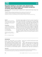
Figure 4. Tumour selectivity of methyl aminolevulinate. The pattern of fluorescence induced by
of PDT for treatment of this disease.
Because of the increasing inci- methyl aminolevulate is clearly limited to abnormal tissue (A), when compared with the outline of the
dence of adenocarcinoma of the tumour as seen under white light (B).
oesophagus and deaths from the
disease, there is pressure to improve screening procedures vulva. Difficulties in light delivery have already been
and hence a need for a simple routine treatment. PDT might resolved, but results so far with aminolevulinic acid have
be well-placed to fill this role, although new systemic drugs been disappointing.41,42 Systemic sensitisers will probably be
that do not have the disadvantages of porfimer sodium or more effective, provided that drugs with greater selectivity
and less photosensitivity can be developed.
temoporfin, will probably be needed.
Bladder cancer
Pituitary tumours
Porfimer sodium has been approved for treatment of
carcinoma in situ of the bladder, but does not seem to be
used widely. This restricted use could be because of reported
difficulties in damage to healthy tissues, which leads to
shrinkage of the bladder and, in some cases, the need for
cystectomy.53,54 However, preliminary data have shown PDT’s
potential, with intravesically applied aminolevulinic acid, to
treat the whole bladder.55 Furthermore, such treatment might
be a simple procedure, with no substantial side-effects.
In theory, bladder carcinoma in situ should be wellsuited to treatment with PDT and is another indication that
could be investigated further when better sensitising agents
become available.
An interesting example of the benefits of PDT in localised
disease is the ablation of pituitary tumours. In a phase I/II
trial,57 use of a transphenoidal approach for light delivery
with systemic porfimer sodium effectively prevented a
second recurrence in 12 patients who had recurrence of
disease after initial resection and radiotherapy.
Early non-small-cell lung cancer
Porfimer sodium has also been approved for treatment of
microinvasive, non-small cell lung cancer in the USA, Japan,
and Europe. Although early disease is often not identified
(up to 80% of lung cancer is already sufficiently advanced as
to be inoperable at time of diagnosis), detection of disease
should improve in the future, at least in patients at high risk,
when PDT will become an important treatment option.56
Intraepithelial neoplasias
PDT should also be very well suited to elimination of
intraepithelial neoplasias, such as those of the cervix and the
Oncology Vol 5 August 2004
Glioblastoma
Several studies have used PDT in an adjuvant setting in
treatment of glioblastoma after surgical resection.58,59
Improvements in both quality of life and survival were
reported, but the trial sizes were small. Early work has used
porfimer sodium, but temoporfin60 has also been used. The
potential benefit of use of PDT in an adjuvant setting for this
disease is the possibility of attaining adequate concentrations
of photosensitiser in the tumour, without the accumulation of
substantial concentrations in healthy brain tissue because of
the blood–brain barrier. This setting is another application
where early trials have shown promise, but where larger wellcontrolled randomised trials are needed.
Other early diseases
There are many other early diseases and precancerous
disorders in which PDT has great potential, provided that
sensitisers can be developed that have little or no skin
photosensitivity, are more selective for neoplastic tissue, and
503
Review
Photodynamic therapy
Table 5. Selected trials for localised cancer or precancerous conditions, 2000–February, 2004
Condition
Photosensitisers
Treatment
Trial type
Patients
Ref
Carcinoma of the lip
Temoporfin
Non-randomised phase II trial
25
20
Barrett’s oesophagus
Aminolevulinic acid
Randomised, double blind,
placebo controlled trial
36
39
Barrett’s oesophagus
Porfimer sodium
40
Aminolevulinic acid
Multicentre, partially blinded
randomised study
Randomised, double blind,
placebo controlled trial
208
Cervical intraepithelial
neoplasia
25
41
Cervical intraepithelial
neoplasia
Aminolevulinic acid
Phase I/II trial
40
42
Dysplasia and early
cancer in Barrett’s
oesophagus
Barrett's oesophagus
Porfimer sodium
Temoporfin (0·15 mg/kg) 96 h before
20 J/cm2 light (652 nm)
30 mg/kg oral aminolevulinic acid
or placebo 4 h before laser
endoscopy. Total light dose
60 J/cm2 (514 nm)
2 mg/kg porfimer sodium 48–72 h
before laser treatment (630 nm)
Topical application of 3%
aminolevulinic acid gel to cervix for
3 h, followed by 100 J/cm2 635 nm
laser light
Topical aminolevulinic acid
(200 mg/mL) for 90 min before
50–150 J/cm2 light (630 nm)
Porfimer sodium alone vs porfimer
sodium with oral prednisone, followed
by 630 nm light
30 mg/kg oral aminolevulinic
acid 4 h before laser endoscopy
10 g of 10% aminolevulinic acid
topically applied 2–3 h before
illumination. Light dose 120 J/cm2
(635 nm)
Topical aminolevulinic acid (20%)
before treatment with laser light
at 100 J/cm2 (635 nm)
Randomised controlled trial
60
43
Phase II trial
40
44
Phase II trial
15
45
Phase II trial
25
46
Aminolevulinic acid
Vulval intraepithelial
neoplasia
Aminoleuvilinic acid
Vulval intraepithelial
neoplasia
Aminolevulinic acid
have a short drug-to-light interval to facilitate a single
treatment in the outpatient or day-patient clinic.
Advanced cancer
PTD has been used for various indications in which a cure is
not feasible, but where survival can be lengthened and
quality of life improved. These indications are the main
non-dermatological disorders for which licensed drugs are
approved. In advanced disease, the intrinsic advantages of
PDT are minimum invasiveness and that it does not restrict
the use of other subsequent treatments, including
retreatment with PDT. However, the imposition on a
patient of a sustained skin photosensitivity should be
weighed against the advantages PDT treatment has on
quality of life. Table 6 shows the details of trials on use of
PDT for advanced cancer.
cancer. The debulking of tumour and subsequent relief of
dyspnoea takes longer after PDT than after laser treatment
with neodynium yttrium-aluminium-garnet (NdYAG),
making PDT less suitable for patients with acute respiratory
distress.79 Another disadvantage of PDT with porfimer
sodium could be the apparent high cost (in excess of £1500
per treatment for a patient who weights 75 kg), although
alternative treatment methods might approach or exceed
this amount when calculated properly. Despite these
limitations, a review and meta-analysis80 of all relevant
clinical trials concluded that bronchoscopic PDT was
beneficial in the treatment of selected patients with
advanced lung cancer. Almost all the 636 patients treated
had relief of symptoms, including improved ventilatory
function and relief of dyspnoea, with few adverse events.
Carcinoma of the oesophagus
Lung cancer
Porfimer sodium is licensed for the reduction of
obstruction and palliation of symptoms for patients with
completely or partially obstructing endobronchial nonsmall-cell lung cancer. Palliation is necessary because
patients can die from obstruction of the airway before they
succumb to the metastasising tumour.77 By maintainance of
the airway, quality of life and survival can be improved.
Trials in our group78 and elsewhere,6 with many patients
(table 6), have shown clearly the benefits of PDT in terms of
quality of life and survival. Compared with alternatives,
PDT is easy to use, which is particularly beneficial for
treatment of smaller bronchi.79 However, there are also
factors that have limited the use of PDT for advanced lung
504
Porfimer sodium is also licensed for palliation of patients with
completely obstructing oesophageal cancer, or for patients
with partial obstruction of the oesophagus who cannot be
treated satisfactorily with NdYAG laser therapy.1 Although
palliative control of obstruction is often necessary, survival
can be short, and whether use of drugs that cause lengthened
skin photosensitivity (such as porfimer sodium and
temoporfin) is justified is debatable. This disorder is thus
another indication in which improved drugs with little or no
skin photosensitivity could have a role.
Head and neck cancer
Temoporfin was licensed in the European Union, Norway,
and Iceland in 2001 as a local treatment for patients with
Oncology Vol 5 August 2004
Review
Photodynamic therapy
Table 6. Selected trials on photodynamic therapy for advanced cancer, 2000–February, 2004
Tumour type
Photosensitiser
Treatment
Brain
Temoporfin
Non-resectable
cholangiocarcinoma
Porfimer sodium
Mesothelioma
Temoporfin
Lung
Haematoporphyrin
derivative and
aminolevulinic acid
Lung
Haematoporphyrin
derivative
Lung or bronchus
Haematoporphyrin
derivative
Head and neck
Temoporfin
Head and neck
Temoporfin
Intraperitoneal
Porfimer sodium
Intraperitoneal
Porfimer sodium
Intraperitoneal
Porfimer sodium
Intraperitoneal
Porfimer sodium
Prostate
Temoporfin
Oesophagus
Aminolevulinic acid
vs haematoporphyrin
derivative
Oesophagus
Porfimer sodium
Hilar bile-duct
Porfimer sodium
Temoporfin (0·15 mg/kg) 96 h
Phase II trial
before treatment. Light dose
20–140 J/cm2 (652 nm)
Patients randomly assigned stenting and
Randomised prospective study
porfimer sodium (2·0 mg/kg) or stenting
alone. 48 h drug-to-light interval.
Light dose 180 J/cm2 (630 nm)
Generally, 0·1 mg/kg temoporfin
Phase I trial
6 days before surgery. Light dose
10 J/cm2 (652 nm)
Oral aminolevulinic acid (60 mg/kg),
Non-randomised study
with 6–8 h drug-to-light interval
(16 patients). Intravenous haematoporphyrin
derivative (2 mg/kg), with 48 h drug-to-light
interval (24 patients). Laser-light dose
100 J/cm2 (630 nm) for both groups
2 mg/kg given intravenously 48 h before
Prospective phase I/II trial to
illumination. Light dose 300 J/cm2
determine effect of hyperbaric
oxygen
2 mg/kg given intravenously 48 h before
Prospective, non-randomised
illumination. Light dose 300 J/cm2
pilot study to determine effect of
hyperbaric oxygen
Temoporfin 96 h before light interval,
Retrospective study
followed by irradiation with 652 nm laser
light (20 J/cm2)
Temoporfin 96 h before light interval,
Prospective phase I/II trial
followed by irradiation with 652 nm laser
light (20 J/cm2).
Mean follow-up of 37 months
2·5 mg/kg, with 48 h drug-to-light interval. Phase II trial
Intraperitoneal tumours resected and
analysed for presence of porfimer sodium
2·5 mg/kg 48 h before treatment. Laser
Phase II trial
light delivered to all intraperitoneal surfaces
2·5 mg/kg 48 h before treatment (532 nm Phase II trial
or 630 nm light used)
2·5 mg/kg 48 h before treatment (630 nm Phase II trial to investigate systemic
laser light used)
capillary leak syndrome, which
occurs after combination of surgery
and photodynamic therapy
0·15 mg/kg 96 h before treatment
Phase I–II trial
(652 nm laser, 100–150 mW)
60 mg/kg oral aminolevulinic acid 6–8 h
Non-randomised
before illumination (22 patients); 2 mg/kg
intravenous haematoporphyrin derivative
48 h before illumination (27 patients).
Light dose 300 J/cm2 of 630 nm laser light
1·5–2·0 mg/kg 48 h drug-to-light interval
Non-randomised
before 630 nm laser light (300–400 J/cm2)
2·0 mg/kg 1–4 days before treatment with Phase II trial
light (630 nm, 242 J/cm2)
advanced head and neck cancer who did not respond to
previous therapies, and who were unsuitable for
radiotherapy or chemotherapy. PDT is an attractive option
for treatment of cancers of the head and neck because of the
good cosmesis achieved and the accurate localisation of
therapy, limiting damage to organs in the head.61
Other applications
Several other indications in advanced cancer, such as such as
non-resectable cholangiocarcinoma, have been investigated
Oncology Vol 5 August 2004
Trial
Patients
Ref
25
61
39
62
26
63
40
64
30
65
40
66
25
67
25
68
12
69
56
70
11
71
65
72
14
73
49
74
77
75
23
76
in PDT trials, although approval for the drugs used has not
yet been obtained.62 Data from a prospective randomised
controlled trial on non-resectable cholangiocarcinoma
showed that PDT used in conjunction with plastic stents
improved quality of life and survival, with a low rate of
adverse side-effects. Patients assigned PDT with porfimer
sodium had decreased serum concentrations of bilirubin,
increased scores in physical function, and decreased
symptoms. The beneficial effects of PDT on the first
39 patients enrolled in the trial were so great that it was
505
Review
deemed unethical to continue with the randomisation
procedure. Use of PDT with temporforin in pancreatic
cancer is under development by Bown and colleagues81 to
provide an alternative treatment for this aggressive disease,
which is commonly unresponsive to chemotherapy or
radiotherapy. Other indications in which PDT is being
assessed include mesothelioma,63,82 and intraperitoneal
tumours.83,84
Why is PDT not a mainstream therapy in
oncology, and what is its future?
It is more than 25 years since PDT was first used in
oncology. Since then, thousands of patients have been
treated. The approach has progressed slowly but surely, and
four drugs have now been licensed for general use. In some
specialties of medicine (eg, dermatological oncology and
ophthalmology), PDT is used widely; however, in other
specialties its use remains marginal. Why has PDT not
achieved a more sustained entry to the therapeutic scene in
oncology, and will it become mainstream in future?
There are many answers to the first question, including
the difficulty in the establishment of the optimum variables
for a treatment that has several components, clinician and
hospital resistance to a new approach, the capital cost of
setting up a PDT centre, and the previous lack of inexpensive
and convenient light sources. However, the main answer
must be that the existing combinations of drugs and light
sources in the applications in which they have been used
have not established clear advantages over alternatives in
large controlled comparative randomised clinical trials. This
situation is probably because the drugs have been effective in
establishing the viability and feasibility of PDT, but have not
been optimum in terms of effectiveness. The development of
PDT could be compared with that of radiotherapy, which
has been used for more than 50 years but is only now
approaching optimum use. The competitive situation for
PDT is even more acute because of the very existence of
radiotherapy.
The effectiveness of PDT with verteprofin for treatment
of macular degeneration (which does not lead to lengthened
skin photosensitivity, is selective, and has a very short drugto-light interval) points the way to similar success in
oncology. To date, most sensitiser development for cancer
treatment seems to have been driven chemically, rather than
biologically or clinically, with a focus on improved optical
properties: focus is needed on soving the problems of early
sensitisers—ie, sustained skin photosensitivity, low
selectivity, and inconveniently long drug-to-light intervals.
Modification of the photosensitising moiety through its
physicochemical properties or improved targeting by
conjugation of the photosensitiser to moieties such as
antibodies, polymers,85,86 and peptide scaffolds, might
overcome the present difficulties.87
In dermatological oncology, PDT is already a routine
treatment, and its use will continue to increase. For internal
lesions, inexpensive and convenient light sources are now
available. However, improved drugs that are more selective
and that can be used conveniently and without sustained
skin photosensitivity are needed. If such drugs can be
506
Photodynamic therapy
Search strategy and selection criteria
Published data up to 2000 were reviewed in a previous article in
The Lancet Oncology. More recent publications were identified
by extensive searching of PubMed, Web of Science, and our
own reference base. Search terms included combinations of:
“Photofrin”, “photodynamic therapy”, “PDT”, “5-aminolaevulinic
acid”, “ALA”, “methyl 5-aminolaevulinate”, “MAL”, “Foscan”,
“porfimer sodium”, “temoporfin”, and “Metvix”. Selection of
material focused on clinical applications that use recently
licensed drugs. Trials included in tables 4–6 were published
between 2000 and February, 2004, and included more than ten
patients.
developed, then the advantages of PDT in terms of
minimum invasion in the body, patient and practitioner
convenience, and health economics will ensure a substantial
future role for this type of treatment in oncology.
Conflict of interest
SBB is a consultant and minority shareholder in Photopharmica Ltd, a
company formed under the auspices of the University of Leeds, UK,
which is involved in development of new-generation photosensitisers,
for which SBB is named on several patent applications. These drugs are
in the early stage of preclinical development in oncology and are not
mentioned in the manuscript.
Acknowledgments
We thank Colin Hopper, Hugh Barr, and Colin Morton for advice and
information about recent clinical trials. We thank Even Angell-Peterson
for permission to use figure 4 and Yorkshire Cancer Research, UK, for
financial support.
References
1 Hopper C. Photodynamic therapy: a clinical reality in the treatment
of cancer. Lancet Oncol 2000; 1: 212–19.
2 Gomer C J, Ferrario A, Fisher AMR, et al. Photodynamic therapy:
basic mechanisms and molecular applications. In: Coohill TP,
Valenzeno DP, eds. Photobiology for the 21st century.
Overland Park Kansas: Valdenmar Publishing Company, 2001:
143–47.
3 Oleinick NL, Morris RL, Belichenko I. The role of apoptosis in
response to photodynamic therapy: what, where, why, and how.
Photochem Photobiol Sci 2002; 1: 1–21.
4 Henderson BW, Dougherty TJ. How does photodynamic therapy
work? Photochem Photobiol 1992; 55: 145–57.
5 Barrett AJ, Kennedy JC, Jones RA, et al. The effect of tissue and
cellular pH on the selective biodistribution of porphyrin-type
photochemotherapeutic agents: a volumetric titration study.
J Photochem Photobiol B 1990; 6: 309–23.
6 Dougherty TJ, Gomer CJ, Henderson BW, et al. Photodynamic
therapy. J Natl Cancer Inst 1998; 90: 889–905.
7 Ochsner M. Light scattering of human skin: a comparison between
zinc (II)- phthalocyanine and photofrin II. J Photochem Photobiol B
1996; 32: 3–9.
8 Star WM. Light dosimetry in vivo. Phys Med Biol 1997; 42:
763–87.
9 Brancaleon L, Moseley H. Laser and non-laser light sources for
photodynamic therapy. Lasers Med Sci 2002; 17: 173–86.
10 Sharman WM, Allen CM, van Lier JE. Photodynamic therapeutics:
basic principles and clinical applications. Drug Discov Today 1999;
4: 507–17.
11 Ackroyd R, Kelty C, Brown N, Reed M. The history of
photodetection and photodynamic therapy. Photochem Photobiol
2001; 74: 656–69.
12 Moriwaki SI, Misawa J, Yoshinari Y, et al. Analysis of
photosensitivity in Japanese cancer-bearing patients receiving
photodynamic therapy with porfimer sodium (Photofrin).
Photodermatol Photoimmunol Photomed 2001; 17: 241–43.
13 Gilson D, Ash D, Driver I, et al. Therapeutic ratio of photodynamic
therapy in the treatment of superficial tumours of skin and
subcutaneous tissues in man. Br J Cancer 1988; 58: 665–67.
Oncology Vol 5 August 2004
Review
Photodynamic therapy
14 Brown SB, Mellish KJ. Verteporfin: a milestone in opthalmology
and photodynamic therapy. Expert Opin Pharmacother 2001; 2:
351–61.
15 Messmer KJ, Abel SR. Verteporfin for age-related macular
degeneration. Ann Pharmacother 2001; 35: 1593–98.
16 Polo L, Valduga G, Jori G, Reddi E. Low-density lipoprotein
receptors in the uptake of tumour photosensitizers by human
and rat transformed fibroblasts. Int J Biochem Cell Biol 2002; 34:
10–23.
17 Preston DS, Stern RS. Nonmelanoma cancers of the skin.
N Engl J Med 1992; 327: 1649–62.
18 Morton CA, Brown SB, Collins S, et al. Guidelines for topical
photodynamic therapy: report of a workshop of the British
Photodermatology Group. Br J Dermatol 2002; 146: 552–67.
19 Chang SC, Bown SG. Photodynamic therapy: applications in
bladder cancer and other malignancies. J Formos Med Assoc 1997;
96: 853–63.
20 Kubler AC, de Carpentier J, Hopper C, et al. Treatment of
squamous cell carcinoma of the lip using Foscan-mediated
photodynamic therapy. Int J Oral Maxillofac Surg 2001; 30: 504–09.
21 Kubler AC, Haase T, Staff C, et al. Photodynamic therapy of
primary nonmelanomatous skin tumours of the head and neck.
Lasers Surg Med 1999; 25: 60–68.
22 Ormrod D, Jarvis B. Topical aminolevulinic acid HCl photodynamic therapy. Am J Clin Dermatol 2000; 1: 133–39.
23 Piacquadio DJ, Chen DM, Farber HF, et al. Photodynamic therapy
with aminolevulinic acid topical solution and visible blue light in
the treatment of multiple actinic keratoses of the face and scalp:
investigator-blinded, phase 3, multicenter trials. Arch Dermatol
2004; 140: 41–46.
24 Szeimies RM, Karrer S, Radakovic-Fijan S, et al. Photodynamic
therapy using topical methyl 5-aminolevulinate compared with
cryotherapy for actinic keratosis: a prospective, randomized study.
J Am Acad Dermatol 2002; 47: 258–62.
25 Freeman M, Vinciullo C, Francis D, et al. A comparison of
photodynamic therapy using topical methyl aminolevulinate
(Metvix) with single cycle cryotherapy in patients with actinic
keratosis: a prospective, randomized study. J Dermatol Treat 2003;
14: 99–106.
26 Smith S, Piacquadio D, Morhenn V, et al. Short incubation PDT
versus 5-FU in treating actinic keratoses. J Drugs Dermatol 2003;
2: 629–35.
27 Pariser DM, Lowe NJ, Stewart DM, et al. Photodynamic therapy
with topical methyl aminolevulinate for actinic keratosis: results
of a prospective randomized multicenter trial. J Am Acad Dermatol
2003; 48: 227–32.
28 Jeffes EW, McCullough JL, Weinstein GD, et al. Photodynamic
therapy of actinic keratoses with topical aminolevulinic acid
hydrochloride and fluorescent blue light. J Am Acad Dermatol 2001;
45: 96–104.
29 Varma S, Wilson H, Kurwa HA, et al. Bowen’s disease, solar
keratoses, and superficial basal cell carcinomas treated by
photodynamic therapy using a large-field incoherent light source.
Br J Dermatol 2001; 144: 567–74.
30 Wang I, Bendsoe N, Klinteberg CA, et al. Photodynamic therapy vs
cryosurgery of basal cell carcinomas: results of a phase III clinical
trial. Br J Dermatol 2001; 144: 832–40.
31 Horn M, Wolf P, Wulf HC, et al. Topical methyl aminolaevulinate
photodynamic therapy in patients with basal cell carcinoma prone
to complications and poor cosmetic outcome with conventional
treatment. Br J Dermatol 2003; 149: 1242–49.
32 Dijkstra AT, Majoie IM, van Dongen JW, et al. Photodynamic
therapy with violet light and topical 6-aminolaevulinic acid in the
treatment of actinic keratosis, Bowen’s disease and basal cell
carcinoma. J Eur Acad Dermatol Venereol 2001; 15: 550–54.
33 Salim A, Leman JA, McColl JH, et al. Randomized comparison of
photodynamic therapy with topical 5-fluorouracil in Bowen’s
disease. Br J Dermatol 2003; 148: 539–43.
34 Morton CA, Whitehurst C, Moore JV, MacKie RM. Comparison of
red and green light in the treatment of Bowen’s disease by
photodynamic therapy. Br J Dermatol 2000; 143: 767–72.
35 Rhodes LE, De Rie M, Enstrom Y, et al. Photodynamic therapy
using topical methyl aminolevulinate vs surgery for nodular basal
cell carcinoma: results of a multicenter randomized prospective
trial. Arch Dermatol 2004; 140: 17–23.
36 Soler AM, Warloe T, Berner A, Giercksky KE. A follow-up study
of recurrence and cosmesis in completely responding superficial
and nodular basal cell carcinomas treated with methyl 5-amino-
Oncology Vol 5 August 2004
60
laevulinate-based photodynamic therapy alone and with prior
curettage. Br J Dermatol 2001; 145: 467–71.
Basset-Seguin N, Ibbotson S, Emtestam Lea. Photodynamic
therapy using Metvix is as efficacious as cryotherapy in BCC, with
better cosmetic results. J Eur Acad Dermatol Venereol 2001; 15: 226.
Allison RR, Mang TS, Wilson BD. Photodynamic therapy for
the treatment of nonmelanomatous cutaneous malignancies.
Semin Cutan Med Surg 1998; 17: 153–63.
Ackroyd R, Brown NJ, Davis MF, et al. Photodynamic therapy for
dysplastic Barrett’s oesophagus: a prospective, double blind,
randomised, placebo controlled trial. Gut 2000; 47: 612–17.
Overholt BF, Lightdale CJ, Wang K, et al. International multicenter
partially blinded randomised study of the efficacy of photodynamic
therapy using porfimer sodium for the ablation of high-grade
dysplasia in Barrett’s esophagus: results of 24 month follow-up.
Gastroenterology 2003; 124 (suppl): 151.
Barnett AA, Haller JC, Cairnduff F, et al. A randomised, doubleblind, placebo-controlled trial of photodynamic therapy using 5aminolaevulinic acid for the treatment of cervical intraepithelial
neoplasia. Int J Cancer 2003; 103: 829–32.
Keefe KA, Tadir Y, Tromberg B, et al. Photodynamic therapy of
high-grade cervical intraepithelial neoplasia with 5-aminolevulinic
acid. Lasers Surg Med 2002; 31: 289–93.
Panjehpour M, Overholt BF, Haydek JM, Lee SG. Results of photodynamic therapy for ablation of dysplasia and early cancer in
Barrett’s esophagus and effect of oral steroids on stricture
formation. Am J Gastroenterol 2000; 95: 2177–84.
Ackroyd R, Brown NJ, Davis MF, et al. Aminolevulinic acidinduced photodynamic therapy: safe and effective ablation of
dysplasia in Barrett’s esophagus. Dis Esophagus 2000; 13: 18–22.
Fehr MK, Hornung R, Schwarz VA, et al. Photodynamic therapy
of vulvar intraepithelial neoplasia III using topically applied
5-aminolevulinic acid. Gynecol Oncol 2001; 80: 62–66.
Hillemanns P, Untch M, Dannecker C, et al. Photodynamic
therapy of vulvar intraepithelial neoplasia using 5-aminolevulinic
acid. Int J Cancer 2000; 85: 649–53.
Regula J, Macrobert AJ, Gorchein A, et al. Photosensitisation and
photodynamic therapy of oesophageal, duodenal, and colorectal
tumours using 5 aminolaevulinic acid induced protoporphyrin IX: a
pilot study. Gut 1995; 36: 67–75.
Barr H, Shepherd NA, Dix A, et al. Eradication of high-grade
dysplasia in columnar-lined (Barrett’s) oesophagus by photodynamic therapy with endogenously generated protoporphyrin IX.
Lancet 1996; 348: 584–85.
Gossner L, Stolte M, Sroka R, et al. Photodynamic ablation of highgrade dysplasia and early cancer in Barrett’s esophagus by means of
5-aminolevulinic acid. Gastroenterology 1998; 114: 448–55.
Ackroyd R, Brown NJ, Vernon D, et al. Photodynamic therapy
(PDT) for Barrett’s oesophagus: a dosimetric pilot study. Br J Surg
1996; 83: 1637.
Ackroyd R, Brown NJ, Reed M. Photodynamic therapy (PDT) for
Barrett’s oesophagus: establishing optimal treatment parameters.
Eur J Surg Oncol 1996; 22: 551.
Kelty CJ, Marcus SL, Ackroyd R. Photodynamic therapy for
Barrett’s esophagus: a review. Dis Esophagus 2002; 15: 137–44.
Nseyo UO, Shumaker B, Klein EA, Sutherland K. Photodynamic
therapy using porfimer sodium as an alternative to cystectomy in
patients with refractory transitional cell carcinoma in situ of the
bladder. J Urol 1998; 160: 39–44.
D’Hallewin MA, Baert L. Long-term results of whole bladder wall
photodynamic therapy for carcinoma in situ of the bladder.
Urology 1995; 45: 763–67.
Waidelich R, Beyer W, Knuchel R, et al. Whole bladder
photodynamic therapy with 5-aminolevulinic acid using a white
light source. Urology 2003; 61: 332–37.
Mathur PN, Edell E, Sutedja T, Vergnon JM. Treatment of
early stage non-small cell lung cancer. Chest 2003; 123 (suppl):
S176–80.
Marks PV, Belchetz PE, Saxena A, et al. Effect of photodynamic
therapy on recurrent pituitary adenomas: clinical phase I/II trial—
an early report. Br J Neurosurg 2000; 14: 317–25.
Muller PJ, Wilson BC. Photodynamic therapy for malignant newly
diagnosed supratentorial gliomas. J Clin Laser Med Surg 1996;
14: 263–70.
Popovic EA, Kaye AH, Hill JS. Photodynamic therapy of brain
tumors. J Clin Laser Med Surg 1996; 14: 251–61.
Zimmermann A, Ritsch-Marte M, Kostron H. mTHPC-mediated
507
37
38
39
40
41
42
43
44
45
46
47
48
49
50
51
52
53
54
55
56
57
58
59
Review
61
62
63
64
65
66
67
68
69
70
71
72
photodynamic diagnosis of malignant brain tumors. Photochem
Photobiol 2001; 74: 611–16.
Lou PJ, Jones L, Hopper C. Clinical outcomes of photodynamic
therapy for head-and-neck cancer. Technol Cancer Res Treat 2003;
2: 311–17.
Ortner ME, Caca K, Berr F, et al. Successful photodynamic therapy
for nonresectable cholangiocarcinoma: a randomized prospective
study. Gastroenterology 2003; 125: 1355–63.
Friedberg JS, Mick R, Stevenson J, et al. A phase I study of Foscanmediated photodynamic therapy and surgery in patients with
mesothelioma. Ann Thorac Surg 2003: 75: 952–59.
Maier A, Tomaselli F, Matzi V, et al. Comparison of 5-aminolaevulinic acid and porphyrin photosensitization for photodynamic
therapy of malignant bronchial stenosis: a clinical pilot study. Lasers
Surg Med 2002; 30: 12–17.
Tomaselli F, Maier A, Sankin O, et al. Acute effects of combined
photodynamic therapy and hyperbaric oxygenation in lung
cancer—a clinical pilot study. Lasers Surg Med 2001; 28:
399–403.
Tomaselli F, Maier A, Pinter H, et al. Photodynamic therapy
enhanced by hyperbaric oxygen in acute endoluminal palliation of
malignant bronchial stenosis (clinical pilot study in 40 patients).
Eur J Cardiothorac Surg 2001; 19: 549–54.
Dilkes MG, Benjamin E, Ovaisi S, Banerjee AS. Treatment of
primary mucosal head and neck squamous cell carcinoma using
photodynamic therapy: results after 25 treated cases. J Laryngol Otol
2003; 117: 713–17.
Copper MP, Tan IB, Oppelaar H, et al. Meta-tetra(hydroxyphenyl)
chlorin photodynamic therapy in early-stage squamous cell
carcinoma of the head and neck. Arch Otolaryngol Head Neck Surg
2003; 129: 709–11.
Menon C, Kutney SN, Lehr SC, et al. Vascularity and uptake of
photosensitizer in small human tumor nodules: implications for
intraperitoneal photodynamic therapy. Clin Cancer Res 2001;
7: 3904–11.
Hendren SK, Hahn SM, Spitz FR, et al. Phase II trial of debulking
surgery and photodynamic therapy for disseminated intraperitoneal
tumors. Ann Surg Oncol 2001; 8: 65–71.
Bauer TW, Hahn SM, Spitz FR, et al. Preliminary report of photodynamic therapy for intraperitoneal sarcomatosis. Ann Surg Oncol
2001; 8: 254–59.
Canter RJ, Mick R, Kesmodel SB, et al. Intraperitoneal photo-
508
Photodynamic therapy
73
74
75
76
77
78
79
80
81
82
83
84
85
86
87
dynamic therapy causes a capillary-leak syndrome. Ann Surg Oncol
2003; 10: 514–24.
Nathan TR, Whitelaw DE, Chang SC, et al. Photodynamic therapy
for prostate cancer recurrence after radiotherapy: a phase I study.
J Urol 2002; 168: 1427–32.
Maier A, Tomaselli F, Matzi V, et al. Does new photosensitizer
improve photodynamic therapy in advanced esophageal carcinoma?
Lasers Surg Med 2001; 29: 323–27.
Luketich JD, Christie NA, Buenaventura PO, et al. Endoscopic
photodynamic therapy for obstructing esophageal cancer: 77 cases
over a 2-year period. Surg Endosc 2000; 14: 653–57.
Berr F, Wiedmann M, Tannapfel A, et al. Photodynamic therapy for
advanced bile duct cancer: evidence for improved palliation and
extended survival. Hepatology 2000; 31: 291–98.
Barber P, Barr H, George J, et al. Photodynamic therapy in the
treatment of lung and oesophageal cancers. Clin Oncol (R Coll
Radiol) 2002; 14: 110–16.
Moghissi K, Dixon K, Stringer M, et al. The place of bronchoscopic
photodynamic therapy in advanced unresectable lung cancer:
experience of 100 cases. Eur J Cardiothorac Surg 1999;
15: 1–6.
Ost D. Photodynamic therapy in lung cancer: a review.
Methods Mol Med 2003; 75: 507–26.
Moghissi K, Dixon K. Is bronchoscopic photodynamic therapy
a therapeutic option in lung cancer? Eur Respir J 2003; 22: 535–41.
Bown SG, Rogowska AZ, Whitelaw DE, et al. Photodynamic
therapy for cancer of the pancreas. Gut 2002; 50: 549–57.
Hahn SM, Smith RP, Friedberg J. Photodynamic therapy for
mesothelioma. Curr Treat Options Oncol 2001; 2: 375–83.
Dougherty TJ. An update on photodynamic therapy applications.
J Clin Laser Med Surg 2002; 20: 3–7.
Pass HI, DeLaney TF, Tochner Z, et al. Intrapleural photodynamic
therapy: results of a phase I trial. Ann Surg Oncol 1994; 1: 28–37.
Brokx RD, Bisland SK, Gariepy J. Designing peptide-based scaffolds
as drug delivery vehicles. J Control Release 2002; 78: 115–23.
Konan YN, Gurny R, Allemann E. State of the art in the delivery of
photosensitizers for photodynamic therapy. J Photochem Photobiol B
2002; 66: 89–106.
Allen CM, Sharman WM, van Lier JE. Photodynamic therapy:
targeting cancer cells with photosensitiser-bioconjugates. In:
Page M, ed. Tumour targeting in cancer therapy. Totowa, NJ:
Humana Press, 2002: 329–61.
Oncology Vol 5 August 2004