BMJ preeclamsia)
Bạn đang xem bản rút gọn của tài liệu. Xem và tải ngay bản đầy đủ của tài liệu tại đây (1.56 MB, 69 trang )
Pre-eclampsia
Straight to the point of care
Last updated: Feb 18, 2020
Table of Contents
Summary
3
Basics
4
Definition
4
Epidemiology
4
Aetiology
4
Pathophysiology
4
Classification
5
Prevention
7
Primary prevention
7
Screening
7
Secondary prevention
7
Diagnosis
9
Case history
9
Step-by-step diagnostic approach
9
Risk factors
12
History & examination factors
14
Diagnostic tests
16
Differential diagnosis
18
Diagnostic criteria
19
Treatment
23
Recommendations
23
Treatment details overview
26
Treatment options
28
Emerging
36
Follow up
37
Recommendations
37
Complications
38
Prognosis
39
Guidelines
41
Diagnostic guidelines
41
Treatment guidelines
42
Online resources
44
Evidence tables
45
References
55
Images
65
Disclaimer
67
Summary
◊ Hypertensive syndrome that occurs in pregnant women after 20 weeks' gestation, consisting of newonset, persistent hypertension with either proteinuria or evidence of systemic involvement.
◊ All pregnant women presenting with hypertension and either proteinuria or evidence of systemic
involvement require close assessment and monitoring for pre-eclampsia and its complications.
◊ Delivery is the definitive treatment; the decision about when and how to deliver should only be made
after a thorough assessment of the risk and benefits to the mother and baby.
◊ Other mainstays of management include antihypertensive therapy, seizure control, and fluid
restriction.
◊ Maternal mortality is highest after delivery, so vigilance should be maintained in the postpartum
period.
◊ Can occur in subsequent pregnancies; therefore, women should be counselled about the risk.
Pre-eclampsia
Basics
BASICS
Definition
A disorder of pregnancy associated with new-onset hypertension (defined as a blood pressure ≥140 mmHg
systolic and/or ≥90 mmHg diastolic), which occurs most often after 20 weeks of gestation and frequently near
term. Although often accompanied by new-onset proteinuria, hypertension and other signs or symptoms of
pre-eclampsia may present in some women in the absence of proteinuria.[1]
Epidemiology
While the exact incidence is unknown, pre-eclampsia has been reported to occur in about 4% of all
pregnancies in the US.[4] When figures include patients who develop pre-eclampsia postpartum, the
incidence is between 2% and 8% of all pregnancies worldwide.[5]
The incidence of severe disease and complications varies. Severe disease, which is associated with an
increased risk of morbidity and mortality, has an incidence of only 0.5% in the developed world,[6] but rises
to over 1% in low-income countries.[7] Similarly, the incidence of complications such as eclampsia is also
variable. In developed countries eclampsia is estimated to affect 5-7 cases in 10,000.[8] However, in lowincome countries the incidence of eclampsia is significantly higher, with estimates in some countries as high
as 100 in 10,000.[8]
Aetiology
Pre-eclampsia is associated with a failure of normal invasion of trophoblast cells leading to maladaptation of
maternal spiral arterioles, and is associated with hyperplacentation disorders such as diabetes, hydatidiform
mole, and multiple pregnancy.[9]
There are numerous risk factors that increase the probability and severity, including primiparity, previous
maternal history or family history, body mass index >30, maternal age >40 years, multiple pregnancy, pregestational diabetes, autoimmune disease, renal disease, chronic hypertension, and an interval of 10
years or more since a previous pregnancy.[10] However, these risk factors do not account for all cases and
complications such as eclampsia, HELLP syndrome (a subtype of severe pre-eclampsia characterised by
haemolysis [H], elevated liver enzymes [EL], and low platelets [LP]), and fetal growth restriction are not
present in all patients.
Pathophysiology
Pre-eclampsia is associated with a failure of the normal invasion of trophoblast cells leading to maladaptation
of maternal spiral arterioles.[9] The maternal arterioles are the source of blood supply to the fetus.
Maladaptation of these vessels can interfere with normal villous development leading to placental
insufficiency and, consequently, fetal growth restriction. The pathophysiology of this insufficient trophoblastic
invasion is likely to be multifactorial, with genetics, immunology, and endothelial dysfunction each playing
a role. The extent and specificity with which placental gene expression changes in pre-eclampsia remains
to be fully understood.[11] Raised levels of pro-inflammatory and anti-angiogenic cytokines in the maternal
circulation may contribute to placental vasoconstriction and subsequent hypoxia.[12] [13]
The systemic maternal response results in vasoconstriction and capillary leaking, leading to hypertension
and complications such as:
4
This PDF of the BMJ Best Practice topic is based on the web version that was last updated: Feb 18, 2020.
BMJ Best Practice topics are regularly updated and the most recent version of the topics
can be found on bestpractice.bmj.com . Use of this content is subject to our disclaimer (.
Use of this content is subject to our) . © BMJ Publishing Group Ltd 2020. All rights reserved.
Basics
Pre-eclampsia
• Cerebral vascular dysregulation and oedema
• Liver vascular dysregulation and oedema
Although the clinical manifestation does not occur until after 20 weeks' gestation, the abnormal physiological
changes may occur from early in the first trimester.[14] This has been suggested by the presence of various
biomarkers, such as:
• Pregnancy-associated plasma protein A
• ADAM12 (a disintegrin and metalloproteinase 12)
• Placental growth factor
• Soluble endoglin
• Soluble fms-like tyrosine kinase 1.[12]
Classification
American College of Obstetricians and Gynecologists:
classification of severity[1]
Disease classification and severity is based on blood pressure (BP) measurement and whether there are
signs of systemic involvement.
• Diagnostic criteria
• BP is 140-159 mmHg systolic and/or 90-109 mmHg diastolic and proteinuria is ≥300 mg/24
hours; or dipstick reading ≥2+ (use only if other quantitative methods not available); or
protein:creatinine ratio is ≥0.3 mg/dL.
• BP is 140-159 mmHg systolic and/or 90-109 mmHg diastolic and, in the absence of proteinuria,
any of the following is present:
• Thrombocytopenia, platelets count <100,000/microlitre
• Serum creatinine ≥1.1 mg/L or a doubling of the serum creatinine concentration in the
absence of other renal disease
• Impaired liver function, elevated blood concentrations of liver transaminases to twice
normal concentration
• Pulmonary oedema
• New-onset headache unresponsive to medication and not accounted for by alternative
diagnoses or visual symptoms.
• Severe features
• BP is ≥160 mmHg systolic and/or ≥110 mmHg diastolic (on two occasions at least 4 hours
apart, unless antihypertensive therapy is initiated before this time
• Thrombocytopenia, platelets count <100,000/microlitre
This PDF of the BMJ Best Practice topic is based on the web version that was last updated: Feb 18, 2020.
BMJ Best Practice topics are regularly updated and the most recent version of the topics
can be found on bestpractice.bmj.com . Use of this content is subject to our disclaimer (.
Use of this content is subject to our) . © BMJ Publishing Group Ltd 2020. All rights reserved.
5
BASICS
• Pulmonary oedema.
Pre-eclampsia
Basics
BASICS
• Serum creatinine ≥1.1 mg/L or a doubling of the serum creatinine concentration in the absence
of other renal disease
• Impaired liver function, elevated blood concentrations of liver transaminases to twice normal
concentration, and severe persistent right upper quadrant or epigastric pain unresponsive to
medication and not accounted for by alternative diagnoses
• Pulmonary oedema
• New-onset headache unresponsive to medication and not accounted for by alternative
diagnoses
• Visual disturbances.
HELLP syndrome is a subtype of severe pre-eclampsia characterised by haemolysis (H), elevated liver
enzymes (EL), and low platelets (LP). The diagnosis and management of HELLP syndrome is not discussed
in detail in this topic.
National Institute for Health and Care Excellence: classification of
severity (UK)[2]
Severity of disease is based on full clinical assessment, including both BP measurement and consideration
of a range of potential concerns, which could include any of the following:
• Sustained systolic blood pressure of ≥160 mmHg
• Any maternal biochemical or haematological investigations that cause concern: for example, a new
and persistent:
• Rise in creatinine (≥90 micromol/L, ≥1 mg/100 mL), or
• Rise in alanine transaminase (>70 IU/L, or twice upper limit of normal range), or
• Fall in platelet count (<150,000/microlitre)
•
•
•
•
•
Signs of impending eclampsia
Signs of impending pulmonary oedema
Other signs of severe pre-eclampsia
Suspected fetal compromise
Any other clinical signs that cause concern.
Severe disease is described as pre-eclampsia:
• With severe hypertension that does not respond to treatment, or
• That is associated with ongoing or recurring severe headaches, visual scotomata, nausea or vomiting,
epigastric pain, oliguria, and severe hypertension, as well as
• Progressive deterioration in laboratory blood tests such as rising creatinine or liver transaminases or
falling platelet count, or failure of fetal growth or abnormal Doppler findings.
6
This PDF of the BMJ Best Practice topic is based on the web version that was last updated: Feb 18, 2020.
BMJ Best Practice topics are regularly updated and the most recent version of the topics
can be found on bestpractice.bmj.com . Use of this content is subject to our disclaimer (.
Use of this content is subject to our) . © BMJ Publishing Group Ltd 2020. All rights reserved.
Pre-eclampsia
Prevention
Primary prevention
Low-dose aspirin (75-150 mg/day orally starting at 11-14 weeks' gestation) reduces the incidence and
severity of pre-eclampsia.[2] [27] [28] The effect appears to be uniform across all risk groups, but its use
should be targeted at high-risk groups such as those with hypertension, diabetes, renal disease, autoimmune
disease, multiple pregnancy, body mass index >30, maternal age >40 years, or interval of ≥10 years since
previous pregnancy.[2] [10] [24] Meta-analyses of randomised controlled trials suggest that the benefits of
aspirin may be limited to prevention of early onset rather than term disease, and only when given at doses of
>100mg/day.[29] [30]
It is important to optimise treatment for hypertension and renal disease prior to pregnancy. Controlled weight
loss reduces the incidence of pre-eclampsia.[2] Exercise in pregnancy should be encouraged in the absence
of complications, including maternal comorbidities, and risk factors for bleeding or premature delivery. A
regular supervised exercise programme may reduce the risk of pre-eclampsia, independently of body mass
index.[31] [32]
Women with hypertension, including those with an isolated elevated diastolic blood pressure at booking,
should be followed up in an increased-frequency surveillance programme.
Screening
All pregnant women should be seen regularly throughout their pregnancy, and have their blood pressure
(BP) measured.[4] If hypertension (defined as BP ≥140 mmHg and/or 90 mmHg) is found, urinalysis is
mandatory.[4] If this persists, there should be a step-up in care to an assessment centre or admission to
a care facility, depending on findings and symptoms. Uterine artery Doppler may be of limited value in
predicting the onset of disease and is therefore not recommended as a stand-alone screening tool.[1] [2] [38]
[44]
Biomarker screening tests reflecting placental development, such as sFlt-1:placental growth factor (PlGF)
ratio or PlGF alone, are available and there is evidence to support the use of PlGF as a diagnostic test.[39]
[40] Studies to determine the sensitivity, specificity, and cut-off values will help to establish the clinical
utility of these biomarkers as screening tests.[45] [46] It is likely that a combination of biomarkers will be
required.[47] Prediction algorithms utilising a combination of biomarkers and maternal history have not yet
shown sufficient sensitivity to be recommended for clinical use.[47] [48] One prospective cohort study of
16,747 pregnancies found that the combination of maternal history, biomarkers, and uterine artery Doppler
could achieve a detection rate of 82.4% (95% CI 76.1% to 88.7%) for preterm pre-eclampsia.[49]
In the UK, at least 50% of patients who develop pre-eclampsia are picked up by screening through antenatal
care.[24] However, in many parts of the developing world, the presentation is mostly acute, due to lack of
screening availability.
Secondary prevention
Low-dose aspirin (starting at 12-14 weeks' gestation) is recommended in subsequent pregnancies. If risk
of pre-eclampsia is thought to be high (e.g., previous early-onset disease, severe disease), the benefits are
clear. However, they are less clear in mild to moderate or late disease, where the outcome is typically good
anyway.[1] [2] [9]
There is some evidence that low molecular weight heparin, with or without aspirin, might reduce the placental
insufficiency in pre-eclampsia, but long-term safety studies are not available.[85] [86]
This PDF of the BMJ Best Practice topic is based on the web version that was last updated: Feb 18, 2020.
BMJ Best Practice topics are regularly updated and the most recent version of the topics
can be found on bestpractice.bmj.com . Use of this content is subject to our disclaimer (.
Use of this content is subject to our) . © BMJ Publishing Group Ltd 2020. All rights reserved.
7
PREVENTION
Epidemiological studies have found that low dietary calcium is associated with pre-eclampsia. One Cochrane
review found that the addition of high-dose calcium (≥1 g daily) reduced the risk of pre-eclampsia and
preterm birth compared with placebo. In populations where dietary calcium intake is low, the World Health
Organization recommends that pregnant women should receive 1.5 g to 2.0 g of supplementary calcium daily
in order to reduce pre-eclampsia severity. However, large, high-quality studies of calcium supplementation
from early pregnancy at a range of doses and in different populations are required.[33] [34] [35] [36]
Prevention
PREVENTION
Pre-eclampsia
8
This PDF of the BMJ Best Practice topic is based on the web version that was last updated: Feb 18, 2020.
BMJ Best Practice topics are regularly updated and the most recent version of the topics
can be found on bestpractice.bmj.com . Use of this content is subject to our disclaimer (.
Use of this content is subject to our) . © BMJ Publishing Group Ltd 2020. All rights reserved.
Pre-eclampsia
Diagnosis
Case history
Case history #1
A 25-year-old pregnant woman presents for her routine antenatal visit. She is at 32 weeks' gestation and
reports no symptoms. On examination, her blood pressure (BP) is 145/95 mmHg and urinalysis reveals
proteinuria (2+). She is referred to the antenatal day unit where a quantitative protein measurement of 1.5
g/24 hours is confirmed. Further laboratory tests reveal elevated liver enzymes; however, platelets and all
other tests are normal.
Case history #2
A 35-year-old woman presents at 37 weeks' gestation with severe headache and acute abdominal pain.
She had a routine antenatal visit 4 days previously with no signs or symptoms reported or observed. On
examination, her BP is 165/110 mmHg and urinalysis reveals proteinuria (3+). She is admitted to hospital
and is started on labetalol.
Other presentations
Pre-eclampsia may also be found on routine examination for lack of fetal movements or generally feeling
unwell. The International Society for the Study of Hypertension in Pregnancy considers uteroplacental
dysfunction, leading to decreased fetal growth, as one of several new-onset conditions that can define
pre-eclampsia when accompanied by gestational hypertension.[3] As such, women whose babies are
affected by fetal growth restriction should be investigated accordingly for pre-eclampsia. Uncommon
symptoms include breathlessness and visual disturbances. All women who present with unusual
symptoms in pregnancy should be investigated for pre-eclampsia.
A diagnosis of pre-eclampsia should be made when there is persistent, new-onset hypertension usually with
proteinuria after 20 weeks' gestation.[1] [2] [3] [37] The absence of hypertension excludes the diagnosis,
although there are related conditions, such as HELLP syndrome, that may present with and without
hypertension. HELLP syndrome is a subtype of severe pre-eclampsia characterised by haemolysis (H),
elevated liver enzymes (EL), and low platelets (LP).[2] The presence of proteinuria is no longer mandatory
in the diagnosis of pre-eclampsia; systemic involvement or fetal growth restriction together with hypertension
are enough to fulfil the diagnosis, even in the absence of proteinuria.[1] [3] After diagnosis, fetal assessment
should be performed with further maternal tests to assess systemic involvement.
If there are signs and symptoms of severe pre-eclampsia or complications, immediate treatment is needed.
On confirmation of the diagnosis, or if there are concerns regarding maternal or fetal health, the woman
should be admitted to an obstetric care facility for management.[1] [2]
History
Pre-eclampsia occurs in women after 20 weeks' gestation.[1] [2] [38] Key risk factors include primiparity,
positive family history, pre-eclampsia in a previous pregnancy, body mass index >30, maternal age >40
This PDF of the BMJ Best Practice topic is based on the web version that was last updated: Feb 18, 2020.
BMJ Best Practice topics are regularly updated and the most recent version of the topics
can be found on bestpractice.bmj.com . Use of this content is subject to our disclaimer (.
Use of this content is subject to our) . © BMJ Publishing Group Ltd 2020. All rights reserved.
9
DIAGNOSIS
Step-by-step diagnostic approach
Pre-eclampsia
Diagnosis
years, multiple pregnancy, gestational hypertension (hypertension developing after 20 weeks' gestation
in the absence of both proteinuria and systemic symptoms), pre-gestational diabetes, polycystic ovary
syndrome, autoimmune disease, renal disease, or chronic hypertension.
The woman may be asymptomatic and diagnosed at a routine clinic visit, or she may present acutely with
the following symptoms.
• Headache: usually frontal; presence of this symptom classifies pre-eclampsia as severe.[1] Occurs
in around 40% of patients with severe disease, and is one of the few symptoms that predict an
increased risk of eclampsia.[24]
• Upper abdominal pain: usually right upper quadrant pain; occurs in around 16% of patients with
severe disease, and is a clinical symptom of HELLP syndrome.[24]
• Visual disturbances: photopsia (perceived flashing lights in the visual fields), scotomata, and retinal
vasospasm are relatively rare, but predict an increased risk of eclampsia. Cortical blindness should
alert the clinician to underlying cerebral oedema.
• Breathlessness: due to pulmonary oedema and may complicate pre-eclampsia. If it occurs after
delivery, it is one of the main causes of maternal mortality.
• Seizures: mandates admission to intensive care unit, stabilisation, and delivery.[1]
• Oliguria: may be associated with increasing oedema. Patient is at most risk postpartum, when
pulmonary oedema is more likely.
The presence of these symptoms, in addition to hypertension with or without proteinuria, classifies preeclampsia as severe.[1] If fetal movements are reduced, there is a need for immediate fetal ultrasound
assessment.
Physical examination
DIAGNOSIS
Hypertension (defined as blood pressure [BP] ≥140 mmHg systolic and/or ≥90 mmHg diastolic) in a
previously normotensive woman is diagnostic.[1] [2] [38] At least 2 measurements should be made, at
least 4 hours apart.[1]
Oedema is very common, but is not discriminatory, and so should not be used in diagnosis. Hyper-reflexia
and/or clonus are rare and have little value in the clinical assessment.[38] Fundoscopy is rarely abnormal,
but, if it is, underlying chronic hypertension is implied.
If the uterus is small for dates, this implies that the amniotic fluid volume is reduced, which may signify
growth restriction, and fetal ultrasound assessment is required. Fetal growth restriction is found in around
30% of women with pre-eclampsia.[24]
Urinalysis
Reagent strip testing can be used to screen for the presence of protein in the urine. Reagent strip testing
with automated readers is more accurate than visual analysis.
Proteinuria in association with elevated blood pressure in the pre-eclampsia range requires referral to a
specialist unit or hospital admission for assessment. In the absence of proteinuria or systemic signs of
pre-eclampsia, an alternative diagnosis should be sought.
The standard diagnostic test for urinary protein estimation is a 24-hour urine collection, with a diagnostic
level considered to be urinary excretion of ≥0.3 g protein/24 hours.[1] [2] [3] The presence of proteinuria
10
This PDF of the BMJ Best Practice topic is based on the web version that was last updated: Feb 18, 2020.
BMJ Best Practice topics are regularly updated and the most recent version of the topics
can be found on bestpractice.bmj.com . Use of this content is subject to our disclaimer (.
Use of this content is subject to our) . © BMJ Publishing Group Ltd 2020. All rights reserved.
Pre-eclampsia
Diagnosis
≥5 g/24 hours is no longer used as a marker of severity, as the level of proteinuria does not relate to
outcome.[1] [2]
Completing 24-hour urine collection is awkward for women; where available, alternative spot tests such as
protein:creatinine ratio (PCR, where a result of ≥30 mg/mmol is diagnostic) and albumin:creatinine ratio
(ACR, where a result of ≥8 mg/mmol is diagnostic) are preferred.[1] [2] [38]
Because the level of proteinuria does not correlate with outcome, there is no benefit in routinely repeating
urinalysis once a diagnosis has been made.[2]
Fetal assessment
Immediate fetal ultrasound assessment is required if fetal movements are reduced or fetal growth
restriction is suspected. Fetal biometry should be used to diagnose or exclude fetal growth restriction,
although growth can only be fully assessed by scans done 2 weeks apart.
Other methods of fetal assessment should be done in all patients initially using the following tests:[1] [2]
[38]
• Umbilical artery Doppler velocimetry is the main assessment tool. It reduces perinatal mortality
and supports better decision-making, leading to more appropriate delivery decisions. It should
be carried out on admission and, if normal, repeated twice weekly. If abnormal, more intensive
monitoring may be required using other means, including Doppler assessment of other fetal vessels
and fetal cardiotocography. Delivery may be necessary within a few days.
[Fig-1]
• Fetal cardiotocography is recommended to assess fetal wellbeing, but is of little prognostic value.
It should be performed initially, and then no more than twice weekly, unless there is a cause for
concern such as vaginal bleeding, reduced fetal movements, or increased severity of disease.
• Amniotic fluid assessment, the single deepest vertical pocket being preferred over the amniotic
index. Can easily be combined with umbilical artery Doppler velocimetry.
Full blood count, serum creatinine, and liver function tests (LFTs) are all useful indicators of disease
progression, and are therefore recommended in all patients after initial urinalysis and fetal assessment.
Elevated serum creatinine implies underlying renal disease. Although elevated serum uric acid is
associated with severe pre-eclampsia, it does not offer diagnostic value. Decreased platelet and
increased transaminase levels are partly diagnostic for HELLP syndrome. The platelet count is the
main criterion used in classifying severity of HELLP syndrome. If the platelet count is <100 × 10⁹/L,
a full coagulation screen and LFTs should be carried out. If the platelet count is ≥100 × 10⁹/L, further
coagulation tests are not typically recommended.
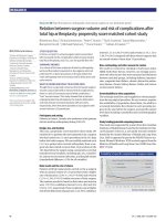
The UK National Institute for Health and Care Excellence (NICE) advocates the use of placental growth
factor (PlGF) testing to exclude a diagnosis of pre-eclampsia (within 14 days of testing) in women
presenting between 20 weeks to 34 weeks plus 6 days of gestation.[2] [39] One cluster-randomised
controlled trial reported faster time to diagnosis and lower incidence of severe maternal adverse outcomes
in maternity units where PlGF results were available, compared with those units where PlGF results were
concealed (control).[40] Median time to pre-eclampsia diagnosis was 4.1 days with concealed testing
versus 1.9 days with revealed testing (time ratio 0.36, 95% CI 0.15 to 0.87; P=0.027). Maternal severe
This PDF of the BMJ Best Practice topic is based on the web version that was last updated: Feb 18, 2020.
BMJ Best Practice topics are regularly updated and the most recent version of the topics
can be found on bestpractice.bmj.com . Use of this content is subject to our disclaimer (.
Use of this content is subject to our) . © BMJ Publishing Group Ltd 2020. All rights reserved.
11
DIAGNOSIS
Other maternal investigations
Pre-eclampsia
Diagnosis
adverse outcomes were reported in 24 (5%) of 447 women in the concealed testing group versus 22 (4%)
of 573 women in the revealed testing group (adjusted odds ratio 0.32, 95% CI 0.11 to 0.96; P=0.043).
Perinatal outcomes were not affected.[40]
NICE also recommends the Elecsys immunoassay sFlt-1/PlGF ratio as an alternative to PlGF-only testing,
although the need for traditional history taking and examination remains paramount. Equally, it remains
unclear whether use of these investigations carries any benefit for perinatal outcomes and, therefore, fetal
wellbeing assessments are required, as described above.
Risk factors
Strong
primiparity
• Pre-eclampsia is strongly associated with primiparity. The incidence is twice as high in these patients
compared with multiparous women. This is thought to be due to the development of tolerance
to specific immunological factors after the first pregnancy, thereby reducing risk in subsequent
pregnancies. These immunological factors are most likely to be associated with placental adaptations,
where the interaction between maternal and paternal immunological factors is most active. However,
some experts believe that pre-eclampsia is driven by systemic circulation of placental debris, which
again allows paternal factors to affect the systemic response.[2] [9] [10] [15] [16]
pre-eclampsia in previous pregnancy
DIAGNOSIS
• The risk of recurrence is around 10% to 50%, although it is thought to be higher in those with previous
early onset (i.e., <30 weeks) or severe disease, and lower for those with mild-moderate or late-onset
disease. Because the risk of recurrence is reduced with a change of partner, the increased risk in
these patients is likely to be due to a failure of tolerance to the specific immunological factor.[2] [9] [10]
family history of pre-eclampsia
• If a mother had pre-eclampsia, the daughter has a 25% chance of developing the condition. Similarly,
if a sister had pre-eclampsia, there is a 1 in 3 chance of developing it. These findings suggest a
genetic component to the condition. Although some studies have suggested associations with various
genetic markers, larger studies are still required.[2] [10] [17] [18]
BMI >30
• Associated with an increased risk of pre-eclampsia. The risk increases as BMI increases, becoming
more significant when BMI is >35.[2] [10] [19]
• The reasons for this are multifactorial, but may include overdiagnosis due to difficulties in measuring
blood pressure, and the fact that adipose tissue is a potent supplier of inflammatory mediators, thereby
making obese women more likely to mount an exaggerated inflammatory response.[10] [20]
maternal age >40 years
• Pregnancy at extremes of maternal age has been associated with pre-eclampsia. Women ≥40
years of age have a 4 times greater risk of morbidity and mortality compared with that of women
<25 years. This is probably due to altered physiological adaptation to pregnancy, and an increase in
comorbidities.[2] [10]
12
This PDF of the BMJ Best Practice topic is based on the web version that was last updated: Feb 18, 2020.
BMJ Best Practice topics are regularly updated and the most recent version of the topics
can be found on bestpractice.bmj.com . Use of this content is subject to our disclaimer (.
Use of this content is subject to our) . © BMJ Publishing Group Ltd 2020. All rights reserved.
Pre-eclampsia
Diagnosis
multiple (twin) pregnancy
• The association between pre-eclampsia and a multiple pregnancy is well documented. The data
are most compelling in twin pregnancies. Morbidity and mortality associated with pre-eclampsia are
increased in patients with a twin pregnancy.[2] [10] [21]
sub-fertility
• Women with sub-fertility are at higher risk of adverse pregnancy outcomes including preeclampsia.[22] This association is independent of maternal age and multiple pregnancy. In
pregnancies where there is a donor embryo, the incidence of pre-eclampsia is significantly higher.[23]
gestational hypertension
• Over 25% of patients with gestational hypertension (hypertension after 20 weeks' gestation in the
absence of both proteinuria and systemic symptoms) go on to develop pre-eclampsia.[1] These
patients should therefore be monitored closely.
pre-existing diabetes
• Diabetes is associated with a larger than average placenta and an increase in inflammatory
vascular disease, so there is a potential risk of both the placental trigger and the degree of maternal
response.[2] [10] [21] [24]
polycystic ovary syndrome (PCOS)
• Women with PCOS may be more likely to develop pre-eclampsia due to their increased risk of obesity,
type 2 diabetes, and treatment for sub-fertility.[2] [10] [21] [25]
autoimmune disease
renal disease
• Women with renal disease may already have hypertension and proteinuria, thereby making the
diagnosis of pre-eclampsia difficult. However, the incidence of pre-eclampsia in women with renal
disease of any type is thought to be around 25%. The presence of any autoimmune disease can
further increase the incidence.[2] [10] [21]
pre-existing cardiovascular disease and chronic hypertension
• The incidence of pre-eclampsia in women with chronic hypertension of any type is thought to be
around 25%. The presence of any autoimmune disease can further increase the incidence.[2] [10] [21]
Weak
interval of ≥10 years since previous pregnancy
• Women with a long interval between pregnancies have an increased risk of pre-eclampsia, but factors
such as age, obesity, and comorbidities may contribute to risk.[2] [15] [17]
This PDF of the BMJ Best Practice topic is based on the web version that was last updated: Feb 18, 2020.
BMJ Best Practice topics are regularly updated and the most recent version of the topics
can be found on bestpractice.bmj.com . Use of this content is subject to our disclaimer (.
Use of this content is subject to our) . © BMJ Publishing Group Ltd 2020. All rights reserved.
13
DIAGNOSIS
• Women with autoimmune disease, especially those with antiphospholipid antibody syndrome, have an
increased risk of pre-eclampsia, although it can be difficult to distinguish the two conditions.[2] [10] [21]
• Patients with autoimmune disease may have pre-existing vascular disease that worsens the preeclampsia, resulting in severe illness.
• An acute postpartum exacerbation can occur, but this is most likely to be due to the underlying
autoimmune disease.
Pre-eclampsia
Diagnosis
high-altitude residence
• The incidence of pre-eclampsia may be increased at high altitudes.[26]
History & examination factors
Key diagnostic factors
>20 weeks' gestation (common)
• Occurs in women after 20 weeks' gestation.[1] [2] [38]
BP ≥140 mmHg systolic and/or ≥90 mmHg diastolic and previously
normotensive (common)
• Hypertension (defined as BP ≥140 mmHg systolic and/or ≥90 mmHg diastolic) in a previously
normotensive woman is diagnostic.[1] [2] [38]
• At least two measurements should be made, at least 4 hours apart.[1]
• Considered severe if BP ≥160 mmHg systolic and/or ≥110 mmHg diastolic.[1]
• Correct cuff size should be used. Systolic measurement is taken as the first sound heard (K1) and
the diastolic measurement is the disappearance of sounds completely (K5). Where K5 is absent, K4
(muffling) should be accepted.
• High systolic BP is associated with stroke and placental abruption.[2]
headache (common)
• Usually frontal headache. Presence of this symptom classifies pre-eclampsia as severe.[1]
• Headache occurs in around 40% of patients with severe disease, and is one of the few factors that
predict an increased risk of eclampsia.[24]
DIAGNOSIS
upper abdominal pain (common)
• Usually right upper quadrant pain. Occurs in around 16% of patients with severe disease, and is a
clinical symptom of HELLP syndrome.[24]
• HELLP syndrome is a subtype of severe pre-eclampsia characterised by haemolysis (H), elevated liver
enzymes (EL), and low platelets (LP) syndrome.
• Presence of this symptom classifies pre-eclampsia as severe.[1]
Other diagnostic factors
reduced fetal movement (common)
• If fetal movements are reduced, there is a need for immediate fetal ultrasound assessment.
fetal growth restriction (common)
• Fetal growth restriction is found in around 30% of patients.[24]
• If the uterus is small for dates, this implies that the amniotic fluid volume is reduced, which may signify
fetal growth restriction.
• Fetal ultrasound assessment is required.
oedema (common)
• Very common, but is not discriminatory and so should not be used in diagnosis.
14
This PDF of the BMJ Best Practice topic is based on the web version that was last updated: Feb 18, 2020.
BMJ Best Practice topics are regularly updated and the most recent version of the topics
can be found on bestpractice.bmj.com . Use of this content is subject to our disclaimer (.
Use of this content is subject to our) . © BMJ Publishing Group Ltd 2020. All rights reserved.
Pre-eclampsia
Diagnosis
visual disturbances (uncommon)
• A relatively rare but concerning symptom that may predict an increased risk of eclampsia.[24]
• Includes photopsia (perceived flashing lights in the visual fields), scotomata, and retinal vasospasm.
Cortical blindness is a rare but critical symptom implying cerebral oedema.
• Fundoscopy is rarely abnormal, but if it is, underlying chronic hypertension is implied.
• Presence of this symptom classifies pre-eclampsia as severe.[1]
seizures (uncommon)
• Rare but critical symptom that indicates eclampsia and mandates admission to intensive care unit,
stabilisation, and delivery.[1]
breathlessness (uncommon)
• Rare presentation associated with pulmonary oedema. If pulmonary oedema occurs after delivery, it is
one of the main causes of maternal mortality.
• Presence of this symptom classifies pre-eclampsia as severe.[1]
oliguria (uncommon)
• Defined as <500 mL urine/day or <30 mL urine in 2 consecutive hours.
• May be associated with increasing oedema. Patient is at most risk postpartum when pulmonary
oedema is more likely.
• Presence of this symptom classifies pre-eclampsia as severe.[1]
hyper-reflexia and/or clonus (uncommon)
• Has poor positive and negative predictability for eclampsia.[38]
DIAGNOSIS
This PDF of the BMJ Best Practice topic is based on the web version that was last updated: Feb 18, 2020.
BMJ Best Practice topics are regularly updated and the most recent version of the topics
can be found on bestpractice.bmj.com . Use of this content is subject to our disclaimer (.
Use of this content is subject to our) . © BMJ Publishing Group Ltd 2020. All rights reserved.
15
Diagnosis
Pre-eclampsia
Diagnostic tests
1st test to order
Test
Result
urinalysis
positive reagent strip test;
urinary excretion of ≥0.3
g protein in 24 hours; or
urine protein:creatinine
ratio ≥30 mg/mmol;
albumin:creatinine ratio
of ≥8 mg/mmol; may be
normal
• Reagent strip testing can be used to screen for the presence of
protein in the urine. Reagent strip testing with automated readers is
more accurate than visual analysis. Proteinuria in association with
elevated blood pressure in the pre-eclampsia range requires referral
to a specialist unit or hospital admission for assessment.
• Standard diagnostic test for urinary protein estimation is 24-hour
urine collection. A urinary excretion of ≥0.3 g protein in 24 hours is
diagnostic.[1] [2] [3]
• The presence of proteinuria ≥5 g/24 hours is no longer used as
a marker of severity, as the level of proteinuria does not relate to
outcome.[1]
• Completing 24-hour urine collection is awkward for women, and,
where available, alternative spot tests such as protein:creatinine
ratio (PCR, where a result of ≥30 mg/mmol is diagnostic) and
albumin:creatinine ratio (ACR, where a result of ≥8 mg/mmol is
diagnostic) are preferred.[1] [2] [38]
• The presence of proteinuria is no longer mandatory in the diagnosis
of pre-eclampsia; systemic involvement or fetal growth restriction
together with hypertension are enough to fulfil the diagnosis, even in
the absence of proteinuria.[1] [3]
DIAGNOSIS
fetal ultrasound
• Provides immediate information about fetal wellbeing, including size
of baby and amniotic fluid volume. If fetal movements are reduced,
there is a need for immediate fetal ultrasound assessment.
• Fetal biometry should be used to diagnose or exclude fetal growth
restriction. Growth can only be fully assessed by scans done 2 weeks
apart.
• Single scan can give an estimation of fetal weight and an assessment
of whether the baby is small for dates, and gives the neonatologist
important information about the need for immediate delivery.
variable depending on
severity; fetal biometry
may reveal fetal growth
restriction
umbilical artery Doppler velocimetry
•
•
•
•
absence of end diastolic
flow is a sign that
The main assessment tool; its use reduces perinatal mortality and
supports better decision-making, leading to more appropriate delivery delivery will probably
be necessary in the near
decisions.
future
[Fig-1]
Should be carried out on admission and, if normal, repeated twice
weekly.[2] Presence of end diastolic flow is reassuring.
If abnormal, more intensive monitoring may be required using other
means, including Doppler assessment of other fetal vessels and
fetal cardiotocography. Delivery is likely to be necessary within a few
days.[2]
New evidence suggests that placental vascular indices obtained from
three-dimensional power Doppler may be predictive of pre-eclampsia.
However, large studies are required to validate its use in the wider
population.[41]
amniotic fluid assessment
16
deepest vertical pocket
≥2 cm implies normality;
This PDF of the BMJ Best Practice topic is based on the web version that was last updated: Feb 18, 2020.
BMJ Best Practice topics are regularly updated and the most recent version of the topics
can be found on bestpractice.bmj.com . Use of this content is subject to our disclaimer (.
Use of this content is subject to our) . © BMJ Publishing Group Ltd 2020. All rights reserved.
Diagnosis
Pre-eclampsia
Test
Result
• Appears to be beneficial (rather than full biophysical profiling) with
the single deepest vertical pocket being preferred over the amniotic
index.
• Easily combined with umbilical artery Doppler velocimetry.[1] [2] [38]
• Can assess fetal wellbeing and inform about the need to deliver
immediately.
fetal cardiotocography
• Assesses immediate fetal wellbeing, but is of little prognostic value.
• Should be used to assess fetal wellbeing initially, and then no more
than twice weekly, unless there is a cause for concern such as
vaginal bleeding, reduced fetal movements, or increased severity of
disease.
FBC
• Useful indicator of disease progression and recommended in all
patients.
• Decreased platelet count is partly diagnostic for HELLP syndrome.
HELLP syndrome is a subtype of severe pre-eclampsia characterised
by haemolysis (H), elevated liver enzymes (EL), and low platelets
(LP).
• If the platelet count is <100x10⁹/L, a full coagulation screen and blood
film should be carried out to diagnose/exclude HELLP syndrome.
liver function tests
<2 cm is associated with
increased fetal morbidity
and delivery should be
considered
no abnormalities in
tracing indicate assured
fetal wellbeing
may reveal low platelet
count
may be elevated
• Useful indicator of disease progression and recommended in all
patients.
• Increased transaminase levels are partly diagnostic for HELLP
syndrome. HELLP syndrome is a subtype of severe pre-eclampsia
characterised by haemolysis (H), elevated liver enzymes (EL), and
low platelets (LP).
serum creatinine
may be elevated
placental growth factor
DIAGNOSIS
• Useful indicator of disease progression and recommended in all
patients.
• Elevated serum creatinine implies underlying renal disease.
• Renal failure is a rare complication, and when it occurs, it is usually
acute tubular necrosis associated with co-existing sepsis or placental
abruption.
low
• Where available, measurement of maternal placental growth factor
may reduce time to diagnosis of pre-eclampsia, with consequent
reduction in incidence of severe maternal adverse outcomes.[39] [40]
Other tests to consider
Test
Result
coagulation screen
typically normal
• Usually normal in a woman with pre-eclampsia. May be abnormal
with advanced disease affecting the liver, or in association with
abruption.
• Should also be carried out as an assessment of risk for interventions
such as spinal or epidural analgesia, or surgical intervention where
excessive bleeding may increase the morbidity or mortality risk.
This PDF of the BMJ Best Practice topic is based on the web version that was last updated: Feb 18, 2020.
BMJ Best Practice topics are regularly updated and the most recent version of the topics
can be found on bestpractice.bmj.com . Use of this content is subject to our disclaimer (.
Use of this content is subject to our) . © BMJ Publishing Group Ltd 2020. All rights reserved.
17
Diagnosis
Pre-eclampsia
Differential diagnosis
DIAGNOSIS
Condition
Differentiating signs / Differentiating tests
symptoms
Chronic hypertension
• Pre-existing hypertension
prior to pregnancy.
• Retinopathy commonly seen
in longstanding disease.
•
Urinalysis: absence of newonset proteinuria.
Gestational hypertension
• No differentiating signs or
symptoms.
•
Urinalysis: absence of
proteinuria.
Epilepsy
• History of epilepsy or
seizures before pregnancy.
• BP normal.
• Focal neurological deficits or
symptoms.
•
Urinalysis: absence of
proteinuria.
Antiphospholipid
antibody syndrome
• History of repeated early
pregnancy loss.
• History of venous
thrombosis, stroke, or
transient ischaemic attack.
•
Lupus anticoagulant:
positive.
Anticardiolipin antibodies:
medium or high titre.
Anti-beta-2-glycoprotein I:
titre >99th percentile.
Thrombotic
thrombocytopenic
purpura
• Presentation before 20
weeks' gestation.
• Thrombosis, purpura, or
spontaneous bleeding.
• Fever.
• Neurological signs (e.g.,
seizures) in the absence
of signs of severe preeclampsia.
•
ADAMTS-13 activity
assay and inhibitor titres:
decreased activity.
Haemolytic uraemic
syndrome
• Presentation before 20
weeks' gestation.
• Microangiopathic haemolytic
anaemia in the absence
of signs of severe preeclampsia.
• Thrombosis.
• Renal failure in the absence
of signs of severe preeclampsia.
• Diarrhoea (especially bloody
diarrhoea), nausea, or
vomiting.
•
Peripheral blood smear:
presence of schistocytes.
FBC: anaemia,
thrombocytopenia.
Renal disease
• History of renal disease
before pregnancy.
•
Serum creatinine: elevated
in the absence of signs of
severe pre-eclampsia.
Liver disease
• History of liver disease
before pregnancy.
• History of alcohol abuse.
• Jaundice.
•
Urinalysis: absence of
proteinuria.
Serum bilirubin: elevated.
18
•
•
•
•
This PDF of the BMJ Best Practice topic is based on the web version that was last updated: Feb 18, 2020.
BMJ Best Practice topics are regularly updated and the most recent version of the topics
can be found on bestpractice.bmj.com . Use of this content is subject to our disclaimer (.
Use of this content is subject to our) . © BMJ Publishing Group Ltd 2020. All rights reserved.
Diagnosis
Pre-eclampsia
Condition
Differentiating signs / Differentiating tests
symptoms
Gallbladder disease
Pancreatic disease
• BP is normal.
• Hepatomegaly.
•
• History of biliary pain before
pregnancy.
• Right upper quadrant (RUQ)
pain is colicky in nature, and
is usually intense, lasting
>30 minutes.
• BP is normal.
• Distended, tender
gallbladder may be palpable.
•
•
•
•
•
•
•
•
History prior to pregnancy.
History of alcohol abuse.
BP is normal.
Steatorrhoea.
Jaundice.
Nausea and vomiting.
•
•
•
•
•
Liver function tests: very high
in cases of viral disease.
Blood glucose: decreased in
cases of acute fatty liver of
pregnancy.
Liver function tests: elevated
alkaline phosphatase,
gamma glutamyl
transpeptidase, and bilirubin.
RUQ ultrasound:
pericholecystic fluid;
distended gallbladder;
thickened gallbladder wall;
gallstones; positive Murphy
sign.
Abdominal ultrasound:
structural/anatomical
changes including cavities;
duct irregularity; contour
irregularity of head/body;
calcification.
Abdominal CT scan:
pancreatic calcifications;
focal or diffuse enlargement
of the pancreas; ductal
dilation; vascular
complications.
Abdominal x-ray: pancreatic
calcifications.
Serum amylase: elevated.
DIAGNOSIS
Diagnostic criteria
American College of Obstetricians and Gynecologists[1]
Disease severity is based on the blood pressure (BP) measurement and whether there are signs of systemic
involvement.
• Diagnostic criteria
• BP is 140-159 mmHg systolic and/or 90-109 mmHg diastolic and proteinuria is ≥300 mg/24
hours; or dipstick reading ≥2+ (use only if other quantitative methods not available); or
protein:creatinine ratio is ≥0.3 mg/dL.
• BP is 140-159 mmHg systolic and/or 90-109 mmHg diastolic and, in the absence of proteinuria,
any of the following is present:
• Thrombocytopenia, platelets count <100,000/microlitre
• Serum creatinine >1.1 mg/L or a doubling of the serum creatinine concentration in the
absence of other renal disease
This PDF of the BMJ Best Practice topic is based on the web version that was last updated: Feb 18, 2020.
BMJ Best Practice topics are regularly updated and the most recent version of the topics
can be found on bestpractice.bmj.com . Use of this content is subject to our disclaimer (.
Use of this content is subject to our) . © BMJ Publishing Group Ltd 2020. All rights reserved.
19
Diagnosis
Pre-eclampsia
• Impaired liver function, elevated blood concentrations of liver transaminases to twice
normal concentration
• Pulmonary oedema
• New-onset headache unresponsive to medication and not accounted for by alternative
diagnoses or visual symptoms.
• Severe features
• BP is ≥160 mmHg systolic and/or ≥110 mmHg diastolic (on two occasions at least 4 hours
apart, unless antihypertensive therapy is initiated before this time)
• Thrombocytopenia, platelets count <100,000/microlitre
• Serum creatinine >1.1 mg/L or a doubling of the serum creatinine concentration in the absence
of other renal disease
• Impaired liver function, elevated blood concentrations of liver transaminases to twice normal
concentration, and severe persistent right upper quadrant or epigastric pain unresponsive to
medication and not accounted for by alternative diagnoses
• Pulmonary oedema
• New-onset headache unresponsive to medication and not accounted for by alternative
diagnoses
• Visual disturbances.
National Institute for Health and Care Excellence (UK)[2]
Severity of disease is based on full clinical assessment, including both blood pressure (BP) measurement
and consideration of a range of potential concerns, which could include any of the following:
DIAGNOSIS
• Sustained systolic blood pressure of ≥160 mmHg
• Any maternal biochemical or haematological investigations that cause concern: for example, a new
and persistent:
• Rise in creatinine (≥90 micromol/L, ≥1 mg/100 mL), or
• Rise in alanine transaminase (>70 IU/L, or twice upper limit of normal range), or
• Fall in platelet count (<150,000/microlitre)
•
•
•
•
•
Signs of impending eclampsia
Signs of impending pulmonary oedema
Other signs of severe pre-eclampsia (see below)
Suspected fetal compromise
Any other clinical signs that cause concern.
Severe disease is described as pre-eclampsia:
• With severe hypertension that does not respond to treatment, or
• That is associated with ongoing or recurring severe headaches, visual scotomata, nausea or vomiting,
epigastric pain, oliguria, and severe hypertension, as well as
• Progressive deterioration in laboratory blood tests such as rising creatinine or liver transaminases or
falling platelet count, or failure of fetal growth or abnormal Doppler findings.
Society of Obstetric Medicine of Australia and New Zealand[38]
20
This PDF of the BMJ Best Practice topic is based on the web version that was last updated: Feb 18, 2020.
BMJ Best Practice topics are regularly updated and the most recent version of the topics
can be found on bestpractice.bmj.com . Use of this content is subject to our disclaimer (.
Use of this content is subject to our) . © BMJ Publishing Group Ltd 2020. All rights reserved.
Pre-eclampsia
Diagnosis
A diagnosis of pre-eclampsia can be made when hypertension arises after 20 weeks' gestation, and is
accompanied by one or more of the following.[38]
• Renal involvement:
• Significant proteinuria: dipstick proteinuria subsequently confirmed by a spot urine
protein:creatinine ratio ≥30 mg/mmol
• Serum or plasma creatinine ≥90 micromol/L
• Oliguria.
• Haematological involvement:
• Thrombocytopenia
• Haemolysis
• Disseminated intravascular coagulation.
• Hepatic involvement:
• Elevated serum transaminases
• Severe epigastric or right upper quadrant pain.
• Neurological involvement:
•
•
•
•
Convulsions (eclampsia)
Hyper-reflexia with sustained clonus
Severe headache
Persistent visual disturbances (e.g., photopsia, scotomata, cortical blindness, retinal
vasospasm)
• Stroke.
fullPIERS (Pre‐eclampsia Integrated Estimate of RiSk) model[42]
Identifies women who are at increased risk of adverse outcomes among those already diagnosed with preeclampsia. The fullPIERS model is a validated tool recommended by the UK National Institute for Health
and Care Excellence (NICE) to help guide decisions on the most appropriate place of care, and can be used
alongside full clinical assessment within 48 hours of hospital admission.
The parameters used to calculate the prediction are:
•
•
•
•
•
•
Gestational age
Presence of chest pain or dyspnoea
Platelet count
Serum creatinine level
Aspartate transaminase (AST) level
Pulse oximetry (SpO₂).
The risk of maternal adverse outcomes is expressed as a percentage. [PRE-EMPT: fullPIERS (Preeclampsia Integrated Estimate of RiSk) - external validation and recalibration] ( />evidence/fullpiers)
This PDF of the BMJ Best Practice topic is based on the web version that was last updated: Feb 18, 2020.
BMJ Best Practice topics are regularly updated and the most recent version of the topics
can be found on bestpractice.bmj.com . Use of this content is subject to our disclaimer (.
Use of this content is subject to our) . © BMJ Publishing Group Ltd 2020. All rights reserved.
21
DIAGNOSIS
• Pulmonary oedema.
• Fetal growth restriction.
• Placental abruption.
Pre-eclampsia
Diagnosis
Prediction of complications in early-onset pre-eclampsia (PREP-S
[survival analysis]) model[43]
PREP-S is a validated risk prediction tool recommended by the UK National Institute for Health and Care
Excellence (NICE) to help guide decisions on the most appropriate place of care for women already
diagnosed with pre-eclampsia, alongside full clinical assessment. PREP-S was internally validated with a UK
population, and is intended for use only up to 34 weeks of pregnancy.
The parameters used to calculate the prediction are:
•
•
•
•
•
•
•
•
•
•
•
•
•
•
Maternal age
Gestational age
Presence of exaggerated tendon reflexes
Presence of pre-existing medical conditions
Protein to creatinine ratio (PCR)
Serum urea concentration
Platelet count
Systolic blood pressure
Treatment with antihypertensive drugs
Treatment with magnesium sulfate
Pulse oximetry (SpO₂)
Alanine transaminase (ALT) level
Serum creatinine level
Timepoint from baseline.
DIAGNOSIS
The risk of maternal adverse outcomes is expressed as a percentage.
22
This PDF of the BMJ Best Practice topic is based on the web version that was last updated: Feb 18, 2020.
BMJ Best Practice topics are regularly updated and the most recent version of the topics
can be found on bestpractice.bmj.com . Use of this content is subject to our disclaimer (.
Use of this content is subject to our) . © BMJ Publishing Group Ltd 2020. All rights reserved.
Pre-eclampsia
Treatment
Recommendations
Management of pre-eclampsia is based on disease severity and progression.
Mainstays of treatment include:
•
•
•
•
•
Monitoring
Deciding on a delivery date and method
Lowering blood pressure (BP)
Controlling seizures
Postpartum fluid management.
The main causes of maternal mortality are stroke and pulmonary oedema; therefore, lowering BP and
managing postpartum fluid are the most important aspects of treatment, regardless of the presence of
other complications such as eclampsia or HELLP syndrome. HELLP syndrome is a subtype of severe preeclampsia characterised by haemolysis (H), elevated liver enzymes (EL), and low platelets (LP).
Management should be in a tertiary-care setting or in consultation with an obstetrician/gynaecologist with
experience in managing high-risk pregnancies.[1] Management differs between countries; however, the basic
principles of management are the same.
Hospital admission
Women with clinical findings that warrant close attention should be managed in an inpatient care
facility.[1] [50] [51] On admission, further assessment is required. BP should be monitored regularly for
rising levels, need for intervention, and response to therapy; however, there is little guidance on how
often this should be performed. A good guide is at least 4 times per day on a ward, or continuously in an
intensive care unit.[50]
In cases of well-controlled mild to moderate disease, outpatient management can be considered,
although close outpatient monitoring in a day unit or equivalent is required.[1] [50] [38] [Evidence C]
Validated risk prediction models such as fullPIERS or PREP-S may be helpful to guide decisions about
admission and thresholds for intervention, alongside full clinical assessment.[50]
Plan for delivery
The definitive treatment for pre-eclampsia is delivery; however, this is not always possible immediately.
Even after delivery, it may take a few days for the condition to resolve completely.
The decision to deliver can only be made after a thorough assessment of the risks and benefits to
both the mother and baby. The main risk to the baby is prematurity, a cause of neonatal morbidity and
mortality.[50] Neonatal healthcare costs also rise significantly with immediate delivery.[52]
If the patient is considered stable (i.e., absence of seizures, controlled hypertension), a conservative
approach is usually taken and the decision to deliver is based on the gestational age.[1] [50] [38]
Early delivery may be indicated if there are concerns regarding maternal condition, such as uncontrolled
hypertension, deteriorating blood test results, reduced oxygen saturation, or signs of placental abruption.
This PDF of the BMJ Best Practice topic is based on the web version that was last updated: Feb 18, 2020.
BMJ Best Practice topics are regularly updated and the most recent version of the topics
can be found on bestpractice.bmj.com . Use of this content is subject to our disclaimer (.
Use of this content is subject to our) . © BMJ Publishing Group Ltd 2020. All rights reserved.
23
TREATMENT
Early delivery
Pre-eclampsia
Treatment
Delivery may also be warranted for fetal indications, such as abnormal umbilical artery Doppler or
cardiotocography concerns.
A rushed delivery in an unstable patient can be dangerous. Treatment with magnesium sulfate and
antihypertensive therapy is necessary before delivery is considered in these patients (i.e., presence
of seizures, uncontrolled hypertension).[1] Delivery should be considered after the patient has been
stabilised.
The decision and plan for delivery should be discussed with senior obstetric, anaesthetic, and neonatal
staff.[50]
Delivery at <34 weeks' gestation
Prolonging the pregnancy is beneficial for the fetus at <34 weeks' gestation, providing that fetal
assessments are satisfactory. One Cochrane review of expectant management versus delivery between
24-34 weeks of gestation found reduced morbidity for the baby for some outcomes.[53] Limited data are
available regarding the effect of preterm delivery on maternal health.
This approach requires careful in-hospital maternal and fetal surveillance.[54] If delivery is required,
intravenous magnesium sulfate and antenatal corticosteroids are recommended before 34 weeks'
gestation to mature fetal lungs.[50]
Delivery at 34-36 weeks plus 6 days' gestation
There is limited evidence to guide management, and decisions regarding delivery should be made on a
case-by-case basis while monitoring maternal and fetal condition.
One study of women with non-severe pre-eclampsia at 34-37 weeks' gestation found that the risk of
adverse maternal outcome did not differ between those randomised to immediate delivery or expectant
management.[55] However, immediate delivery significantly increased the risk of neonatal respiratory
distress syndrome.[55] When the results of this study were combined with those of another randomised
trial, planned earlier delivery was associated with a reduction in maternal morbidity and mortality for
pregnancies at more than 34 weeks' gestation.[56] [57] The review authors acknowledged that the data
are limited.[57]
If disease severity in the mother increases, then immediate delivery will be required. In order to reduce
respiratory distress syndrome, antenatal corticosteroids are recommended before 34 weeks' gestation to
mature fetal lungs.[50] There is likely to be benefit from the administration of antenatal corticosteroids at
34-36 weeks, but this is unclear, and a decision should be made based on the specific case.[50]
Delivery at >37 weeks' gestation
Delivery is the most sensible approach, and is indicated within 24-48 hours.
TREATMENT
Mode of delivery
The mode of delivery depends on the gestational age, and is determined by individual patient factors.[1]
[50] [38]
At <32 weeks' gestation, caesarean is the most likely mode of delivery. Attempted vaginal delivery may
fail, cause significant fetal morbidity, or be unsafe in a severely ill mother.[1]
24
This PDF of the BMJ Best Practice topic is based on the web version that was last updated: Feb 18, 2020.
BMJ Best Practice topics are regularly updated and the most recent version of the topics
can be found on bestpractice.bmj.com . Use of this content is subject to our disclaimer (.
Use of this content is subject to our) . © BMJ Publishing Group Ltd 2020. All rights reserved.
Pre-eclampsia
Treatment
At >32 weeks' gestation the decision should be made on a case-by-case basis, depending on maternal
and fetal condition and the mother’s preferences.[50]
If a caesarean is performed, regional anaesthesia is preferred if tolerated and there is no coagulopathy.
If a general anaesthetic is used, care should be taken to prevent the hypertensive response to intubation
and extubation, and the risk of laryngeal oedema.[58]
Management of hypertension
Antihypertensive therapy should be started when the BP is ≥160 mmHg systolic and/or ≥110 mmHg
diastolic or when BP is persistently ≥140 mmHg systolic and/or ≥90 mmHg diastolic.[1] [50]
With severe hypertension (BP ≥160 mmHg systolic and/or ≥110 mmHg diastolic)
For the management of acute-onset, severe hypertension in a critical care setting, intravenous labetalol,
intravenous hydralazine, and oral nifedipine can be used first line. Second-line options (e.g., combination
therapy, alternative drugs) can be discussed with a specialist if the patient does not respond to first-line
therapies.[1] [50] [59] [Evidence C]
For ongoing control of hypertension or treatment of non-severe hypertension (BP persistently ≥140 mmHg
systolic and/or ≥90 mmHg diastolic)
Oral monotherapy is effective in most cases. Some women may, however, require combination
therapy with two different antihypertensive agents, or intravenous therapy, depending on the clinical
circumstances. There is no need to reduce the BP too quickly or by too much; the aim is to stop the rise
and reduce the BP gradually to <140 mmHg systolic and <90 mmHg diastolic.
Preferred antihypertensive agents
Labetalol is considered the antihypertensive of choice, and is effective as monotherapy in 80% of
patients.[1] [50] [6] [59] [Evidence C] It appears to be safe and effective in pregnant women for the
management of pre-eclampsia; however, it should be avoided in women with asthma or any other
contraindication to its use.[1]
Oral nifedipine may be as effective as intravenous labetalol, and can be considered in cases of
deteriorating hypertension previously controlled with oral labetalol.[60]
Hydralazine is widely used to manage severe hypertension in pregnancy; however, it can produce an
acute fall in BP and should be used along with plasma expansion.[50] Smaller, more frequent doses may
be used.
Methyldopa is widely used in lower- and middle-income countries and remains an acceptable alternative
if labetalol or nifedipine are not suitable.[50] [51] [Evidence C] Methyldopa should be avoided in the
postpartum period due to its association with depression; women already taking methyldopa should
change to an alternative antihypertensive treatment within 2 days of delivery.[50] [61]
This PDF of the BMJ Best Practice topic is based on the web version that was last updated: Feb 18, 2020.
BMJ Best Practice topics are regularly updated and the most recent version of the topics
can be found on bestpractice.bmj.com . Use of this content is subject to our disclaimer (.
Use of this content is subject to our) . © BMJ Publishing Group Ltd 2020. All rights reserved.
TREATMENT
Other oral antihypertensive agents may be acceptable depending on setting and clinical
circumstances.[50]
25