The use of CT and MRI in the characterization of intracranial mass
Bạn đang xem bản rút gọn của tài liệu. Xem và tải ngay bản đầy đủ của tài liệu tại đây (751.49 KB, 12 trang )
Imaging, 19 (2007), 173–184
The use of CT and MRI in the characterization of intracranial mass
lesions
R M S CARTER,
BSc, MRCS
and P M PRETORIUS,
MSc, FRCR
Department of Neuroradiology, West Wing, John Radcliffe Hospital, Headley Way, Headington,
Oxford OX3 9DU, UK
Summary
N Clinical and demographic information should be combined with imaging findings
to arrive at a specific diagnosis or a short differential diagnosis.
N The age of the patient and anatomical location of the lesion(s) are the most useful
discriminating factors.
N Intravenous administration of contrast material is indicated when an intracranial
mass lesion is detected on CT or MRI.
N MRI is the preferred imaging modality for the evaluation of intracranial mass
lesions, but CT is often performed at the initial presentation for practical reasons.
Abstract. Intracranial mass lesions are an important cause
of neurological morbidity and a common indication for cranial
imaging. Given the wide range of pathological processes that
can present as intracranial mass lesions, the radiologist has an
important role in limiting the differential diagnosis in an
individual case in order to inform the clinical decision-making
process. This review illustrates the use of cranial CT and MRI,
including diffusion weighted imaging (DWI), in the
characterization of intracranial mass lesions. A detailed
description of the imaging appearances of all mass lesions is
beyond the scope of this review, but we hope to provide the
reader with a rational approach to the complex task of
producing a short differential diagnosis.
The management of intracranial mass lesions was
revolutionized by the development of CT and MRI. The
pivotal role of the radiologist in the diagnostic workup of
these patients is summarized in Box 1.
The prognosis and management of different intracranial mass lesions vary widely and the radiologist should
therefore always strive to provide the referring clinician
with as short a differential diagnosis as possible. Being
able to exclude certain possibilities with confidence from
the differential diagnosis is also very helpful.
Some radiologists remain reluctant to offer a short
differential diagnosis, reasoning that the pathologist will
have the final say in any case. This is an out-dated and
unnecessarily defeatist view for three reasons. (1) Several
brain masses have pathognomonic or virtually pathognomonic appearances (e.g. cerebral aneurysms [1, 2], lipoma
of the corpus callosum [3], dermoid and epidermoid
tumours [4–7], Lhermitte-Duclos disease (Figure 1) [8],
most vestibular schwannomas [9–11] and most meningiomas [12, 13]), rendering biopsy obsolete or even contraindicated unless surgery is indicated for clinical reasons.
(2) To minimize surgical morbidity, most brain biopsies
are performed through a small burr-hole using a biopsy
needle yielding a tiny fragment of tissue. This approach is
obviously prone to sampling errors. In a large glioma, for
Imaging, Volume 19 (2007) Number 2
DOI: 10.1259/imaging/
64168868
’ 2007 The British Institute of
Radiology
instance, the biopsy may only yield tissue corresponding
to a World Health Organization (WHO) grade II lesion
while the imaging may show contrast enhancement and
necrosis indicating a grade IV lesion. (3) Certain histological features are ambiguous and should be interpreted in
the light of the imaging findings, e.g. Rosenthal fibres are
typically found in pilocytic astrocytomas, but can also be
seen in reactive gliosis [14].
CT and MRI can be used to obtain information about
several features of a mass lesion, including the location,
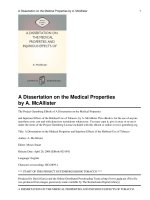
Figure 1. Using location and specific imaging characteristics
of a lesion to obtain an accurate diagnosis. Axial T2 weighted
MRI of a 35-year-old female with a 6-week history of
occipital headache shows the characteristic ‘‘tiger-striped’’
or laminated appearance of Lhermitte-Duclos disease, as
demonstrated by hyperintense right-sided hemispheric
expansion with parallel linear striations on the surface.
173
R M S Carter and P M Pretorius
size and extent, tissue composition and state of the
blood–brain barrier. Chemical and pathophysiological
information obtained from advanced imaging techniques, such as 1H MR spectroscopy and perfusion
imaging, also has a role to play in certain cases but,
since these techniques are not currently widely available,
we have decided to concentrate on modalities and
techniques available in most district general hospitals.
What is a mass lesion and what is mass effect?
For the purposes of this review, we include any discrete
intracranial lesion (or lesions) causing mass effect. Mass
effect refers to anatomical distortion of tissues surrounding the lesion or anatomical distortion and/or enlargement of the structure in which the lesion arises. Judging
the amount of mass effect a lesion exerts is a very
subjective exercise since measurable phenomena, such as
midline shift, are absent in most cases. The amount of
mass effect in an individual case depends on the size and
nature of the lesion. Not surprisingly, small lesions
generally exert less mass effect than larger lesions, and
in certain parts of the intracranial compartment, such as
the deep cerebral white matter, small mass lesions may
have no appreciable mass effect on imaging studies. The
effect of the nature of the lesion on the amount of mass
effect is more complex. In brain metastases, for example,
the mass effect is caused not only by proliferating
neoplastic cells pushing away surrounding normal brain
tissue but also by an increase in interstitial water
(vasogenic oedema) in and around the tumour. Acute
infarcts, on the other hand, cause mass effect by virtue of
an increase in intracellular water (cytotoxic oedema) [15].
It should come as no surprise that brain tumours
(particularly metastases) are generally associated with
more mass effect than a similar sized acute infarct.
Our visual appreciation of mass effect is further
influenced by the exact location of the lesion. For
example, a lesion in the brain stem would appear to
have more mass effect than a similar sized lesion in the
deep cerebral white matter, since changes in the relatively
small size and predictable contours of the brainstem are
more noticeable on imaging studies.
How do you generate a short differential
diagnosis?
To come up with a differential diagnosis, the radiologist integrates information obtained from imaging
studies — CT and MRI for the purposes of this review —
with demographic and clinical information about the
patient.
With experience, this process of assigning different
levels of importance to various pieces of information
becomes intuitive, but it remains one of the most
complex cognitive tasks we perform as radiologists. In
this process, it is important to make use of all the
relevant information available, not only on the images
but also on the request card.
Certain bits of information have greater discriminating
value than others. For example, the age of the patient
strongly influences the differential diagnosis in many
174
cases while the patient’s sex is usually irrelevant except
in the very specific instances mentioned below.
Radiology trainees in particular may find our proposed
checklist of eight questions (Box 2) useful when trying to
come up with a short differential diagnosis of an
intracranial mass. The fact that four of the eight
questions refer to clinical and demographic information
reflects the importance of this information. The reader
should also keep in mind that the relevance of the
answer to one question may depend on the answer to
another question. For example, the anatomical location of
a mass has different implications in different age groups.
We shall now briefly discuss the relevance of each of
the eight questions using examples. This discussion is far
from comprehensive, but hopefully serves to illustrate
the usefulness of this approach.
How old is the patient?
The age of the patient is of particular importance in
distinguishing between different types of intra-axial
cerebral neoplasms. Although most neoplasms have a
wide age range, their distribution is often very skewed
within that range. For example, the most common primary
intra-axial brain tumour, glioblastoma multiforme (GBM)
has an age range extending from infancy to the ninth
decade (and probably beyond), but the overwhelming
majority of GBMs occur in adults, with 70% occurring in
patients between the ages of 45 years and 70 years [16]. A
solitary intra-axial ring-enhancing supratentorial mass
lesion in a middle-aged or elderly person is therefore
most likely to be either a metastasis or a GBM while the
same appearance in a child is more likely to represent a
primitive neuroectodermal tumour [17] or an infectious
condition, such as a bacterial abscess or a tuberculoma. On
the other hand, an enhancing cerebellar mass in a 3-yearold child is very likely to represent a primary tumour
(astrocytoma, medulloblastoma or ependymoma) [17]
while a similar appearance in a 60-year-old person is
much more likely to represent a metastasis (Figure 2).
Extra-axial neoplasms are uncommon in children but
the imaging appearances of the two most common types,
namely meningiomas and vestibulocochlear schwannomas (acoustic neuromas), are so characteristic that it
rarely causes confusion when encountered in a child. The
presence of either of these tumours in a child should
raise a suspicion of neurofibromatosis type 2 [11].
How did the patient present?
The clinical presentation can occasionally be very
helpful in distinguishing between different intracranial
mass lesions. As a general rule, dramatic imaging
appearances in an eloquent brain area without dramatic
neurological deficits, is more in favour of tumour than
either infarction or demyelination.
For example, a WHO grade II or III astrocytoma can
have a very similar appearance to an infarct on CT and
MRI but the symptomatology is distinctly different, with
tumours usually presenting with seizures and/or headaches and/or gradual onset of relatively mild neurological deficits (Figures 3 and 4) [18], while infarcts
Imaging, Volume 19 (2007) Number 2
CT and MRI for intracranial mass lesions
Figure 2. The relevance of patient age in tumours in a particular location. Axial gadolinium enhanced T1 weighted images
through the posterior fossa in (a) a 6-year-old boy and (b) a 62-year-old man, both presenting with recent onset of cerebellar
symptoms and signs. Based on the ages of the patients, a primary tumour was considered most likely in the child, whereas a
metastasis was suspected in the older patient. Histology confirmed a medulloblastoma in the child (a) and a non small-cell lung
cancer metastasis in the man (b).
typically present with a sudden onset of dramatic motor
and/or sensory deficits. Infarcts and demyelination only
rarely present with seizures [19].
Figure 3. Combining clinical and imaging information to
reach the correct diagnosis. This 35-year-old man has a 12year history of temporal lobe epilepsy. The coronal gadolinium enhanced T1 weighted image shows an intra-axial,
cortically based, partially cystic mass with eccentric rim
enhancement. The long history of seizures attributable to
this lesion points towards an indolent neoplasm rather than
a more aggressive rim enhancing lesion, such as a metastasis,
glioblastoma multiforme (GBM) or abscess. In this location,
ganglioglioma and dysembryoplastic neuroepithelial tumour
(DNET) are the most likely candidates. The enhancement
characteristics strongly favour a ganglioglioma and this was
confirmed histologically.
Imaging, Volume 19 (2007) Number 2
Does the patient have a known disease, syndrome
or malignancy?
Only a small minority of patients presenting with an
intracranial mass lesion have a predisposing condition,
but knowledge of the condition can be extremely helpful.
A previous diagnosis of a malignancy with a tendency
to metastasize to the central nervous system (CNS), such
as bronchogenic carcinoma, breast carcinoma, melanoma
or bowel carcinoma, obviously increases the likelihood of
enhancing intracranial lesions being metastases [20].
Figure 4. Combining clinical and imaging information to
reach the correct diagnosis. Coronal T2 weighted image of a
7-year-old child with precocious puberty and a long history
of gelastic seizures shows a small, well defined hyperintense
mass arising from the inferior aspect of the hypothalamus.
The location and clinical features enable the diagnosis of
hypothalamic hamartoma to be made, obviating the need
for histological diagnosis.
175
R M S Carter and P M Pretorius
There are two rare exceptions to this rule and both
involve lesions of midline structures. First, lymphocytic
hypophysitis is a rare non-neoplastic cause of an
enlarged enhancing pituitary gland and or pituitary
stalk and is at least eight times more common in women
than men [31]. We have to stress that the other causes of
this appearances, such as pituitary adenoma,
Langerhan’s cell histiocytosis, sarcoidosis and germinoma, are more common in both sexes. Second,
germinomas in boys and men tend to occur in the pineal
gland while the same tumours in women and girls are
more common in the suprasellar region [16]. It goes
without saying that the great (more than one hundredfold) gender difference in the incidence of breast
carcinoma has an influence on how we search for the
primary tumour in cases of cerebral metastases from an
unknown primary.
Where is the lesion/s located?
Figure 5. Diagnosing specific histological tumour types as
features of a predisposing genetic syndrome. Axial gadolinium enhanced T1 weighted image demonstrates bilateral,
homogeneously enhancing, extra-axial, cerebellopontine
angle masses with extension into the IAMs. This characteristic appearance of bilateral vestibular schwannomas is
diagnostic of neurofibromatosis type 2 (NF2). Further
enhancing lesions seen bilaterally within Meckel’s cave are
therefore likely to represent bilateral meningiomas or
trigeminal schwannomas.
Even a solitary intra-axial enhancing mass in such a
patient should be considered a metastasis until proven
otherwise. On the other hand, similar appearances in a
patient with AIDS would raise the suspicion of primary
CNS lymphoma or toxoplasmosis [21].
Neurocutaneous syndromes (particularly von HippelLindau (VHL) [22], neurofibromatosis types 1 and 2 [11,
23] and tuberous sclerosis [24–26]) predispose to particular tumours. Knowledge about these conditions allows
the radiologist to make a confident diagnosis of a specific
histological tumour type in many cases (Figures 5 and 6).
At the time of the patient’s first presentation with an
intracranial tumour, the diagnosis of the neurocutaneous
syndrome may not yet have been made but additional
imaging features often give clues to the underlying
genetic condition.
Examples of conditions predisposing to non-neoplastic
mass lesions include cyanotic heart lesions and pulmonary arteriovenous malformations, which predispose to
brain abscesses [27, 28], and adult polycystic kidney
disease and coarctation of the aorta, which predispose to
cerebral aneurysms [29, 30].
Is the patient male or female?
While there are slight sex differences in the incidence of
many intracranial mass lesions, the differences are generally too small to use sex as a meaningful discriminator.
176
The location of a mass is a highly discriminating
imaging feature and the radiologist should always strive
to get as much information as possible about the exact
location of the tumour from the imaging studies. The
following anatomical descriptors are particularly useful
and should be employed when describing the location of
a mass.
Intra-axial vs extra-axial
The term ‘‘intra-axial’’ implies that a mass arises
within the neuraxis, i.e. within the substance of the CNS
(brain or spinal cord). Extra-axial lesions can arise within
the skull, meninges, cranial nerves or blood vessels.
Extra-axial lesions are easier to classify accurately than
intra-axial lesions and often have a pathognomonic
appearance [9–13] (Figure 5). The ability to distinguish
intra-axial from extra-axial masses is arguably the single
most discriminating imaging feature that can be elicited.
It can also be a surprisingly difficult distinction to make
in some cases. Intra-axial masses are completely surrounded by brain tissue except for rare instances of
tumours with an exophytic component. Lesions within
the cerebral or cerebellar white-matter or the deep greymatter structures can easily be classified as intra-axial.
However, when a lesion involves the cortex it should be
carefully evaluated to decide whether it is protruding
into the cortex from outside (extra-axial) or arising
within the cortex or subcortical white-matter (intraaxial). Imaging features strongly suggestive of an extraaxial origin include ‘‘trapped’’ cerebrospinal fluid (CSF)
and or pial blood vessels between the lesion and the
cortex, and a buckled or concertina appearance of the
underlying cortex (Figure 7). The presence of oedema is
not particularly useful in distinguishing between intraaxial and extra-axial lesions as it can be present or absent
in either [13, 32, 33].
Supra-tentorial vs infra-tentorial
This is an easy distinction to make and, in combination
with the patient’s age, it is particularly useful in
distinguishing between different types of intra-axial
tumours. For example, GBMs and metastases together
Imaging, Volume 19 (2007) Number 2
CT and MRI for intracranial mass lesions
Figure 6. Diagnosing specific histological tumour types as features of a predisposing genetic syndrome. (a) Axial T2 weighted and (b)
gadolinium-enhanced T1 weighted MR images in an 8-year-old boy with learning difficulties, epilepsy and a 2-month history of
headaches. There is a well circumscribed, avidly enhancing mass in a subependymal location associated with the frontal horn of the
right lateral ventricle. In addition, there are bilateral small subependymal nodules in the trigones of the lateral ventricles as well as a
cortical/subcortical area of high signal on T2 in the left frontal lobe. These features, particularly given the clinical history, are
pathognomonic of tuberous sclerosis (TS). The enhancing mass therefore represents a subependymal giant cell astrocytoma.
account for more than half of all intrinsic neoplasms in
adults. While both these tumour types occur more
frequently in the supratentorial compartment, only
approximately 1% of GBMs are infratentorial vs approximately 15% of brain metastases. An enhancing cerebellar
mass lesion in an adult is therefore much more likely to
represent a metastasis than a GBM [16].
A cystic lesion with an enhancing mural nodule in the
cerebellum of a child suggests a pilocytic astrocytoma
while the same appearance in an adult or a patient with
VHL syndrome suggests a haemangioblastoma [16, 17,
22, 34]. In the supratentorial compartment, particularly
in the temporal lobe of a child or adult with epilepsy the
same appearance is suggestive of a ganglioglioma
(Figure 3) [17, 35].
Specific sites
Certain lesions are specific to — or at least more
common in — certain intracranial locations (Figures 1–6,
8 and 9); for example, pituitary adenomas are limited to
the anterior pituitary gland. Similarly, certain sites give
rise to a limited number of lesions; for example, a mass
lesion in the jugular foramen is likely to represent a
glomus tumour, schwannoma, meningioma or metastasis [36]. Short differential diagnoses for mass lesions in
the following locations can be found in standard
textbooks of neuroradiology: pituitary/suprasellar
region, cerebello-pontine angle, brain stem, pineal,
intraventricular, jugular foramen, cavernous sinus.
Are the lesions solitary or multiple?
Multiple enhancing intra-axial lesions suggest haematogenous dissemination of a malignant or infectious
process or a multifocal inflammatory process, such as
Imaging, Volume 19 (2007) Number 2
Figure 7. Intra-axial vs extra-axial. Axial T2 weighted MR image
demonstrating a right frontal pole meningioma (extra-axial)
and a right posterior temporal lobe glioblastoma (intra-axial).
Note that there is a cleft of cerebrospinal fluid trapped between
the meningioma and the cortex (arrow), while the glioblastoma
is deep to the cortex. The presence of oedema is not particularly
useful in distinguishing between intra-axial and extra-axial
lesions since it can be present or absent in either.
177
R M S Carter and P M Pretorius
Figure 8. Axial T2 weighted MR images in two different patients demonstrating the importance of location vs signal
characteristics. (a) Colloid cyst of the third ventricle. The shape (well defined and round) and location anteriorly in the third
ventricle establishes the diagnosis in this case. Colloid cysts are typically non-enhancing and can be hypointense, isointense or
hyperintense on T2 weighted images; therefore the signal characteristics are less helpful than the location in making the
diagnosis. (b) Cavernoma of the mid-brain. An intra-axial lesion anywhere in the central nervous system with these signal
characteristics is most likely to represent a cavernoma. The peripheral low signal is due to haemosiderin staining while the
central high signal denotes the presence of extra-cellular methaemoglobin, indicating a more recent episode of haemorrhage.
In this case, the signal characteristics are more useful than the location of the lesion, as cavernomata occur throughout the
neuraxis and look the same in any location.
multiple sclerosis or acute disseminated encephalomyelitis (ADEM).
Primary intra-axial brain tumours are usually solitary
although ‘‘satellite nodules’’ are occasionally seen distant
from the main tumour bulk in high-grade lesions such as
GBMs [37].
Metastatic spread of primary CNS neoplasms is very
uncommon with the exception of metastatic seeding of
the CSF spaces. For example, an intra-axial tumour, such
as a cerebellar medulloblastoma, could give rise to one or
more metastatic deposits in the intracranial or spinal
subarachnoid space or within the ventricular system.
Figure 9. Using age and location to achieve the correct diagnosis: a 4-year-old mute child with cranial nerve palsies. (a) Sagittal
midline fluid attenuated inversion recovery (FLAIR) image shows a uniform high signal mass expanding the pons, characteristic
of a pontine astrocytoma. Pontine astrocytomas are usually of the diffuse fibrillary type and are at least World Health
Organization (WHO) grade II, whereas astrocytomas in the midbrain and medulla are usually pilocytic astrocytomas (WHO grade
I). (b) Axial post-gadolinium T1 weighted image demonstrates patchy contrast enhancement which suggests a WHO grade III
lesion.
178
Imaging, Volume 19 (2007) Number 2
CT and MRI for intracranial mass lesions
Figure 10. Utilizing specific imaging characteristics to reach the correct diagnosis. This 30-year-old man has a history of periodic
headaches and now presents with acute meningeal signs. (a) Sagittal midline unenhanced T1 weighted image showing a large
hyperintense and heterogeneous mass with several, similarly strongly hyperintense foci scattered within the sub-arachnoid
space. (b) Axial CT image confirms multiple, midline low attenuation foci associated with the falx, confirming these to be fat
droplets of a ruptured dermoid cyst. Intracranial dermoids usually contain a varying combination of lipid, liquid cholesterol,
whorls of hair, calcifications and decomposed epithelial cells producing typical appearances, as illustrated in this case.
Other tumours associated with this pattern of dissemination include ependymomas, germinomas, pilocytic astrocytomas and glioblastomas [38, 39].
What are the imaging characteristics on unenhanced CT or MRI?
The density of a mass on CT gives some useful
information about its constituents. Fluid within a cystic
or necrotic lesion can easily be distinguished from
adipose tissue in a lipoma or lipid material in a dermoid
cyst (Figure 10). However, it is important to keep in
mind that fat can be misinterpreted as gas since the
narrow window levels employed when interpreting
brain CT examinations render fat much darker (and
therefore closer in appearances to gas) than on the wider
window settings employed when viewing CT images of
other body parts. It is therefore prudent to widen the
window levels whenever a ‘‘black’’ lesion is seen on a
brain CT scan to distinguish between fat and air.
The density of the solid component of an intra-axial
tumour on unenhanced CT scans reflects the cellularity
and extracellular water content of the tumour. WHO
grade I and II astrocytomas are generally quite low
density compared with normal white-matter [34, 40]
while hypercellular tumours, such as lymphoma, germinoma and medulloblastoma, are usually denser than
normal white matter [41, 42].
Calcification and acute haemorrhage within a mass is
more easily and more reliably detected on CT than MRI
[43, 44].
Diffusion weighted imaging (DWI) has a well established role in acute stroke imaging, but it also gives very
useful information about certain intracranial mass
Imaging, Volume 19 (2007) Number 2
lesions. Two circumstances where DWI is particularly
useful are in the evaluation of ring enhancing intra-axial
mass lesions and CSF signal extra-axial mass lesions.
The differential diagnosis of ring enhancing intracranial mass lesions include metastases, GBM, acute
inflammatory demyelination, bacterial abscess and a
number of other infectious conditions, such as toxoplasmosis, tuberculoma and cysticercosis. In clinical practice,
it is often important to distinguish between a bacterial
brain abscess on the one hand and a tumour on the other
hand since the former requires urgent surgical aspiration
and intravenous antibiotics while the latter can often be
managed with oral corticosteroids and elective surgical
biopsy (Figure 11). DWI can be used to help distinguish
between these two entities since pus in bacterial
abscesses demonstrates markedly restricted diffusion
(bright on b51000 s mm–2 images and dark on apparent
diffusion coefficient (ADC) map) while necrotic tumour
material demonstrates facilitated diffusion (dark on
b51000 s mm–2 images and bright on ADC map), except
in rare instances where haemorrhage has occurred into
the necrotic tumour centre [45–47].
An extra-axial mass lesion returning signal isointense
or nearly isointense to CSF on T1 and T2 weighted
imaging is likely to be either an arachnoid cyst or an
epidermoid tumour. Diffusion is facilitated in arachnoid
cysts while epidermoid tumours demonstrate restricted
diffusion (Figure 12) [48, 49].
What are the enhancement characteristics?
Contrast uptake within brain tissue implies disruption
of the blood–brain barrier (e.g. in acute infarcts and
demyelinating lesions) or, in the case of neoplasms, it
179
R M S Carter and P M Pretorius
Figure 11. The use of diffusion-weighted imagine (DWI) in intra-axial ring enhancing lesions. (a) A contrast-enhanced CT scan
of the brain in an elderly woman with headache and a right-sided visual field defect shows a ring enhancing lesion with
surrounding vasogenic oedema. (b) A DWI (b51000) image of the same patient shows restricted diffusion (high signal) within
the fluid content of the lesion. Surgical aspiration confirmed an abscess. (c) An axial contrast enhanced T1 weighted MR image in
an elderly lady with a left sided visual field defect demonstrates an intra-axial ring enhancing lesion. (d) The DWI (b51000)
image shows free diffusion (low signal) of the fluid content. Histology confirmed a GBM with a necrotic centre.
implies angioneogenesis with increased endothelial
permeability of the abnormal tumour capillaries [50].
Different morphological patterns of contrast enhancement are recognized. Descriptive terms, such as nonenhancement, uniform enhancement, patchy enhancement, gyriform enhancement and ring enhancement
(also called rim or peripheral enhancement) are in
common use and give useful information about the
nature of the lesion.
As a general rule, most intracranial tumours —
whether primary or metastatic — show some form of
contrast enhancement. The notable exceptions are WHO
grade II astrocytomas and oligodendrogliomas
(Figures 9 and 13) [16, 51, 52], and gliomatosis cerebri
— a rare, diffusely infiltrating glial tumour that is
usually grade III.
180
Relatively uniform enhancement is seen in most solid
tumours including meningiomas, schwannomas, pituitary
adenomas and pineal tumours, such as germinomas. CNS
lymphomas typically show uniform enhancement in
immunocompetent patients while ring-enhancing lesions
have been described in immunodeficient patients [42].
Ring enhancement also occurs in aggressive tumours
such as GBMs and metastases (around areas of necrosis)
as well as in abscesses and some inflammatory demyelinating lesions (Figure 11). Other infectious lesions, such
as toxoplasmosis, cysticercosis and tuberculomas, can
also demonstrate ring enhancement.
Inflammatory demyelinating lesions only enhance
during the active phase of demyelination [53–55]. In
multiple sclerosis, this phase usually lasts less than 3
months for individual lesions. All the patterns of
Imaging, Volume 19 (2007) Number 2
CT and MRI for intracranial mass lesions
Figure 12. The use of diffusion-weighted imaging (DWI) in cerebrospinal fluid (CSF) signal extra-axial masses. (a) Axial T2
weighted MR image demonstrates a CSF signal mass lesion in the cisterna magna displacing the medulla posteriorly. (b) The
lesion is bright on a b51000 DWI image indicating restricted diffusion. The lesion therefore represents an epidermoid rather
than an arachnoid cyst.
enhancement (including no enhancement) listed in Box 2
have been described in demyelinating lesions.
Incomplete ring enhancement (also referred to as a
‘‘broken ring’’) is an unusual pattern of enhancement
and is strongly associated with inflammatory demyelination (Figure 14) including multiple sclerosis although
it has been described in infectious and neoplastic
conditions [56, 57].
Gyriform enhancement within the cerebral cortex
occurs in infarcts and certain encephalitic conditions
that affect the cortex [58, 59].
Conclusions
One of the most important roles of the radiologist in the
diagnostic pathway of a patient with an intracranial mass
is to provide the clinicians with a short differential
diagnosis. The radiologist should integrate all the relevant
information available on the images as well as the request
card to achieve this. Certain bits of information have a
higher discriminating value than others and the radiologist should give greater weight to such information when
formulating the differential diagnosis.
Figure 13. Contrast enhancement gives information about tumour grade. (a) Axial T1 weighted image in a patient with a
longstanding grade II astrocytoma shows an intra-axial mass with relatively uniform hypointense signal compared with whitematter. (b) Following gadolinium administration, there is a focal area of enhancement anteromedially (arrow) within the mass
indicating anaplastic transformation to a grade III astrocytoma.
Imaging, Volume 19 (2007) Number 2
181
R M S Carter and P M Pretorius
Figure 14. Tumefactive demyelination. This 61-year-old man with no history of multiple sclerosis presented with progressive
right sided hemiparesis and left sided facial numbness. The left-sided pontine mass lesion returns high signal on (a) T2 and low
signal on (b) T1 weighted imaging. There is incomplete ring enhancement seen on (c) the axial and (d) sagittal gadolinium
enhanced T1 weighted images. Given the patient’s age, a tumour was suspected despite the enhancement pattern. Stereotactic
biopsy demonstrated demyelination with no evidence of a tumour.
182
Imaging, Volume 19 (2007) Number 2
CT and MRI for intracranial mass lesions
Box 1
The role of the radiologist:
N
N
N
N
N
Confirm the presence of a mass lesion/lesions
Determine whether there are any complications
needing urgent attention, e.g. hydrocephalus, tonsillar
herniation, compression of the anterior optic pathways
Offer a short differential diagnosis:
N Try to distinguish neoplastic from non-neoplastic masses
N Inform the referring clinicians urgently if the
differential diagnosis includes infectious conditions, such as bacterial abscess or tuberculosis
Liaise with the neuropathologist in cases where a
biopsy is performed, particularly if there are
ambiguous histological findings
Provide information relevant to the surgical management:
N Relationship of tumour to major arteries and
venous sinuses and whether those structures are
patent
N Relationship to eloquent brain areas such as the
motor-cortex and other important structures
such as cranial nerves
N In cases of obstructive hydrocephalus, sagittal
high resolution midline T2 weighted sequences
are needed to determine whether a third
ventriculostomy can be performed safely
Box 2
Eight questions to help limit the differential
diagnosis of intracranial masses:
1. How old is the patient?
2. How did the patient present?
3. Does the patient have a disease or syndrome, e.g. a
phacomatosis or AIDS or a known malignancy
elsewhere?
4. Is the patient male or female?
5. Where is the lesion located?
- Intra-axial vs extra-axial
- Supra-tentorial vs infra-tentorial
- Specific sites: brainstem, pituitary, suprasellar,
pineal, intra-ventricular, cerebellopontine angle/
IAM
6. Are the lesions solitary or multiple?
7. What are the imaging characteristics on unenhanced CT/MRI?
8. What are the enhancement characteristics?
- None
- Solid
- Ring
- Smooth
- Irregular
Imaging, Volume 19 (2007) Number 2
References
1. Biondi A, Scialfa G, Scotti G. Intracranial aneurysms: MR
imaging. J Neuroradiol 1988;30:214–8.
2. Donovan JL, Nesbit GM. Distinction of masses involving
the sella and suprasellar space: specificity of imaging
features. AJR Am J Roentgenol 1996;167:597–603.
3. Ichikawa T, Kumazaki T, Mizumara S, Kijima T, Motohashi
S, Gocho G. Intracranial lipomas. J Nippon Med Sch
2000;67:388–91.
4. Wilms G, Casselman J, Demaerel Ph, Plets C, Haene I De,
Baert AL. CT and MRI of ruptured intracranial dermoids. J
Neuroradiol 1991;33:149–51.
5. Kallmes DF, Provenzale JM, Cloft HJ, McClendon RE.
Typical and atypical MR imaging features of intracranial
epidermoid tumors. AJR Am J Roentgenol 1997;169:883–7.
6. Davidson HD, Ouchi T, Steiner RE. NMR imaging of
congenital intracranial germinal layer neoplasms. J
Neuroradiol 1985;27:301–3.
7. Tampieri D, Melanson D, Ethier R. MR imaging of
epidermoid cysts. AJNR Am J Neuroradiol 1989;10:351–6.
8. Ashley DG, Zee CS, Chadrasoma PT, Segall HD. LhermitteDuclos disease: CT and MR findings. J Comput Assist
Tomogr 1990;14:984–7.
9. Curati WL, Graif M, Kingsley DP, Niendorf HP, Young IR.
Acoustic neuromas: Gd-DTPA enhancement in MR imaging. Radiology 1986;158:447–51.
10. Eldevik OP, Gabrielsen TO, Jacobsen EA. Imaging findings
in schwannomas of the jugular foramen. AJNR Am J
Neuroradiol 2000;21:1139–44.
11. Shu HH, Mirowitz SA, Wippold FJ. Neurofibromatosis: MR
imaging findings involving the head and spine. AJR Am J
Roentgenol 1993;160:159–64.
12. Goldsher D, Litt AW, Pinto RS, Bannon KR, Kricheff II.
Dural ‘‘tail’’ associated with meningiomas on Gd-DTPAenhanced MR images: characteristics, differential diagnostic
value, and possible implications for treatment. Radiology
1990;176:447–50.
13. Buetow MP, Buetow PC, Smirniotopoulos JG. Typical,
atypical, and misleading features in meningioma.
Radiographics 1991;11:1087–106.
14. Dinda AK, Sarkar C, Roy S. Rosenthal fibres: an immunohistochemical, ultrastructural and immunoelectron microscopic study. Acta Neuropathol 1990;79:456–60.
15. Klatzo I. Pathophysiological aspects of brain edema. Acta
Neuropathol 1986;72:236–9.
16. Chapter 1: Astrocytic Tumours. In: Kleihous P, Cavenee
WK, editors. WHO Classification of Tumours. Pathology
and genetics. Tumours of the Nervous System. Lyon,
France: IARC Press, 2000:176–84.
17. Rickert CH, Paulus W. Epidemiology of central nervous
system tumors in childhood and adolescence based on the
new WHO classification. Childs Nerv Syst 2001;17:503–11.
18. Zentner J, Wolf HK, Ostertun B, Hufnagel A, Campos MG,
Solymosi L, et al. Gangliogliomas: clinical, radiological, and
histopathological findings in 51 patients. J Neurol
Neurosurg Psychiatry 1994;57:1497–502.
19. Bladin CF, Alexandrov AV, Bellavance A, Bornstein N,
Chambers B, Cote´ R, et al. Seizures after stroke: a
prospective
multicenter
study.
Arch
Neurol
2000;57:1617–22.
20. Merchut MP. Brain metastases from undiagnosed systemic
neoplasms. Arch Intern Med 1989;149:1076–80.
21. Levy RM, Pons VG, Rosenblum ML. Central nervous
system mass lesions in the acquired immunodeficiency
syndrome. J Neurosurg 1984;61:9–16.
22. Maher ER, Yates JRW, Harries R, Benjamin C, Harris R,
Moore T, et al. Clinical features and natural history of von
Hippel-Lindau disease. QJM 1990;77:1151–63.
183
R M S Carter and P M Pretorius
23. Martuza RL, Eldridge R. Neurofibromatosis 2 (bilateral
acoustic neurofibromatosis). N Engl J Med 1998;318:
684–8.
24. McMurdo SK, Moore SG, Brant-Zawadzki M, Berg B, Koch
T, Newton T, et al. MR imaging of intracranial tuberous
sclerosis. AJR Am J Roentgenol 1987;148:791–6.
25. Braffman BH, Bilaniuk LT, Naidich TP, Altman NR, Post
JD, Quencer RM, et al. MR imaging of tuberous sclerosis:
pathogenesis of this phakomatosis, use of gadopentetate
dimeglumine, and literature review. Radiology 1992;183:
227–38.
26. Nixon JR, Houser OW, Gomez MR, Okazaki H. Cerebral
tuberous sclerosis: MR imaging. Radiology 1989;170:869–73.
27. Kagawa M, Takeshita M, Yato S, Kitamura K. Brain abscess
in congenital cyanotic heart disease. J Neurosurg
1983;58:913–7.
28. Brydon HL, Akinwunmi J, Selway R, Ul-Haq I. Brain
abscesses associated with pulmonary arteriovenous malformations. Br J Neurosurg 1999;13:265–269.
29. Wakabayashi T, Fujita S, Ohbora Y, Suyama T, Tamaki N,
Matsumoto SJ. Polycystic kidney disease and intracranial
aneurysms. Early angiographic diagnosis and early operation for the unruptured aneurysm. Neurosurgery
1983;58:488–91.
30. Mercado R, Lopez S, Cantu C, Sanchez A, Revuelta R,
Gomez-Llata S, et al. Intracranial aneurysms associated
with unsuspected aortic coarctation. J Neurosurg
2002;97:1221–5.
31. Thodou E, Asa SL, Kontogeorgos G, Kovacs K, Horvath E,
Ezzat S. Clinical case seminar: lymphocytic hypophysitis:
clinicopathological findings. J Clin Endocrinol Metab
1995;80:2302–11.
32. Goldman CK, Bharara S, Palmer CA, Vitek J, Tsai JC, Weiss
HL, et al. Brain edema in meningiomas is associated with
increased vascular endothelial growth factor expression.
Neurosurgery 1997;40:1269–77.
33. Kimelberg HK. Current concepts of brain edema. Review of
laboratory investigations. J Neurosurg 1995;83:1051–9.
34. Lee YY, Tassel PV, Bruner JM, Moser RP, Share JC. Juvenile
pilocytic astrocytomas: CT and MR characteristics. AJR Am
J Roentgenol 1989;152:1263–70.
35. Demierre B, Stichnoth FA, Hori A, Spoerri O. Intracerebral
ganglioglioma. J Neurosurg 1986;65:177–82.
36. Weber AL, McKenna MJ. Radiologic evaluation of the
jugular foramen. Anatomy, vascular variants, anomalies,
and tumors. Neuroimaging Clin N Am 1994;4:579–98.
37. Heuch I, Blom GP. Glioblastoma multiforme in three family
members, including a case of true multicentricity. J Neurol
1986;233:142–4.
38. Bryan P. CSF seeding of intra-cranial tumours: a study of 96
cases. Clin Radiol 1974;25:355–60.
39. Onda K, Tanaka R, Takahashi H, Takeda N, Ikuta F.
Symptomatic cerebrospinal fluid dissemination of cerebral
glioblastoma. J Neuroradiol 1990;32:146–50.
40. Le Roux PD, Berger MS, Wang K, Mack LA, Ojemann GA.
Low grade gliomas: comparison of intraoperative
ultrasound characteristics with preoperative imaging studies. J Neurooncol 1992;13:189–98.
41. Chang T, Teng MM, Guo WY, Sheng WC. CT of pineal
tumors and intracranial germ-cell tumors. AJR Am J
Roentgenol 1989;153:1269–74.
184
42. Lee YY, Bruner JM, Van Tassel P, Libshitz HI. Primary
central nervous system lymphoma: CT and pathologic
correlation. AJR Am J Roentgenol 1986;147:747–52.
43. Bradley WG Jr, Waluch V, Yadley RA, Wycoff RR.
Comparison of CT and MR in 400 patients with suspected
disease of the brain and cervical spinal cord. Radiology
1984;152:695–702.
44. Butzer JF, Cancilla PA, Cornell SH. Computerized axial
tomography of intracerebral hematoma. A clinical and
neuropathological study. Arch Neurol 1976;33:206–14.
45. Desprechins B, Stadnik T, Koerts G, Shabana W, Breucq C,
Osteaux M. Use of diffusion-weighted MR imaging in
differential diagnosis between intracerebral necrotic tumors
and cerebral abscesses. AJNR Am J Neuroradiol 1999;20:
1252–7.
46. Noguchi K, Watanabe N, Nagayoshi T, et al. Role of
diffusion-weighted echo-planar MRI in distinguishing
between brain abscess and tumour: a preliminary report.
Neuroradiology 1999;41:171–4.
47. Buktea Y, Paksoy Y, Genc E, Uca AU. Role of diffusionweighted MR in differential diagnosis of intracranial cystic
lesions. Clin Radiol 2005;60:375–83.
48. Tsuruda JS, Chew WM, Moseley ME, Norman D. Diffusionweighted MR imaging of the brain: value of differentiating
between extra-axial cysts and epidermoid tumors. AJNR
Am J Neuroradiol 1990;11:925–34.
49. Laing AD, Mitchell PJ, Wallace D. Diffusion-weighted
magnetic resonance imaging of intracranial epidermoid
tumours. Australas Radiol 1999;43:16–9.
50. Sage MR. Blood–brain barrier: phenomenon of increasing
importance to the imaging clinician. AJR Am J Roentgenol
1982;138:887–98.
51. Kreth FW, Faist M, Rossner R, Volk B, Ostertag CB,
Christoph B, et al. Supratentorial World Health
Organization Grade 2 astrocytomas and oligoastrocytomas.
A new pattern of prognostic factors. Cancer 1997;79:370–9.
52. Earnest F 4th, Kelly PJ, Scheithauer BW, Kall BA, Cascino
TL, Ehman RL, et al. Cerebral astrocytomas: histopathologic
correlation of MR and CT contrast enhancement with
stereotactic biopsy. Radiology 1988;166:823–7.
53. Weinstein MA, Lederman RJ, Rothner AD, Duchesneau
PM, Norman D. Interval computed tomography in multiple
sclerosis. Radiology 1978;129:689–94.
54. Grossman RI, Gonzalez-Scarano F, Atlas SW, Galetta S,
Silberberg DH. Multiple sclerosis: gadolinium enhancement
in MR imaging. Radiology 1986;161:721–5.
55. Barkhof F, Filippi M, Miller DH, Scheltens P, Campi A,
Polman CH, et al. Comparison of MRI criteria at first
presentation to predict conversion to clinically definite
multiple sclerosis. Brain 1997;120:2059–69.
56. Masdeu JC, Moreira J, Trasi S, Visintainer P, Cavaliere R,
Grundman M. The open ring. A new imaging sign in
demyelinating disease. J Neuroimaging 1996;6:104–7.
57. Masdeu JC, Quinto C, Olivera C, Tenner M, Leslie D,
Visintainer P. Open-ring imaging sign. Highly specific for
atypical brain demyelination. Neurology 2000;54:1427–33.
58. Lee KY, Cho WH, Kim SH, Kim HD, Kim IO. Acute
encephalitis associated with measles: MRI features. J
Neuroradiol 2003;45:100–6.
59. Ida M, Mizunuma K, Hata Y, Tada S. Subcortical low
intensity in early cortical ischemia. AJNR Am J Neuroradiol
1994;15:1387–93.
Imaging, Volume 19 (2007) Number 2