COMPARISION OF CURCUMIN NANOEMULSION DROPS SIZE BETWEEN HOMINIZATION AND ULTRASONICATION SUPPORTING
Bạn đang xem bản rút gọn của tài liệu. Xem và tải ngay bản đầy đủ của tài liệu tại đây (471.6 KB, 9 trang )
<span class='text_page_counter'>(1)</span><div class='page_container' data-page=1>
<b>COMPARISION OF CURCUMIN NANOEMULSION DROPS SIZE </b>
<b>BETWEEN HOMINIZATION AND ULTRASONICATION SUPPORTING </b>
HO THI THANH THUY1, PHAM THI KIM NGOC1, NGUYEN THI THANH TU,
HUYNH THI THANH TRUC, NGUYEN VAN HAI1, TRAN QUANG HIEU1,2*
1
<i> Faculty of Food Technology-Saigon Technology University </i>
<i>2</i>
<i>Basic Science Department-Saigon Technology University </i>
<i> </i>
<b>Abstracts. In this paper, curcumin nano-emulsions were successfully prepared by combining </b>
hominization and ultra-sonication methods which have been mentioned. The optimal conditions for the
hominization method have been established as follows: 3% emulsifier concentration, 20,000 rpm of
capacity in 60 minutes, the average size of nano drops was 78 nm. Meanwhile, the optimal conditions of
the ultrasonic method are also constructed as follows: 2% emulsifier concentration, 450 w/g of ultrasonic
power, 20 kHz frequency, the average size of the droplet was 58 nm. Nano-emulsion system has been
stable after 4 months of cold storage.
<b>Keywords. nano emulsion, curcumin, hominization, sonication. </b>
<b>1. INTRODUCTION </b>
Curcumin, a natural yellow phenolic compound, is present in Curcuma longa Linn (turmeric). It is a
natural antioxidant and has shown many pharmacological activities such as inflammatory,
anti-microbial, anti-cancer, and anti-Alzheimer in both preclinical and clinical studies. Moreover, curcumin
has hepatoprotective, nephroprotective, cardioprotective, neuroprotective, hypoglycemic, and antidiabetic
activities and it also suppresses thrombosis as well as protects against myocardial infarction [1].
Nanotechnology is increasingly considered to be the technology of the future. Among the wide
applications of nanotechnology is the use of nanoparticles for enhancing the bioavailability and the
solubility of lipophilic compounds such as curcumin in drug delivery systems. Therefore, applying
nanoparticles gained immense popularity in the last decade due to their potential to improve the
therapeutic effects ofencapsulated drugs by protecting drugs from enzymatic degradation. Providing their
controlled release and prolonged blood circulation, changing their pharmacokinetics, decreasing their
toxicity, and limiting their nonspecific uptake [2,3]. Over a period of time, numerous emphases have been
given to develop the biodistribution of natural curcumin, but it is only just recently that the application of
the field of nanotechnology has considerably enhanced its therapeutic effects. Nanoparticles such as
liposomes [3], micelles[4], nanogels [5], niosomes [6], cyclodextrins [7], dendrimers[8], chitosan [9], and
solid lipids[10] are emerging as one of the useful alternatives that have been shown to deliver therapeutic
concentrations of curcumin. The use of the above nanoparticle has improved main problems of curcumin
such as low solubility, instability, poor bioavailability, and rapid metabolism in cancers, wound healing,
Alzheimer’s disease, epilepticus, ischemia diseases, inflammatory diseases. In this work, we prepared
nano-emulsions with the support of surfactant Tween 80 and two ultrasound and hominization techniques
that were also used to enhance the dispersion of curcumin in water solvents.
<b>2. MATERIALS AND METHODS </b>
<b>2.1. Chemicals and equipment </b>
</div>
<span class='text_page_counter'>(2)</span><div class='page_container' data-page=2>
(v/v), Acetone, Ethyl acetate, DPPH (2,2-diphenyl-1-picrylhydrazyl), Trolox
(6-hydroxy-2,5,7,8-tetramethylchroman-2-carboxylic acid), Curcumin and Silica gel were purchased from Sigma–Aldrich Pty
Ltd.
Yamato RE301 vacuum rotary evaporator, VCX 750 Ultrasound System, IKA T 25 digital
ULTRA-TURRAX®, Vis Spectrometer, Thermo Genesys 10S Vis and HLPC/MS Agilent 1260 with
UV-Agilent probe have been used in this research.
<b>2.2. Preparation of turmeric extract </b>
Curcumin was extracted from turmeric with the aid of ultrasound that was reported in previous work
[12]. The optimal extraction process of curcumin had been formulated from the following specifications:
solvent for the extraction process was 100% alcohol 960, the proportion of ingredients/solvent was 1/50,
extraction time was 60 minutes, extraction temperature was 60°C, the ultrasonic power for extraction was
300 W/g TP and the optimum ultrasonic time was 90 seconds. The extract was filtered and concentrated
using a rotary evaporator under reduced pressure to increase the curcumin content (20 brix). Final
curcumin content in the turmeric extract was measured using HPLC analysis.
<b>2.3. Preparation of turmeric nanoemulsions </b>
To prepare the nanoemulsions, the oil phase was first prepared by dissolving 2% (v/v) turmeric extract
in sachi oil. The aqueous phase was prepared by mixing Tween 80 with distilled water. The mixture was
subjected to magnetic stirring at 60oC for 1 h to obtain a coarse emulsion. Nanoemulsions were then
prepared by further homogenizing the coarse emulsion. The mixture was subjected to high-speed
homogenization at 5,000-25.000 rpm for 10 mins or to ultrasonication (US) with a Vibra Cell (VCX-750,
Sonics & Materials, Inc., USA) operated at 750 W and 40% amplitude for 10 mins.
<b>2.4. Determination of curcumin content </b>
To determine the curcumin content, the sample was diluted in methanol, vortex and filtered through a
0.45 µm membrane filter for HPLC analysis. A high-performance liquid chromatograph (Agilent 1260
HPLC) fitted with a UV absorbance detector and an Eclipse Plus C18 column (4.6×250 mm, 5 μm;
Agilent) was used. The mobile phase was acetonitrile and 2% acetic acid (70:30, v/v), pumped at a flow
rate of 0.8 mL/min. A sample volume of 20 μL was injected, and the detector was operated at 465 nm.
<b>2.5. Determination of efficiency formation curcumin nanoemulsion (E%) </b>
The calculation of curcumin content in nanoemulsions was performed by a direct method as
previously described by H. Rachmawati et al.,[13]. The nanoemulsion was centrifuged at 14,000 rpm
for20 mins and 5 mL of DMSO was added to 10 µL of supernatant to extract the curcumin. Curcumin
concentration was then measured using an HPLC-UV (Agilent 1260). The efficiency formation curcumin
nanoemulsion was calculated using the following equation:
%E = A-Ad/A
A: Total amount of curcumin applied in preparing nanoemulsion
Ad: Amount of curcumin was extracted into DMSO
<b>2.6. Determination of nano-emulsion stability </b>
The most common method to evaluate the stability according to previous researches is the thermal
stress test. To experience the inseparability of the system, we have also carried out a centrifugal stress test
at 3500 rpm for 30 minutes of the samples which were stored at 10oC. This stability test procedure is
similar to that mentioned by Bernardi with the study on nano-emulsions of rice bran oil. Moreover, the
stability of nano-emulsions was studied by the changes in mean droplet diameter throughout 120 days of
storage at 15oC.
<b>2.7. Particle size measurements </b>
</div>
<span class='text_page_counter'>(3)</span><div class='page_container' data-page=3>
transparency of the final nano-emulsion systems, the measurements were performed by the intensity-time
fluctuations of the laser beam scattered from the samples at 25oC and an angle at 90o. The samples were
maintained as the initial state to avoid indetermination of the droplet in systems when the concentration of
the system was extremely diluted.
<b>2.8. Transmission electron microscopy (TEM) </b>
The particle size and droplet shape nano-emulision were determined by transmission electron
microscopy (TEM; HT 7700, Hitachi, Ltd., Japan). The samples were diluted 10-fold, and a drop of the
diluted sample was applied onto a carbon-coated 300-mesh copper grid, which was kept under ambient
conditions for 30 s. Phosphotungstic acid was applied to the grid for 10 s as a negative staining agent, and
the grid was then dried overnight and imaged.
<b>3. RESULTS AND DISCUSSION </b>
<b>3.1 Effect of concentration surfactant Tween 80 </b>
According to McClements, DJ 2010 [16], the formation of nanoparticles in the emulsion system
depends on the dispersion rate of the emulsion system (the time of adsorption of the emulsifier on the
surface to divide and collide between droplets together). As the Tween 80 concentration increases, the
dispersion rate also increases, the emulsifier will quickly adsorb to the dividing surface to form a layer
around the particles before the collision between the droplets. The system occurs and forms particles of
small size. In addition, the effect of forming particles of small size depends on the rheological properties
of the grain and the surrounding environment. The study of Kentish, S et al 2008 [14], indicated that the
viscosity ratio of the continuous dispersion phase in the appropriate range will better support the
formation of 54 nanoparticles.
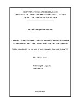
Table 1: Mean of nanocurcumin drops size at different Tween concentrations
Concentration
(%, w/v)
Hominization method
(m)
Ultrasound method
(m)
0.5
1
20.0263 0.095a
11.9255 0.230b
15.2262 0.097a
6.5169 0.131b
2 9.5789 0.108d 2.8208 0.007c
3
4
9.0657 0.209e
10.8139 0.176c
0.1138 0.004e
0.4191 0.008d
<i>In the same column, different values indicate differences according to the columns (p< 0.05) </i>
</div>
<span class='text_page_counter'>(4)</span><div class='page_container' data-page=4>
Figure 1. Diameter of nano emulsion at differences of Tween concentraions
The results of our research are quite consistent with the data from other scientists. For example,
nano-curcumin obtained from Kentish, S. research, also reached the average particle size of 0.135µm at 5%
Tween 40 concentration in 400W ultrasonic capacity, frequency of 20 kHz. The study of Ahmed, K. et al.
2012, for the average size of nano-curcumin reached 0.174µm at the concentration of emulsifiers (lipids)
of 10% (w / v). The study of D. Shailendiran et al., 2011 [17], for the average particle size reached
0.120µm with 5 minutes of ultrasound conditions. Another study by Sari. T. P and coworkers 2015, an
emulsion system of 0.04% CUR with a mixture of 2% Tween 80 and 0.5% whey protein concentrate 70
in ultrasonic conditions at 4oC in 15 minutes was carried out and reached the average particle size of
0.141µm [18].
(a) Hominization (b) Ultrasonication
Figure 2. Nano curcumin emulsion at Tween concentrations difference
<b>3.2. Effect of capacity to nano drops size </b>
The influence of equipment capacity on the process of creating nano-curcumin was presented in Table
2 and Figure 3. For the hominization method, when the rate increases from 5,000 to 20,000 rounds/min,
the average particle size tends to decrease sharply from 9.067 µ m to 0.619 µm. This could explain that
when the hominization rate increased, intermolecular bonds in both solvents and dispersants were cut off
and substances were easily distributed to each other and formed new systems with smaller dimensions
and more stable surface energy.
Our results were better than other studies using the hominization method. For example, the study of
Wang, X., J and colleagues 2008 at the rate of <13,000 rpm, the emulsion system was unsustainable and
phase separation after 24 hours of storage and large size 2-20 µm [20]. Another study by Giang P.V et al.,
0.0 0.5 1.0 1.5 2.0 2.5 3.0 3.5 4.0 4.5
0
5
10
15
20
Size of
na
no
(
m
)
Tween concentration (%)
</div>
<span class='text_page_counter'>(5)</span><div class='page_container' data-page=5>
2013 at the speed of 18,000 rpm for 15 minutes, the particle size only reached 0.269 µm.
Table 2: Mean of nano curcumin drops size at differences capacity
Capacity
(rpm)
Hominization method
(m)) Capacity (W/g)
Ultrasound method
(m)
5.000
10.000
9.067 0.014 a
1.934 0.006b
150
300
0.419 0.002 a
0.088 0.001b
15.000
20.000
1.253 0.013c
0.619 0.001d
450
600
0.058 0.002d
0.076 0.001c
<i>In the same column, different values indicate differences according to the columns (p< 0.05)</i>
Figure 3. Average particle size distribution at the hominization capacities: 5,000 rpm (a), 10,000 rpm (b),
15,000 rpm (c) and 20,000 rpm (d)
The results also showed that the ultrasound method is suitable for creating nano-emulsion.
Figure 4 indicated that drops size depends heavily on ultrasonic capacity. The average size of
drops decreased significantly from 0.419µm to 0.058 µm when the capacity increases from 150
- 450W/g. According to 1978's Li & Fogler, the reason could be explained that low-frequency
(20 - 100kHz) ultrasound waves exert a strong effect on the surface that divides between the
two phases, making it unstable leading to the explosion of dispersed phase (oil phase) in
continuous phase (water phase); In addition, the air bubbles (cavitation) in the liquid
environment (gas invasion) through each cycle (-) and cycle (+) appears. At that time, the
dispersed particles in the continuous phase are increasingly divided into the smallest [15].
However, the average particle size of 600W/g has an increasing tendency (0.076µm). This
result is also consistent with the study of Kentish, S., and colleagues [14]. This can be
explained by the high ultrasonic power for a certain period time, the "cavitation" bubble
explosion reaches critical and saturated point. At that time, the air bubbles formed not much
and had only the main stirring. The emitted ultrasonic energy (Bjerknes energy) is increased to
push the emulsion particles to enter junctions and collide with each other to recreate larger
sized particles according to Pangu & Feke in 2004 [17].
(a) <sub>(b)</sub><sub> </sub>
</div>
<span class='text_page_counter'>(6)</span><div class='page_container' data-page=6>
Figure 4. Average particle size distribution at the ultrasonic capacities: 150W/g (a), 300 W/g (b), 450W/g (c) and
600W/g (d)
The morphology of curcumin nanoemulsions using transmission electron microscopy (TEM) was
shown in Figure 5. Observation of nanoemulsion by TEM imaging is likely the best method to study
specimen’ morphology, purity and particle size distribution in non-solid disperse systems. As presented in
Figure 5, the spherically oil drops were monodispersed with uniform particle size, confirming the particle
size in Table 1 which was measured by using a dynamic light scattering instrument.
<b>Figure 5. Transmission electron microscopy (TEM) analysis of curcumin nanoelmusion </b>
<b>3.3. The efficiency of nano formation </b>
Nano-forming efficiency was calculated according to the formula in section 2.5. The results showed
that nano-elmusion production efficiency was quite high. The efficiency of the hominization method at
20,000 rpm reached 72.4 ± 5.0%, while the efficiency of the ultrasonic method was 87.5 ± 7.5%.
<b>3.4. Nano-emulsion stability </b>
The impact of storage time on the stability of a representative nano-emulsions: 0.5% curcumin, 2%
coconut oil, and 3% Tween 80 were examined. Figure 6 represents the droplet size of the emulsion at the
beginning and that after 4 months. The results in Figure 6 suggested that the mean droplet diameter did
not increase during storage time (during 4 months) and even has tended to get smaller. Droplet size
measurements were a good indicator of the formulation stability. It should be noted that, after a certain
time, the droplet diameter remained stable. Figure 7 indicated that the nanosystem was stable at pH
values from 3 to 7.
(a) (b)
</div>
<span class='text_page_counter'>(7)</span><div class='page_container' data-page=7>
Figure 6. Droplet size of nano curcumin emulsions before and after 4 months stored at 15oC.
Figure 7: Distribution of system size of curcumin nano at different pH
<b>4. CONCLUSIONS </b>
After a period of research on preparing nano curcumin by two methods, we found that the ultrasonic
method gave the size of the system smaller than the hominization method. However, the advantage of the
hominization method is the lower equipment cost, resulting in lower product costs compared to the
ultrasonic method. Compared with previously published studies, the nanoparticle size in this work was
smaller. The results showed that one of the two methods above was suitable to prepare nano curcumin
with the investigated parameters.
<b>ACKNOWLEDGMENT </b>
This project is funded by the Department of Science and Technology of Tien Giang Province under
Grant no DTYD03/17, 2018-2019.
<b>REFERENCES </b>
0.00 0.02 0.04 0.06 0.08 0.10
0
2
4
6
8
10
12
14
16
Distribu
tion
%
Diameter(m)
pH5
</div>
<span class='text_page_counter'>(8)</span><div class='page_container' data-page=8>
[1]. Ornchuma Naksuriya, Siriporn Okonogi, Raymond M. Schiffelers, Wim E. Hennink ,
Curcumin nanoformulations: A review of pharmaceutical properties and preclinical studies and clinical data related
to cancer treatment, Bio. Mat. 35 (2014) pp. 3365-3383
[2]. R. A. Freitas Jr., “What is nanomedicine?” Nanomedicine:Nanotechnology, Biology, and Medicine, vol. 1, no.
1, 2005, pp. 2–9.
[3]. Ahmed, K., Li, Y., McClements, D. J., & Xiao, H.. Nanoemulsion-and emulsion-based delivery systems for
curcumin: encapsulation and release properties. Foo. Chem., 132(2), (2012), pp.799-807.
[4]. R. Raveendran, G. Bhuvaneshwar, and C. P. Sharma, “In vitro cytotoxicity and cellular uptake of
curcumin-loaded Pluronic/ Polycaprolactone micelles in colorectal adenocarcinoma cells,” J. of Bio. Appli, vol. 27, no. 7,
2013, pp. 811-827.
[5]. S. Mangalathillam, N. S. Rejinold, A. Nair, V.-K. Lakshmanan,S. V. Nair, and R. Jayakumar, “Curcumin loaded
chitin nanogels for skin cancer treatment via the transdermal route,” Nanoscale,vol. 4, no. 1, 2012, pp. 239–250.
[6]. S. Mandal, C. Banerjee, S. Ghosh, J. Kuchlyan, and N. Sarkar, “Modulation of the photophysical properties of
curcumin in nonionic surfactant (Tween-20) forming micelles and niosomes:
a comparative study of different microenvironments,” J. Phy. Chem. B, vol. 117, no. 23, 2013, pp. 6957–6968,.
[7]. S. Rahman, S. Cao, K. J. Steadman,M.Wei, and H. S. Parekh, “Native and 𝛽-cyclodextrin-enclosed curcumin:
entrapment within liposomes and their in vitro cytotoxicity in lung and colon cancer,” Drug Del., vol. 19, no. 7,
2012, pp. 346–353.
[8]. S. Debnath, D. Saloum, S. Dolai et al., “Dendrimer-curcumin conjugate: a water soluble and effective cytotoxic
agent against breast cancer cell lines,” Anti-Cancer Agents in Med Chem., vol. 13, no. 10, 2013 pp. 1531–1539.
[9]. F. Akhtar, M. M. A. Rizvi, and S. K. Kar, “Oral delivery of curcumin bound to chitosan nanoparticles cured
Plasmodium yoelii infected mice,” Biotech. Adv, vol. 30, no. 1, 2012, pp. 310–320.
[10]. V. Kakkar, S. Singh, D. Singla, and I. P. Kaur, “Exploring solid lipid nanoparticles to enhance the oral
bioavailability of curcumin,”Mole.r Nutri. and Foo. Res, vol. 55, no. 3, 2011, pp. 495–503.
[11].
Ipar VS
,Dsouza A
,Devarajan PV
, Enhancing Curcumin Oral Bioavailability ThroughNanoformulations,
Eur. J. Drug. Metab. Pharmacokinet.
Feb 15, 2019 ,doi: 10.1007/s13318-019-00545-z.[12]. N.V.Hai, N. T. Cong, L. Q. Tri, N. T. Sang, T. Q. Hieu, Optimizing the process of extracting curcumin from
Curcuma Longa L. with the aid of ultrasonic waves, International Conference on Advanced Technology in Food
Science and Biotechnology, ISBN: 978-604-67-1137-7,2018, pp 18-30.
[13]. H. Rachmawati, L. Meylina, A. Rahma, and Y. C. Sumirtapur, Size-Dependent of Oil Droplet of Curcumin
Nanoemulsion on the In Vivo Release Kinetic of Curcumin After Oral and Intravenous Administrations in Animal
Model, Adv. Sci., Eng. and Med., Vol. 6, 2014, pp. 959–964.
[14]. S. Kentish., T. J. Wooster, M. Ashokkumar, S. Balachandran, R.Mawson andL. Simons, The use of ultrasonics
for nanoemulsion preparation. Inn. Foo. Sci & Emer. Tech. 9(2), 2008, 170-175.
[15]. Li, M. K., & Fogler, H. S. Acoustic emulsification. Part 1. The instability of the oil-water interface to form the
initial droplets. J. of Fl. Mech. 88(3), 1978, pp. 499-511.
</div>
<span class='text_page_counter'>(9)</span><div class='page_container' data-page=9>
[17]. D. Shailendiran, N. Pawar, A. Chanchal, R.P. Pandey, H. B. Bohidar, andA. K. Verma, Characterization and
antimicrobial activity of nanocurcumin and curcumin. In Nanoscience, Technology and Societal Implications
(NSTSI), IEEE, 2011 International Conference 2011, pp. 1-7.
[18]. T. P. Sari, B. Mann, R. Kumar, R. R. B. Singh, R. Sharma, M. Bhardwaj, andS. Athira, Preparation and
characterization of nanoemulsion encapsulating curcumin. Foo. Hydroc. 43, 2015, pp.540-546.
[19]. G.D. Pangu, D. L. Feke, Acoustically aided separation of oil droplets from aqueous emulsions. Chem. Eng.
Sci. 59(15),2004,pp.3183-3193.
[20]. X. Wang, Y. Jiang, Y.W. Wang, M.T. Huang, C. T. Ho, and Q. Huang, Enhancing anti-inflammation
activity of curcumin through O/W nanoemulsions. Foo. Chem. 108(2), 2008, pp.419-424.
<b>SO SÁNH KÍCH THƯỚC HẠT NANO CURCUMIN NHŨ TƯƠNG ĐƯỢC ĐIỀU </b>
<b>CHẾ BẰNG PHƯƠNG PHÁP ĐỒNG HĨA VÀ SIÊU ÂM </b>
<b>Tóm tắt: Trong bài báo này, hệ nano nhũ tương curcumin đã được điều chế thành công bằng cả hai </b>
phương pháp đồng hóa và siêu âm. Các điều kiện tối ưu cho phương pháp đồng hóa được thiết lập như
sau: nồng độ dầu sachi 2%, nồng độ chất nhũ hóa 3%, cơng suất 20.000 vịng/phút, thời gian đồng hóa là
60 phút, kích thước trung bình của hệ nano là 78 nm. Trong khi đó, các điều kiện tối ưu của phương pháp
siêu âm cũng được xây dựng như sau: nồng độ dầu sachi 2%, nồng độ chất nhũ hóa 2%, cơng suất siêu
âm 450 w/g, tần số 20 kHz, kích thước trung bình của giọt nước là 58 nm. Hệ thống nhũ tương nano đã
ổn định sau 4 tháng bảo quản lạnh.
<b>Từ khóa: hệ nhũ tương, curcumin, đồng hóa, siêu âm. </b>
</div>
<!--links-->
Tài liệu Báo cáo Y học: Role of electrostatics in the interaction between plastocyanin and photosystem I of the cyanobacterium Phormidium laminosum ppt
- 10
- 674
- 0


.push({});</script> </div> </div> </div> <div class="vf_link_relate px-2 my-2"> <h2 class="vf_doc_relate text-2xl font-bold my-4">Tài liệu liên quan</h2> <ul class="grid grid-cols-12 gap-2"> <li class="col-span-6 md:col-span-2"> <div class="card-doc " onclick="actionDocRelated(this)"> <a class="card-doc-img" href="https://text.123docz.net/document/1065142-tai-lieu-bao-cao-y-hoc-role-of-electrostatics-in-the-interaction-between-plastocyanin-and-photosystem-i-of-the-cyanobacterium-phormidium-laminosum-ppt.htm" title="Tài liệu Báo cáo Y học: Role of electrostatics in the interaction between plastocyanin and photosystem I of the cyanobacterium Phormidium laminosum ppt"> <i class="icon i_type_doc i_type_doc2"></i> <img class="lazy" src="data:image/gif;base64,R0lGODlhAQABAIAAAP///wAAACH5BAEAAAAALAAAAAABAAEAAAICRAEAOw==" data-src="https://media.store123doc.com/images/document/14/br/ey/medium_eyl1392920503.jpg" width="124" height="179" alt="Tài liệu Báo cáo Y học: Role of electrostatics in the interaction between plastocyanin and photosystem I of the cyanobacterium Phormidium laminosum ppt" onerror="this.src=)