- Trang chủ >>
- Văn Mẫu >>
- Văn Bản Mẫu
Determination of total polyphenol, saponin contents, antioxidant and antibacterial activities of Melastoma malabathricum leaves by liquid-liquid extraction
Bạn đang xem bản rút gọn của tài liệu. Xem và tải ngay bản đầy đủ của tài liệu tại đây (345.58 KB, 8 trang )
<span class='text_page_counter'>(1)</span><div class='page_container' data-page=1>
<i>DOI: 10.22144/ctu.jen.2020.002 </i>
<b>Determination of total polyphenol, saponin contents, antioxidant and antibacterial </b>
<i><b>activities of Melastoma malabathricum leaves by liquid-liquid extraction </b></i>
Huynh Thanh Duy, Nguyen Van Thanh, Le Nhu Thuy, Nguyen Trieu Nhat Uyen and Nguyen Duc Do*
<i>Biotechnology Research and Development Institute, Can Tho University, Vietnam </i>
<i>*Correspondence: Nguyen Duc Do (email: ) </i>
<b>Article info. </b> <b> ABSTRACT </b>
<i>Received 08 Jul 2019 </i>
<i>Revised 04 May 2020 </i>
<i>Accepted 31 Mar 2020</i>
<i><b> The present study is aimed to investigate the total polyphenol and saponin </b></i>
<i>contents, antioxidant and antibacterial activities of Melastoma </i>
<i>malabath-ricum leaves extracts. These fractions were carried out using hexane, </i>
<i>n-hexane:ethyl acetate (ratio 1:1), ethyl acetate and methanol solvents. The </i>
<i>crude extract (CE) exhibited the highest amount of total polyphenol content </i>
<i>by using the Folin-Ciocalteu assay. The n-hexane fraction (F1) showed the </i>
<i>highest total saponin content which was determined by vanillin/H2SO4</i>
<i>method. The antioxidant activity of extracts was evaluated by using three </i>
<i>different methods including DPPH radical scavenging assay, hydrogen </i>
<i>per-oxide assay (H2O2) and reducing power assay (Fe3+). The results showed </i>
<i>that the activity of the methanol fraction (F4) against DPPH was the </i>
<i>strong-est, the CE extract was the highest reducing power and the ethyl acetate </i>
<i>frac-tion (F3) illustrated the strongest antioxidant capacity through H2O2 assay. </i>
<i>Furthermore, all extracts were also tested for the antibacterial activity by </i>
<i>agar well diffusion method at a concentration of 100 mg/mL. F4 showed the </i>
<i>highest activity against Escherichia coli and Staphylococcus aureus, while </i>
<i>there were no significant differences among all extracts when testing with </i>
<i>Lactobacillus acidophilus. These findings provide important evidence that </i>
<i>there is a correlation between the polyphenol content and antioxidant </i>
<i>capac-ity and antibacterial activcapac-ity. Besides, the saponin content was no </i>
<i>contribu-tion to antioxidant and antibacterial abilities. </i>
<i><b>Keywords </b></i>
<i>Antibacterial, antioxidant, </i>
<i>fraction, Melastoma </i>
<i>mala-bathricum, polyphenol, </i>
<i>sapo-nin </i>
Cited as: Duy, H.T., Thanh, N.V., Thuy, L.N., Uyen, N.T.N and Do, N.D., 2020. Determination of total
<i>polyphenol, saponin contents, antioxidant and antibacterial activities of Melastoma malabathricum </i>
<i>leaves by liquid-liquid extraction. Can Tho University Journal of Science. 12(1): 8-15. </i>
<b>1 INTRODUCTION </b>
Plants derived natural products are the source of
most active components of medications, which in
turn play a significant role in the treatment or
pre-vention of human illnesses. The tropical plants have
been investigated intensively during the last decades
in order to evaluate the possibility of developing
new, sustainable, natural and affordable cosmetics
<i>and drugs (Alnajar et al., 2012). </i>
</div>
<span class='text_page_counter'>(2)</span><div class='page_container' data-page=2>
Plant constituents, namely flavonoid and phenolic
compounds and ginsenosides are broadly distributed
and have been reported to exert multiple biological
effects including antioxidant, anti-inflammatory,
anticarcinogenic and anticancer activities (Sharma
<i>et al., 2011; Lu et al., 2009). </i>
Melastomataceae plants originate in the tropic and
subtropical regions, with a total of more than 4,000
species in the world. In the Southeast Asian region
<i>alone, the genus Melastoma comprises 22 species, </i>
two subspecies, and three varieties. It is native to
tropical and temperate Asia and the Pacific Islands.
The plant is one of the most common weeds that
grow wildly and abundantly throughout the tropics,
especially in the moist areas, and can be found in the
Indian Ocean Islands, throughout South and
South-East Asia, China, Taiwan, Australia, and the South
Pacific Ocean. Various scientific papers were
<i>pub-lished on pharmacological properties of Melastoma </i>
<i>malabathricum (Mua), the detailed and careful </i>
anal-ysis revealed that it exhibited promising an
<i>anti-in-flammatory, antifungal and antioxidant (Joffry et </i>
<i>al., 2012). In addition, the most recent research on </i>
<i>M. malabathricum revealed that its bioactive </i>
con-stituents exhibited free radical scavenging,
anti-in-flammatory, antibacterial and antiviral activities
<i>(Alnajar et al., 2012). The antibacterial and </i>
<i>phyto-chemical screening of Memecylon umbellatum </i>
Burm leaves extract was shown to inhibit the growth
<i>of Escherichia coli and Staphylococcus aureus </i>
<i>(Killedar et al., 2012). Basing on the latest </i>
refer-ences, there are no such phytochemical reports
<i>con-cerning M. malabathricum, so the present study was </i>
designed to determine total polyphenol and saponin
contents, antioxidant and bacterial inhibition
<i>property of extract of wild Melastoma </i>
<i>malabathri-cum found in the Mekong Delta of Vietnam. </i>
<b>2 MATERIALS AND METHODS </b>
<b>2.1 Materials </b>
<i>2.1.1 Plant materials </i>
<i>The healthy M. malabathricum leaves, which were </i>
not damaged by disease, insects or mechanical
in-jury, were collected in the early morning from Hung
Phu, Cai Rang district, Can Tho city.
<i>2.1.2 Microorganisms </i>
Microorganisms used in this study were both
<i>Gram-negative bacteria (E. coli) and Gram-positive </i>
<i>bacte-ria (L. acidophilus and S. aureus). All the stock </i>
cul-tures were obtained from the Molecular Biology lab,
Biotechnology Research and Development Institute,
Can Tho University, Vietnam. Lysogeny broth (LB)
was used as the media for the culturing of bacterial
strains.
<i>2.1.3 Chemicals </i>
For natural compounds extraction, these chemicals
were used ethanol (Vietnam), n-hexane (Vietnam),
ethyl acetate (Vietnam), methanol (Vietnam),
dis-tilled water, Na2SO4 anhydrous (China). Testing for
antioxidant activity used FeCl3.6H2O (China),
H2SO4 (China), gallic acid (China), Folin –
Ciocal-teu (Germany), Na2CO3 (China), H2O2 30%
(China), NaH2PO4.2H2O (China), Na2HPO4.12H2O
(China), K3[Fe(CN)6] (China), CCl3COOH (China),
2,2-diphenyl-1-picrylhydrazyl (DPPH)(US),
ascor-bic acid (China). Antibacterial tests used peptone
(India), yeast extracts (Germany).
<b>2.2 Methods </b>
<i>2.2.1 Fractionation procedure </i>
<i>A total of 700 g of dried M. malabathricum leaves </i>
were ground with a blender and extracted with 3.5 L
ethanol 96% and combined with ultrasonic at 120W
within 60 minutes. The extract was filtrated with
Na2SO4 anhydrous and then evaporated in a rotary
evaporator under reduced pressure, collected 50 g
<i>crude extract of M. malabathricum (CE). Next, 20 g </i>
of CE was dissolved with 50 mL of distilled water
before extracting with of n-hexane (F1),
n-hex-ane:ethyl acetate (1:1) (F2), ethyl acetate (F3) and
the residue (F4) which was mixed with methanol,
respectively by using liquid/liquid extraction. All
solutions were rotated and vacuumed until the
sol-vent has evaporated. Finally, the factions were
stored at -4o<b><sub>C. </sub></b>
<i>2.2.2 Determination of total polyphenol content </i>
<i>(TPC) </i>
The TPC was estimated according to the procedure
of Yadav and Agarwala (2011) with a few
modifi-cations. Briefly, the reaction mixture contained 0.1
mL of plant extract with 1 mL of Folin – Ciocalteu
reagent and 1 mL of 2% solution Na2CO3 were then
added. The absorbance was measured at 765 nm
af-ter 45 minutes of incubation at room temperature.
Gallic acid was used as standard with concentration
ranging from 20 to 120 μg/mL (blank sample was
methanol). All the samples were measured in
tripli-cate. The results were expressed as gallic acid
equiv-alent (mg GAE/g of extracted compound) and
deter-mined from standard curve (y = ax + b) of gallic
acid.
</div>
<span class='text_page_counter'>(3)</span><div class='page_container' data-page=3>
Where, C: total polyphenol content (mg GAE/g of
extracted compound); c: x value from gallic acid
standard curve (μg/mL); V: volume of samples
(mL); m: mass of samples in V (g).
<i>2.2.3 Determination of total saponin content </i>
<i>(TSC) </i>
The TSC was estimated according to the procedure
<i>of Hiai et al. (1976) with a few modifications. 0.5 </i>
mL of samples was mixed with 0.2 mL of 4% (w/v)
vanillin reagent and then 1.8 mL of 70% (v/v)
sul-furic acid was added. After that, the mixture in test
tubes was shaken before incubating at 60o<sub>C for 10 </sub>
minutes in a water bath, then for color development
cooled in an ice-water bath for 15 minutes. Blank
sample was mixtures of vanillin solution and
sulfu-ric acid. The absorbance was measured at 550 nm.
Ginsenoside (Rb1, Rg1, Rg3) was used as standard
with concentration ranging from 10 to 60 μg/mL.
All the samples were measured in triplicate. Result
was expressed as ginsenoside (Rb1, Rg1, Rg3)
equivalent (mg/g of extracted compound) and
deter-mined from standard curve (y = ax + b) of
ginseno-side (Rb1, Rg1, Rg3).
Total saponin content: 𝑆 = 𝑐. 𝑉/𝑚
Where, S: total saponin content (mg/g of extracted
compound); c: x value from saponin (Rb1, Rg1,
Rg3) standard curve (μg/mL); V: volume of samples
(mL); m: mass of samples in V (g).
<i>2.2.4 DPPH (2,2-diphenyl-1-picrylhydrazyl) </i>
<i>scavenging assay </i>
The free radical scavenging activity was determined
by the DPPH assay described by Blois (1958) with
a few modifications. Briefly, 1.5 mL of the extract
or ascorbic acid (control sample) at different
con-centrations in methanol was added to 0.5 mL of 0.1
mM DPPH solution into test tubes. The mixture was
then incubated in darkness at room temperature for
30 minutes, and its absorbance pouring into a
cu-vette was measured at 517 nm. Ascorbic acid was
used as a control sample with concentration ranging
from 0.5 to 2.5 μg/mL. Blank sample was a mixture
of methanol and DPPH solution. All the samples
were measured in triplicate. The percentage of the
DPPH radical scavenging was calculated as the
fol-lowing equation:
% scavenging of DPPH free radicals = [(𝐴𝑜 −
𝐴)/𝐴<sub>𝑜</sub>]. 100
Where, Ao: the absorbance of blank sample; A: the
absorbance of extracts or ascorbic acid.
The standard curve (y = ax + b) of ascorbic acid or
extracts was established from percentage inhibition
at its different concentrations. Then, the IC50 value
of extracts or ascorbic acid was calculated.
<i>2.2.5 Hydrogen peroxide (H2O2) scavenging </i>
<i>assay </i>
The ability of plant extracts to scavenge hydrogen
peroxide can be estimated according to the method
<i>of Sharma et al. (2011). A solution of hydrogen </i>
per-oxide (40 mM) was prepared in phosphate buffer
(0.05 M pH 7.4). Briefly, 0.5 mL of the extracts or
ascorbic acid (control sample) at different
concen-trations in phosphate buffer was added to 2.5 mL of
hydrogen peroxide into test tubes. After 10 minutes,
the absorbance was measured at 230 nm against a
blank sample containing samples in phosphate
buffer without hydrogen peroxide. Ascorbic acid
was used as a control sample with concentration
ranging from 50 to 100 μg/mL. All the samples were
measured in triplicate. The percentage of
hydro-gen peroxide scavenging was calculated as the
following equation:
% scavenging of H2O2 free radicals = [(𝐴𝑜 −
𝐴)/𝐴<sub>𝑜</sub>]. 100
Where, Ao: the absorbance of blank sample; A: the
absorbance of extracts or ascorbic acid.
The standard curve (y = ax + b) of ascorbic acid or
extracts was established from percentage inhibition
at its different concentrations. Then, the IC50 value
of extracts or ascorbic acid was calculated.
<i>2.2.6 Reducing power assay (Fe3+<sub>) </sub></i>
The method was based on the principle of increase
in the absorbance of reaction mixtures that indicated
the power of the samples and was described by
<i>Singhal et al. (2014). Briefly, 90 μL of a test sample </i>
solution in distilled water, 225 μL of 0.2M
phos-phate buffer (pH 6.6) and 225 μL of 1% (w/v)
po-tassium ferricyanide were added into test tubes. The
resulting mixture was incubated at 50o<sub>C for 20 </sub>
</div>
<span class='text_page_counter'>(4)</span><div class='page_container' data-page=4>
% scavenging of Fe3+<sub> = [(𝐴 − 𝐴</sub>
𝑜)/𝐴𝑜]. 100
Where, Ao: the absorbance of blank sample; A: the
absorbance of extracts or ascorbic acid.
The standard curve (y = ax + b) of ascorbic acid or
extracts was established from percentage inhibition
at its different concentrations. Then, the IC50 value
of samples or ascorbic acid was calculated.
<i>2.2.7 Antibacterial activity </i>
Antibacterial activity was determined by using the
<i>agar well diffusion method (Balouiri et al., 2016). </i>
All fractions and crude extracts were prepared in
di-methyl sulfoxide (DMSO) and 100 mg/mL of each
was used for activity, DMSO was used as negative
control and ampicillin 0.5 mg/mL was used as
<i>con-trol sample for E. coli and S. aureus, and ampicillin </i>
<i>0.05 mg/mL as the control sample for L. </i>
<i>acidophi-lus. The LB agar plates were uniformly smeared </i>
with the suspension of bacteria. Wells (6 mm
diam-eter) were created, to which 20 μL of different
sam-ples were loaded into each well. The plates were
incubated at 37o<sub>C for 24 hours, after that the test </sub>
ma-terials having antibacterial activity inhibited the
growth of microorganisms and a clear, distinct zone
of inhibition was visualized surrounding the well.
<b>2.3 Statistical analysis </b>
The data were processed by Excel 2010 software
and analyzed by Minitab (version 16), ANOVA
analysis. The mean values were compared by the
Tukey test. All the samples of each assay were
meas-ured in triplicate.
<b>3 RESULTS AND DISCUSSIONS </b>
<b>3.1 Determination of TPC and TSC </b>
The total polyphenol content was highest in CE (430
mg GAE/g extract), which was no significant
differ-ence with F4 (420 mg GAE/g extract) and F3 (344
mg GAE/g extract). F1 (97.5 mg GAE/g extract)
was the lowest total polyphenol content. No
signifi-cant differences were found between F1 and F2 (128
<b>mg GAE/g extract). The solvent with higher polarity </b>
would extract a higher amount of total polyphenol
<i>content from M. malabathricum leaves. Table 1 </i>
exhibited the results of total polyphenol and saponin
<i>contents of crude extract and fractions from M. </i>
<i>mala-bathricum leaves. </i>
<i>Polar solvent extract (methanol extract) from M. </i>
<i>malabathricum leaves contained higher TPC than </i>
non-polar solvent extract (chloroform extract)
<i>(Za-karia et al., 2011). According to Handique and </i>
<i>Gogoi (2016), the dry powders of M. </i>
<i>malabathri-cum leaves were extracted in n-hexane, ethyl acetate </i>
and methanol by using Soxhlet extraction. The
re-sults were a similar trend to this study that the
meth-anol extract contained the highest TPC with 9.6 mg
GAE/g extract while the lowest was found in
n-hex-ane extract (0.798 mg GAE/g extract). The TPC of
all extracts in the present study was many times
<i>higher than M. malabathricum extract of this report </i>
due to different extraction and quantification
meth-ods were employed.
<b>Table 1: TPC and TSC of crude extract and </b>
<i><b>frac-tions of M. malabathricum leaves</b></i>
<b></b>
<b>Treat-ments </b>
<b>TPC (mg GAE/g </b>
<b>extract) </b>
<b>TSC (mg/g </b>
<b>ex-tract) </b>
CE 430.0±15.30a <sub>41.3±1.78</sub>d
F1 97.5±6.56c <sub>95.2±1.98</sub>a
F2 128.0±7.65c <sub>71.1±1.74</sub>b
F3 344.0±2.85b <sub>58.5±2.90</sub>c
F4 420.0±3.27ab <sub>32.6±2.00</sub>d
<i>Where: M. malabathricum crude extract (CE). n-hexane </i>
<i>fraction (F1), n-hexane:ethyl acetate (1:1) fraction (F2), </i>
<i>ethyl acetate fraction (F3) and the residue (F4). Values </i>
<i>are expressed as mean ± standard deviation for triplicate. </i>
<i>Values with the same superscripts in the same column are </i>
<i>not significantly different at 99% level of confidence based </i>
<i>on Tukey test (p<0.01). TPC was calculated based on the </i>
<i>standard curve of gallic acid: y = 0.0036x – 0.0009, R2<sub> = </sub></i>
<i>0.9919. TSC was calculated based on the standard curve </i>
<i>of ginsenoside (Rb1, Rg1, Rg3): y = 0.0176x + 0.019; R2</i>
<i>= 0.9934. </i>
</div>
<span class='text_page_counter'>(5)</span><div class='page_container' data-page=5>
Shibata’s group, and named as Rx according to
theirmobility on TLC plates, with polarity
<i>de-creasing from index “a” to “h” (Nag et al., </i>
2012). This report indicates that Rb1
com-pound is more polar than Rg1 and Rg3 so that
<i>the M. malabathricum</i> extracts maybe contain a
high amount of Rg1 and Rg3 compounds.
<b>3.2 Determination of antioxidant activities </b>
In the present study, the radical scavenging ability
<i>of the crude extract and fractions of M. </i>
<i>malabathri-cum leaves was studied against DPPH, H</i>2O2 and
Fe3+<sub>. The results were expressed by IC</sub>
50 (the
con-centration of extract required to quench 50% of free
radicals under the given experimental conditions).
The extract with lower IC50 value has stronger
anti-oxidant potential.
Based on the results shown in Table 2, IC50 was the
lowest in F4 (1.61 μg/mL) which indicated that this
extract had the strongest antioxidant ability to
scav-enge DPPH radical. No significant differences were
found between ascorbic acid (1.52 μg/mL), CE
(1.65 μg/mL) and F4. F1 was the highest IC50 value
(8.29 μg/mL) and had the weakest antioxidant
<i>ca-pacity. The M. malabathricum leaves were extracted </i>
with polar solvents showed higher antioxidant
ca-pacity than non-polar solvent. The methanol extract
<i>of M. malabathricum had strongest antioxidant </i>
ca-pacity with IC50 value at 37.26 μg/mL while the
n-hexane extract was the weakest (IC50 value at 314.51
μg/mL) compared to the IC50 value of Trolox (as a
control) was 29.19 μg/mL (Handique and Gogoi,
2016). The results of the report above shows a
sim-ilar trend with this study on the effect of solvents
extraction on antioxidant activity.
<b>Table 2: IC50</b><i><b> values (μg/mL) of ascorbic acid, crude extract and fractions of M. malabathricum in </b></i>
<b>dif-ferent assays </b>
<b>Treatments </b>
<b>IC50 values (μg/mL) </b>
<b>DPPH scavenging assay </b> <b>Hydrogen peroxide </b>
<b>(H2O2) scavenging assay </b>
<b>Reducing power (Fe3+<sub>) </sub></b>
<b>assay </b>
CE 1.65±0.04d <sub>37.5±0.15</sub>d <sub>3.42±0.05</sub>c
F1 8.29±0.16a <sub>50.0±0.83</sub>b <sub>24.2±1.26</sub>a
F2 7.13±0.20b <sub>41.2±0.56</sub>c <sub>23.0±2.71</sub>a
F3 2.35±0.05c <sub>33.5±0.38</sub>e <sub>9.15±0.18</sub>b
F4 1.61±0.01d <sub>35.6±0.05</sub>ed <sub>4.10±0.24</sub>c
Ascorbic acid 1.52±0.02d <sub>88.7±1.22</sub>a <sub>2.14±0.16</sub>c
<i>Where: M. malabathricum crude extract (CE). n-hexane fraction (F1), n-hexane:ethyl acetate (1:1) fraction (F2), ethyl </i>
<i>acetate fraction (F3) and the residue (F4). IC50 values are expressed as mean ± standard deviation for triplicate. Values </i>
<i>with the same superscripts in the same column are not significantly different at 99% level of confidence based on Tukey test </i>
<i>(p<0.01). </i>
In the H2O2 scavenging assay, all extracts had an
an-tioxidant activity stronger than ascorbic acid.
Partic-ularly, IC50 value was the highest in F1 (50 μg/mL)
which showed the weakest antioxidant capacity. F3
(IC50 value at 33.5 μg/mL) had the strongest H2O2
free radical scavenging, which was no significant
difference with F4 (IC50 value at 35.6 μg/mL). No
significant differences were found between CE (IC50
value at 37.5 μg/mL) and F4. The results also
<i>showed that M. malabathricum leaves were </i>
ex-tracted with low polarity solvents had stronger
anti-oxidant capacity than non-polar solvents and high
<i>polarity solvents. According to Ruskin et al. (2017), </i>
<i>Canthium coromandelicum leaves were extracted </i>
with different solvents, namely chloroform, ethyl
acetate and ethanol. The results showed a similar
trend with this study on the peroxide scavenging
ca-pacity had the strongest in ethyl acetate extract.
Based on the results of reducing power (Fe3+<sub>) assay, </sub>
the IC50 value was the highest in F1 (24.2 μg/mL)
which was no significant difference with F2 (IC50
value at 23 μg/mL). F4 was the lowest in IC50 value
(4.1 μg/mL) indicating that this extract had the
strongest antioxidant capacity. No significant
differ-ences were found between CE (IC50 value at 3.42
μg/mL), F4 (IC50 value at 4.1 μg/mL) and ascorbic
acid (IC50 value = 2.14 μg/mL). According to Sudan
<i>et al. (2014), fractions of Arisaema jacquemontii </i>
</div>
<span class='text_page_counter'>(6)</span><div class='page_container' data-page=6>
was the weakest (150 mmol/g dry weight). The
re-sults also showed a similar trend to the present study
that samples were extracted with polar solvents had
stronger a reducing power capacity than non-polar
solvents.
<i>Through three antioxidant assays, M. malabathricum </i>
leaves which were extracted with high polar
sol-vents (ethanol and methanol), had the strongest
an-tioxidant capacity in DPPH and reducing power
as-say, whereas the H2O2 scavenging activity was the
strongest when the extract was fractionated by
me-dium polar solvents (ethyl acetate). Polyphenol
con-tents act as antioxidants because they have hydroxyl
groups that can release protons in the form of
hydro-gen ions (Marjoni and Zulfisa, 2017). According to
Hadique and Gogoi (2016), polyphenol contents are
potential antioxidants and free radical-scavengers,
hence there should be a close correlation between
the content of polyphenol and antioxidant activity.
Moreover, polyphenols are very valuable plant
con-stituents in the scavenging of free radicals, due to
their several phenolic hydroxyl groups. The amount
of polyphenolic compound increases, antioxidant
activity increases as well (Saha and Verma, 2016).
The results of this study also showed a similar trend
to the reports above on the total polyphenol content
that is significantly correlated with antioxidant
ca-pacity. Indeed, CE and F4 contained a high amount
of polyphenol content, so they had strong
antioxi-dant activity. However, the TPC was not a
correla-tion with H2O2 scavenging activity because the
dif-ferences in H2O2 scavenging capacity among the
ex-tracts can be attributed to the structural features of
their active components, which determine their
<i>elec-tron-donating abilities (El-Chaghaby et al., 2014). In </i>
addition, different methods to measure antioxidant
ac-tivity with various mechanisms may lead to different
<i>observations (Sun et al., 2005) so that the results of </i>
dif-ferent antioxidant assays (DPPH, H2O2 and reducing
power) were different. Furthermore, the antioxidant
activity of a plant does not rely solely on phenolic
compounds, but also on other substances such as
<i>ca-rotenoids, vitamins and minerals (Tan et al., 2011). </i>
Besides, the TSC was not related to the antioxidant
<i>capacity (Table 1 and 2). According to Lee et al. </i>
(2016), antioxidant activity was not proportional to
ginsenoside Rg1 content and significant correlation
was observed. Considering ginsenoside’s chemical
structures, they are not electron-rich compounds
like phenolic compounds, which are stabilized by
the resonance delocalization of the unpaired
elec-trons comprising the ring. Thus, ginsenosides are
not easily prone to enter into efficient
electron-do-nation reactions with oxidizing agents. Because of
the weak degree of electron-donating ability of
gin-senosides, they are probably poor radical scavengers
<i>in an antioxidant assay (Chae et al., 2010). </i>
<b>3.3 Antibacterial activity </b>
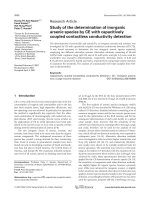
The Figure 1 exhibited the results of the
antibacterial activity of crude extract and frations
<i>from M. malabathricum leaves </i>
<b> (1) (2) (3) </b>
<i><b>Fig. 1: The antibacterial activity of fractions of M. malabathricum leaves against E. coli (1), L.</b></i>
<i><b>acidophilus (2) and S. aureus (3) at the concentration in 100 mg/mL </b></i>
<i>All extracts from M. malabathricum leaves had </i>
activity against three testing bacteria (Table 3). CE
and F4 exhibited the greatest inhibitory activity
<i>against E. coli and S. aureus which were not </i>
significantly different to those and ampicillin
<i>(p<0.05). The effectiveness against E. coli, L. </i>
<i>acidophilus and S. aureus were arranged in </i>
descending order of CE, F4, F3, F2 and F1. The
<i>antimicrobial effect of all extracts against L. </i>
<i>acidophilus were not significantly different (Fig. 1). </i>
</div>
<span class='text_page_counter'>(7)</span><div class='page_container' data-page=7>
<i><b>Table 3: The antibacterial activity of fractions and crude extract of M. malabathricum against various </b></i>
<b>bacteria in the concentration of 100 mg/mL after 24 hours of culture </b>
<b>Treatments </b> <b>Inhibition zone diameter (mm) </b>
<i><b>Escherichia coli </b></i> <i><b>Lactobacillus acidophilus </b></i> <i><b>Staphylococcus aureus </b></i>
CE 8.8±0.3a <sub>13.0 ±0.0</sub>b <sub>13.3±1.0</sub>a
F1 2.9±0.3c <sub>11.7±0.6</sub>b <sub>9.0±0.5</sub>b
F2 4.3±0.6bc <sub>11.7±0.6</sub>b <sub>10.0±0.1</sub>ab
F3 7.4±1.2ab <sub>12.2±0.3</sub>b <sub>11.7±0.6</sub>ab
F4 8.5±1.0a <sub>12.3±0.6</sub>b <sub>12.7±2.3</sub>a
Ampicillin 6.3±0.8ab <sub>15.3±1.2</sub>a <sub>12.0±1.0</sub>ab
<i>Where: M. malabathricum crude extract (CE). n-hexane fraction (F1), n-hexane:ethyl acetate (1:1) fraction (F2), ethyl </i>
<i>acetate fraction (F3) and the residue (F4). The diameters of inhibition zone values are expressed as mean ± standard </i>
<i>devi-ation for triplicate. Values with the same superscripts in the same column are not significantly different at 95% level of </i>
<i>con-fidence based on Tukey test (p<0.05). Ampicillin (0,5 mg/mL) was used as a control sample for E. coli and S. aureus, and </i>
<i>ampicillin 0.05 mg/mL as a control sample for L. acidophilus. </i>
<i>The bacterial inhibitory ability of M. malabathricum </i>
extract was due to the compounds it contained.
Ac-cording to Cowan (1999), phytochemical
com-pounds such as phenolics, tannins, flavonoids,
alka-loids and quinone had the antimicrobial activity. In
particular, the F4 and CE, which were extracted in
high polarity solvents contained high polyphenol
content (Table 1). The reason why polyphenol had
the antibacterial activity as polyphenols were known
as a factor that inactivated cellular enzymes or
caused changes in membrane permeability (Moreno
<i>et al., 2006). In addition, phenolic compounds could </i>
form ligands with many metal ions such as ferric or
cupric ions, which could cause iron deprivation in
bacteria or formed hydrogen bonds with vital
pro-teins such as microbial enzymes and thus inhibited
many enzymes (Scalbert, 1991). Therefore, bacteria
would be inhibited their growth and population.
<i>Ac-cording to Killedar et al. (2012) using high polarity </i>
solvents such as ethanol and methanol exhibited
<i>po-tent inhibitory E. coli and S. aureus of the extracted </i>
<i>from Memecylon umbellatum (Melastomaceae </i>
<i>fam-ily) leaves, which was supported by the findings of </i>
<i>this study. Another report of Alwash et al. (2013) </i>
detected kaempferol (Kf) compound in the methanol
<i>extract of M. malabathricum L. leaf. This compound </i>
<i>inhibited Staphylococcus sp. with the value of the </i>
zone of the inhibition was 15.67 ± 0.58 mm and
MIC value 0.25 mg/mL. Kaempferol is a natural
fla-vonol found in many plants. Therefore, flavonoid
<i>compound present in M. malabathricum extract may </i>
be kaempferol and this compound has bioactive
po-tential activity.
<b>4 CONCLUSIONS </b>
In this present study, the correlation between
poly-phenol content and antioxidant capacity and
antimi-crobial activity in solvents with different polarity
and ratio were found. Besides, the saponin content
might not be attributed to antioxidant and
<i>antibacte-rial activities. The crude extract of M. </i>
<i>malabathri-cum is one promising source of the natural </i>
antioxi-dants as well as antimicrobial agents. Isolation and
identification of active compounds in the crude
ex-tract are needed in future research which could be
used for agriculture and pharmacy.
<b>REFERENCES </b>
Alnajar, Z.A.A., Abdulla, M.A., Ali, H.M., Alshawsh,
M. A., and Hadi, A. H. A., 2012. Acute toxicity
eval-uation, antibacterial, antioxidant and
<i>immunomodu-latory effects of Melastoma malabathricum. </i>
Mole-cules. 17(3): 3547-3559.
Alwash, M.S., Ibrahim, N., and Ahmad, W.Y.W., 2013.
Identification and mode of action of antibacterial
<i>components from Melastoma malabathricum Linn </i>
leaves. American Journal of Infectious
Dis-eases. 9(2): 46-58.
Balouiri, M., Sadiki, M., and Ibnsouda, S.K., 2016.
Methods for in vitro evaluating antimicrobial
activ-ity: A review. Journal of Pharmaceutical
Analy-sis. 6(2): 71-79.
Blois, M.S., 1958. Antioxidant determinations by the use of
a stable free radical. Nature. 181: 1199–1200.
Chae, S., Kang, K.A., Youn, U., Park, J.S., and Hyun,
J.W., 2010. A comparative study of the potential
an-tioxidant activities of ginsenosides. Journal of Food
Biochemistry. 34(s1): 31-43.
</div>
<span class='text_page_counter'>(8)</span><div class='page_container' data-page=8>
El-Chaghaby, G.A., Ahmad, A.F., and Ramis, E.S., 2014.
Evaluation of the antioxidant and antibacterial
<i>proper-ties of various solvents extracts of Annona squamosa L. </i>
leaves. Arabian Journal of Chemistry. 7(2): 227-233.
Handique, J.G., and Gogoi, D., 2016. Antioxidant
activi-ties of the medicinal plants used for preparation of
fermentation cakes of “Haanj”, the rice based
alco-holic beverage of Ahom community people of
As-sam, India. Int J Pharmacog Phytochem Res. 8(2):
217-222.
Hiai, S., Oura, H. and Nakajima T., 1976. Color reaction of
some sapogenins and saponins with vanillin and sulfuric
acid. Planta Med. 29(2): 116-22.
<i>Joffry, S.M., Yob, N.J., Rofiee, M.S., et al., 2012. </i>
<i>Me-lastoma malabathricum (L.) Smith ethnomedicinal </i>
uses, chemical constituents, and pharmacological
properties: a review. Evidence-Based
Complemen-tary and Alternative Medicine, 48 pages.
Killedar, S.G., and Harinath, N., 2012. Antimicrobial
and phytochemical screening of different leaf
<i>ex-tracts of Memecylon umbellatum Burm. International </i>
Research Journal of Pharmacy. 3(2): 188-192.
<i>Kim, J.H., Hong, Y.H., Lee, J.H., et al., 2005. A role for </i>
the carbohydrate portion of saponin Rg3 in Na+
channel inhibition. Molecules & Cells (Springer
Sci-ence & Business Media BV). 19(1): 137-142.
<i>Lee, J.W., Mo, E.J., Choi, J.E., et al., 2016. Effect of </i>
Korean Red Ginseng extraction conditions on
antiox-idant activity, extraction yield, and ginsenoside Rg1
and phenolic content: Optimization using response
surface methodology. Journal of Ginseng
Re-search. 40(3): 229-236.
<i>Lee, S.M., Bae, B.S., Park, H.W., et al., 2015. </i>
<i>Character-ization of Korean Red Ginseng (Panax ginseng </i>
Meyer): history, preparation method, and chemical
composition. Journal of Ginseng Research. 39(4):
<b>384-391. </b>
Lu, J.M., Yao, Q., and Chen, C., 2009. Ginseng
com-pounds: an update on their molecular mechanisms
and medical applications. Current Vascular
Pharma-cology. 7(3): 293-302.
Marjoni, M.R., and Zulfisa, A., 2017. Antioxidant
activ-ity of methanol extract/fractions of senggani leaves
<i>(Melastoma candidum D. Don). Pharmaceutica </i>
Ana-lytica Acta. 8(557): 2-6.
Moreno, S., Scheyer, T., Romano, C.S., and Vojnov,
A.A., 2006. Antioxidant and antimicrobial activities
of rosemary extracts linked to their polyphenol
com-position. Free Radical Research. 40(2): 223-231.
Nag, S.A., Qin, J., Wang, W., Wang, M.H., Wang, H., &
Zhang, R., 2012. Ginsenosides as anticancer agents:
in vitro and in vivo activities, structure-activity
rela-tionships, and molecular mechanisms of
ac-tion. Frontiers in Pharmacology. 3: 25.
Ruskin, S.R., Vasanthakumari, B. and Citarasu, T., 2017.
<i>In vitro antioxidant activity of various leaf extracts </i>
<i>of Canthium coromandelicum (Burm.f.) Alston. </i>
Asian Journal Pharmaceutical and Clinical Research.
10(5): 214-218.
Saha, S., and Verma, R.J. 2016. Antioxidant activity of
<i>polyphenolic extract of Terminalia chebula Retzius </i>
fruits. Journal of Taibah University for Science.
10(6): 805-812.
Scalbert, A., 1991. Antimicrobial properties of
tan-nins. Phytochemistr., 30(12): 3875-3883.
Sharma, H.K., and Kumar, A., 2011. Evaluation of total
phenol, flavonoid and in vitro antioxidant activity of
<i>methanolic extract of leaves of Melastoma </i>
<i>malabath-ricum Linn. Asian Journal of Chemistry. 23(1): 434. </i>
Singhal, M., Paul, A., and Singh, H.P., 2014. Synthesis
and reducing power assay of methyl semicarbazone
derivatives. Journal of Saudi Chemical Society.
18(2): 121-127.
Sudan, R., Bhagat, M., Gupta, S., Singh, J., and Koul,
A., 2014. Iron (Fe II) chelation, ferric reducing
anti-oxidant power, and immune modulating potential of
<i>Arisaema jacquemontii (Himalayan Cobra Lily). </i>
Bi-oMed Research International. 2014: 7.
Sun, T., and Ho, C.T., 2005. Antioxidant activities of
buckwheat extracts. Food Chemistry. 90(4): 743-749.
Tan, P.W., Tan, C.P., and Ho, C.W., 2011. Antioxidant
prop-erties: Effects of solid-to-solvent ratio on antioxidant
<i>compounds and capacities of Pegaga (Centella </i>
<i>asiat-ica). International Food Research Journal. 18: 557-562. </i>
Yadav, R.N.S., and Agarwala M., 2011. Phytochemical
analysis of some medicinal plants. Journal of Phytology.
3(12): 10-14.
</div>
<!--links-->