Big Data on Real-World Applications. Chapter 5: PESSCARA: An Example Infrastructure for Big Data Research
Bạn đang xem bản rút gọn của tài liệu. Xem và tải ngay bản đầy đủ của tài liệu tại đây (1.09 MB, 15 trang )
<span class='text_page_counter'>(1)</span><div class='page_container' data-page=1>
PESSCARA: An Example Infrastructure for Big Data Research
<b>RESEARCH-ARTICLE </b>
Panagiotis Korfiatis and Bradley Erickson∗
Show details
<b>Abstract </b>
Big data requires a flexible system for data management and curation which has to be intuitive, and it should
also be able to execute non-linear analysis pipelines suitable to handle with the nature of big data. This is
certainly true for medical images where the amount of data grows exponentially every year and the nature of
images rapidly changes with technological advances and rapid genomic advances. In this chapter, we describe
a system that provides flexible management for medical images plus a wide array of associated metadata,
including clinical data, genomic data, and clinical trial information. The system consists of open-source Content
Management System (CMS) that has a highly configurable workflow; has a single interface that can store,
manage, enable curation, and retrieve imaging-based studies; and can handle the requirement for data auditing
and project management. Furthermore, the system can be extended to interact with all the modern big data
analysis technologies.
<b>Keywords: big data, data analysis, content management system, curation, 3D imaging, workflows, REST API </b>
<b>1. Introduction </b>
Big data is the term applied for data sets that are large and complex, rendering traditional analysis methods inadequate.
‘Large’ can be defined in many ways, including both the number of discrete or atomic elements, but also, the actual size
in terms of bytes can also be important [1]. A single image can be viewed as being one datum, but in other cases may be
viewed to have multiple data elements (i.e. each pixel). An image can be as small as 10s of bytes, but typically is megabytes,
but can be several orders of magnitude larger. Furthermore, most research requires many images, and usually further
processing on each image must be done, yielding an enormous amount of data to be managed. For example, generating
filtered versions of one 15 MB image can lead to several GB depending on the filters that been applied. Additionally, when
the information is combined with metadata like genomic information or pathology imaging, the data increase exponentially
in size [2–4].
Current popular non-medical imaging applications are as simple as determining if a certain animal is present in a picture.
In some cases, medical imaging applications can be as simple: is there a cancer present in this mammogram? In most cases,
though, the task is more complex: is the texture of the liver indicating hepatic steatosis, or is the abnormality seen on this
brain MRI due to a high grade glioma, multiple sclerosis, a metastasis, or any of a number of other causes. In some respects,
the problem is similar, but other aspects are different. The stakes are also much higher.
</div>
<span class='text_page_counter'>(2)</span><div class='page_container' data-page=2>
routine clinical practice. Thus, as with other medical data mining efforts, collecting, transforming, and linking the medical
record information to the images is a substantial and non-trivial effort [5].
Finally, once one has the images and appropriate medical history collected, the actual processing of the image data must
begin. In many cases, multiple image types can be collected for a part of the body, and ‘registering’ these with each other
is essential, such that a given x, y, z location in one image is the same tissue as in another image. Since most body tissues
deform, this transformation is non-trivial. And tracking the tissues through time is even more challenging, particularly if
the patient has had surgery or experienced other things that substantially changed their shape. Once the images are
registered, one can then begin to apply more sophisticated algorithms to identify the tissues and organs within the image,
and once the organs are known, one can then begin to try to determine the diagnosis.
One of the challenging tasks when dealing with big data when there are multiple associations, like medical images and
metadata originating from a variety of sources, is management and curation [6]. Without proper organization, it is very
challenging to extract meaningful results [7]. Big data analytics based on well-organized and linked data sets plays a
significant role in aiding the exploration and discovery process as well as improving the delivery of care [8–10].
In this chapter, we describe a system we have constructed based on years of experience attempting to perform the above
analysis. We believe that this system has unique properties that will serve as a basis for moving medical imaging solidly
into the ‘big data’ world, including flexible means to represent complex data, a highly scalable storage structure for data,
graphical workflows to allow users to efficiently operate on large data sets, and integration with GPU-based grid computers
that are critical to computing on large image sets [11].
<b>2. Unique requirements of medical image big data </b>
<b>2.1. IMAGE DATA FORMATS: DICOM, NIFTI, OTHERS </b>
Most people are familiar with photographic standards for image files—JPEG, TIFF, PNG, and the like. These are designed
to serve the needs of general photography, including support for RGB colour scheme, compression that saves space at the
cost of perfect fidelity, and a simple header describing some of the characteristics of the photograph and camera.
</div>
<span class='text_page_counter'>(3)</span><div class='page_container' data-page=3>
continues to evolve to support new imaging modalities and capabilities, and also new technical capabilities (e.g. RESTful
interfaces). For many years, DICOM defined each image as its own ‘object’ and thus its own file. While was fine for
radiographics images, it was more problematic for multi-slice image techniques like CT and MR that naturally produce
images that are effectively three dimensional (3D). DICOM does support 3D image formats and also image annotation
methods, but adoption of these has been slow, leading to use of other file formats for imaging research [13].
An early popular file format for medical image research was the Analyze© file format which had one small (384 bytes)
header file, and a separate file which consisted of only image pixel data. The header proved too limiting for some uses,
specifically its representation of image orientation, and was extended, resulting in the Neuroimaging Informatics
Technology Initiative (NIfTI) file format (see There are other
formats including Nearly Raw Raster Data (NRRD) (see that are also used in
medical image research.
In most cases, each file format is able to represent the relevant information fairly well. There are many tools to convert
between the various formats. The main advantage of these alternative formats is that a complete three or more dimensional
data set is stored in a single file, compared to the popular 2D DICOM option which can requires many 10s to 1000s of
files. Which file is selected is largely driven by the applications one expects to use, and the file formats they support.
<b>2.2. CONNECTING IMAGES WITH IMAGE-SPECIFIC METADATA AND OTHER DATA </b>
One of the major concerns when managing big data originating from medical practice is the data privacy. Data privacy is
a critical issue for all people, but in most jurisdictions, there are specific requirements for how medical and health
information must be kept private. One of the early comprehensive regulations on medical data privacy was the Health
Insurance Portability and Accountability Act (HIPAA) [14]. It specified what data were considered private and could not
be exposed without patient consent, and penalties for when such data breeches occurred. In the case of textual medical
data, even a casual reader can quickly determine if protected Health Information (PHI) is within a document.
Medical images are more difficult to assess because DICOM images contain tags as part of the header that are populated
with PHI during the normal course of an imaging examination. Releasing such medical images with that information in
tact without patient consent would represent a breech of HIPAA. Removing these tags, and inserting some other identifier
such as for research is straightforward to do in most cases. However, in some cases, vendors may also place PHI in
non-standard locations of the header or may include it as part of the pixel information in the image. In some cases, this is done
for compatibility with older software. In other cases, hospitals have been known to put PHI in fields that were designated
for other purposes, to address their unique workflow needs. It is these exceptional cases that make de-identification more
challenging. Fortunately, putting PHI into non-standard locations is declining as awareness of these problems is becoming
better known.
</div>
<span class='text_page_counter'>(4)</span><div class='page_container' data-page=4>
Recognition algorithms, but they may have false negatives and positives due to the actual image contents looking like a
character, or obscuring a character. Fortunately, the practice of burning in PHI is also declining.
When study of big data is conducted for clinical purposes, it may be appropriate to perform the research directly on medical
records with the true medical record identifiers. This avoids the need for de-identification, which can be slow and expensive
for some types of data. The medical record number usually makes it easy to tie various pieces of information for a subject
together. However, having PHI directly accessible by computer systems beyond the Electronic Health Record (EHR)
[15,16] represents increased risk of HIPAA or equivalent violation and therefore is discouraged.
Working on de-identified data substantially reduces the risk of releasing PHI during the course of big data research. This
means that the de-identification step must be tailored for the type of data and that the de-identification also be coordinated
so that the same study identifier is used. While not complex in concept, implementation can be more difficult if there is a
strong need for rapid data access. The challenge is that when a new patient arrives in an emergency room, their true identity
may not be known for some time, but medical tests and notes will be generated with a ‘temporary ID’. How and when that
temporary ID is changed to the final ID can be very different, and in some cases, a single temporary ID cannot be used in
all systems.
Misidentified patients (e.g. same name) and correction of their data are similar problems. And cases where there is more
than one subject (e.g. the foetus in a mother) also represent challenges that are manageable but must be considered up
front. Obstetrical ultrasound images are nearly always of the foetus, but usually are collected under the identifier of the
mother. In the case of twins, it can be challenging to know which foetus is seen on a given image, and such a notation is
usually done by annotating the image (burning into pixels) rather than in a defined tag that is reliably computed.
<b>2.3. COMPUTATIONAL ENVIRONMENT </b>
Currently, there is no standard or expected computational environment used for image and metadata analysis. Researchers
utilize a variety of operating systems, programming languages, and libraries (and versions of libraries). Furthermore, the
tools can be deployed as command line executable, GUIs or more recently as web-based applications. There is a plethora
of computational tools available but setting them up and maintaining them poses challenges. Setting up the appropriate
environment is challenging since the user has to anticipate all the specific libraries and parameters that will be used during
later computational steps. This is made more challenging because not all tools are available on any single platform. There
is also an expectation of sharing data and algorithms, which also complicates long-term support of a platform.
</div>
<span class='text_page_counter'>(5)</span><div class='page_container' data-page=5>
computations are unique to imaging, later steps that include classification and characterization or more generally analytical
methods are similar to other big data efforts originating from different fields [20].
<b>3. PESSCARA design </b>
We have developed the Platform to Enable Sharing of Scientific Computing Algorithms and Research Assets
(PESSCARA) to address the challenges we see with big data in medical imaging. The central component of PESSCARA
is a Content Management System (CMS) that stores image data and metadata as objects. The CMS we chose is TACTIC
(), an open-source CMS with a Python API to access objects [21]. The Python API allows
efficient development and testing of image processing routines on large sets of image objects [22]. TACTIC manages both
project data and files, with project data stored in the database and files stored in the file system. TACTIC can store any
type of data and image data format, including file formats commonly used in medical research, such as Analyze, NRRD,
NifTI, and DICOM. The properties assigned to the image objects can be used to select the subset of images to be processed,
define the way that images are processed, and to capture some or all of the results of processing. TACTIC also has a
workflow engine that can execute a series of graphically defined steps. Finally, it has project management facilities that
can address planning, data auditing, and other aspects of project management.
</div>
<span class='text_page_counter'>(6)</span><div class='page_container' data-page=6>
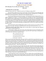
<i>FIGURE 1. </i>
PESSCARA architecture. Most image analysis systems consist only of a data archive. PESSCARA includes this and allows
for both federated and local data archives. PESSCARA also has an Asset Manager that allows flexible tagging of data,
easy browsing of the data, and a workflow engine for processing data based on tags. Workflows and components of
workflows are created in the development environment, and workflows are also executed in that same environment.
<b>3.1. DATABASES VS CONTENT MANAGEMENT </b>
</div>
<span class='text_page_counter'>(7)</span><div class='page_container' data-page=7>
additional capabilities make a CMS an excellent tool to use for big data research, since such data are complex and require
metadata in order to assure proper processing and interpretation, thus leading to meaningful information [6,23].
PESSCARA is designed to link image and associated metadata with the computational environment. It allows users to
focus on the content rather than database tables and gives great flexibility in assigning meaning to the various assets.
Content in our example (discussed later in this chapter) consists of image data, metadata, biomarker information, notes,
and tags.
TACTIC tracks the content creation process, which in the case of medical image research means the original acquired
image, and all of its subsequent processing steps until the final measured version. TACTIC allows tracking of data
check-in and checkout by providcheck-ing a mechanism to identify changes; it also employs a versioncheck-ing system to record the history
of the changes to specific content. It also includes user logins and authentication, allowing tracking of who performed
certain steps and when. Our adaptation of TACTIC for medical image research purposes was straightforward because
medical images are digital content.
PESSCARA has a very flexible data-handling schema (Figure 2) that can easily address the heterogeneous data that are a
part of ‘big data’, so it can adapt as new requirements emerge. It is easy to add other components to this schema to address
other needs, for instance when genomic data need to be processed, rather than simply included as data.
All the data are available through a Representational State (REST) API designed to scale based on the requests issued from
the analytical applications. Some of this is a part of TACTIC, though more of the management of computational tasks is
through other components like sergeant and the grid engine (seeFigure 1).
<b>FIGURE 2. </b>
</div>
<span class='text_page_counter'>(8)</span><div class='page_container' data-page=8>
level tag that equates to the institutional research board identifier, or essentially the project number. Each of these has a
context that has permitted methods and workflows that can be applied.
<b>3.2. WORKFLOW </b>
When dealing with a large number of assets (data and metadata of any kind), it is crucial to have a mechanism that can
automate and efficiently execute a specific series of actions on the data. In general, the workflows in medical imaging
research tend to be linear and simple to implement. For example, a data importation/curation task typically begins by
classifying the incoming image data based on their type, converting the data to a format suitable for subsequent analyses,
placing new images on a queue for human quality control where the system then displays selected images and enables the
reviewer to approve or reject them.
PESSCARA supports such workflows, which may be developed either as Python code, or developed graphically using the
provided tool (Figure 3). PESSCARA users may design workflows and set the events that trigger workflows and define
the users who are allowed to perform human steps. Tasks within the workflow can be calls to REST APIs, Python code,
or notifications.
The workflows can be initialized based on events that can be either automated or manually controlled by a user or a
prespecified group.
<b>FIGURE 3. </b>
Snapshot of the pipeline creation tool. The pipeline workflow is used to depict the steps that a particular series need to
undergo.
<b>3.3. GRID COMPUTING </b>
</div>
<span class='text_page_counter'>(9)</span><div class='page_container' data-page=9>
REST API, making it easier for people to utilize an application without the hustle of setting up and configuring binaries or
executable. In the case of PESSCARA, a ‘step’ can be a call to sergeant, which in turn, could launch a grid job that might
result in the processing of a large group of images utilizing the grid engine. This is, in fact, a common thing for us to do in
our research efforts.
Cloud computing has been emerging as a good way to address computational challenges in modern big data research. This
is because it is a way that a small research laboratory can access large computers, and the pay-as-you-go model provides
flexibility for any size user. Cloud computing also addresses one of the challenges relating to transferring and sharing data,
because data sets and analysis results held in the cloud can be shared with others just by providing credentials so they may
also access the instance in the cloud.
The PESSCARA design allows us to leverage such cloud-computing resources. PESSCARA is engineered to support
architectures such as MapReduce, Spark, and Storm [24–26] that are popular constructs in cloud computing. These
technologies enable researchers to utilize data for fast analysis, with the end goal to translate scientific discovery into
applications for clinical settings.
<b>3.4. MULTI-SITE SYNCHRONIZATION </b>
Content synchronization is an important requirement for multi-centre clinical trials and settings with multiple collaborators.
TACTIC offers a powerful mechanism to synchronize data among servers hosting the databases and users, ensuring that
changes are always up to date and that the correct version of the content is used. Encryption and decryption through a
public- and private-key mechanism are used for all data transfers.
This is a particularly important feature for scientists, since ‘data’ include not just the raw data, but also all the metadata
(which can be at least as laborious to create) and processed versions of data. PESSCARA achieves this via the content
management system using the object capabilities, meaning that the visibility of what is shared and synchronized is very
flexible and straightforward to administer.
We decided NOT to use this synchronization for algorithms, primarily because other tools such as github
(www.github.com) already provide this capability, and specialized capabilities like merging of code—something that is
not as easily done with a CMS, unless a special module was written for ‘code’ objects. Since github has already done this,
we preferred to let users select the tool of their choice for code sharing and management.
<b>4. Using PESSCARA </b>
<b>4.1. DATA IMPORTATION, CURATION, EDITING </b>
</div>
<span class='text_page_counter'>(10)</span><div class='page_container' data-page=10>
Subsequently, CTP is used to de-identify the data for compliance with HIPAA. Tags that should be removed from the
DICOM object are configured through a lookup table. In addition, CTP provides a log of all actions, which meets the
logging requirements in 21 CFR part 11. During the de-identification process, a table with the correspondence between
patient identifier and research identifier is kept and securely maintained. This table is useful for adding information to the
patient dataset, such as tags from the pathology reports and survival information. In addition, when data corresponding to
follow-up studies of patients who have been de-identified are included, CTP will assign the same research identifiers.
Although CTP is capable of removing PHI, it can appear in many unexpected locations (e.g. burned-in pixel values). For
this reason, PESSCARA is typically configured to place imported images in a ‘quarantine’ zone until the assigned user
reviews the data. In our case, an important step of image importation is converting images from DICOM to NIfTI because
most image processing packages do not deal well with native DICOM files. The tiPY library includes a routine to perform
this conversion.
Once data have been imported into TACTIC and some initial workflows have been completed (i.e. for image series
classification, or querying databases to gather additional information such as genomics or survival information), TACTIC
workflow places the object on a queue for data quality inspection. At this point, information missing can be added
manually, and poor quality items can be censored.
The project management element of PESCARA enables project managers to monitor resource usage and progress. This
can allow tracking of resources used to support accurate billing and know individual effort. One can also assign total
expected counts and thus calculate fractional completion.
To ensure data security, PESSCARA regularly backs up all parameter files used by CTP, dcm4che, the virtual machine
running TACTIC, and the file storage area. This exists as just another workflow and thus is flexible in what is included,
frequency, and how it is performed.
<b>4.2. CREATING IMAGE PROCESSING MODULES/DOCKERS </b>
Distribution of image analysis algorithms, particularly when developed in small research laboratories, is challenging since
currently there is not standardized image analysis development environment. When the user employs the PESSCARA
infrastructure, they are working with a standardized environment that usually enables easy deployment of the algorithm.
However, for algorithms that are not easy to be implemented in the PESSCARA environment (i.e. the LINUX host running
PESSCARA), there is support for docker containers () to perform ‘steps’ of a workflow.
</div>
<span class='text_page_counter'>(11)</span><div class='page_container' data-page=11>
execute. A disadvantage is that currently Microsoft Windows and Apple OS X applications are not supported; though,
Windows support has been announced.
For development purposes, PESSCARA supports a majority of tools used in the image processing community, including
ITK, Slicer3D, FSL, and others. However, for algorithm development, Python is the preferred language for PESSCARA.
Python is a very approachable, readable language that includes a number of powerful tools including Numpy, Matplotlib,
scikit-learn, nipype, RPy, and pandas. The Jupyter Notebook development framework extends Python and is at the core of
a substantial shift in the methodology of science, enabling iteration, documentation, and sharing of science. This
philosophy is in perfect alignment with PESSCARA. It promotes reproducible research (i.e. provenance tracking of the
entire history from input data, algorithms used, intermediate calculations, and results). Its interactive capabilities means
that code that code already run can have its results used rather than re-running the code.
While Python is the ‘first language’ of PESSCARA, there are many libraries and developers that depend on other
languages, including non-Python tools such as ITK, FSL, ANTs, Slicer, and others. Furthermore, Jupyter enables
development in many different languages including R, C++, and Julia. [27].
A Jupyter Notebook (which includes code, data, and results) can be easily shared by simply giving the URL and login
credentials to your audience. In addition, the Results/Output and comments (including LaTex and Markdown) can be
integrated into the Notebook to document what has been done in a long-term and shareable way.
The basic model for such ‘shared science’ is import/export. The user often starts by importing other investigators’
Notebooks, but they may also start their own. They can then develop in their own ‘sandbox’, and when they feel they have
something to share, they can ‘export’ it, which makes it publicly visible and available to be imported by others. Exporting
the code in conventional Python format is also supported. They can also save all code and results as HTML for publishing
on the web, or as PDF as a ‘final’ document to be saved in an electronic laboratory notebook [28].
Based on this architecture, the algorithms can be utilized by a variety of cloud services and important characteristic to
consider when large amount of data are involved.
<b>4.3. CREATING AND EXECUTING WORKFLOWS </b>
As noted above, workflow is critical in modern science. One must be able to execute the research process consistently.
When dealing with ‘big data’, efficiency is also essential. In the following section, we show a multi-centre implementation
of a workflow created with PESSCARA (Figure 4). The application will be aimed at developing imaging biomarkers for
differentiating between progression and pseudoprogressions in case of glioblastoma multiforme (a type of malignant brain
tumour) using large data sets and then applying the findings from a large data set to a live clinical trial and ultimately
routine clinical practice.
</div>
<span class='text_page_counter'>(12)</span><div class='page_container' data-page=12>
computational environment. Once the code and the workflows have been established, the clinical configuration is created
containing only the workflows and the computational environed to support them.
Following is an example of how the two configurations of PESSCARA can work.
<b>FIGURE 4. </b>
Translation of workflows created with PESSCARA for a multi-centre set-up. Each of n Centers collects image data and
sends via CTP software. The same CTP software also acts as a receiver at the Central Analysis Lab, where CTP sends it
to PESSCARA for analysis. We expect there would be a separate instance of PESSCARA for a clinical trial to minimize
the chance that a developer would alter data or impact performance.
</div>
<span class='text_page_counter'>(13)</span><div class='page_container' data-page=13>
<b>FIGURE 5. </b>
Example workflow. In this case, images are first identified by the Series Classifier. Once they are labelled, Data Curation
is performed, in this example at a remote centre (Center 3). Then, human-assisted segmentation is performed, and
biomarkers and then computed. This is again reviewed by a human, and if acceptable, the measurements are sent to the
central data collection.
</div>
<span class='text_page_counter'>(14)</span><div class='page_container' data-page=14>
Once data curation is finished, a notification is sent to centre 2 where the tumour segmentation is performed. The Image
analyst can get the data either through the web page or through a link, to perform the tumour segmentation task. Once this
is completed, the step(s) responsible for perfusion analysis computation as well as the registration of the tumour ROI to
the perfusion image is executed. Once the data are reviewed and found acceptable, the imaging biomarkers extracted from
perfusion are assigned to the appropriate tags for that examination. Once this step in completed, the data metadata and all
analytics extracted are available for analysis utilizing any kind of ‘big data’ analysis methodology. This may be simply
stored for later group analysis or may be made available for immediate clinical decision-making. All the data and metadata
created during the execution of the workflow are backed up to a different server for protection over data loss.
<b>4.4. CURRENT STATUS AND NEXT STEPS </b>
Currently, the system is under development with further optimization needed to enhance its security features. Additionally,
further resources are needed to provide the users with more resources for faster testing and support for algorithms with
higher computational requirements. The system has been undergoing rapid development—the documentation and training
resources have not kept up.
We hope that the next phases will see further connections of PESSCARA with non-imaging data repositories;
improvements in the workflow engine enable a wider variety of algorithms on a wider variety of platforms and greater
connections to clinical systems.
We do intend to provide the basic system as open access tools through github so researchers will be able to set the same
environment locally with more resources. We also hope to provide a simple demonstration environment
() that will allow prospective users to test the PESSCARA environment.
<b>5. Conclusion </b>
Big data techniques will lead to an improved model of healthcare delivery with the potential to achieve better clinical
outcomes and increased efficiency. However, appropriate infrastructure is needed to enable the data collection and curation
especially in case of heterogeneous (with respect to data) environments such as healthcare.
</div>
<span class='text_page_counter'>(15)</span><div class='page_container' data-page=15>
<b>6. Acknowledgements </b>
</div>
<!--links-->
<a href=' /><a href=' /><a href=' /><a href=' /><a href=' /><a href=' /><a href=' /><a href=' /><a href=' /><a href=' /><a href=' /><a href=' /><a href=' /> ÔN THI HSG LOP 5
- 5
- 487
- 2


.push({});</script> </div> </div> </div> <div class="vf_link_relate px-2 my-2"> <h2 class="vf_doc_relate text-2xl font-bold my-4">Tài liệu liên quan</h2> <ul class="grid grid-cols-12 gap-2"> <li class="col-span-6 md:col-span-2"> <div class="card-doc " onclick="actionDocRelated(this)"> <a class="card-doc-img" href="https://text.123docz.net/document/382335-on-thi-hsg-lop-5.htm" title="ÔN THI HSG LOP 5"> <i class="icon i_type_doc i_type_doc1"></i> <img class="lazy" src="data:image/gif;base64,R0lGODlhAQABAIAAAP///wAAACH5BAEAAAAALAAAAAABAAEAAAICRAEAOw==" data-src="https://media.store123doc.com/images/document/13/ne/us/medium_usf1369375815.jpg" width="124" height="179" alt="ÔN THI HSG LOP 5" onerror="this.src=)