- Trang chủ >>
- Nông - Lâm - Ngư >>
- Thú y
Molecular detection and characterization of anaplasma platys and ehrlichia canis in dogs from northern colombia
Bạn đang xem bản rút gọn của tài liệu. Xem và tải ngay bản đầy đủ của tài liệu tại đây (386.25 KB, 6 trang )
Veterinary Microbiology 233 (2019) 184–189
Contents lists available at ScienceDirect
Veterinary Microbiology
journal homepage: www.elsevier.com/locate/vetmic
Molecular detection and characterization of Anaplasma platys and Ehrlichia
canis in dogs from northern Colombia
Risa Pesapanea, Janet Foleya, Richard Thomasb, Lyda R. Castrob,
a
b
T
⁎
Department of Medicine and Epidemiology, School of Veterinary Medicine, University of California, Davis, CA 95616, USA
Grupo de Investigación Evolución, Sistemática y Ecología Molecular (GIESEMOL), Universidad del Magdalena, Santa Marta, Magdalena, Colombia
A R T I C LE I N FO
A B S T R A C T
Keywords:
Anaplasmosis
Ehrlichiosis
Domestic dogs
South America
Tick-borne disease
Zoonosis
Ehrlichia canis and Anaplasma platys are intracellular tick-transmitted bacteria that infect dogs; there is evidence
for limited zoonotic potential as well. The prevalence of E. canis in Colombia has been evaluated in different
regions; however little is known about the prevalence or distribution of A. platys. Neither pathogen has been
studied in the Magdalena region, thus the purpose of our study was to assess the prevalence of these pathogens in
dogs attending veterinary clinics from the cities of Santa Marta and Ciénaga, and to assess possible associated
risk factors for infection. A. platys and E. canis infections in blood were evaluated by Taqman PCR assays. E. canis
was detected in 26/170 (15.3%, 95% CI 10.4%–21.8%) and A. platys in 34/168 (20.2%, 95% CI 14.6%–27.3%)
of all dogs tested. Eleven dogs (6.5%, 95% CI 3.4–11.7%) were coinfected with both pathogens. Sequencing
results showed low diversity within E. canis and within A. platys strains, however a strain of E. canis detected in
our study area is genetically distinct from strains reported in another city of Colombia. Our results suggest that
for A. platys, Santa Marta dogs were at greater risk than Ciénaga dogs, and that purebred dogs were at slightly
lower risk in both areas. The confirmation of these pathogens in northern Colombia should cause concern for the
possible co-transmission of these agents to humans or animals in the region.
1. Introduction
The pathogenic bacteria Ehrlichia canis and Anaplasma platys cause
canine monocytic ehrlichiosis and canine cyclic thrombocytopenia,
respectively, and are both transmitted by the brown dog tick
(Rhipicephalus sanguineus sensu lato), which is widely distributed
around the world (Dantas-Torres, 2010). These diseases mainly affect
canines, in the case of ehrlichiosis causing potentially fatal, chronic
multi-systemic disease with possible hemorrhage, pancytopenia, and
lymphadenopathy. Molecular testing of symptomatic human patients
detected DNA of these bacteria in Venezuela (Perez et al., 2006; ArragaAlvarado et al., 2014), Mexico (Silva et al., 2014), and Costa Rica
(Bouza-Mora et al., 2017). Therefore, testing dogs for these pathogens is
very important to support management of the health of the domestic
dog population, and also allows dogs to act as sentinels for human
health (Jones et al., 2018).
Although both of these pathogens have been reported in Colombia,
studies have been limited to particular regions, and differences in
methodologies used make the results difficult to compare. As a consequence, eco-epidemiological aspects and distribution of these pathogens are unclear. Very little is known about A. platys in Colombia: one
⁎
study documented an A. platys PCR prevalence of 2.2% in blood from
91 dogs in the cities of Bogotá, Villavicencio and Bucaramanga (VargasHernandez et al., 2016), and a 53% seroprevalence was found among
dogs in Barranquilla (McCown et al., 2014).
In contrast, there is more evidence for E. canis from various regions
of Colombia including the Caribbean coastal areas. The Caribbean
studies confirmed E. canis in ticks in the department of Cordoba using
DNA sequencing (Miranda and Mattar, 2015); and studies in Barranquilla reported seroprevalence of 74–83% (n = 223) (McCown et al.,
2014, 2015). Prevalences of 28% and 6% of E. canis and Anaplasma sp.
respectively were observed by immunochromatography combined with
blood smear analyses among 184 dogs attending veterinary clinics
(Badillo-Viloria et al., 2017).
No such studies are available for the department of Magdalena in
northern Colombia although environmental and socio-economic conditions of this region could support these tick-borne diseases (TBDs).
The purpose of this study was to detect and characterize A. platys and E.
canis from dog blood samples from the cities of Santa Marta and
Ciénaga in the department of Magdalena, Colombia, using molecular
techniques.
Corresponding author.
E-mail address: (L.R. Castro).
/>Received 7 November 2018; Received in revised form 15 April 2019; Accepted 1 May 2019
0378-1135/ © 2019 Elsevier B.V. All rights reserved.
Veterinary Microbiology 233 (2019) 184–189
R. Pesapane, et al.
2. Methods
GenBank were aligned using the program MEGA 7.0 with the ClustalW
algorithm (Thompson et al., 1994).
For phylogenetic reconstruction, Bayesian inference and maximum
likelihood analyses were performed in the MrBayes 3.2.2 (Ronquist
et al., 2012) and RAxML 8.0.24 (Stamatakis, 2006) programs, respectively. The best nucleotide substitution model for each of the datasets
was selected using the Partition Finder program (Lanfear et al., 2012),
with the Bayesian Information Criterion. The GTR + G was selected as
the best model for the 16S gene of the E. canis sequences. The GTR
model was selected for the 16S gene of the A. platys sequences.
2.1. Samples
Dogs were included in this study by convenience sampling of patients visiting two different veterinary clinics, one in the city of Santa
Marta (11°15′56″N, 74°12′09″W) and one in the city of Ciénaga
(11°00′12″N, 74°15′33″W) in the department of Magdalena, northern
Colombia between January and November 2017. Both cities are located
2 m above sea level and close to the Sierra Nevada mountains. The
climate is warm and dry, with an annual rainfall of 362 mm and
622 mm respectively and an average temperature of 28 °C. All animals
included in this study were privately owned dogs with an outdoor or
mixed indoor-outdoor lifestyle. Some dogs appeared healthy, while
others had different clinical signs suggestive of TBDs. Most of the
owners were not aware of TBDs and no tick control measures had been
used on these dogs. Data on age, sex, breed, and locality were recorded
for each dog. Blood samples were drawn from the jugular vein into
ethylenediaminetetraacetic acid (EDTA) tubes and kept at −20 °C until
DNA extraction.
2.5. Data analysis
Data were maintained in Excel (Version 15.34, Microsoft, Redmond,
WA, USA) and all statistical analyses were performed in R (Version
3.4.3, R Core Team, 2017). Prevalence and 95% confidence intervals
were calculated with the function prop.test. Univariate and multivariable logistic regression analyses, as well as Spearman’s rank coefficient correlation, were used to assess potential risk factors for either E.
canis or A. platys. The strengths of association were assessed through
odds ratios, p-values (≤0.05) and 95% confidence intervals. Factors
included in multivariable analyses were location, sex, age, and breed.
Dogs were first grouped by months of age (0–4, 5–14, 15–60, and > 60)
and later as dependent juveniles (< 4 months) or independent adults
(> 4 months). Dog breeds were recorded categorically as either purebred (e.g. Golden Retriever, Jack Russell, etc.) or mixed breed. The best
model was chosen based on lowest AIC score after a backwards stepwise
approach. A chi-square test was used to determine whether coinfection
occurred more frequently than would be expected by chance.
2.2. DNA extraction
DNA was extracted from blood using the DNeasy Blood and Tissue
kit (Qiagen, Valencia, CA, USA) following manufacturer instructions.
2.3. Amplification and sequencing
Real-time PCR amplification was performed using 1 u L of extracted
DNA in a 12 u L reaction of Maxima Probe/ROX qPCR Master Mix
(ThermoFisher, Waltham, MA, USA) with previously published dsb
primers for the detection of E. canis (Doyle et al., 2005) and proprietary
16S rRNA primers for A. platys based on GenBank ID EU004823.1 designed and validated by the Real-time PCR Research and Diagnostics
Core Facility at UC Davis (www.vetmed.ucdavis.edu/taqmanservice).
For both assays, the thermal cycling protocol consisted of 50 °C for
2 min, then 95 °C for 10 min followed by 50 cycles at 95 °C for 15 s and
60 °C for 1 min. Each reaction contained a positive sample (a sample
that had been amplified and for which DNA sequencing confirmed
presence of pathogen-specific DNA) and molecular-grade water as negative controls. Results of real-time PCR were considered positive if
they had a cycle threshold (CT) value < 40 and a characteristic amplification curve. A CT value > 40 was only considered positive if
confirmed with sequencing.
For phylogenetic analyses, the 16S rDNA region of A. platys and E.
canis were amplified from positive dog blood by conventional PCR
using 3-5uL of extracted DNA in 25 u L reactions of Amplitaq Gold DNA
Polymerase (ThermoFisher, Waltham, MA, USA) as previously described by Pinyoowong et al. (2008) and Inayoshi et al. (2004), respectively. Using the same reagents, the dsb gene for E. canis was also
amplified with primers from Labruna et al. (2007). All reactions included molecular-grade water as a control. Amplifications were followed by 1% agarose-gel electrophoresis and were visualized with
GelRed® (Biotium, Hayward, CA, USA) under UV-light. PCR products
were purified with either ExoSAP-IT PCR Product Cleanup Reagent
(ThermoFisher, Waltham, MA, USA) or QiaQuick Gel Extraction
(Qiagen, Valencia, CA, USA) and sequenced in both forward and reverse directions on an ABI 3730 sequencer (Davis Sequencing). Sequences were verified using BLAST search of GenBank (NCBI; http://
blast.ncbi.nlm.nih.gov/Blast.cgi). Manual edits and alignments were
performed in the program CLC Main Workbench v.7.6.2 (Qiagen, Valencia, CA, USA).
3. Results
3.1. Real-Time PCR
Blood from 170 dogs (n = 34 Ciénaga, n = 136 Santa Marta) successfully yielded DNA for screening of TBD (Table 1). Forty-nine dogs
were PCR positive for at least one pathogen for an overall TBD prevalence of 28.8% (95% CI 22.2–36.3%) across sites in the department of
Magdalena. PCR-positive results included 17 samples (12 E. canis and 5
A. platys) with CT values above 40 that were confirmed as genus Ehrlichia or Anaplasma through DNA sequencing. E. canis was detected in
26/170 dogs (15.3%, 95% CI 10.4%–21.8%) and A. platys in 34/168
dogs (20.2%, 95% CI 14.6%–27.3%). Eleven dogs (6.5%, 95% CI
3.4–11.7%) were coinfected with both pathogens, but coinfection was
less prevalent than expected (p = 0.005). Prevalence of both TBD was
higher in Santa Marta than Ciénaga (Table 1).
3.2. Risk factor assessment for TBD
Dogs had 5.0 times greater odds of being PCR positive for A. platys if
they resided in Santa Marta, but slightly lower odds if they were
purebred in the univariate logistic regression model (Table 2). The
adjusted odds ratio for A. platys was 4.7 for purebred dogs in Santa
Table 1
Sample sizes, demography, and real-time PCR results of privately-owned domestic dogs from two cities in the district of Magdalena, northern Colombia that
were screened for Ehrlichia canis and Anaplasma platys between January and
November 2017.
Location
Ciénaga
Santa Marta
Totals
2.4. Phylogenetic analysis
The sequences obtained in this study and others available in
185
N
34
136
170
Sex
F/M
16/18
75/61
91/79
Breed
Mixed
Pure
9
52
61
25
84
109
E. canis
Pos/N (%)
A. platys
Pos/N (%)
0/34 (0%)
26/136 (19.1%)
26/170 (15.3%)
2/34 (5.8%)
32/134 (23.8%)
34/168 (20.2%)
Veterinary Microbiology 233 (2019) 184–189
R. Pesapane, et al.
but of the 7 bp that did align, 5 were polymorphic (29% identity). The
lack of overlap among sequences generated between these two Colombian studies can be attributed to the use of different primers targeting different regions within 16S.
Three E. canis Santa Marta 16S rDNA amplicons were sequenced and
a complete sequence of 1360 bp was obtained for each one. All sequences were confirmed as E. canis and then aligned and compared to
each other as well as 18 other E. canis strains reported from Brazil,
Turkey, Thailand, India, Venezuela, Italy, Greece, Taiwan, South Africa,
Japan, China, USA, Spain, Peru, Israel, and Colombia (Sup Table 2).
Two of the three E. canis isolates were 100% identical to one another
and to E. canis from Brazil (EF195135). The third E. canis isolate differed by a single nucleotide polymorphism from cytosine to thymine at
nucleotide position 859. Santa Marta sequences differed from isolates
from Brazil (EF195134), Italy (EU439944), Venezuela (AF373612-3),
Greece (EF011110-1), India (JX861392), Turkey (KJ513197), Thailand
(EU263991), and Taiwan (GU810149) by a single nucleotide polymorphism at position 905. Several of the remaining E. canis strains
showed very close identity (99.71–99.85%) but more polymorphisms
were observed between E. canis Santa Marta and Lima (94.18%), Israel
(93.96%), China “Gxht67” (70.77%). Again there was little sequence
overlap between our fragment and the fragment from a strain previously reported from Colombia (JN368080, Vargas et al., 2012), yet 2
of 6 bp were polymorphic (67% identity) (Sup Table 2). Both this Colombian isolate and the Chinese isolate were substantially shorter in
length, just 362 bp and 967 bp respectively, limiting comparability.
All dsb sequences were confirmed as E. canis, then aligned and
compared to each other as well as 11 other E. canis strains available in
GenBank: USA (AF403710), Thailand (KY576856), Brazil (GU586135,
KP167596, DQ460716, DQ460715), Costa Rica (KR732921), Cameroon
(DQ124254), Argentina (MF805005, K253450), and Mexico
(KU323869). The four Santa Marta isolates were 100% identical to one
another. Similarly, Santa Marta strains were identical to 10 of the
comparative E. canis strains and differed only from Mexico by 2 bp
(data not shown). Due to this lack of variability in the dsb region, these
sequences were not used in downstream phylogenetic analyses.
Table 2
Univariate and adjusted multivariable analysis of risk factors that were significantly associated with being PCR positive for Anaplasma platys among privately-owned domestic dogs from two cities in the district of Magdalena,
Colombia (n = 168).
Risk Factors
Odds Ratio
P-value
95% CI
Location
Ciénaga
Santa Marta
Ref
5.0
0.01
1.1–22.8
Breed
Mixed
Pure
Location + Breed
Ref
0.46
4.7
0.01
0.01
0.21–1.0
1.0–21.6
Marta in the multivariable logistic regression model (Table 2). Neither
sex nor age were significantly associated with A. platys. There were no
significant predictors for infection with E. canis using either univariate
or multivariable logistic regression models.
3.3. Amplification, sequencing and Molecular characterization of A. platys
and E. canis from Colombian dogs
Approximately 1.4 kb amplicons corresponding to the targeted 16S
rRNA gene fragment were obtained from two A. platys PCR-positive and
three E. canis positive dogs in Santa Marta and were deposited in
GenBank under accession numbers: MK121782, MK138362,
MK138376, MK138377, MK138374. Amplicons of 410 bp corresponding to the targeted dsb gene fragment were obtained from four
E.canis positive dogs in Santa Marta and were deposited in GenBank
under accession numbers: MK783023, MK783024, MK783025,
MK783026.
Two A. platys Santa Marta 16S rDNA amplicons were sequenced and
a complete sequence of 1371 bp was obtained for each one. Both sequences were confirmed as A. platys, then aligned and compared to each
other as well as 14 other A. platys strains reported from France,
Thailand, India, Zambia, Cuba, Japan, Spain, China, Venezuela, USA,
Croatia, Italy, and Colombia (Sup Table 1). The two A. platys isolates
were 100% identical to one another and differed by only one single
nucleotide polymorphism at position 7 from A. platys from France
(AF303467), Thailand (EF139459), India (KT982643), Zambia
(LC269822), and Cuba (KX792089). Most of the remaining A. platys
strains showed very close identity ranging from 99.27 to 99.85%. Our
16S fragment had very little sequence overlap with a strain previously
reported from Colombia (KF576217, Vargas-Hernandez et al., 2016)
3.4. Phylogenetic analysis of Ehrlichia and Anaplasma
Phylogenetic analyses were performed using a complete 1360–1371
bp fragment of Ehrlichia or Anaplasma 16S rDNA from this study along
with type or reference sequences from 16 E. canis or 10 A. platys isolates, respectively, for which this fragment was also available in
GenBank. The closely related species of E. chaffeensis, E. ewingii, and A.
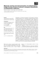
Fig. 1. Tree topology of phylogenetic analyses by Maximum Likelihood and Bayesian Inference including sequences of the 16S gene of Anaplasma platys obtained in
this work and others from GenBank. The numbers correspond to the values of posterior probabilities, bootstrap values were not included but were < 50%.
186
Veterinary Microbiology 233 (2019) 184–189
R. Pesapane, et al.
Fig. 2. Bayesian Inference phylogenetic analyses including 16S genes of Erhlichia canis obtained in this work and others from GenBank. The numbers correspond to
the values of posterior probabilities.
with A. platys and E. canis range from pronounced anemia to a reduction in circulating platelets (thrombocytopenia) (Gaunt et al., 2010). In
coinfections, one of the two hemopathogens usually predominates,
which causes most of the conditions and clinical signs in individuals (Al
Izzi et al., 2013). Gaunt et al. (2010) found that A. platys infection
tended to be more persistent in coinfected dogs than in dogs lacking E.
canis infection. They also concluded that coinfections should be considered in dogs with atypically severe or unusual clinical presentations.
Our data and analyses supported the low diversity within E. canis
and A. platys strains already reported in other studies using the 16S
rRNA gene (Pinyoowong et al., 2008; Unver et al., 2001). Interestingly,
although there was little sequence overlap between our strains and the
only 16S sequence previously reported in Colombia, there were polymorphisms in the overlapping section. The phylogenetic analysis also
placed the previously reported Colombian strain in a different clade
than our Santa Marta sequences. A similar result was recently reported
by Daramola et al., 2018, who found molecularly different strains of E.
canis within Nigeria also using 16S, and by Namboopphaa et al. (2018)
who also reported two different genogroups of E. canis in Thailand.
Our results also corroborated the low diversity in the dsb gene as
previously reported (Cicuttin et al., 2016; Namboopphaa et al., 2018),
making it useful for molecular detection and corroboration of E. canis
but not informative for phylogenetics (Namboopphaa et al., 2018). Due
to these reasons, resolution of the trees is poor and with low values of
bootstraps/posterior probabilities suggesting that more data, and also
other more variable regions, such as the gp36 gene (Namboopphaa
et al., 2018), are needed to increase confidence of these relationships.
Infection with E. canis or A. platys in dogs is associated with different risk factors, with studies reporting higher prevalence among
males than females and among older dogs than young ones (Vieira
et al., 2013; Barrantes-Gonzalez et al., 2018). We did not find significant predictors for infection with E. canis. For A. platys, Santa Marta
dogs were at greater risk than Ciénaga dogs, and purebred dogs were at
slightly lower risk in both areas. Ciénaga is a smaller, more rural city
compared to Santa Marta. However, we have no explanation for why
phagocytophilum were used as outgroups. The resulting phylogenetic
tree for A. platys (Fig. 1) revealed that there is global mixture forming a
monophyletic clade with no general geographic trend. The resulting
phylogenetic tree for E. canis (Fig. 2) similarly revealed a worldwide
mixture. Our sequences group with sequences from Peru and Brazil, but
with low support.
4. Discussion
We have molecularly identified A. platys and E. canis for the first
time in dogs in Magdalena, Colombia, with prevalences of 20.2% and
15.3%, respectively. Although ehrlichiosis and anaplasmosis are frequently diagnosed and treated by veterinarians in the region, the frequencies of these pathogens which we detected were low relative to
findings in other regions of Colombia. Possibly explanations could be
differing detection methodologies or differing underlying ecologies,
possibly related to climatic conditions. Studies in which samples were
obtained from feral dogs (McCown et al., 2014, 2015; Vargas et al.,
2012) generally showed higher prevalence of infection in comparison to
studies like ours in which samples came from owned dogs seen at veterinary clinics (e.g. Badillo et al., 2017). Typically, serostudies would
yield higher prevalence than PCR (McCown et al., 2014, 2015; Vargas
et al., 2012). In relation to weather conditions, PCR-prevalence detected by McCown et al. (2015) was higher in Barranquilla and Cartagena, when compared to Medellin, with the first two cities being tropical and associated with wetland ecosystems, while Medellin is located
approximately 1500 m above sea level, and has a cooler tropical
weather. Santa Marta has similar environmental conditions to Cartagena and Barranquilla.
E. canis and A. platys share the same tick vector, and dogs may
become infected with both pathogens, either simultaneously or sequentially. We found 11 dogs coinfected with both E. canis and A.
platys, as has been reported in Brazil, with coinfection in 3.4% of dogs
in Espírito Santo (Vieira et al., 2018), and 16.1% of dogs in Recife
(Ramos et al., 2010). The clinical implications in individuals coinfected
187
Veterinary Microbiology 233 (2019) 184–189
R. Pesapane, et al.
Center of Excellence for Vector-Borne Diseases funded by the U.S.
Centers for Disease Control and Prevention (Cooperative Agreement
1U01CV000516). We specially thank Antonio Villamizar from the veterinary clinic Origen Animal in Santa Marta, and Danilo Lombardi
from the veterinary clinic Fincas y Marcotas in Cienaga. We also thank
Adriana Santodomingo for her assistance in collecting the samples.
our results contradict other studies that report that regions with less
urbanization and a lower socioeconomic status had higher prevalence
of pathogens such as E. canis, when compared to urban areas (Vieira
et al., 2013; Barrantes-Gonzalez et al., 2018; Dantas-Torres et al.,
2018). Barrantes-Gonzalez et al. (2018) also found greater risk of infection in mixed-breed dogs, suggesting this was due to ecological
factors rather than immunological factors. Owners of purebred dogs
might be more cautious or restrictive of their dogs´ movements, and
mixed-breeds are more likely to be kept outside. However, at least one
study has shown that certain breeds are more susceptible to heavy infestations by the vector, R. sanguineus s.l. (Louly et al., 2009), so the
possibility of some influence of physiological differences between
breeds or purebreds and mixed breeds cannot be discounted.
All our samples are from dogs with owners attending veterinary
clinics. The proximity between humans and dogs has been suggested as
a possible risk factor for human infection with E. canis and A. platys
(Jones et al., 2018). Although A. platys is typically considered pathogenic only in animals, it has been detected in the blood of humans and
dogs of the same household in Chicago (Breitschwerdt et al., 2014), and
in two women with chronic symptoms in Venezuela (Arraga-Alvarado
et al., 2014). E. canis is distributed worldwide and is considered a
serious, potentially fatal canine pathogen. Although initially human
ehrlichiosis was thought to be caused by E. canis with which is crossreacts on serology, most cases are now known to be associated with E.
chaffeensis. Nevertheless, E. canis infections have been confirmed in
human patients from Venezuela (Perez et al., 2006) and Costa Rica
(Bouza-Mora et al., 2017). The molecular identification of E. canis and
A. platys in Santa Marta and Ciénaga, two cities of northern Colombian
reinforces the importance of alerting the veterinary community, dog
owners, and public health authorities to prevent the risk of transmission
of these vector-borne pathogens among dogs and other hosts.
Appendix A. Supplementary data
Supplementary material related to this article can be found, in the
online version, at doi: />References
Al Izzi, S., Martin, D.S., Chan, R.Y.Y., Leutenegger, C.M., 2013. Babesia canis vogeli,
Ehrlichia canis, and Anaplasma platys infection in a dog. Vet. Clin. Pathol. 42,
471–475.
Arraga-Alvarado, C.M., Qurollo, B.A., Parra, O.C., Berrueta, M.A., Hegarty, B.C.,
Breitschwerdt, E.B., 2014. Case report: Molecular evidence of Anaplasma platys infection in two women from Venezuela. Am. J. Trop. Med. Hyg. 91, 1161–1165.
Badillo-Viloria, M., Diaz-Perez, A., Orozco-Sanchez, C., de Lavalle-Galvis, R., 2017.
Infection by Ehrlichia canis and Anaplasmasp. in dogs attended in veterinary clinics,
Barranquilla, Colombia. Rev. MVZ Córdoba 22, 6023–6033.
Barrantes-Gonzalez, A.V., Jimenez-Rocha, A.E., Romero-Zúñiga, J.J., Dolz, G., 2018.
Serology, molecular detection and risk factors of Ehrlichia canis infection in dogs in
Costa Rica. Ticks Tick Borne Dis. 7, 1245–1251.
Bouza-Mora, L., Dolz, G., Solórzano-Morales, A., Romero-Ziga, J.J., Salazar-Sánchez,
L., Labruna, M.B., Aguiar, D.M., 2017. Novel genotype of Ehrlichia canis detected in
samples of human blood bank donors in Costa Rica. Ticks Tick Borne Dis. 8, 36–40.
Breitschwerdt, E.B., Hegarty, B.C., Qurollo, B.A., Saito, T.B., Maggi, R.G., Blanton, L.S.,
Bouyer, D.H., 2014. Intravascular persistence of Anaplasma platys, Ehrlichia chaffeensis, and Ehrlichia ewingii DNA in the blood of a dog and two family members.
Parasit. Vectors 7, 7–298.
Cicuttin, G.L., De Salvo, M.N., Gury Dohmen, F.E., 2016. Molecular characterization of
Ehrlichia canis infecting dogs, Buenos Aires. Ticks Tick Borne Dis. 7, 954–957.
Dantas-Torres, F., 2010. Biology and ecology of the brown dog tick, Rhipicephalus sanguineus. Parasit. Vectors 3.
Dantas-Torres, F., da Silva, Y.Y., da Oliveira Miranda, D.E., da Silva Sales, K.G., Aguiar
Figueredo, L., Otranto, D., 2018. Ehrlichia spp. infection in rural dogs from remote
indigenous villages in north-eastern Brazil. Parasit. Vectors 11, 139.
Daramola, O.O., Takeet, M.I., Oyewusi, K.I., Oyekunle, M.A., Talabi, A.O., 2018.
Detection and molecular characterisation of Ehrlichia canis in naturally infected dogs
in south west Nigeria. Acta Vet. Hung. 66, 85–95.
Doyle, C.K., Labruna, M.B., Breitschwerdt, E.B., Tang, Y.W., Corstvet, R.E., Hegarty, B.C.,
Bloch, K.C., Li, P., Walker, D.H., McBride, J.W., 2005. Detection of medically important Ehrlichia by quantitative multicolor TaqMan real-time polymerase chain reaction of the dsb gene. J. Mol. Diagn. 7, 504–510.
Gaunt, S., Beall, M., Stillman, B., Lorentzen, L., Diniz, P., Chandrashekar, R.,
Breitschwerdt, E., 2010. Experimental infection and co-infection of dogs with
Anaplasma platys and Ehrlichia canis: hematologic, serologic and molecular findings.
Parasit. Vectors 3, 33.
Inayoshi, M., Naitou, H., Kawamori, F., Masuzawa, T., Ohashi, N., 2004. Characterization
of Ehrlichia species from Ixodes ovatus ticks at the foot of Mt. Fuji, Japan. Microbiol.
Immunol. 48, 737–745.
Jones, E.H., Hinckley, A.F., Hook, S.A., Meek, J.I., Backenson, B., Kugeler, K.J., Feldman,
K.A., 2018. Pet ownership increases human risk of encountering ticks. Zoonoses
Public Health 65, 74–79.
Labruna, M.B., McBride, J.W., Camargo, L.M.A., Aguiar, D.M., Yabsley, M.J., Davidson,
W.R., Stromdahl, E.Y., Williamson, P.C., Stich, R.W., Long, S.W., Camargo, E.P.,
Walker, D.H., 2007. A preliminary investigation of Ehrlichia species in ticks, humans,
dogs, and capybaras from Brazil. Vet. Par. 143, 189–195.
Lanfear, R., Calcott, B., Ho, S.Y.W., Guindon, S., 2012. PartitionFinder: combined selection of partitioning schemes and substitution models for phylogenetic analyses. Mol.
Biol. Evol. 29, 1695–1701.
Louly, C.C.B., Soares, S., Silveira, D., Neto, O., Silva, A., Borges, L., 2009. Differences in
the susceptibility of two breeds of dogs, English cocker spaniel and beagle, to
Rhipicephalus sanguineus (Acari: Ixodidae). Int. J. Acarol. 35, 25–32.
McCown, M.E., Alleman, A., Sayler, K.A., Chandrashekar, R., Thatcher, B., Tyrrell, P.,
Stillman, B., Beall, M., Barbet, A.F., 2014. Point prevalence survey for tick-borne
pathogens in military working dogs, shelter animals, and pet populations in northern
Colombia. J. Spec. Oper. Med. 14, 53–57.
McCown, M.E., Monterroso, V.H., Cardona, W., 2015. Monitoreo de Ehrlichia canis,
Anaplasma phagocytophilum, Borrelia burgdorferi, y Dirofilaria immitis en perros de tres
ciudades en Colombia. Ces Med. Vet. Zootec. 10, 224–231.
Miranda, J., Mattar, S., 2015. Molecular detection of Anaplasma sp. and Ehrlichiasp. in
ticks collected in domestical animals, Colombia. Trop. Biomed. 32, 726–735.
Namboopphaa, B., Rittipornlertraka, A., Tattiyapongb, M., Tangtrongsupc, S.,
Tiwananthagorna, S., Chungd, Y.-T., Sthitmateea, N., 2018. Two different genogroups of Ehrlichia canis from dogs in Thailand using immunodominant protein
genes. Infect. Genet. Evol. 63, 116–125.
Perez, M., Bodor, M., Zhang, C., Xiong, Q., Rikihisa, Y., 2006. Human infection with
5. Conclusion
Our study confirms for the first time the presence of E. canis and A.
platys in dogs from Magdalena in northern Colombia. Ehrlichiosis is
frequently diagnosed and treated by veterinarians in the region, however, we found low frequencies of these pathogens in comparison to
other regions of Colombia. This result indicates that unless confirmatory or diagnostic tests are performed, practitioners should not
assume a case to be canine ehrlichiosis, even if some signs may appear
to be indicative of this disease. We reported new sequences of E. canis,
molecularly different from the only sequence available in GenBank for
Colombia. Also, we report the first 16S sequences for A. platys from
Colombia, which, as expected, are very conserved with others from
around the world. We also found dogs to be coinfected with both pathogens, which should be considered by veterinarians, as it has been
reported that coinfection with two or more tick-borne pathogens may
make it difficult to associate a specific clinical sign to a particular canine vector-borne disease. Our results raise questions about possible cotransmission of these agents to other animals in the region.
Declarations of interest
The authors declare that they have no conflict of interest.
Ethical approval
Permission for manipulating the animals was approved by
Universidad del Magdalena Ethical Committee (Acta 001–18).
Acknowledgments
This study has been funded by the patrimonial fund for research
(Fonciencias) of Universidad del Magdalena [VIN2018137]. JEF and RP
acknowledge funding support from the Pacific Southwest Regional
188
Veterinary Microbiology 233 (2019) 184–189
R. Pesapane, et al.
Thompson, J.D., Higgins, D.G., Gibson, T.J., 1994. CLUSTAL W: improving the sensitivity
of progressive multiple sequence alignment through sequence weighting, positionspecific gap penalties and weight matrix choice. Nucleic Acids Res. 22, 4673–4680.
Unver, A., Perez, M., Orellana, N., Huang, H., Rikihisa, Y., 2001. Molecular and antigenic
comparison of Ehrlichia canis isolates from dogs, ticks, and a human in Venezuela. J.
Clin. Microbiol. 39, 2788–2793.
Vargas-Hernández, G., André, M.R., Faria, J.L.M., Munhoz, T.D., Hernandez-Rodriguez,
M., Machado, R.Z., Tinucci-Costa, M., 2012. Molecular and serological detection of
Ehrlichia canis and Babesia vogeli in dogs in Colombia. Vet. Parasitol. 186, 254–260.
Vargas-Hernandez, G., André, M.R., Cendales, D.M., Sousa, K.C.Mde, Gonỗalves, L.R.,
Rondelli, M.C.H., Machado, R.Z., Tinucci-Costa, M., 2016. Molecular detection of
Anaplasma species in dogs in Colombia. Rev. Bras. Parasitol. Veterinária 25, 459–464.
Vieira, R.F., Vieira, T.S., do Nascimento, D.A., Martins, T.F., Krawczak, F.S., Labruna,
M.B., Chandrashekar, R., Marcondes, M., Biondo, A.W., Vidotto, O., 2013. Serological
survey of Ehrlichia species in dogs, horses and humans: zoonotic scenery in a rural
settlement from southern Brazil. Rev. Inst. Med. Trop. Sao Paulo 55, 335–340.
Vieira, F., de, T., Acosta, I.C.L., Martins, T.F., Filho, J.M., Krawczak, F., da, S., Barbieri,
A.R.M., Egert, L., Fernandes, D.R., Braga, F.R., Labruna, M.B., 2018. Tick-borne infections in dogs and horses in the state of Espírito Santo, Southeast Brazil. Vet.
Parasitol. 249, 43–48.
Ehrlichia canis accompanied by clinical signs in Venezuela. Ann. N. Y. Acad. Sci. 1078,
110–117.
Pinyoowong, D., Jittapalapong, S., Suksawat, F., Stich, R.W., Thamchaipenet, A., 2008.
Molecular characterization of Thai Ehrlichia canis and Anaplasma platys strains detected in dogs. Infect. Genet. Evol. 8, 433–438.
R Core Team, 2017. R: a Language and Environment for Statistical Computing. URL. R
Foundation for Statistical Computing, Vienna, Austria. />Ramos, R., Ramos, C., Araújo, F., Oliveira, R., Souza, I., Pimentel, D., Galindo, M.,
Santana, M., Rosas, E., Faustino, M., 2010. Molecular survey and genetic characterization of tick-borne pathogens in dogs in metropolitan Recife (north-eastern Brazil).
Parasitol. Res. 107, 1115–1120.
Ronquist, F., Teslenko, M., Van der Mark, P., Ayres, D.L., Darling, A., Höhna, S., Larget,
B., Liu, L., Suchard, M.A., Huelsenbeck, J.P., 2012. MrBayes3.2: efficient Bayesian
phylogenetic inference and model choice across a large model space. Syst. Biol. 61,
539–542.
Silva, Á.B., Canseco, S.P., de la Torre, M., del, P.G., Silva, A.M., Mayoral, M.Á., Mayoral,
L.P.C., Martínez, J.L., Pérez-Campos, E., 2014. Infección humana asintomática por
contacto con perros. Un caso de ehrlichiosis humana. Gac. Med. Mex. 150, 171–174.
Stamatakis, A., 2006. RAxML-VI-HPC: maximum likelihood-based phylogenetic analyses
with thousands of taxa and mixed models. Bioinformatics 22, 2688–2690.
189