Drugs and Poisons in Humans - A Handbook of Practical Analysis (Part 24)
Bạn đang xem bản rút gọn của tài liệu. Xem và tải ngay bản đầy đủ của tài liệu tại đây (123.56 KB, 4 trang )
2.62.6
© Springer-Verlag Berlin Heidelberg 2005
II.2.6 Lysergic acid diethylamide
( LSD)
by Shinichi Suzuki
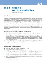
Introduction
Lysergic acid diethylamide (LSD, lysergide) is known as one of the most powerful hallucinogenic
drugs of abuse. LSD was explosively abused in U.S.A. in the latter half of the 1960s. In Japan,
the amount of seizure of LSD is much smaller than that of methamphetamine. However, in
recent years, it has been increasing markedly; 3,500 tablets of LSD were seized in 1996, while
53,043 tablets in 2000 (about 15-fold increase), arousing a serious concern about the phenom-
enon.
LSD is a compound chemically modi ed from ergot alkaloid produced by a bacterium
Claviceps purpurea. LSD is one of the compounds synthesized by the reactions of lysergic acid
isolated from the ergot with various amines at the Rockefeller Institute in the 1930s; LSD was
synthesized as the 25th compound and thus called LSD-25. e strong hallucinogenic activity
of LSD was con rmed by Albert Hofmann who did synthesize the compound; the pharmaco-
logical activity can be obtained by oral administration of as small as 20–75 µg of LSD. e ab-
sorption of LSD from the digestive tract is rapid and distinct visual hallucination takes place
45 min–1 h a er oral intake of about 20 µg of LSD [1]. e hallucination becomes most marked
2–3 h a er the intake and lasts for 8–12 h.
In this chapter, a detection method by TLC and con rmatory analysis by GC/MS for LSD
are presented.
Reagents and their preparation
• LSD can be obtained from each local bureau of drug enforcement o cers under an o cial
transfer process.
• p-Dimethylbenzaldehyde reagent: a 0.125-g aliquot of p-dimethylaminobenzaldehyde
(Wako Pure Chemical Industries, Ltd., Osaka, Japan and other manufacturers) is dissolved
in 100 mL of 65 % sulfuric acid solution, followed by the addition of 0.1 mL of 5 % ferric
chloride aqueous solution.
• Dragendor reagent [2]: there are various modi cations of its preparation; the most typical
method is described here. A 0.85-g aliquot of bismuth subnitrate is dissolved in a mixture
of 40 mL distilled water and 10 mL acetic acid to prepare “A” solution. A 8-g aliquot of
potassium iodide is dissolved in 20 mL distilled water to prepare “B” solution. en, a mix-
ture of A/B/acetic acid/distilled water (1:1:4:20, v/v) is prepared.
• Iodoplatinate reagent [2]: A 1-mL aliquot of 10 % platinic chloride solution is mixed with
25 mL of 4 % potassium iodide solution and 24 mL distilled water.
226 Lysergic acid diethylamide (LSD)
Pretreatments
LSD is seized in the forms of tablets, capsules and paper sheets. e latter is most popular;
a paper sheet which had absorbed LSD can be cut into pieces along perforation lines. One of
the pieces is put in the mouth and sucked.
LSD shows strong bluish uorescence; therefore, LSD can be easily located on a TLC plate,
and the LSD fraction can be obtained for puri cation by LC under an ultraviolet light.
For a piece of paper, which is suspected to contain LSD, the drug is extracted by adding 1 %
tartaric acid aqueous solution and by shaking it for 5 min. is procedure is repeated six times
and the tartarate extract solutions are combined. A er the pH of the solution is adjusted to 8.5,
the solution is extracted with an appropriate amount of chloroform four times. e combined
chloroform extract is evaporated to dryness; the residue is dissolved in a small amount of
methanol to be subjected to further analysis.
TLC analysis
Analytical conditions
TLC plate: a usual silica gel plate, for example, Kieselgel (0.25 µm thickness, Merck, Darm-
stadt, Germany and other manufacturers).
Developing solvents: (A) acetone/chloroform (4:1, v/v); (B) chloroform/methanol/n-hex-
ane (4:2:1, v/v).
Assessment of the method
e R
f
values of LSD were 0.29 with the (A) solvent system and 0.55 with the (B) system. e
colors of the spot of LSD are blue-purple with the p-dimethylaminobenzaldehyde reagent,
orange with the Dragendor reagent and purple with the iodoplatinate reagent.
MS by the direct inlet method
Under the ultraviolet light at 365 nm, the fraction showing strong uorescence is obtained by
TLC or LC, and extracted with chloroform. e resulting residue is subjected to analysis by the
direct inlet method of EI-MS.
Analytical conditions
Any type of an MS instrument can be used; electron energy: 20 eV; ionization current: 110 µA;
measurements: full-scan mode.
227
Assessment and some comments on the method
An EI mass spectrum of LSD is shown in > Fig. 6.1. e molecular ion appears as the base
peak. Fragment peaks can be observed at m/z 221, 207, 181 and 167. e fragmentation path-
ways are shown in
> Fig. 6.2. e con rmation of LSD should be made with each mass spec-
trum.
LSD is easily decomposed by light; all procedure is preferably made under shading from
light.
EI mass spectrum of LSD.
⊡ Figure 6.1
Fragmentation pathways for ions observable in the EI mass spectrum of LSD.
⊡ Figure 6.2
MS by the direct inlet method
228 Lysergic acid diethylamide (LSD)
Toxicity, and concentrations in blood and urine
e acute toxicity of LSD is generally low; it does almost not cause death.
In this chapter, detection and identi cation methods have been described for seized items,
and not for human specimens. It is actually not easy to detect LSD from blood and urine, because
its amount to be ingested is as small as about 10 µg and LSD is rapidly metabolized in human
bodies. As metabolites of LSD, 2-oxo-LSD, 3-, 13- and 14-hydroxy-LSDs and an N-de-ethyl-LSD
are known [3]. A er oral ingestion of 160 µg LSD, the maximum blood concentration of LSD
was 9 ng/mL and its half-life is said to be about 2 h. e concentrations of LSD of LSD-abusing
patients, who had been brought to a critical care medical center were 0.5–1.9 ng/mL in blood and
0.2–7.7 ng/mL in urine [4].
References
1) Tu AT (1999) Principle of Toxicology – Science of Poisons –. Jiho Inc., Tokyo, pp 108–109 (in Japanese)
2) The Pharmaceutical Society of Japan (ed) (1992) Standard Methods of Chemical Analysis in Poisoning – With
Commentary –. Nanzando, Tokyo, p 43 and p 151 (in Japanese)
3) Cody JT (2000) Hallucinogens. In: Bogusz MJ (ed) Handbook of Analytical Separations, Vol. 2, Forensic Science.
Elsevier, Amsterdam, pp 143–162
4) Karch SB (1996) The Pathology of Drug Abuse, 2nd edn. CRC Press, Boca Raton, pp 267–270