Efficacy of fungicides and bioagents against Pythium aphanidermatum causing rhizome rot of turmeric
Bạn đang xem bản rút gọn của tài liệu. Xem và tải ngay bản đầy đủ của tài liệu tại đây (371.93 KB, 7 trang )
<span class='text_page_counter'>(1)</span><div class='page_container' data-page=1>
<i><b>Int.J.Curr.Microbiol.App.Sci </b></i><b>(2017)</b><i><b> 6</b></i><b>(11): 4312-4320 </b>
4312
<b>Original Research Article </b>
<b>Efficacy of Fungicides and Bioagents against </b>
<i><b>Pythium aphanidermatum</b></i>
<b>Causing Rhizome Rot of Turmeric </b>
<b>P.G. Chavan*, K.T. Apet and R.S. Borade </b>
Department of Plant Pathology, Vasantrao Naik Marathwada Krishi Vidyapeeth,
Parbhani - 431 402, Maharashtra, India
<i>*Corresponding author </i>
<i> </i>
<i><b> </b></i> <i><b> </b></i><b>A B S T R A C T </b>
<i><b> </b></i>
<b>Introduction </b>
Turmeric (Curcuma longa L.) is one of the
major spices cultivated for its underground
rhizome belongs to family Zingiberaceae. It is
originated from Tropical South Asia. This is
also called as ‘hidden Lilly’ or ‘golden spice’
or ‘turmeric of commerce’ or ‘Indian saffron’
or ‘Haldi’. Turmeric is the third largest spice
produced in the country and it accounts for
about 14 % of total spices produced in India.
India is the world’s largest producer of
turmeric and apparently accounts for more
than 80 per cent of the world’s production,
followed by China, Indonesia, Bangladesh,
and Thailand (Selvan <i>et al., 2002). The area, </i>
production and productivity of turmeric in
India has been reported to be 175.73 and
185.58 thousand hectares, 959.35 and 943.33
thousand tones and 5459 and 5083 kg/ha,
respectively, during year 2014-15 and
2015-16 (Anonymous, 202015-16). The total area in
Maharashtra under turmeric was 11.0
thousand hectares, with production 11.0
thousand tones and productivity of 1000
kg/ha, respectively (Anonymous, 2015).
Rhizome rot (<i>Pythium aphanidermatum</i>) is one of the most wide spread, destructive
disease of turmeric (<i>Curcuma longa </i>L.), which accounts for about 30 to 80 per cent yield
losses. All the fungicides tested significantly inhibited mycelial growth of <i>P. </i>
<i>aphanidermatum</i>, over untreated control. Average mycelial growth inhibition recorded
with the test systemic fungicides was ranged from 73.32 (Propiconazole) to 100
(Metalaxyl) per cent. However, it was cent per cent with Metalaxyl (100 %), followed by
Carbendazim (97.67 %), Azoxystrobin (94.55 %), Thiophanate methyl (94.15 %),
Fosetyl-AL (86.64 %), Hexaconazole (85.76 %) and Difenconazole (82.85). Whereas, it was
comparatively minimum with Propiconazole (73.32 %) and Penconazole (81.14 %).
Average mycelial growth inhibition recorded with the test non systemic and contact
fungicides was ranged from 50.94 (Metalaxyl 8 % WP + Mancozeb 64 % WP) to 100
(Carbendazim 12 WP + Mancozeb 63 WP) per cent. However, Carbendazim 12 WP +
Mancozeb 63 WP gave cent per cent (100 %) mycelial inhibition. The next fungicides with
significantly least mycelial growth were Copper oxychloride (97.36 %), followed by
Chlorothalonil (76.16 %), Mancozeb (70.62 %). However, Metalaxyl 8 % WP +
Mancozeb 64 % WP and Cymoxanil 8 % + Mancozeb 64 % WP were found less effective
with minimum mycelial inhibition of 50.94 and 55.23 per cent, respectively.
<b>K e y w o r d s </b>
Curcuma longa,
<i>Pythium</i>
<i>aphanidermatum</i>,
Fungicides,
Bioagents,
Management.
<i><b>Accepted: </b></i>
30 September 2017
<i><b>Available Online:</b></i>
10 November 2017
<b>Article Info </b>
<i>International Journal of Current Microbiology and Applied Sciences </i>
<i><b>ISSN: 2319-7706</b></i><b> Volume 6 Number 11 (2017) pp. 4312-4320 </b>
</div>
<span class='text_page_counter'>(2)</span><div class='page_container' data-page=2>
<i><b>Int.J.Curr.Microbiol.App.Sci </b></i><b>(2017)</b><i><b> 6</b></i><b>(11): 4312-4320 </b>
4313
Turmeric is prone to many fungal, bacterial,
viral and nematode diseases. Among all
diseases rhizome rot caused by <i>P. </i>
<i>aphanidermatum </i> is most destructive and
widespread disease causes very high crop loss
under favorable conditions (Rathaiah, 1982).
The disease has been reported to causes more
than 60 per cent mortality of seedlings both in
nursery and field condition and about 50-80
per cent losses during storage (Nirmal, 1992);
rhizome rot resulted in yield loss of 50%
(Rajalakshmi et al., 2016).
<b>Materials and Methods </b>
<i><b>In vitro </b></i><b>evaluation of fungicides </b>
Efficacy of nine systemic fungicides and six
non-systemic / combi fungicides was
evaluated <i>in vitro </i>at various concentrations
against <i>P. </i> <i>aphanidermatum, </i> applying
Poisoned food technique (Nene and
Thapliyal, 1993) and using Potato dextrose
agar (PDA) as basal culture medium. Based
on active ingredient, requisite quantity of the
test fungicides was calculated, mixed
separately thoroughly with autoclaved and
cooled (40 oC) PDA medium in conical flasks
to obtain desired concentrations. This PDA
medium amended separately with the test
fungicides was then poured (20 ml / plate)
aseptically in Petri plates (90 mm dia.) and
allowed to solidify at room temperature. For
each of the test fungicide and its desired
concentrations, three plates / treatment /
replication were maintained. After
solidification of the PDA medium, all the
plates were inoculated aseptically by placing
in the centre a 5 mm culture disc obtained
from actively growing 7 days old pure culture
of <i>P. aphanidermatum </i>and incubated in an
inverted position at 28 ± 2 oC. Petri plates
filled with plain PDA (without any fungicide)
and inoculated with the pure culture disc of P.
<i>aphanidermatum </i> were maintained as
untreated control.
Observations on radial mycelial growth /
colony diameter were recorded at an interval
of 24 hours and continued till untreated
control plates were fully covered with
mycelial growth of the test pathogen. Per cent
inhibition of the test pathogen with the test
fungicides over untreated control was
calculated by applying following formula
(Vincent, 1927).
C – T
Per cent inhibition = --- X 100
C
Where,
C = growth of the test fungus in untreated
control plate
T = growth of the test fungus in treated plate
<i><b>In vitro</b></i><b> evaluation of bioagents </b>
Eight fungal and two bacterial bioagents were
evaluated in vitro against P. aphanidermatum,
applying Dual Culture Technique (Dennis and
Webster, 1971). Seven days old cultures of
the test bioagents and test pathogen (P.
<i>aphanidermatum) grown on PDA were used </i>
for the study. Two 5 mm culture discs, one
each of the test pathogen and test bioagents
were cut out with sterilized cork borer and
placed at equidistance, exactly opposite to
each other on autoclaved and solidified PDA
medium in Petri plates and three plates were
incubated at 28 ± 2 oC. PDA plates inoculated
alone with pure culture disc (5 mm) of the test
pathogen were maintained as untreated
control. The experiment is designed in CRD
and all treatments replicated thrice.
</div>
<span class='text_page_counter'>(3)</span><div class='page_container' data-page=3>
<i><b>Int.J.Curr.Microbiol.App.Sci </b></i><b>(2017)</b><i><b> 6</b></i><b>(11): 4312-4320 </b>
4314
cent inhibition of the test pathogen with the
test bioagent, over untreated control was
calculated by applying following formula
(Arora and Upaddhyay, 1978).
Colony growth in Control plate –
Colony growth in intersecting plate
Per cent
Growth Inhibition = --- X100
Colony growth in control plate
<b>Results and Discussion </b>
<i><b>In vitro </b></i><b>evaluation of systemic fungicides </b>
<b>Mycelial inhibition </b>
Results (Table 1) revealed that all the
systemic fungicides tested (each @ 500, 1000
and 1500 ppm) significantly inhibited
mycelial growth of <i>P. aphanidermatum, over </i>
untreated control.
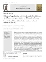
Further, per cent mycelial inhibition was
increased with increase in concentrations of
the fungicides tested (Fig. 1).
At 500 ppm, mycelial growth inhibition was
ranged from 62.72 (Propiconazole) to 100
(Metalaxyl) per cent. However, Metalaxyl
gave cent per cent (100 %) mycelial
inhibition. The next best fungicides found
were Carbendazim (93.01 %), followed by
Azoxystrobin (90.70 %), Thiophanate methyl
(90.35 %), Fosetyl-AL (81.72 %),
Hexaconazole (81.18 %) and Difenconazole
(74.81 %). However, Propiconazole and
Penconazole were found less effective with
minimum mycelial inhibition of 62.72 and
73.40 per cent, respectively.
At 1000 ppm, the trend was same as at 500
ppm and mycelial growth inhibition was
ranged from 75.06 (Propiconazole) to 100
(Metalaxyl and Carbendazim) per cent. It was
cent per cent with the fungicides Metalaxyl
and Carbendazim (each 100 %). In the order
of merit the next most effective fungicides
with significantly maximum mycelial
inhibition were Azoxystrobin (92.96 %),
followed by Thiophanate methyl (92.09 %),
Fosetyl-AL (86.73%), Hexaconazole (85.53
%), Difenconazole (84.16 %), Penconazole
(82.99 %) and Propiconazole (75.06 %).
At 1500 ppm, mycelial growth inhibition was
ranged from 82.17 (Propiconazole) to 100
(Metalaxyl, Carbendazim, Azoxystrobin and
Thiophanate methyl) per cent. However, it
was cent per cent with the fungicides
Metalaxyl, Carbendazim, Azoxystrobin and
Thiophanate methyl (each 100 %). The next
most effective fungicides were Fosetyl-AL
(91.46 %), followed by Hexaconazole (90.57
%), Difenconazole (89.59 %), Penconazole
(87.02 %) and Propiconazole (82.17 %).
Average mycelial growth inhibition recorded
with the test systemic fungicides was ranged
from 73.32 (Propiconazole) to 100
(Metalaxyl) per cent. However, it was cent
per cent with Metalaxyl (100 %), followed by
Carbendazim (97.67 %), Azoxystrobin (94.55
%), Thiophanate methyl (94.15 %),
Fosetyl-AL (86.64 %), Hexaconazole (85.76 %) and
Difenconazole (82.85). Whereas, it was
comparatively minimum with Propiconazole
(73.32 %) and Penconazole (81.14 %).
<i><b>In vitro </b></i> <b>evaluation of non-systemic and </b>
<b>combi-fungicides </b>
<b>Mycelial inhibition </b>
</div>
<span class='text_page_counter'>(4)</span><div class='page_container' data-page=4>
<i><b>Int.J.Curr.Microbiol.App.Sci </b></i><b>(2017)</b><i><b> 6</b></i><b>(11): 4312-4320 </b>
4315
<b>Table.1 </b><i>In vitro </i>bioefficacy of systemic fungicides against<i> P. aphanidermatum </i>
*: Mean of three replications, Dia: Diameter, Av.: Average Figures in Parentheses are angular transformed values
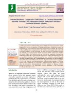
<b>Table.2 </b><i>In vitro</i> evaluation of non-systemic/contact fungicide
*: Mean of three replications, Dia.: Diameter, Av.: Average Figures in parentheses are angular transformed values
<b>Tr. </b>
<b>No. </b> <b>Treatments </b>
<b>Colony Dia. *(mm) at </b>
<b>ppm </b> <b>Av. </b>
<b>(mm) </b>
<b>% Inhibition* at ppm </b> <b>Av. </b>
<b>inhibition </b>
<b>(%) </b>
<b>500 </b> <b>1000 </b> <b>1500 </b> <b>500 </b> <b>1000 </b> <b>1500 </b>
<b>T1</b> Carbendazim 50 WP 6.29 00.00 00.00 2.10
93.01
(74.67)
100.00
(90.00)
100.00
(90.00)
97.67
(81.22)
<b>T2</b> Metalaxyl 50 WP 00.00 00.00 00.00 00.00
100.00
(90.00)
100.00
(90.00)
100.00
(90.00)
100.00
(90.00)
<b>T3</b> Hexaconazole 5 EC 16.94 13.02 8.49 12.82
81.18
(64.29)
85.53
(67.64)
90.57
(72.12)
85.76
(67.83)
<b>T4</b>
Difenconazole 25
EC 22.67 14.26 9.37 15.43
74.81
(59.87)
84.16
(66.55)
89.59
(71.18)
82.85
(65.54)
<b>T5</b> Penconazole 10 EC 23.94 15.31 11.68 16.98
73.40
(58.95)
82.99
(65.64)
87.02
(68.88)
81.14
(64.26)
<b>T6</b>
Thiophanate methyl
70 WP 8.68 7.12 0.00 5.27
90.35
(71.90)
92.09
(73.67)
100.00
(90.00)
94.15
(76.00)
<b>T7</b> Azoxystrobin 23 SC 8.37 6.34 0.00 4.90
90.70
(72.24)
92.96
(74.61)
100.00
(90.00)
94.55
(76.50)
<b>T8</b> Fosetyl-AL 80 WP 16.45 11.94 7.69 12.03
81.72
(64.69)
86.73
(68.64)
91.46
(73.01)
86.64
(68.56)
<b>T9</b>
Propiconazole 25
EC 33.55 22.45 16.05 24.02
62.72
(52.37)
75.06
(60.04)
82.17
(65.02)
73.32
(58.90)
<b>T10</b> Control 90.00 90.00 90.00 90.00
00.00
(00.00)
00.00
(00.00)
00.00
(00.00)
00.00
(00.00)
<b>S.E.+ </b> <b>0.23 </b> <b>0.21 </b> <b>0.17 </b> <b>0.20 </b> <b>0.25 </b> <b>0.24 </b> <b>0.20 </b> <b>0.23 </b>
<b>C.D.(P=0.01) </b> <b>0.75 </b> <b>0.70 </b> <b>0.59 </b> <b>0.68 </b> <b>0.83 </b> <b>0.75 </b> <b>0.75 </b> <b>0.78 </b>
<b>Tr. </b>
<b>No. </b> <b>Treatments </b>
<b>Colony Dia. *(mm) at </b>
<b>ppm </b> <b>Av. </b>
<b>(mm) </b>
<b>% Inhibition* at ppm </b> <b>Av. </b>
<b>inhibition </b>
<b>(%) </b>
<b>1500 </b> <b>2000 </b> <b>2500 </b> <b>1500 </b> <b>2000 </b> <b>2500 </b>
<b>T1</b> Chlorothalonil 75 WP 26.38 21.25 16.74 21.46
70.69
(57.22)
76.39
(60.93)
81.40
(64.45)
76.16
(60.77)
<b>T2</b>
Copper oxychloride 50
WP 7.12 00.00 00.00 2.37
92.09
(73.67)
100.00
(90.00)
100.00
(90.00)
97.36
(80.66)
<b>T3</b>
Cymoxanil 8 % +
Mancozeb 64 % WP 48.97 39.86 32.04 40.29
45.59
(42.47)
55.71
(48.28)
64.40
(53.37)
55.23
(48.00)
<b>T4</b> Mancozeb 50WP 31.54 26.35 21.44 26.44
64.96
(53.70)
70.72
(57.24)
76.18
(60.79)
70.62
(57.18)
<b>T5</b>
Metalaxyl 8 % WP +
Mancozeb 64 % WP 53.64 43.68 35.14 44.15
40.40
(39.47)
51.47
(45.84)
60.96
(51.33)
50.94
(45.54)
<b>T6</b>
Carbendazim 12% WP +
Mancozeb 63 % WP 00.00 00.00 00.00 00.00
100.00
(90.00)
100.00
(90.00)
100.00
(90.00)
100.00
(90.00)
<b>T7</b> Control (untreated) 90.00 90.00 90.00 90.00
00.00
(00.00)
00.00
(00.00)
00.00
(00.00)
00.00
(00.00)
<b>S.E.+ </b> <b>0.30 </b> <b>0.20 </b> <b>0.24 </b> <b>0.25 </b> <b>0.33 </b> <b>0.23 </b> <b>0.24 </b> <b>0.27 </b>
</div>
<span class='text_page_counter'>(5)</span><div class='page_container' data-page=5>
<i><b>Int.J.Curr.Microbiol.App.Sci </b></i><b>(2017)</b><i><b> 6</b></i><b>(11): 4312-4320 </b>
4316
<b>Table.3 </b><i>In vitro </i>bioefficacy of bioagents against <i>P. aphanidermatum </i>
*-Mean of three replications, Dia.: Diameter, Figures in Parentheses are angular transformed values
<b>Fig.1 </b><i>In vitro</i> bioefficacy of systemic fungicides against <i>P. aphanidermatum </i>
<b>Tr. No. </b> <b>Treatments </b> <b>Colony Dia.of test pathogen </b>
<b>* (mm) </b> <b>% Inhibition </b>
T1 <i>Trichoderma viride </i> 12.77 85.81 (67.87)
T2 <i>T. harzianum </i> 17.64 80.40 (63.72)
T3 <i>T. hamatum </i> 23.94 73.40 (58.95)
T4 <i>T. longibrachiatum </i> 21.86 75.71 (60.47)
T5 <i>T. (Gliocladium) virens </i> 19.58 78.24 (62.20)
T6 <i>T. koningii </i> 15.22 83.09 (65.72)
T7 <i>Aspergillus niger </i> 19.34 78.51 (62.38)
T8 <i>T. lignorum </i> 32.14 64.29 (53.30)
T9 <i>Pseudomonas fluorescens </i> 51.71 42.54 (40.71)
T10 <i>Bacillus subtilis </i> 47.66 47.04 (43.31)
T11 Control (untreated) 90.00 0.00 (0.00)
<b>S.E. + </b> <b>0.55 </b> <b>0.61 </b>
</div>
<span class='text_page_counter'>(6)</span><div class='page_container' data-page=6>
<i><b>Int.J.Curr.Microbiol.App.Sci </b></i><b>(2017)</b><i><b> 6</b></i><b>(11): 4312-4320 </b>
4317
<b>Fig.2 </b><i>In vitro</i> bioefficacy of non-systemic and combi-fungicides against <i>P. aphanidermatum </i>
</div>
<span class='text_page_counter'>(7)</span><div class='page_container' data-page=7>
<i><b>Int.J.Curr.Microbiol.App.Sci </b></i><b>(2017)</b><i><b> 6</b></i><b>(11): 4312-4320 </b>
4318
At 1500 ppm, mycelial growth inhibition was
ranged from 40.40 (Metalaxyl 8 % WP +
Mancozeb 64 % WP) to 100 (Carbendazim 12
WP + Mancozeb 63 WP) per cent. However,
Carbendazim 12 WP + Mancozeb 63 WP
gave cent per cent (100 %) mycelial
inhibition.
The next best fungicides found were Copper
oxychloride (92.09 %), followed by
Chlorothalonil (70.69 %) and Mancozeb
(64.96 %). However, Metalaxyl 8 % WP +
Mancozeb 64 % WP and Cymoxanil 8 % +
Mancozeb 64 % WP were found less effective
with minimum mycelial inhibition of 40.40
and 45.59 per cent, respectively.
At 2000 ppm, mycelial growth inhibition was
ranged from 51.47 (Metalaxyl 8 % WP +
Mancozeb 64 % WP) to 100 (Carbendazim 12
WP + Mancozeb 63 WP and Copper
oxychloride) per cent. However, Carbendazim
12 WP + Mancozeb 63 WP and Copper
oxychloride gave cent per cent (100 %)
mycelial inhibition. The next best fungicides
found were Chlorothalonil (76.39 %),
followed by Mancozeb (70.72 %) and
Cymoxanil 8 % + Mancozeb 64 % WP (55.71
%). However, Metalaxyl 8 % WP +
Mancozeb 64 % WP was found less effective
with minimum mycelial inhibition of 51.47
per cent.
At 2500 ppm, mycelial growth inhibition was
ranged from 60.96 (Metalaxyl 8 % WP +
Mancozeb 64 % WP) to 100 (Carbendazim 12
WP + Mancozeb 63 WP and Copper
oxychloride) per cent. However, Carbendazim
12 WP + Mancozeb 63 WP and Copper
oxychloride gave cent per cent (100 %)
mycelial inhibition. The next fungicides with
significantly least mycelial growth were
Chlorothalonil (81.40 %), followed by
Mancozeb (76.18 %), Cymoxanil 8 % +
Mancozeb 64 % WP (64.40 %) and Metalaxyl
8 % WP + Mancozeb 64 % WP (60.96 %).
Average mycelial growth inhibition recorded
with the test non systemic and contact
fungicides was ranged from 50.94 (Metalaxyl
8 % WP + Mancozeb 64 % WP) to 100
(Carbendazim 12 WP + Mancozeb 63 WP)
per cent. However, Carbendazim 12 WP +
Mancozeb 63 WP gave cent per cent (100 %)
mycelial inhibition. The next fungicides with
significantly least mycelial growth were
Copper oxychloride (97.36 %), followed by
Chlorothalonil (76.16 %), Mancozeb (70.62
%). However, Metalaxyl 8 % WP +
Mancozeb 64 % WP and Cymoxanil 8 % +
Mancozeb 64 % WP were found less effective
with minimum mycelial inhibition of 50.94
and 55.23 per cent, respectively.
<i><b>In vitro </b></i><b>evaluation of bioagents against </b><i><b>P. </b></i>
<i><b>aphanidermatum</b></i>
Results (Fig. 3 and Table 3) revealed that all
the bioagents evaluated exhibited fungistatic /
antifungal activity against P. aphanidermatum
and significantly inhibited its growth, over
untreated control. Of the antagonists tested, T.
<i>viride </i>was found most effective with highest
mycelial growth inhibition (85.81%) of the
test pathogen. The second and third
inhibitoriest antagonists found were <i>T. </i>
<i>koningii and T. harzianum with and inhibition </i>
of 83.09 and 80.40 per cent, respectively.
These were followed by <i>Aspergillus niger </i>
(78.51 %), T. (Gliocladium) virens (78.24 %),
<i>T. longibrachiatum </i> (75.71 %), <i>T. hamatum </i>
(73.40 %), <i>T. lignorum (64.29 %). However, </i>
<i>P. fluorescens </i> and <i>Bacillus subtilis </i> were
found less effective with minimum mycelial
inhibition of 42.54 and 47.04 per cent,
respectively.
</div>
<!--links-->
insider computer fraud an in depth framework for detecting and defending against insider it attacks
- 506
- 687
- 0

.push({});</script> </div> </div> </div> <div class="vf_link_relate px-2 my-2"> <h2 class="vf_doc_relate text-2xl font-bold my-4">Tài liệu liên quan</h2> <ul class="grid grid-cols-12 gap-2"> <li class="col-span-6 md:col-span-2"> <div class="card-doc " onclick="actionDocRelated(this)"> <a class="card-doc-img" href="https://text.123docz.net/document/1639933-insider-computer-fraud-an-in-depth-framework-for-detecting-and-defending-against-insider-it-attacks.htm" title="insider computer fraud an in depth framework for detecting and defending against insider it attacks"> <i class="icon i_type_doc i_type_doc2"></i> <img class="lazy" src="data:image/gif;base64,R0lGODlhAQABAIAAAP///wAAACH5BAEAAAAALAAAAAABAAEAAAICRAEAOw==" data-src="https://media.store123doc.com/images/document/2014_07/03/medium_vqq1404378647.jpg" width="124" height="179" alt="insider computer fraud an in depth framework for detecting and defending against insider it attacks" onerror="this.src=)