Biomarkers and Acute Kidney Injury
Bạn đang xem bản rút gọn của tài liệu. Xem và tải ngay bản đầy đủ của tài liệu tại đây (2.17 MB, 36 trang )
<span class='text_page_counter'>(1)</span><div class='page_container' data-page=1>
<b>Biomarkers and Acute Kidney Injury</b>
<b>William T McGee MD MHA FCCM</b>
</div>
<span class='text_page_counter'>(2)</span><div class='page_container' data-page=2>
<b>Disclosure</b>
<b>Speakers Bureau: </b>
</div>
<span class='text_page_counter'>(3)</span><div class='page_container' data-page=3>
<b>AKI </b>
<b>Needed a Better Way to Assess AKI</b>
<b>Needed a Better Way to Monitor Kidney Stress In Real </b>
<b>Time</b>
<b>Use of ScR and Urine Output is Too Slow and Lagging</b>
<b>Increasing Biomarker Presence In Clinical Care</b>
<b>AKI Is Expensive and Has Negative Effect on Metrics</b>
<b>and Patients!!</b>
</div>
<span class='text_page_counter'>(4)</span><div class='page_container' data-page=4>
<b>AKI Is Common And Deadly: Pneumonia</b>
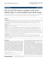
*Genetic and Inflammatory Markers of Sepsis (GenIMS) study.
<i>[1] Murugan R, Karajala-Subramanyam V, Lee M, et al. Acute Kidney Injury in Non-Severe Pneumonia is Associated with an Increased Immune Response and Lower Survival. Kidney Int. </i>
2010;77:527-535.
<b>AKI was found to be prevalent (34%) in a study of 1836 hospitalized patients with </b>
<b>community acquired pneumonia*,1</b>
Severe AKI (n = 189, 10%)
Moderate AKI (n = 135, 7%)
Mild AKI (n = 307, 17%)
No AKI (n = 1205, 66%)
<b>Mortality in </b>
</div>
<span class='text_page_counter'>(5)</span><div class='page_container' data-page=5>
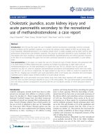
<b>AKI Is Common And Deadly: Sepsis</b>
<b>Sepsis was identified as the presumed etiology in 19% of AKI cases in the ICU in one </b>
<b>study2</b> <b><sub>and septic shock was found to be a contributing factor to AKI in 48% of cases </sub></b>
<b>in another study.3</b>
<b>Hospital </b>
<b>Mortality3</b>
[<i>2] Mehta RL, Pascual MT, Soroko S, et al. Spectrum of Acute Renal Failure in the Intensive Care Unit: the PICARD Experience. Kidney Int. 2004;66:1613-1621.</i>
<i>[3] Uchino S, Kellum JA, Bellomo R, et al. Acute Renal Failure in Critically Ill Patients: a Multinational, Multicenter Study. JAMA. 2005;294:813-818</i>
<b>28%</b>
<b>57%</b>
<b>0%</b>
<b>10%</b>
<b>20%</b>
<b>30%</b>
<b>40%</b>
<b>50%</b>
<b>60%</b>
</div>
<span class='text_page_counter'>(6)</span><div class='page_container' data-page=6>
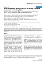
<b>AKI Is Common And Deadly: Major </b>
<b>Surgery (NSQIP data)</b>
[15] Bihorac A et al. National Surgical Quality Improvement Program Underestimates the Risk Associated with Mild and Moderate Postoperative Acute
<i>Kidney Injury. Crit Care Med. 2013;41(11):2570-2583.</i>
0,6% 3%
7%
26%
0%
10%
20%
30%
No AKI Mild (Risk) Moderate
(Injury) (Failure)Severe
3%
6%
11%
29%
0%
10%
20%
30%
No AKI Mild (Risk) Moderate
(Injury) (Failure)Severe
<b>Hospital Mortality</b> <b>90-Day Mortality</b>
In a single-center cohort of 27,841 adult surgical patients undergoing major surgery, it was
identified that hospital and 90-day mortality were significantly higher among patients with AKI
</div>
<span class='text_page_counter'>(7)</span><div class='page_container' data-page=7>
<b>AKI IS PREVALENT IN CT SURGERY</b>
<i>[16] Hobson CE et al. Acute Kidney Injury is Associated with Increased Long-Term Mortality After Cardiothroacic Surgery. Circulation.</i>
2009;119:2444-2453.
<i>N = 2973 CT Surgery Patients16</i>
0%
10%
20%
30%
40%
50%
60%
70%
80%
All Types Isolated CABG Valve Surgery Aortic Surgery Thoracic
Surgery TransplantHeart
<b>% of </b>
<b>CT</b>
<b> S</b>
<b>ur</b>
<b>ger</b>
<b>y </b>
<b>w</b>
<b>ith </b>
<b>A</b>
<b>K</b>
<b>I</b>
Moderate
AKI Severe AKI
Mild AKI
No AKI
</div>
<span class='text_page_counter'>(8)</span><div class='page_container' data-page=8>
<i>[9] Kidney Disease: Improving Global Outcomes (KDIGO) Acute Kidney Injury Work Group. KDIGO Clinical Practice Guideline for Acute Kidney Injury. Kidney Inter, Suppl. 2012; 2:1-138</i>
<i>[10]Tolwani A. Continuous Renal-Replacement Therapy for Acute Kidney Injury. N Engl J Med. 2012;367:2505-2514.</i>
Acute Kidney Injury (AKI) is a rapid (typically within about 48 hours) loss of kidney function9
• 96% of AKI does NOT require RRT10
RIFLE/AKIN/KDIGO criteria were validated over the past decade and provide a standardized
definition of AKI
The criteria are based on increases and serum creatinine and decreases in urine output and stratify
AKI into three severity levels:9
1. Mild AKI (RIFLE-R or Stage 1)
2. Moderate AKI (RIFLE-I or Stage 2)
3. Severe AKI (RIFLE-F or Stage 3)
The criteria are good for epidemiological studies but difficult to apply at the bedside; AKI thus
remains largely a clinical diagnosis9
8
</div>
<span class='text_page_counter'>(9)</span><div class='page_container' data-page=9>
<b>AKI Identification In the ICU Can Be Inconsistent</b>
[4] Massicottee-Azarniouch, Magder S, Goldberg P, Alam A. Acute Kidney Injury in the Intensive Care Unit: Risk Factors and Outcomes of Physician Recognition Compared with KDIGO Classification. Poster presented at: Society of Critical
Care Medicine; February 2016; Orlando, FL.
<b>AKI Reported by ICU Staff</b> <b>No AKI Reported by ICU Staff</b>
<b>2,393 patients admitted to academic hospital ICU in Montreal, Canada from January 2006 through December 2011. KDIGO AKI </b>
<b>calculated from SCr values. Physician definition of AKI was determined by asking ICU staff if a patient had “acute renal failure”.4</b>
<b>“ICU physicians only identified a small proportion of the patients with AKI. Many of the severe forms </b>
<b>of AKI, which were most associated with adverse outcomes, were missed by the physician reporting.”</b>
<b>79% of the cases </b>
<b>of Moderate and </b>
<b>Severe AKI Were </b>
<b>Not Identified by </b>
<b>Reporting </b>
<b>Physician</b>
<b>0</b>
<b>100</b>
<b>200</b>
<b>300</b>
<b>400</b>
<b>500</b>
<b>Moderate AKI</b> <b>Severe AKI</b>
<b>Numb</b>
<b>er </b>
<b>of P</b>
<b>atien</b>
<b>ts</b>
</div>
<span class='text_page_counter'>(10)</span><div class='page_container' data-page=10>
Without Better Tools, The Best Doctors Are Challenged With AKI
AKI IS DEADLY, COSTLY, AND PREVALENT… AND LOCAL
<b>2016 ANNUAL</b> <b>AKI DIAGNOSES6,7,8*</b>
<b>AVG</b> <b>COST</b>
<b>INCREASE</b> <b>/ </b>
<b>PATIENT7</b>
<b>AVG</b> <b>LENGTH</b> <b>OF</b>
<b>STAY</b> <b>INCREASE</b> <b>/ </b>
<b>PATIENT7</b>
<b>READMISSION</b> <b>RATE</b>
<b>INCREASE9</b>
<b>2200 ICU ADMISSIONS</b>
<b>476 ESTIMATED</b> <b>MODERATE/SEVERE</b> <b>ICU DIAGNOSES</b>
<b>$29,800</b> <b>10.4 DAYS</b> <b>15.9%</b>
<b>Although Often Under-reported5<sub>, AKI Hits Home:</sub></b>
<b>A Typical 500-Bed Hospital</b>
<b>$16.2 M</b>
<b>ILLION</b><b>COST</b> <b>INCREASE</b>
<b>5,450 D</b>
<b>AYS</b><b>LENGTH OF</b> <b>STAY</b>
<b>INCREASE</b>
<b>86 P</b>
<b>ATIENTS</b><b>READMISSION</b> <b>INCREASE</b>
<b>ANNUAL</b> <b>ICU IMPACT: </b>
<b>MODERATE/SEVERE</b> <b>AKI</b>
[5] Massicottee-Azarniouch, Magder S, Goldberg P, Alam A. Acute Kidney Injury in the Intensive Care Unit: Risk Factors and Outcomes of Physician Recognition Compared with KDIGO Classification. Poster presented at: Society of Critical Care Medicine;
February 2016; Orlando, FL.
[6] American Hospital Directory Database, accessed Dec 2017 on 7,104 hospitals, data on file
[7] Hobson CE, Ozrazgat-Baslanti T, Kuxhausen A, et. al. Cost and Mortality Associated With Postoperative Acute Kidney Injury. Annals of Surgery. 2014;00:1–8
[8] SCCM: />
</div>
<span class='text_page_counter'>(11)</span><div class='page_container' data-page=11>
AKI IS A SPECTRUM OF KIDNEY DECLINE
AND EARLY IDENTIFICATION IS KEY TO
POTENTIALLY STOP THE PROGRESSION
11
Figure adapted from: [1] Lewington AJP, Certa J, Mehta RL Raising Awareness of Acute Kidney Injury: A Global Perspective of a
<i>Silent Killer. Kidney Int. 2013;84(3):457-467.</i>
<i>[19] Kellum JA, Chawla LS. Cell-Cycle Arrest and acute kidney injury: the light and dark sides. Nephrol Dial Transplant. 2016;1:16-22</i>
Kidney
Stress Decreased Function
Asymptomat
ic Symptomatic (Diagnosis)
</div>
<span class='text_page_counter'>(12)</span><div class='page_container' data-page=12>
<i>SUBOPTIMAL DIAGNOSTIC TOOLS MAKE AKI </i>
IMPROVEMENT DIFFICULT
12
Figure adapted from: [1] Lewington AJP, Certa J, Mehta RL Raising Awareness of Acute Kidney Injury: A Global Perspective of a Silent Killer.
<i>Kidney Int. 2013;84(3):457-467.</i>
<i>[20] Martensson J et al. Novel Biomarkers of Acute Kidney Injury and Failure: Clinical Applicability. Brit J Anesth. 2012;109(6):843-50.</i>
<i>[21] Wlodzimirow KA, et al. A comparison of RIFLE with and without urine output criteria for acute kidney injury in critically ill patients. Critical </i>
<i>Care. 2012;16:R200.</i>
[22] Gould CV, et al. Guideline for Prevention of Catheter-Associated Urinary Tract Infections. HICPAC. 2009.
Kidney
Stress Decreased Function
Asymptom
atic Symptomatic (Diagnosis)
<b>Serum Creatinine</b>
<b>• Lagging indicator20</b>
<b>• Only elevates after 50% of </b>
<b>kidney function lost20</b>
<b>• Non-diagnostic for up to </b>
<b>52% of moderate and </b>
<b>severe AKI21</b>
<b>Urine Output</b>
<b>• Lagging indicator21</b>
<b>• Tedious to measure21</b>
<b>• Affected by HAI </b>
<b>initiatives22</b>
Wouldn’t it be nice to identify
Kidney Stress BEFORE the
dysfunction occurs?
</div>
<span class='text_page_counter'>(13)</span><div class='page_container' data-page=13>
<i>[10] Kashani K, et al. Discovery and validation of cell cycle arrest biomarkers in human acute kidney injury. Crit Care. 2013;17:R25.</i>
<i>[11] Bihorac A, et al. Validation of Cell-Cycle Arrest Biomarkers for Acute Kidney Injury Using Clinical Adjudication. Am J Respir Crit Care </i>
<i>Med. 2014;189(8):932-939.</i>
<b>340 Biomarkers Evaluated </b>
<i>including NGAL & KIM-1</i>
<b>Discovered in 1,200+ Patients</b>
<i>including sepsis, shock, major </i>
<i>surgery and trauma patients</i>
<b>Urinary [TIMP-2]*[IGFBP-7] stood out as the </b>
<b>best-performing biomarkers to predict development </b>
of moderate or severe AKI within 12 hours10
Candidates identified through hypothesis based
on AKI pathophysiology and evaluated individually
and in combinations of 2-4 biomarkers10
<b>Validated in 500+ Critically Ill </b>
<i><b>Patients from Intended Use </b></i>
<i>Population</i>
<i>Patients had diverse ICU admissions (surgery, </i>
<i>sepsis, trauma) and common comorbidities </i>
<i>(including CKD, diabetes, heart disease) </i>11
</div>
<span class='text_page_counter'>(14)</span><div class='page_container' data-page=14>
TIMP-2 and IGFBP7 Outperform Existing Biomarkers
</div>
<span class='text_page_counter'>(15)</span><div class='page_container' data-page=15>
<b>Tissue Inhibitor of Metalloproteinase-2 (TIMP-2)</b>
<b>Insulin-like Growth Factor Binding Protein-7 (IGFBP-7)</b>
TIMP-2 and IGFBP-7 are:14
• <b>Biomarkers of cellular stress in the early phase of tubular cell injury caused by a </b>
<i>wide variety of insults (inflammation, ischemia, oxidative stress, drugs, and </i>
<i>toxins)</i>
• <b>Involved in G1 cell-cycle arrest that prevent cells from dividing until damage </b>
can be repaired
• <b>Both biomarkers appear as “alarm” proteins from other nearby cells</b>
This may help explain why urinary TIMP-2 and IGFBP-7 correspond to risk of AKI.
<i>[12] TIMP-2 figure adapted from: Tuuttila A et al. Three-dimensional structure of human tissue inhibitor of metalloproteinases-2 at 2.1 A resolution. J Mol Biol. 1998;284:1133-1140.</i>
[13] IGFBP-7 figure adapted from: ModBase: Database of Comparative Protein Structure Models [accessed 2014 December 10]. Available from:
<i>[14] Gocze I, et al. Urinary Biomarkers TIMP-2 and IGFBP7 Early Predict Acute Kidney Injury After Major Surgery. PLoS ONE. 2015;10(3).</i>
</div>
<span class='text_page_counter'>(16)</span><div class='page_container' data-page=16>
Getting Ahead of AKI: Measure Kidney Stress Before Damage Occurs
More complete information = personalized medicine
opportunity to consider
kidney-protecting strategies
before it’s too late
<b>Gauges the Risk of </b>
<b>Injury before </b>
<b>Damage Occurs </b>
<b>• Specific to AKI11</b>
<b>• Fast & Simple: 20 min urine test11</b>
<b>• Commercially Available in USA11</b>
<b>• Peer-reviewed evidence11</b>
<b>• Complementary to HAI and QI </b>
<b>initiatives</b>
<b>• Easy, cost effective to Implement</b>
<b>• Baystate Medical Center</b>
</div>
<span class='text_page_counter'>(17)</span><div class='page_container' data-page=17>
<b>Building An Algorithm Around </b>
<b>NephroCheck: Assessment & Prevention</b>
<b>• When Do I Order the NephroCheck Test?</b>
<b>• Sepsis</b>
<b>• CVI</b>
<b>• Shock</b>
<b>• Logistics</b>
• <b>How Long Does it Take To Get the Results?</b>
</div>
<span class='text_page_counter'>(18)</span><div class='page_container' data-page=18>
<b>• Who to Test</b>
<b>• When to Test</b>
<b>• What Will You Do Different</b>
<b>• Different Treatments Sepsis, Shock, Cardiovascular?</b>
<b>• Reassessment After Intervention</b>
<b>• Trend or Treatment Success</b>
<b>• Value of the Negative Test</b>
<b>Building An Algorithm Around </b>
</div>
<span class='text_page_counter'>(19)</span><div class='page_container' data-page=19>
<b> Team Members: Will order test based on inclusion </b>
<b>criteria</b>
<i><b>Goal: To identify patients at risk of AKI before serum </b></i>
creatinine increases and take pre-emptive action to
mitigate incidence of AKI and decrease acute hemodialysis
(volume overload, hyperkalemia, severe acidemia).
24 - 48 hours prior to ICU admission history of
hypotension, use of vasopressors or nephrotoxic
medications, acute respiratory failure, sepsis,
and high-risk surgery.
<i><b>ALL CV surgery</b></i>
</div>
<span class='text_page_counter'>(20)</span><div class='page_container' data-page=20>
CLINICAL RISK FACTORS FOR AKI ARE COMMON BUT NOT
RELIABLE FOR ESTABLISHING THE RISK PROFILE FOR AN
INDIVIDUAL PATIENT
[9] Kidney Disease: Improving Global Outcomes (KDIGO) Acute Kidney Injury Work Group. KDIGO Clinical Practice Guideline for Acute Kidney Injury. Kidney inter., Suppl. 2012; 2:
1-138.
[11] Murugan R et al. Acute Kidney Injury in Non-Severe Pneumonia is Associated with an Increased Immune Response and Lower Survival. Kidney Int. 2010;77:527-535.
[13] Uchino S et al. Acute Renal Failure in Critically Ill Patients: a Multinational, Multicenter Study. JAMA. 2005;294:813-818.
[23] Ronco C, Ricci Z. The concept of risk and the value of novel markers of acute kidney injury. Crit Care. 2013;17:117-118.
<b>Patient Risk Factors9</b>
• Dehydration or volume depletion
• Advanced Age
• Female gender
• Black race
• CKD
• Chronic Disease (heart, lung, liver)
• Diabetes Mellitus
• Cancer
• Anemia
<b>Acute Risk Factors9,11,13</b>
• Sepsis
• Pneumonia
• Cardiogenic Shock
• Major Surgery
• Cardiac Surgery
• Nephrotoxic Drugs
• Radiocontrast Agents
</div>
<span class='text_page_counter'>(21)</span><div class='page_container' data-page=21>
<b>Early recognition and management of patients at risk is paramount </b>
<b>since there are no specific therapies to reverse established AKI.</b>
<b>9</b><b>As compared to AMI, AKI does not provide early signs and </b>
<b>symptoms sufficient to guide risk assessment.</b>
<b>23</b><b>Current methods for risk assessment are insufficient, placing </b>
<b>substantial numbers of patients at serious risk of death and </b>
<b>morbidity.</b>
<b>9,26</b>21
<b>A BETTER WAY TO IDENTIFY PATIENTS AT RISK FOR </b>
<b>AKI IS PARAMOUNT</b>
[9] Kidney Disease: Improving Global Outcomes (KDIGO) Acute Kidney Injury Work Group. KDIGO Clinical Practice Guideline for Acute Kidney Injury. Kidney inter., Suppl. 2012; 2: 1-138.
[23] Ronco C, Ricci Z. The Concept of Risk and the Value of Novel Markers of Acute Kidney Injury. Crit Care. 2013;17:117-118.
</div>
<span class='text_page_counter'>(22)</span><div class='page_container' data-page=22>
NEW TECHNOLOGY, ADVANCE WARNING ENABLES
BETTER OUTCOMES
22
<i><b>What if we could get ahead of </b></i>
<b>AKI? </b>
<i><b>Instead of saying, “wait and </b></i>
<i><b>see</b></i><b>…”</b>
<b>• Early Warning</b>
<b>• Stratify Patient Risk</b>
<b>• Attention on “At Risk” </b>
<b>patients </b>
</div>
<span class='text_page_counter'>(23)</span><div class='page_container' data-page=23>
<b>Target patients – High risk for AKI such as: </b>
<b>CV surgery</b>
- Shock – septic, cardiogenic, hemorrhagic
- Acute decompensated CHF with cardiogenic shock
- ARDS for P/F ratio <200
- Oliguria – persistent after resuscitation
<b>Exclusion: </b>
- Age < 18 years
- Previous renal transplant
- Known stage 2 or 3 AKI
</div>
<span class='text_page_counter'>(24)</span><div class='page_container' data-page=24>
THE CLINICAL CUTOFF WAS SELECTED TO IDENTIFY THE
MAJORITY OF PATIENTS AT RISK FOR MODERATE-SEVERE AKI
*For moderate-severe AKI in the next 12 hours.
<b>The NEPHROCHECK®</b> <b>Test cutoff (AKIRISK®</b> <b>Score > 0.3) was prospectively </b>
<b>selected prior to validation studies to achieve*:</b>
<b>High sensitivity and negative predictive value are important in risk </b>
<b>assessment to ensure that:</b>
• <b>The majority of patients who will develop AKI test positive </b>
• <b>Few patients with a negative test result will be at risk of developing AKI</b>
<b>Study A (408 patients)</b> <b>Study B (126 patients)</b>
High sensitivity 92% 76%
Acceptable specificity 46% 51%
High negative predictive
value 96% 88%
</div>
<span class='text_page_counter'>(25)</span><div class='page_container' data-page=25>
<b>A Quantitative N</b>
<b>EPHRO</b>
<b>C</b>
<b>HECK</b>
<b>®</b><b>Test Provides Confidence to </b>
<b>Identify the Majority of Patients at Risk for AKI</b>
<b>High sensitivity and negative predictive value for confidence in identifying the majority of patients at risk for AKI.</b>
Confidence the AKIRISK® <b>Score is not elevated due to common comorbidities such as </b>
CKD, diabetes, surgery, sepsis and trauma.
<i>Results from Study A and B are not statistically different (p>0.05)</i> 25
<b>AK</b>
<b>IR</b>
<b>ISK</b>
<b>®</b> <b>Sc</b>
</div>
<span class='text_page_counter'>(26)</span><div class='page_container' data-page=26>
Recent published literature has discussed the role of the N
EPHROC
HECK®Test used in
conjunction with clinical judgement and includes recommendations on preferred
kidney sparing strategies to help prevent kidney damage.
19<b>Published Recommendations to Help Prevent Kidney Damage </b>
</div>
<span class='text_page_counter'>(27)</span><div class='page_container' data-page=27>
With dynamic measurement of the risk for AKI, there will be the opportunity to initiate timely
and appropriate preventative therapies and monitoring in the ICU, for those patients who are
judged to be at high risk of AKI.
49As well, less costly interventions are easy and reasonable to implement if risk is identified,
such as considering:
9,19,49•
Discontinuing nephrotoxins or changing dosage
•
Volume status & perfusion pressure
•
Hemodynamic monitoring
•
Monitoring frequency of serum creatinine and urine output
•
Earlier nephrology consult.
<b>Acute Kidney Injury (AKI) is a Significant Opportunity to </b>
<b>Improve Quality of Patient Care</b>
<i>[9] Kidney Disease: Improving Global Outcomes (KDIGO) Acute Kidney Injury Work Group. KDIGO Clinical Practice Guideline for Acute Kidney Injury. Kidney Inter, Suppl. 2012; 2:1-138</i>
<i>[19] Kellum JA, Chawla LS. Cell-Cycle Arrest and acute kidney injury: the light and dark sides. Nephrol Dial Transplant. 2016;1:16-22</i>
</div>
<span class='text_page_counter'>(28)</span><div class='page_container' data-page=28></div>
<span class='text_page_counter'>(29)</span><div class='page_container' data-page=29>
<b>LOW AKI RISK: ≤ 0.3</b> <b>HIGH AKI RISK: > 0.3</b>
- Standard of Care – document
UOP AND Foley remove ASAP
- Monitor & document hourly UOP & consider Foley insertion/maintain
- Daily Serum BUN, Cr. - Serum BUN, Cr. Q 12
- Urine Na, Cr, Eos x 1
- Consider repeat NephroCheck in 12hrs
- CI/SVV/SVI not monitored <sub>- Mandatory hemodynamic monitoring for CI/SVV/SVI Q8 </sub><b><sub>POP algorithm</sub></b>
- Check IVC compressibility with US
- Goals -- MAP>70, SVV < 13, CI >2.0
- Vasopressors (phenylephrine, nor-epinephrine, vasopressin)
- Inotrope support (dobutamine or milrinone)
- Low threshold for inotropes if CI < 2, ScvO2 < 70, and/or Lactic Acid increasing
despite adequate MAP and volume expansion
- Diuretics or fluids as needed
based on <b>physiology</b>
- Diuretics and fluids to be utilized ONLY after determining fluid status <b>POP</b>
- For oliguria or hypotension, IVV expansion <b>SV optimization POP</b>
- Crystalloid, blood products rarely albumin, clinical decision
</div>
<span class='text_page_counter'>(30)</span><div class='page_container' data-page=30>
<b>LOW AKI RISK: ≤ 0.3</b> <b>HIGH AKI RISK: > 0.3</b>
- Repeat NephroCheck if new
insult (Same incl/excl criteria)
- Consider repeat NephroCheck testing at 12 hr intervals if there is a need to
reevaluate renal stress/intervention
- Avoid & resolve hypervolemia (> 10% fluid gain)
<b>address hemodynamics using physiology POP</b>
- Can use ACEI/ARB
- Cautious use of NSAIDs
- No NSAIDS or ACEi/ARB
- Avoid all unnecessary IV IODINATED contrast dye studies
- Minimize exposure to nephrotoxins (i.e. vancomycin, piperacillin/tazobactam,
aminoglycosides)
- ADJUST MEDICATION DOSING ANTICIPATING DECREASED GFR ESPECIALLY IF UOP
REMAINS LOW
<b>ICU Interventions – AKI Bundle Based on NephroCheck AKI Risk Score</b>
<b>Consider RRT/CRRT Initiation: Inadequate UOP > 12-24 hrs, symptomatic volume </b>
</div>
<span class='text_page_counter'>(31)</span><div class='page_container' data-page=31>
Patient History:
• 61 yo Male with DM, CAD-stent, Afib, COPD
– Creatinine 1.0
– MICU With Active GI Bleed
– Intubated, Central IV Access, A-line Placed
– Bedside UGI Endoscopy For Active Bleed—Vessel Cauterized
– Rebleed > IR for Embolization
• Day 1
– Fluid Resuscitation
Initial NC 0.31 (high risk AKI)
– BP 70/30, CI 1.5 / SVV 20 (Fluid Responsive)
– 7 Units RBC’s and 8 Liters Crystalloid
Clinical Status:
• The physician managing the patient realized an early bump in NC
required extremely aggressive fluid resuscitation, especially with such
high blood loss. Concern was “over-resuscitation” of fluid.
</div>
<span class='text_page_counter'>(32)</span><div class='page_container' data-page=32>
Clinical Issue:
ã NephroCheckđ Test Interpretation: Elevated AKIRisk® Score (> 0.3) Indicates
Higher Risk for Mod/Sev AKI
– AKIRisk Score 0.31 (high kidney stress)
Assessment & Intervention:
• After Successful Resuscitation and Embolization
• CI 2.4 / SVV 10
• Day 2
• CI 2.2 / SVV 11
• Max Creatinine Over Extended Stay
• Creatinine 1.1 Max With NO AKI
Patient Outcome:
• Patient Completely Recovered with no Kidney Damage
</div>
<span class='text_page_counter'>(33)</span><div class='page_container' data-page=33>
Patient History:
• 84 yo Female with Hx of Sjogrens Disease and Arterio-Venous
Malformations Throughout Bowel
• 95Kg pt Admitted to ED With Hgb 7.6 With GI Bleed/Pain
Clinical Status:
• Day 1--Admitted to MICU
• Hgb Fell to 6, BP 60/40, Unresponsive, Shock
• Intubated and Resuscitated Aggressively with 2 pRBC’s, Fluid and
Pressors
• Taken to Interventional Radiology for Embolization
</div>
<span class='text_page_counter'>(34)</span><div class='page_container' data-page=34>
Clinical Issue:
ã NephroCheckđ Test Interpretation: Elevated AKIRisk® Score (>
0.3) indicates higher risk for mod/sev AKI
–
AKIRisk Score 0.04 (low kidney stress)
Assessment & Intervention:
• Day 2: Successful Resuscitation
• Extubated
• Creatinine 1.2, Hgb 9.5
• Pt Transferred to Floor on Day 3
• Surprised to Learn of Low Renal Stress but This Gave Confidence
to Quickly Move pt on to Lower Level of Care:
Throughput!
Key Take Away – Value of Negative Score
</div>
<span class='text_page_counter'>(35)</span><div class='page_container' data-page=35>
<b>Conclusions: Real Time in the MICU With Sepsis</b>
<b>• When we test with NephroCheck: As soon sepsis is suspected</b>
• Nurse Alerts
• Hospitalist Training
<b>• Why we test: AKI shows up with Sepsis more than any other reported DRG</b>
• Real Time Picture of the Kidneys
• Did Interventions Make a Difference
• Positive Predictive of the Negative Test
• Helps with Family Discussion of Care Plan
<b>• What we do with the results: Including kidney care in sepsis treatment</b>
• Maintain MAP
• Volume Status
• Review Medications
</div>
<span class='text_page_counter'>(36)</span><div class='page_container' data-page=36>
<b>Advice For Implementing At Your Hospital</b>
<b>Build A Team </b>
<b>Lab, Nephrology, CT Surgeon, Nursing, Administration</b>
<b>Education </b>
<b>Before Implementation</b>
<b>After Adoption</b>
<b>Feedback To the Team Of Success!!! </b>
</div>
<!--links-->
<a href=' />