- Trang chủ >>
- Y - Dược >>
- Truyền nhiễm
Giáo trình Java cơ bản: Phần 1 - Trường Đại Học Quốc Tế Hồng Bàng
Bạn đang xem bản rút gọn của tài liệu. Xem và tải ngay bản đầy đủ của tài liệu tại đây (374.71 KB, 7 trang )
<span class='text_page_counter'>(1)</span><div class='page_container' data-page=1>
<i><b>Int.J.Curr.Microbiol.App.Sci </b></i><b>(2017)</b><i><b> 6</b></i><b>(11): 3457-3469 </b>
3457
<b>Original Research Article </b>
<b>Cultural and Morphological Characterization of </b>
<i><b>Rhizoctonia solani</b></i>
<b> f. sp. </b>
<i><b>sasakii</b></i>
<b> Isolates Collected from Different Districts of Andhra Pradesh </b>
<b>Bindu Madhavi Gopireddy1*, G. Uma Devi2, K.Vijayakrishna Kumar1, </b>
<b>T. Ramesh Babu1 and T.C.M.Naidu1</b>
1
Acharya N G Ranga Agricultural University, Guntur, Andhra Pradesh, India
2
Professor Jayasankar Telangana State Agricultural University, Hyderabad, Telangana, India
<i>*Corresponding author </i>
<i><b> </b></i> <i><b> </b></i><b>A B S T R A C T </b>
<i><b> </b></i>
<b>Introduction </b>
Maize (<i>Zea mays</i> L.) is one of the most
versatile emerging crops having wider
adaptability under varied agro-climatic
conditions. Globally, maize is known as
queen of cereals due to its high genetic yield
potential. Among the potential factors that
limit maize production, fungal diseases are
reported to cause extensive crop yield
reduction in many countries and are
considered as a priority in disease
management practice (Agrios, 2005). Of
different fungal diseases affecting maize
cultivation, banded leaf and sheath blight
(BLSB) incited by <i>Rhizoctonia solani </i>f. sp.
<i>sasakii </i>Exner (<i>Thanatephorus sasakii </i>(Shirai)
Tu and Kimbrough) (Tu and Kimbrrough,
1978) is an economically significant disease
causing huge losses in all crop growing areas
of the world. Increased incidence of BLSB
has been observed in rice fallow maize crop
(zero tillage) in different districts of Andhra
Pradesh. Effective management of BLSB in
maize is possible only when the pathogen is
eliminated completely or the propagules are
brought down below economic threshold
limits at field level. Control measures used
were partly effective because <i>R. soiani </i>is able
to produce sclerotia that can persist in the soil
for at least two years (Ou, 1985). The pace of
<i>International Journal of Current Microbiology and Applied Sciences </i>
<i><b>ISSN: 2319-7706</b></i><b> Volume 6 Number 11 (2017) pp. 3457-3469 </b>
Journal homepage:
Sixty BLSB affected maize samples were collected from three districts and pathogen was
isolated and identified based on morphological, cultural and sclerotial characters using
standard descriptions of IMI. Light microscopic studies revealed that all the isolates of <i>R. </i>
<i>solani </i>f. sp. <i>sasakii</i>are characteristically branched out at right angle in the distal end of the
cell and showed a characteristic constriction at the point of branching. Formation of
septum near the point of origin of the branch / adjacent to branch was present in most of
the isolates except for isolates RS 44, RS 48, RS 58 and RS 59, where in the septum was
slightly away from the origin of branching. The hyphal width of all the 60 isolates varied
from 5.05 μm (RS 52) to 7.98 μm (RS 15). Out of 60 maize isolates, eight isolates <i>i.e., </i>RS
7, RS 8, RS 9, RS 10, RS 11, RS 12 RS 16 and RS 26 produced barrel shaped monilliod
cells and the remaining isolates produced irregular shaped monilliod cells. Clamp
connections were absent in all the isolates, multinucleate and the number of nuclei per cell
varied from five to seven in the isolates.
<b>K e y w o r d s </b>
Maize, BLSB,
<i>Rhizoctonia solani</i> f.
sp. <i>sasakii</i>,
Morphological,
Cultural
characterization.
<i><b>Accepted: </b></i>
26 September 2017
<i><b>Available Online: </b></i>
10 November 2017
</div>
<span class='text_page_counter'>(2)</span><div class='page_container' data-page=2>
<i><b>Int.J.Curr.Microbiol.App.Sci </b></i><b>(2017)</b><i><b> 6</b></i><b>(11): 3457-3469 </b>
3458
development and durability of resistant
varieties had been slow and unreliable despite
tremendous advancements in the field of plant
genetic engineering. Variability of the
pathogen plays major role in resistance
breeding hence the cultural and
morphological characterization of the isolates
was studied.
<b>Materials and Methods </b>
To study the morphology of hyphae of each
BLSB pathogenic isolate, four day old-fungal
hyphae grown on PDA medium was taken
and stained with 0.1 per cent lactophenol
cotton blue on microscopic slides for
recording, angle of branching, septation,
presence of monilioid cells, presence of clamp
connections, type of septum and hyphal width
and number of nuclei using Olympus CX31
microscope with ProgRes. CT3 image
analyser.
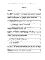
<b>Cultural characteristics of </b> <i><b>Rhizoctonia </b></i>
<i><b>solani</b></i><b> f. sp. s</b><i><b>asakii</b></i><b> isolates (Fig. 1) </b>
<b>Growth of fungal pathogen </b>
The isolates were grown on PDA medium in
Petri dishes at 27 +2ºC until the hyphae
reached the periphery of the petridishes for
determination of color, abundance of
mycelium, zonation and sclerotial characters.
<b>Colony characters </b>
Abundance of mycelium was compared with
the key given by Burpee <i>et al., </i>(1980).
The abundance was characterized into the
following four categories.
Slight: Aerial mycelium does not obscure
surface mycelium
Moderate: Aerial mycelium obscure surface
mycelium and does not touch the cover of
Petri dishes
Abundant: Aerial mycelium obscure surface
mycelium and touches the cover of Petri
dishes
No aerial mycelium
Colony color was determined with the help
Munsell’s soil colour chart (Munsell color
Company, Inc., 1954).
The culture and key color cards were placed
side by side against white background under
sunlight for comparison (Burpee <i>et al., </i>1980).
Observations for colony color were recorded
10 days after incubation.
Based on the colony pigmentation, the
cultures were assigned to different groups
based on dominant spectral color.
<b>Sclerotial characteristics of </b> <i><b>Rhizoctonia </b></i>
<i><b>solani</b></i><b> f. sp. </b><i><b>sasakii </b></i><b>isolates </b>
Sclerotial characteristics were also compared
with the key given by Burpee <i>et al.,</i> (1980).
<b>Location of sclerotia </b>
Based on location of sclerotial production in
the culture the isolates of <i>R. solani</i> f. sp.
<i>sasakii</i> were categorized into following
groups
Aerial: Sclerotia formed with in aerial
mycelium
Embedded: Sclerotia formed with in substrate
<b>Size of sclerotia </b>
</div>
<span class='text_page_counter'>(3)</span><div class='page_container' data-page=3>
<i><b>Int.J.Curr.Microbiol.App.Sci </b></i><b>(2017)</b><i><b> 6</b></i><b>(11): 3457-3469 </b>
3459
the Petri dishes under the stereo binocular
microscope and were classified as (a) Large
(b) Small
<b>Colour of the sclerotia </b>
It was categorized in to 4 groups a) Light
brown (b) Brown (c) Dark brown (d) Deep
dark brown
<b>Location </b> <b>and </b> <b>Pattern </b> <b>of </b> <b>sclerotial </b>
<b>formation </b>
Location of sclerotia produced by different
isolates in the Petri dishes containing PDA
were observed and recorded as sclerotia
produced on the surface of agar (aerial) or
submerged in the medium. The pattern of the
sclerotial production was also studied and the
isolates were divided into different categories
based on their distribution on the culture. The
production of sclerotia by different isolates
was recorded as more or less circular manner
concentrated towards periphery; irregularly
scattered but more towards the centre of the
colony; irregular very sparsely scattered and
scattered irregularly all over the colony
surface. Sclerotial types were mainly divided
into two categories as follows.
Sclerotiawith rough border
Sclerotiawith smooth border
<b>Results and Discussion </b>
<b>Morphological </b> <b>variability </b> <b>among </b> <b>the </b>
<b>isolates of </b><i><b>Rhizoctonia solani </b></i><b>f. sp. </b><i><b>sasakii </b></i><b>of </b>
<b>maize</b>
Morphological characters are the important
basic factors for identification of a fungus and
its variability. Studies on morphological
characteristics of <i>R. solani</i> f. sp. <i>sasakii </i>maize
isolates (60 numbers) and RS 61 were studied
and the results are presented in Table 1.
Light microscopy studies revealed that all the
isolates of <i>R. </i> <i>solani </i> f. sp. <i>sasakii </i>
characteristically branched out at right angle
in the distal end of the cell (Plate 1).
<b>Constriction at the point of branching </b>
All isolates showed a characteristic
constriction at the point of branching.
Formation of septum near the point of origin
of the branch / adjacent to branch was present
in most of the isolates while in isolates RS 44,
RS 48, RS 58, RS 59 the septum was slightly
away from the origin of branching.
<b>Hyphal width </b>
The hyphal width of all the 60 isolates varied
from 5.05 μm (RS 52 from Krishna district) to
7.98 μm (RS 15 from Prakasam district). The
hyphal widths of most of the isolates were at
par with each other. However, the differences
in hyphal width observed among the other
isolates were non- significant.
<b>Monilioid cells </b>
In addition to ordinary vegetative hyphae, <i>R. </i>
<i>solani</i>produces simple or branched chains of
short broad cells, which may be hyaline or
brown, barrel shaped, pyriform, irregular, or
lobate known as monilioid cell (Plate1). Out
of 60 maize isolates, eight isolates <i>i.e., </i>RS 7,
RS 8, RS 9, RS 10, RS 11, RS 12 RS 16 and
RS 26 produced barrel shaped monilioid cells.
The remaining isolates produced irregular
shaped monilioidcells .
Clamp connections: were absent in all the
isolates
</div>
<span class='text_page_counter'>(4)</span><div class='page_container' data-page=4>
<i><b>Int.J.Curr.Microbiol.App.Sci </b></i><b>(2017)</b><i><b> 6</b></i><b>(11): 3457-3469 </b>
3460
taxonomical importance which were
described by the previous workers Duggar
(1915), Matsumato (1921), Thomas (1925).
<b>Number of nuclei</b>
All the isolates under present investigation
were found to be multinucleate and the
number of nuclei per cell varied from five to
seven. Isolates RS 21, RS 23, RS 25, RS 26,
RS 27 had statistically maximum number (7)
of nuclei per cell (Table 1).
The classification of <i>Rhizoctonia solani</i>has
been done on the basis of hyphal and cultural,
morphology, nuclear condition, hyphal
anastomosis and morphology of teleomorphs.
The present findings on morphological
variability among <i>R. solani </i>isolates are in
accordance with Sneh <i>et al., </i>(1991), Amita
Singh <i>et al., </i>(1999), Meena <i>et al.,</i> (2001) and
Srinivas (2002) in maize, Singh <i>et al., </i>(2002)
and Basu <i>et al., </i>(2004) in rice.
<b>Cultural variability of </b><i><b>R. solani </b></i><b>isolates </b>
<b>Colony colour</b>
The colour of the colony varied from white to
dark brown. Based on pigmentation dominant
spectral colour from Munsell’s soil colour
chart (1954), the cultures were assigned to
five colour groups with respective shade
numbers <i>i.e, </i> Group I- white, Group
II-yellowish white, Group III-pale brown and
Group IV brown and Group V dark brown
(Table 1).
Among the 60 isolates studied, 11 isolates
belonged to group I, 9 isolates with yellowish
white were assigned in group II, 22 isolates in
Group III, ten isolates in group IV and eight
in Group V. The variation in the colour of the
colony might be attributed to the production
of pigments by the pathogen. The differences
in the intensity of the colour might also
correspond to the amount of pigments
released by respective isolate in the media.
The colour production may also be due to
release of other secondary metabolites like
toxins. Amita Singh <i>et al., </i>(1999) assigned
Munsell’ssoil colour chart shade number to
the colony colour of <i>R. solani </i>isolates from
rice, maize, soybean, mung beans and cotton.
Further, Akhtar <i>et al., </i>(2009) stated that the
colony colour of <i>R. solani </i>maize isolates <i>Hc </i>
and <i>It </i>were brown whereas the isolates <i>Bc, Jr </i>
and <i>Rf </i>had white colour. Studies on cultural
characteristics revealed that the colony colour
of different <i>R. solani </i> isolates varied from
white to brown on PDA (Khodayari <i>et al., </i>
2009). The results are also in agreement with
the observations of other researchers (Sneh <i>et </i>
<i>al.,</i> 1991; Sweetingham and Mac Nish, 1994;
Amita Singh <i>et al., </i>1999). Srinivas (2002)
categorised maize isolates of <i>R. solani</i>causing
BLSB disease based on colony pigmentation.
<b>Abundance of mycelium</b>
Among 60 isolates, 27 isolates produced
abundant mycelium, while 18 isolates have
moderate mycelium and the remaining 15
isolates recorded slight/ sparse mycelium.
<b>Colony diameter and growth rate </b>
The data presented in Table 1 on colony
diameter and growth rate revealed that there
were significant differences among the
isolates after 72 hours of incubation on PDA
medium. Among the 60 isolates, 29 isolates
recorded as fast growers (more than 40 mm
growth) and 21 as moderate (35-40mm
growth) and ten recorded slow growth (30-35
mm) after 72 h of incubation.
The cultural characteristics studied among the
</div>
<span class='text_page_counter'>(5)</span><div class='page_container' data-page=5>
<i><b>Int.J.Curr.Microbiol.App.Sci </b></i><b>(2017)</b><i><b> 6</b></i><b>(11): 3457-3469 </b>
3461
Distinct differences were observed in the
colony appearance and the isolates were
categorised into different groups based on
texture and abundance of mycelium. The
difference in the colony growth was distinct
in 27 isolates. These isolates produced
abundant aerial cottony mycelial growth
which may be due to the inherent nature of
these isolates to go for quick and profuse
mycelial growth in early stages of growth
before setting the sclerotia.
Similar observations have been made by Toda
<i>et al.,</i> (1999) who divided <i>Rhizoctonia </i>AG-D
isolates into two subgroups AG-D (I) and
AG-D (II), based on the results of cultural
characteristics; Srinivas (2002) categorised
the <i>R. solani </i>f. sp. <i>sasakii </i>isolates from maize
based on texture and abundance of their
mycelia growth and colony appearance.
Similarly Guleria <i>et al., </i>(2007) used cultural
characters for differentiating the <i>R. solani </i>
isolates from rice.
Significant variations with respect to colony
growth and growth rate were also recorded
among the isolates under the study. The
isolates RS 34, RS 26, RS 25 having fast
growth rate were found more virulent as they
induced susceptible reaction on maize. Meena
<i>et al., </i>(2003) observed that the fast growing
isolate of <i>R. solani </i>from maize was found to
be more virulent on a susceptible maize
cultivar.
Similarly, Guleria <i>et al., </i>(2007), Thind and
Aggarwal (2008) and Khodaryari <i>et al., </i>
(2009) stated that the <i>R. solani </i>isolates from
rice were fast growing with >20 mm mycelia
growth rate per day indicating their fast
growing nature. Rapid growth rate among <i>R. </i>
<i>solani </i> isolates have also been reported by
Peltier (1916), Matz (1921), Matsumoto
(1934) and Parmeter and Whitney (1970).
<b>Fig.1 </b>Cultural and sclerotial characteristics of different isolates of
</div>
<span class='text_page_counter'>(6)</span><div class='page_container' data-page=6>
<i><b>Int.J.Curr.Microbiol.App.Sci </b></i><b>(2017)</b><i><b> 6</b></i><b>(11): 3457-3469 </b>
3462
<b>Table.1 </b>Cultural characteristics of different isolates of <i>Rhizoctonia solani</i> f.sp. <i>sasakii</i> collected
from Prakasam, Guntur and Krishna districts of Andhra Pradesh
<b>Isolate </b> <b>Hyphal </b>
<b>width </b>
<b>(µm) </b>
<b>Number </b>
<b>of nuclei </b>
<b>Colour of </b>
<b>the </b>
<b>colony </b>
<b>Colony </b>
<b>diameter </b>
<b>after </b>
<b>72h (mm) </b>
<b>Growth </b>
<b>rate* </b>
<b>Growth </b>
<b>pattern </b>
<b>Time taken </b>
<b>to initiate </b>
<b>Sclerotia </b>
<b>(days) </b>
RS 1 5.35 6 Pale brown 66 Fast Moderate 4
RS 2 5.40 5 Pale brown 57 Fast Moderate 4
RS 3 6.05 5 White 45 moderate Abundant 4
RS 4 5.83 5 White 69 Fast Moderate 5
RS 5 7.05 6 White 44 moderate Slight 5
RS 6 7.27 6 White 29 slow Slight 6
RS 7 6.45 6 Pale brown 75 Fast Moderate 4
RS 8 5.95 5 Pale brown 78 Fast Abundant 4
RS 9 6.10 5 White 42 Moderate Slight 5
RS 10 5.12 6 White 30 Slow Slight 6
RS 11 5.37 6 White 45 Moderate Slight 5
RS 12 5.55 5 White 44 Moderate Slight 5
RS 13 6.65 6 Pale 45 Moderate Abundan 5
RS 14 7.23 6 Pale brown 22 slow Moderate 7
RS 15 7.98 6 Pale brown 29 slow Slight 7
RS 16 6.45 6 White 64 Fast Abundant 4
RS 17 5.08 5 Pale brown 40 Moderate Abundant 5
RS 18 5.11 5 White 39 Moderate Moderate 5
RS 19 5.86 5 White 28 slow Slight 6
RS 20 6.65 7 Pale brown 39 Moderate Moderate 5
RS 21 5.84 7 Yellowish
white
65 Fast Abundant 4
RS 22 5.48 6 Pale brown 69 Fast Abundant 4
</div>
<span class='text_page_counter'>(7)</span><div class='page_container' data-page=7>
<i><b>Int.J.Curr.Microbiol.App.Sci </b></i><b>(2017)</b><i><b> 6</b></i><b>(11): 3457-3469 </b>
3463
RS 24 5.23 6 Pale brown 41 Moderate Abundant 4
RS 25 6.15 7 Pale brown 66 Fast Moderate 5
RS 26 6.87 7 Yellowish
white
69 Fast Abundant 4
RS 27 5.21 7 Yellowish
white
70 Fast Abundant 4
RS 28 5.65 6 Yellowish
white
66 Fast Abundant 4
RS 29 6.51 6 Pale brown 62 Fast Abundant 4
RS 30 7.57 6 Pale brown 60 Fast Moderate 4
RS 31 7.14 5 Pale brown 44 Moderate Moderate 4
RS 32 6.16 5 Yellowish
white
51 Fast Moderate 4
RS 33 5.20 5 Pale brown 55 Fast Moderate 4
RS 34 6.80 6 Yellowish
white
26 slow Abundant 5
RS 35 7.12 6 Yellowish
white
61 Fast Abundant 4
RS 36 7.05 6 Pale brown 72 Fast Slight 6
RS 37 6.55 7 Pale brown 29 Slow Slight 6
RS 38 5.33 5 Yellowish
white
46 Fast Moderate 4
RS 39 5.84 6 Pale brown 55 Fast Abundant 4
RS 40 6.81 6 Yellowish
white
41 Moderate Moderate 5
RS 41 6.25 6 Brown 32 Moderate Slight 5
RS 42 6.13 6 Dark brown 34 Moderate Slight 5
RS 43 5.68 6 Brown 30 Moderate Slight 5
RS 44 5.26 6 Pale brown 31 Moderate Slight 6
RS 45 5.11 5 Dark brown 40 Fast Abundant 5
RS 46 6.60 5 Dark brown 42 Fast Abundant 5
RS 47 6.47 6 Dark brown 44 Fast Abundant 6
RS 48 6.80 6 Pale brown 44 Fast Abundant 6
RS 49 7.55 6 Brown 45 Fast Abundant 6
RS 50 5.45 6 Dark brown 50 Fast Abundant 6
RS 51 5.83 6 Dark brown 54 Fast Abundant 6
RS 52 5.05 6 Brown 52 Fast Abundant 6
RS 53 6.25 6 Brown 36 Moderate Abundant 8
RS 54 7.30 6 Dark brown 32 Slow Moderate 10
RS 55 7.87 6 Brown 38 Moderate Abundant 8
RS 56 6.48 5.5 Brown 39 Moderate Abundant 8
</div>
<!--links-->
Báo cáo thực hiện công khai của trường đại học quốc tế Hồng Bàng
- 81
- 1
- 2


.push({});</script> </div> </div> </div> <div class="vf_link_relate px-2 my-2"> <h2 class="vf_doc_relate text-2xl font-bold my-4">Tài liệu liên quan</h2> <ul class="grid grid-cols-12 gap-2"> <li class="col-span-6 md:col-span-2"> <div class="card-doc " onclick="actionDocRelated(this)"> <a class="card-doc-img" href="https://text.123docz.net/document/192519-bao-cao-thuc-hien-cong-khai-cua-truong-dai-hoc-quoc-te-hong-bang.htm" title="Báo cáo thực hiện công khai của trường đại học quốc tế Hồng Bàng"> <i class="icon i_type_doc i_type_doc2"></i> <img class="lazy" src="data:image/gif;base64,R0lGODlhAQABAIAAAP///wAAACH5BAEAAAAALAAAAAABAAEAAAICRAEAOw==" data-src="https://media.store123doc.com/images/document/13/rc/ds/medium_FhEXJHbVCu.jpg" width="124" height="179" alt="Báo cáo thực hiện công khai của trường đại học quốc tế Hồng Bàng" onerror="this.src=)